a subpial, transitory germinal zone forms chains of neuronal … · neural cell adhesion molecule;...
TRANSCRIPT

94 (2006) 168–180www.elsevier.com/locate/ydbio
Developmental Biology 2
A subpial, transitory germinal zone forms chains of neuronal precursors inthe rabbit cerebellum
Giovanna Ponti a, Paolo Peretto b, Luca Bonfanti a,c,⁎
a Department of Veterinary Morphophysiology, University of Turin, Via Leonardo da Vinci 44, 10095 Grugliasco, Italyb Department of Animal and Human Biology, University of Turin, Via Accademia Albertina 13, 10153 Turin, Italy
c Rita Levi Montalcini Center for Brain Repair, Italy
Received for publication 3 January 2006; revised 20 February 2006; accepted 22 February 2006Available online 3 April 2006
Abstract
Protracted neurogenesis occurs at different postnatal stages in different brain locations, whereby leading to site-specific adult neurogenesis insome cases. No spontaneous genesis of neurons occurs in the cerebellum after the postnatal genesis of granule cells from the external germinallayer (EGL), a transitory actively proliferating zone which is thought to be exhausted before puberty. Here, we show the protracted genesis ofnewly generated neuronal precursors in the cerebellar cortex of young rabbits, persisting beyond puberty. Neuroblasts generated within an activelyproliferating subpial layer thus extending the postnatal EGL are arranged to form thousands of tangential chains reminiscent of those responsiblefor cell migration in the forebrain subventricular zone. These subpial chains cover the whole cerebellar surface from the 2nd to the 5th month oflife, then disappearing after puberty. In addition, we describe the appearance of similar groups of cells at the end of granule cell genesis in themouse cerebellum, here limited to the short period of EGL exhaustion (4–5 days). These results show common features do exist in the postnatalreorganization of secondary germinal layers of brain and cerebellum at specific stages, parallel to differences in the slowing down of cerebellarneurogenesis among mammalian species.© 2006 Elsevier Inc. All rights reserved.
Keywords: Development; Neurogenesis; Migration; Meninges; PSA-NCAM
Introduction
Adult neurogenesis persists throughout life in two restrictedbrain areas: the dentate gyrus of the hippocampus and theforebrain subventricular zone (SVZ) (Gage, 2000; Alvarez-Buylla and Garcia-Verdugo, 2002). Unlike the forebrain, themammalian cerebellum is considered incapable of any sponta-neous cell genesis after the exhaustion of the external germinallayer (EGL). The EGL is a secondary actively proliferating zonemade up of cell precursors which originate from the germinal
Abbreviations: SVZ, subventricular zone; PSA-NCAM, polysialylatedneural cell adhesion molecule; BrdU, 5-bromo-2′-deoxyuridine; ML, molecularlayer; GL, inner granular layer; EGL, external germinal layer; SPL, subpiallayer; DCX, doublecortin; CNS, central nervous system.⁎ Corresponding author. Dip. di Morfofisiologia Veterinaria, Via Leonardo da
Vinci, 44, 10095-Grugliasco (TO) Italy. Fax: +39 011 6709138.E-mail address: [email protected] (L. Bonfanti).
0012-1606/$ - see front matter © 2006 Elsevier Inc. All rights reserved.doi:10.1016/j.ydbio.2006.02.037
trigone following tangential subpial displacement (Altman andBayer, 1997), then leading to delayed genesis of the granule cellpopulation in the postnatal developing cerebellar cortex. In allspecies studied, this transitory germinal zone is formed byseveral layers of granule cell precursors which progressivelyreduce their thickness as the cells migrate deep into the cortex toform the inner granule layer (GL). In atricial mammals(Sanchez-Villagra and Sultan, 2001), the EGL is active in thepre- and postnatal period, then disappearing parallel to or beforeaccomplishing postnatal granule cell genesis, which occurs atspecific ages: postnatal day 21 in mice (Fujita et al., 1966) and22 in rats (Altman, 1969), postnatal month 3 in primates (Rakic,1971) and 11 in humans (Abraham et al., 2001). After EGLexhaustion, direct contact between the pial surface andBergmann glial endfeet on one side and parallel fibers on theother is thought to characterize the external part of the molecularlayer (ML) (Altman and Bayer, 1997). Although in rabbits theend of cell genesis and the neurochemical maturation of the

169G. Ponti et al. / Developmental Biology 294 (2006) 168–180
cerebellum have been fixed around the second postnatal month(Smith, 1963; Lossi et al., 1995), here, we describe the existenceof a secondary germinal matrix persisting beyond puberty insubpial position. This subpial layer originating from a structuralmodification of the EGL is capable of generating neuronalprecursors which assemble to form tangential chains reminis-cent of the forebrain SVZ, namely the main adult neurogeneticarea of the mammalian brain (Gage, 2000; Alvarez-Buylla andGarcia-Verdugo, 2002). In addition, since rodents and lago-morphs belong to distinct mammalian orders, we have studiedthe last phases of mouse EGL revealing a tendency to formchain-like structures similar to rabbit SPL chains, here restrictedto a very short period in coincidence with the exhaustion of thegerminative layer.
Materials and methods
5-Bromo-2′-deoxyuridine (BrdU) injections and tissue preparation
Experimentation was conducted in accordance with current EU and Italianlaws (Italian Ministry of Health, authorization n. 66/99-A). Twenty-two peri-puberal (3–5 months old; Charles River, Milan) New Zealand White rabbits(Orictolagus cuniculus) were used for light microscopy. Seventeen receivedintraperitoneal injections of BrdU (Sigma; 40 mg/kg). Eight received a singleinjection, five were killed after 2 h and three after 5 days. Nine rabbitsreceived one daily injection for 5 days and were killed 2 h, 5 and 10 days(n = 3 each) after the last injection. In addition, two 4-month-old rabbits forelectron microscopy, two 6-month-old and four adult (1 and 2 year old)rabbits for light microscopy, were used. Twelve postnatal rabbits (10, 15, 23,30, 40, 75 days old) were treated with BrdU for 2 h. Cerebella from 18 young(P15, P18, P21, P24, P26; P28; n = 3 for each age) CD-1 mice (CharlesRiver, Italy) were also used for light microscopy. In addition, two P21 micewere used for electron microscopy.
Animals were anesthetized with a ketamine/xylazine solution (100 mg/kgbody weight: 33 mg/kg body weight) and perfused as previously described(Luzzati et al., 2003; Peretto et al., 2005). Cerebella were extracted carefully topreserve the pia mater, postfixed 6 h (light microscopy), frozen at −80°C andcryostat (16 μm thick) or vibratome (100 μm thick) sectioned in series, alongsagittal and coronal orientation. Tissues for electron microscopy were postfixed2 h, cut sagittally using a blade (about 300 μm thick), then fixed in osmium-ferrocyanide for 1 h, stained en bloc with 1% uranyl acetate, dehydrated,embedded in Araldite and processed as previously described (Luzzati et al.,2003). Ultra-thin sections were examined with a Philips CM10 transmissionelectron microscope. Semithin sections for light microscopy (1 μm thick) werestained with 1% toluidine blue and 0.5% NaHCO3.
Immunohistochemistry
Immunohistochemical reactions were carried out by using single peroxidaseand double immunofluorescence methods on cryostat sections incubatedovernight at room temperature with primary antibodies: (i) anti-BrdU, 1/600(monoclonal, Harlan Laboratories, Haslett, MI); (ii) anti-Ki67, 1/300 (MIB1,monoclonal, Immunotech, Luminy); 1/2000 (polyclonal, Novocastra); (iii) anti-laminin, 1/200 (polyclonal, Chemicon); (iv) anti-PSA-NCAM, diluted 1/3500(monoclonal IgM; G. Rougon, Marseille, France); (v) anti-class III β-tubulin, 1/1000 (TU-J1, monoclonal and polyclonal, Babco, Richmond, VA); (vi) anti-human neuronal protein HuC/D, 1/200 (monoclonal, Molecular Probes); (vii)anti-DCX, 1/750 (polyclonal goat, Santa Cruz); (viii) anti-glial fibrillary acidicprotein, 1/1000 (GFAP, polyclonal, DAKO); (ix) anti-vimentin, 1/800(monoclonal, DAKO); (x) anti-Ng2 chondroitin sulfate proteoglycan, 1/250(polyclonal, Chemicon); anti-O4, 1/200 (monoclonal, Chemicon). All poly-clonal antisera used did not give any problem on the rabbit cerebellar tissue. Fordouble staining, indirect immunofluorescence procedures using FITC–avidin + Cy3 conjugated antibodies were used. Antibodies were diluted in0.01 M PBS, pH 7.4, containing 0.5% Triton X-100. Fluorescent specimens
mounted in 1,4-diazabicyclo[2.2.2]octane (Dabco, Sigma) were observed with alaser scanning Olympus Fluoview confocal system.
Electron microscopic serial reconstructions
The electron microscopic reconstruction of two rabbit SPL chains wascarried out on serial ultra-thin sections (80 nm thick) collected onto Formvar-coated grids. Micrographs from the sixth section in the sequence were drawn todefine the contour of single cells and processes. The digital images have beenadded in sequence on Adobe Photoshop software. Drawings from 43 levels wereexamined to follow the behavior of the chain across a total length of 21 μm. Thereconstruction of a mouse subpial chain-like structure was performed using thesame method, although in this case the drawing of one section every 12 (about1 μm interval) was enough, due to the presence of a small number of cellprocesses. A total of 16 sections, up to a total length of 16 μm, were considered.
Cell countings and statistical analysis
The amount of newly generated cells in the cerebellum of 12 3-month-oldrabbits was analyzed by counting the number of BrdU-immunoreactive nuclei inparasagittal cryostat sections, at 2 h (animals n = 3), 5 (n = 6: 3 after a singleinjection, 3 after 5 injections) and 15 (n = 3 after 5 injections) days survivaltimes. A total of 200 mm of pial surface and underlying cortex was analyzed in 3representative sections for each animal, cut at different medial–lateral levels.Immunoreactive nuclei present on the cerebellar surface (cut nuclei wereconsidered only on one side of the section) and in cerebellar cortical layers (on asingle focal plane) were counted. Images of the cortical areas were imported onImage proplus, then countings of total nuclei, nuclei densities and nuclei/numberof cells (on cresyl violet-stained sections) were performed in the SPL (5 μmbeneath the pial surface), ML and GL. Parallel sections were used to estimate thepercentage of BrdU/PSA-NCAM double-labeled cells detectable in the SPL at2 h and 5 days survival times (total cells counted = 7291).
The number of proliferating cells in each cerebellar layer has been analyzedby one-way ANOVAwith main factor time and repeated measurements factor oflayer (SPL, ML, GL). Statistical analyses of data were performed by the SPSSstatistical software (version 12.01 for Windows). A value of P ≤ 0.05 wasaccepted as statistically significant.
The number of PSA-NCAM-immunoreactive cells in each layer wascounted in an area corresponding to 10 mm of pial surface in representative thickvibratome sections (animals n = 3; total cells counted = 6341). In the SPL, due tothe presence of chains, 2 bipolar cells/chain were considered.
Results
A proliferative layer persists in the young rabbit cerebellum
By using endogenous (Ki67 antigen) and exogenouslyadministered (5-bromo-2′-deoxyuridine, BrdU) markers ofcell proliferation a consistent amount of newly generated cellswere found in the cerebellar cortex of peri-puberal (3- to 5-month-old) rabbits (Fig. 1). BrdU detected at 2-h survival timewas used to identify s-phase cells, and Ki67 confirmed thislabeling as cell proliferation and not DNA repair (Kee et al.,2002; Rakic, 2002). In BrdU/Ki67 double labelings, bothantigens were concentrated at the same sites, all BrdU+ cellsresulting double-stained. As expected, Ki67 revealed a highernumber of cells (Fig. 1A), due to its estimated permanence inthe cell cycle (Kee et al., 2002). Most proliferating cells wereobserved along the cerebellar surface (Fig. 1). Double stainingsfor proliferation markers and laminin (Fig. 1B), and semithinsections from resin-embedded material (Fig. 1C) showed thatthe vast majority of proliferating elements occupied a specificsubpial position, which did not affect the meninges. Dividing

Fig. 1. Cell proliferation in the young rabbit cerebellum (sagittal sections). Newly generated cells, detected with Ki67 (A, red) and BrdU at 2-h (A, B, green; F, brown)or 5-day (G, brown) survival time, are concentrated in the SPL. Their subpial position is confirmed in double labeling with laminin (B, red), in semithin sections (C,arrowhead) and electron microscopy (D). (g) Bergmann glial endfeet; bv, meningeal blood vessel; p, pial cell; m, mitosis; bl, basal lamina. (E) Apoptotic figure in theSPL; ab, apoptotic bodies; a, arachnoid cell; sas, subarachnoid space; c, collagen. (H–J) Quantitative analysis of BrdU+ cells in the SPL, ML and GL at differentsurvival times. 2 h, 2 hours; 5d, 5 days; (1i) single BrdU injection; (5i), five daily injections. Scale bars: 10 μm (A–C); 2 μm (D, E); 50 μm (F, G).
170 G. Ponti et al. / Developmental Biology 294 (2006) 168–180
cells were arranged to form a single non-continuous layer(12.14 ± 3.39 BrdU+ nuclei/mm; Figs. 1F and 2M), irregularlycovering the whole cerebellar surface. We will refer to this thinregion which persists beyond puberty as the subpial layer (SPL),to distinguish it from the postnatal EGL (see below). Electronmicroscopy confirmed the occurrence of mitotic figures in theSPL, at the interface between the basal lamina of the pia materand the ML (Fig. 1D), thus providing independent evidence (seeRakic, 2002) for cell proliferation at this level. Some figureswith typical morphological signs of apoptotic cells were alsodetectable in the SPL (Fig. 1E).
Local cell proliferation was rare in cerebellar cortical layersoutside the SPL (Fig. 1F). The cell density of BrdU+ elementsafter 2-h survival was 700 times higher in the SPL than incortical tissue (Fig. 1I). The ANOVA for repeated measures
revealed no significative reduction of the number of BrdU+nuclei, but an interaction between time and their distribution incellular layers considered, namely SPL, ML, GL (F = 11.215;P = 0.005). Furthermore, the one-way ANOVA in each cellularlayer indicated a reduction of BrdU+ nuclei in the SPL between2-h and 5-day survival time (F = 8.699; P = 0.042). Noremarkable differences were observed in different medial–lateral or rostral–caudal extensions of the cerebellum, indicat-ing a remarkable homogeneity of this process in the entirecerebellar cortex. Being cell proliferation of the SPL widelyscattered over a large surface area and not concentrated within adiscrete anatomical site (as is the case for the forebrain SVZneurogenesis; see Peretto et al., 1999; Gage, 2000; Alvarez-Buylla and Garcia-Verdugo, 2002), we treated other animalswith 5 daily injections of BrdU, in order to detect a larger

Fig. 2. Organization of the SPL in peri-puberal rabbits (sagittal sections). Immunocytochemistry for PSA-NCAM (A, B, D, brown; F, J, green; G–I, red) identifytangentially oriented chain-like structures in the SPL of the entire cerebellar cortex (A, B, D, F–J). Numerous PSA-NCAM+ cells are also detectable in the ML (B,cresyl violet counterstained). (C) Regularly arranged, subpial cells (arrows) in a semithin section. (E–G) Chains of cells in the SPL show antigenic features of neuronalprecursors, being immunoreactive for doublecortin (DCX, E), HuC/D protein (Hu, F), and class III β-tubulin (β-tub, G). (H, I) These cells are newly generated: doublestaining with BrdU (green) and PSA-NCAM (red) at different postinjection stages (2 h and 5 days) shows local cell proliferation among chains in the SPL (H), whereasthe newly generated cells become part of the chains in the subsequent 5 days (I). (J–J′) The SPL chains are oriented in a medial–lateral way, following rows of GFAP+Bergmann glial endfeet. Scale bars: 200 μm (A); 50 μm (B); 20 μm (C–J).
171G. Ponti et al. / Developmental Biology 294 (2006) 168–180
population of newly generated cells. In animals which hadreceived the same number of BrdU injections but were killedafter 2 weeks, a remarkable decrease in newly generated cellswas observed in the SPL, whereas many had survived in the MLand GL (Fig. 1H). In this case, the ANOVA for repeatedmeasures revealed a highly significative interaction betweentime and distribution of BrdU+ nuclei in cellular layers
(F = 37.17; P = 0.000) as well as a remarkable reduction intheir number (F = 92.76; P = 0.001). The one-way ANOVA ineach cellular layer indicated a significative reduction of BrdU+nuclei in all layers considered, notably in the SPL (SPL,F = 84.373, P = 0.001; ML, F = 12.96, P = 0.023; GL,F = 11.022, P = 0.029). These data suggest that the cell genesisoccurring in subpial position could be followed by displacement

172 G. Ponti et al. / Developmental Biology 294 (2006) 168–180
through the cortical tissue, although only a small populationactually survives after 2 weeks. The total number of BrdU+cells/layer (Fig. 1H) as well as their percentage comparedwith the entire cell population in each layer was higher inthe ML than in the granule layer at all survival times (Fig.1J). It is important to note that cell density was similar in thetwo cortical layers (Fig. 1I), due to a larger extension of theML.
In 6-month-old rabbits, the occurrence of dividing cells haddramatically decreased, even on the cerebellar surface, and inadult (1 and 2 years old) animals only scattered BrdU+ or Ki67+nuclei were occasionally detected (not shown).
Newly generated neuronal precursors in the SPL formthousands of small chains
In order to explore the nature and arrangement of newlygenerated cells in the SPL, we performed immunocytochemis-try by using antibodies raised against specific neurogenesis-associated molecules. Firstly, we detected the polysialylatedform of the neural cell adhesion molecule (PSA-NCAM), aglycoprotein strictly associated to the entire population of adult-generated neuronal precursors in the hippocampus (Seki andArai, 1991; Bonfanti et al., 1992) and SVZ (Bonfanti andTheodosis, 1994; Rousselot et al., 1995; Doetsch and Alvarez-Buylla, 1996; Petreanu and Alvarez-Buylla, 2002; Luzzati et al.,2003), required for their migration (Ono et al., 1994; Hu, 2000).Unlike the cerebellum of rodents after the EGL exhaustion,which lacks PSA-NCAM (Bonfanti et al., 1992, and this study;see Fig. 6), in rabbits, we found many immunoreactive cells,most of which were localized on the subpial surface (Figs. 2A,B, D). A remarkable number of PSA-NCAM+ cells withneuronal-like morphology were also present in the corticaltissue, most of which were localized within the ML (Fig. 2B;see below). A semiquantitative analysis of the relative amountof PSA-NCAM+ cells in 3-month-old rabbits indicated that85% (898.33 ± 15.21 chains, namely 1796.67 ± 30.42 cells in10 mm) of them are confined within the SPL, 12%(251 ± 11.60) in the ML, and only 3% (66 ± 3.00) in theremaining layers (Purkinje cell layer, inner granule layer andwhite matter).
Thus, the attention was focused on the SPL, where all thecells appeared as tangentially oriented, bipolar elementsgrouped to form chain-like structures (Fig. 2D), reminiscentof the chains of neuroblasts described in the SVZ of rodents(Lois et al., 1996) and rabbits (Luzzati et al., 2003; Ponti et al.,unpublished). These chain-like structures were homogeneouslypresent on the whole cerebellar extent in all animals in whichthere had been no damage to the cerebellar surface duringdissection. About 7000 of these structures were present alongthe perimeter of a single representative sagittal section of thevermis (spaced about 10–20 μm from one another in 3-month-old animals; Figs. 2A, B, D), regularly arranged with a prevalentmedial–lateral orientation. In GFAP/PSA-NCAM doublestainings, they appeared in close contact and regular relation-ships with rows of Bergmann endfeet, contacting themtransversely at the junction with the pia mater (Figs. 2J, J′, J″).
To establish their real nature as chains, these aggregates wereanalyzed with electron microscopy (Fig. 3). Small subpial cells(about 3–4 μm in diameter) sharing exactly the same pattern ofthe PSA-NCAM+ chain-like structures were detectable insagittal semithin sections of resin-embedded lamellae (Figs. 1C,2C, and 3C). The ultrastructural study revealed the typicalcytology of neuroblasts (Lois et al., 1996; Jankovski and Sotelo,1996; Doetsch et al., 1997; Peretto et al., 1997; Luzzati et al.,2003) (SVZ type A cells, Lois et al., 1996), with a large nucleusand a thin halo of electrondense cytoplasm, and confirmed thelocalization of these cells at the basal lamina/ML interface (Fig.3). One or two, rarely three cell bodies were associated withdifferently sized processes containing the same type ofcytoplasm, thus forming aggregates which were clearlyrecognizable from the surrounding axons and glia. Cerebellarcoronal sections (longitudinal to these aggregates) revealedtheir identity as continuous, tangential chains of bipolar-shapedelements with leading and trailing processes (Fig. 3B). Furthercharacterization was obtained by carrying out a serialultrastructural reconstruction of two subpial tangential chainsalong a 21 μm tract of cerebellar surface (Figs. 3C–G). Due tothe existence of many thin cell processes intermingled with cellbodies, a careful analysis involving 43 parasagittal levels wasperformed in order to follow the processes of individual cells. Avery elongated bipolar neuroblast was entirely reconstructedwithin a chain (the cell colored in red in Fig. 3). The serialreconstruction showed that several long processes of SPLneuroblasts are closely associated with few cell bodies, thusforming very thin chains. The SPL chains were in direct contactwith the glia limitants formed by the Bergmann endfeet andwith the parallel fibers, rarely coming into direct contact withbasal lamina. As already visible in light microscopy (see Figs.2D, E, J), and confirmed by the ultrastructural reconstruction(Fig. 3), different chains can contact one another at some pointsto form a subpial network.
In addition to ultrastructural cytology, the neuronal nature ofsubpial cells was confirmed by immunoreactivity for themicrotubule binding protein doublecortin (DCX), a markerspecifically associated to neuroblasts and differentiating healthyneurons produced in adult neurogenic areas (Nacher et al., 2001;Brown et al., 2003; Rao and Shetty, 2004), the early neuronalmarker class III β-tubulin (Menezes and Luskin, 1994; Perettoet al., 1997; Alvarez-Buylla and Garcia-Verdugo, 2002) andHuC/D protein (Goldman, 1997). As described for PSA-NCAM, staining for these markers also clearly revealed thetangential chains (Figs. 2E–G). The glial markers O4, Ng2,vimentin, and GFAP were used to evaluate the putative occur-rence of cell precursors of the glial lineage in the SPL. After acareful search for colocalizations with BrdU+, no associationswere found in either early- or late-generated cells (not shown).For these reasons, we conclude that cell genesis in the youngrabbit SPL is devoted to the production of neuronal precursors.
Since cell proliferation and chains of neuroblasts are both insubpial position, we combined local cell proliferation markers(Ki67 and 2-h survival BrdU) and PSA-NCAM to understandtheir mutual relationships. Most of the locally dividing cellswere interposed among the chains (Fig. 2H), whereas many
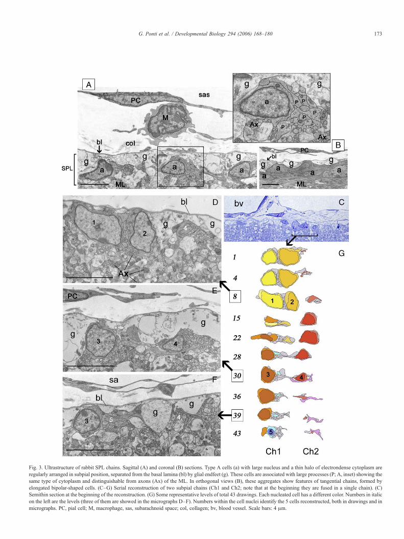
Fig. 3. Ultrastructure of rabbit SPL chains. Sagittal (A) and coronal (B) sections. Type A cells (a) with large nucleus and a thin halo of electrondense cytoplasm areregularly arranged in subpial position, separated from the basal lamina (bl) by glial endfeet (g). These cells are associated with large processes (P; A, inset) showing thesame type of cytoplasm and distinguishable from axons (Ax) of the ML. In orthogonal views (B), these aggregates show features of tangential chains, formed byelongated bipolar-shaped cells. (C–G) Serial reconstruction of two subpial chains (Ch1 and Ch2; note that at the beginning they are fused in a single chain). (C)Semithin section at the beginning of the reconstruction. (G) Some representative levels of total 43 drawings. Each nucleated cell has a different color. Numbers in italicon the left are the levels (three of them are showed in the micrographs D–F). Numbers within the cell nuclei identify the 5 cells reconstructed, both in drawings and inmicrographs. PC, pial cell; M, macrophage, sas, subarachnoid space; col, collagen; bv, blood vessel. Scale bars: 4 μm.
173G. Ponti et al. / Developmental Biology 294 (2006) 168–180

174 G. Ponti et al. / Developmental Biology 294 (2006) 168–180
immunoreactive nuclei resided inside the chains in BrdU-treated animals which survived 5 days (Fig. 2I). Accordingly,the number of BrdU/PSA-NCAM double-labeled cells in theSPL progressively increased at subsequent survival times. Thisconfirms that the SPL is made up of two cell compartments:actively dividing cells which do not yet express PSA-NCAM(Bonfanti and Theodosis, 1994; Rousselot et al., 1995), andchains of PSA-NCAM-immunoreactive cells containing thenewly generated elements (with a 1/10 proportion, respective-ly). These compartments are part of the same population ofnewly generated cells forming a fragmented monolayer which isstructurally different from the postnatal EGL.
Transition from EGL to SPL and postpuberal follow-up of theSPL
In the present study, we show that the proliferative capacityof the postnatal cerebellum in rabbits does not disappear as early
Fig. 4. Transition from EGL to SPL (A–M), and its further follow-up in the adult rapremigratory layer (M) around P30 (summarized in M) is followed by the appearasubsequent postnatal stages (E, F, H, L–N). Cresyl violet (A–C), PSA-NCAM (brown(red) (J–L) stainings. (C–L) Parasagittal sections. (N) Follow-up of SPL and postpucoincides with differences in the ML. Black dots, proliferating cells; black lines, chainsome PSA-NCAM+ (O) and DCX+ (P) neuronal-shaped cells are detectable in the
as previously thought (Smith, 1963). The cerebellar EGL,although sharing features with the SVZ and hippocampus(Hatten, 1999), has so far been considered as a transientgerminal zone destined to disappear after accomplishinggranule cells genesis (Smith, 1963; Altman and Bayer, 1997).Yet, in the rabbit, the occurrence of the SPL does not simplyseem to be the persistence of an EGL since importantdifferences characterize its structure as well as ML cellcomposition throughout its existence.
In order to identify the transition time from postnatal EGL toperi-puberal SPL, we followed its histology, contents ofproliferating cells and PSA-NCAM distribution in youngrabbits at different postnatal stages (Fig. 4). Similarly topostnatal rodents (Dey et al., 1999, and this study, see belowand Fig. 5), PSA-NCAM was absent in the proliferative EGLbut present in the ML and granule layer as a diffuse reaction thatreaches its high intensity at the limit between EGL and ML, inassociation with cells of the premigratory layer (Figs. 4D and 6).
bbit (N–P). Fragmentation of the proliferative layer (P) and its fusion with thence of chains. The distance among proliferating cells and chains increases at, D–F), PSA-NCAM (red)/BrdU (green) (G–I), and PSA-NCAM (green)/GFAPberal neurogenesis in the rabbit cerebellum. Transition between EGL and SPLs. In older (1 year, O; 2 years, P) animals a SPL is no longer detectable, althoughML. Scale bars: (A–L) 20 μm; (O, P) 10 μm.
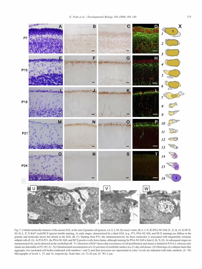
Fig. 5. Cellular/molecular features of the mouse EGL at the end of granule cell genesis. (A, E, I, M, Q) cresyl violet; (B, F, J, N, R) PSA-NCAM; (C, G, K, O, S) DCX;(D, H, L, P, T) Ki67 (red)/DCX (green) double staining. At early stages, characterized by a thick EGL (e.g., P7), PSA-NCAM, and DCX stainings are diffuse in thegranule and molecular layers but absent in the EGL (B, C). Starting from P15, the immunoreactivity for these molecules is associated with tangentially oriented,subpial cells (F, G). At P18-P21, the PSA-NCAM- and DCX-positive cells form chains, although staining for PSA-NCAM is faint (J, K, N, O). At subsequent stages noimmunoreactivity can be detected in the cerebellum (R–T). Detection of Ki67 shows that coexistence of cell proliferation and chains is limited to P18 (L), whereas onlychains are detectable at P21 (P). (U–X) Ultrastructural reconstruction of a 16 μm tract of cerebellar surface in a 21-day-old mouse. (X) Drawings of a subpial chain-likeaggregate; two nucleated cell bodies (indicated with numbers 1 and 2) and their processes are represented in color. Levels are indicated with italic numbers. (U–W)Micrographs of levels 1, 15, and 16, respectively. Scale bars: (A–T) 20 μm; (U–W) 2 μm.
175G. Ponti et al. / Developmental Biology 294 (2006) 168–180
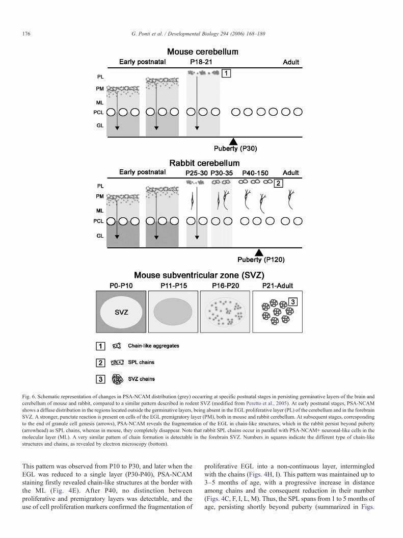
Fig. 6. Schematic representation of changes in PSA-NCAM distribution (grey) occurring at specific postnatal stages in persisting germinative layers of the brain andcerebellum of mouse and rabbit, compared to a similar pattern described in rodent SVZ (modified from Peretto et al., 2005). At early postnatal stages, PSA-NCAMshows a diffuse distribution in the regions located outside the germinative layers, being absent in the EGL proliferative layer (PL) of the cerebellum and in the forebrainSVZ. A stronger, punctate reaction is present on cells of the EGL premigratory layer (PM), both in mouse and rabbit cerebellum. At subsequent stages, correspondingto the end of granule cell genesis (arrows), PSA-NCAM reveals the fragmentation of the EGL in chain-like structures, which in the rabbit persist beyond puberty(arrowhead) as SPL chains, whereas in mouse, they completely disappear. Note that rabbit SPL chains occur in parallel with PSA-NCAM+ neuronal-like cells in themolecular layer (ML). A very similar pattern of chain formation is detectable in the forebrain SVZ. Numbers in squares indicate the different type of chain-likestructures and chains, as revealed by electron microscopy (bottom).
176 G. Ponti et al. / Developmental Biology 294 (2006) 168–180
This pattern was observed from P10 to P30, and later when theEGL was reduced to a single layer (P30-P40), PSA-NCAMstaining firstly revealed chain-like structures at the border withthe ML (Fig. 4E). After P40, no distinction betweenproliferative and premigratory layers was detectable, and theuse of cell proliferation markers confirmed the fragmentation of
proliferative EGL into a non-continuous layer, intermingledwith the chains (Figs. 4H, I). This pattern was maintained up to3–5 months of age, with a progressive increase in distanceamong chains and the consequent reduction in their number(Figs. 4C, F, I, L, M). Thus, the SPL spans from 1 to 5 months ofage, persisting shortly beyond puberty (summarized in Figs.

177G. Ponti et al. / Developmental Biology 294 (2006) 168–180
4M, N). Bearing in mind that in all the species studied, thereduction of EGL to a monolayer is a crucial step which isthought to be followed by its disappearance (Fujita, 1967; Fujitaet al., 1966; Altman, 1969; Rakic, 1971; Abraham et al., 2001),and that in rabbits, this stage coincides with the first appearanceof tangential chains, the shift between EGL and SPL was fixedbetween P30 and P40 (Figs. 4M, N). Such a shift is reminiscentof the modifications occurring in the forebrain SVZ of rodentsat the end of the first month of life, leading to formation of therostral migratory stream chains from an homogeneous mass ofneuronal precursors in the neonatal SVZ (Peretto et al., 2005;see Fig. 6).
From the sixth month of life, a typical SPL was no longerdetectable (see Fig. 4N). At this age, only scattered KI67+nuclei and PSA-NCAM+ cells were present at the cerebellarsurface (not shown). In addition, some cells immunoreactive forDCX and PSA-NCAM were still detectable in the ML even inolder animals (1- and 2-year-old rabbits; Figs. 4O, P),suggesting that a low level of cell genesis persists in thecerebellar cortex after the disappearance of a recognizable SPL.
Small chain-like aggregates in the mouse EGL at the end ofgranule cell genesis
At early postnatal stages (e.g., P7; see Fig. 5), the mouseEGL displayed morphological and molecular features similar tothat of other species, including the rabbit. It was made up ofseveral layers of cells (Fig. 5A), the PSA-NCAM staining beingabsent in the proliferative layer and present as a dense,intercellular reaction in the granule layer (Fig. 5B).
Towards the end of granule cell genesis (P15–P18), the EGLwas firstly reduced to a monolayer of densely packed cellscovering the whole cerebellar surface, as revealed by cresylviolet stainings (Figs. 5E, I). By using the markers PSA-NCAMand doublecortin in immunocytochemical stained specimens,some tangentially oriented cells (Fig. 5F) or chain-like stripes(Fig. 5G), respectively, were visible starting from P15.
A few days later, starting from P18, this layer becamediscontinuous (Fig. 5I). As the distance between cellsprogressively increased, some discrete clusters displaying aregular arrangement were visible in parasagittal sections. Thepattern of immunostaining characterized by chain-like struc-tures showing the same medio-lateral arrangement described inthe rabbit became more evident at P18 (Figs. 5J, K). Thesestructures were still detectable at P21 (Figs. 5N, O), when theywere clearly distinguishable with anti-DCX antibodies, whereasthe staining for PSA-NCAM was fainter and less well defined.
As previously described in literature (Fujita, 1967; Fujita etal., 1966), cell proliferation in the proliferative layer of themouse EGL (Figs. 5D, H) had dramatically decreased at P18(Fig. 5L), and no dividing cells were present from P21 onward(Figs. 5P, T).
An ultrastructural analysis carried out on parasagittalsections of the mouse cerebellar surface at P21 (Figs. 5U–X)revealed the existence of single neuroblasts oriented along amedial–lateral direction, forming small clusters with processesof other cells of the same type. A serial reconstruction of a
single tract of one of these clusters confirmed that someneuroblasts tend to form small chains (Fig. 5Y). These chainswere even smaller than those described in the rabbit SPL anddid not involve several cell bodies at the same level, with eachcell body being surrounded by a small number of processes. Asobserved in rabbits, even mouse chains were frequentlyadjacent to the Bergmann glia endfeet.
On examining the cerebellar surface at P24 and subsequentstages, no cell aggregates were observed in cresyl violetspecimens (Fig. 5Q), and no PSA-NCAM or DCX immunor-eactivities (Figs. 5R, S) were detectable, apart from some DCX+chain-like structures, which were restricted to the surface of theventral part of lamellae I and X, close to the fourth ventricle, upto P24–26 (not shown). This immunostaining had completelydisappeared at P28. Thus, the persistence of mouse chain-likestructures, and therefore an SPL-like structure, can be estimatedin a very short time (4–5 days), without any overlappingproliferative activity.
Discussion
The postnatal follow-up of EGL is different in lagomorphs androdents
Here, we show that at the end of the first month of life theclassical morphology of the EGL in the rabbit cerebellum isreplaced by a transitory germinal zone (SPL), characterized byactive cell proliferation and production of neuroblasts, persist-ing beyond puberty. A remarkable feature of the SPL is thepresence of chains of neuronal precursors morphologically andantigenically similar to those described in the forebrain SVZ(Lois et al., 1996; Jankovski and Sotelo, 1996; Doetsch et al.,1997; Peretto et al., 1997, 1999; Gage, 2000; Alvarez-Buyllaand Garcia-Verdugo, 2002; Luzzati et al., 2003). We show thatthese chains in the rabbit SPL form one of the twocompartments (dividing cells and chains) of the samepopulation of newly generated cells appearing as a fragmentedmonolayer, which is structurally different from the postnatalEGL.
Both SPL compartments showed homogeneous topograph-ical distribution throughout the cortical extension, thusexcluding the possibility of a restricted area of the cerebellumbeing the source of newly generated cells and their reaching theentire surface area by chain migration. Indeed, no cellproliferation was detectable in the ependymal regions of theIVth ventricle. From the end of granule cell genesis (1st/2ndmonth of age) up to puberty (4th–5th month), the neurogeneticprocess in the SPL is remarkable. Then between the 5th and 6thmonth, it almost completely disappears. Throughout this periodof time, the SPL is accompanied by the occurrence of numerousPSA-NCAM+ cells in the cortical tissue, particularly concen-trated within the ML, thus suggesting that the persistent SPL islinked to differences concerning the subjacent layers (Ponti etal., unpublished). Among the mammalian species studied so far,neither the existence of a secondary germinal matrix similar tothe rabbit SPL, nor the occurrence of cells expressing markerslinked to structural plasticity (PSA-NCAM, see Bonfanti et al.,

178 G. Ponti et al. / Developmental Biology 294 (2006) 168–180
1992) and neurogenesis (DCX) in the cerebellar cortex havebeen described. This suggests that the long-lasting occurrenceof an actively proliferating SPL is specific to the rabbit. Toinvestigate such a difference in detail, we re-examined the lastphases of EGL exhaustion in the mouse to find that afragmentation of the EGL premigratory layer does occur atthe end of granule cell genesis, just before the EGLdisappearance, leading to the formation of small chain-likeaggregates. Nevertheless, in the mouse, the occurrence of thesestructures is limited to a very short period of time (4–5 days),followed by their complete disappearance after P21. No PSA-NCAM+ or DCX+ cells can be detected in the whole cerebellarcortex after this stage. Even the immunostaining which revealschain-like aggregates in the mouse cerebellum (particularlyconcerning PSA-NCAM) is very faint, a fact that could explainhow these structures had previously gone undetected. Thus, thefragmentation of the EGL appears to be a new findingrepresenting a common pattern concerning EGL postnatal fatein different mammals. This fragmentation, as well as granulecell genesis as it is known in mammals, do follow a commonpattern as indicated by a similar diffuse intercellular distribu-tion of molecules not specifically associated to granule cellprecursors in the early postnatal cerebellum of both species(Dey et al., 1999, and this study; see Fig. 6 and below).Nevertheless, substantial differences do exist in mice andrabbits after the end of granule cell genesis. First of all, thetemporal window of chain persistence is very different (a fewdays in mice, more than four months in rabbits) and notcomparable owing to the different developmental periods inthe two species. Indeed, in all altricial species studied, the endof both EGL and granule cell genesis do occur before pubertywhereas the rabbit SPL is still detectable beyond puberty.Secondly, the long-lasting rabbit SPL chains are formed by theclose association of two-three cell bodies and many cellprocesses and strongly express plasticity-linked molecules,thus displaying true features of chains, despite being smallerthan their counterpart in the forebrain SVZ (Lois et al., 1996;Jankovski and Sotelo, 1996; Doetsch et al., 1997; Peretto et al.,1997, 1999). On the other hand, the mouse chain-likestructures are ill-defined aggregates faintly expressing PSA-NCAM and rapidly vanishing in a matter of days. Finally, andmost importantly, the mouse chain-like structures occur at astage when cell proliferation in the EGL is highly down-regulated (at P18) or it is already exhausted (at P21), whereasthe rabbit SPL chains are continuously refilled with newborncells.
These findings show that the postnatal fate of cerebellar EGLcan vary remarkably if different mammals are concerned, thussuggesting that the persistence of secondary germinal zonesshould be re-evaluated in different species. For example, it hasbeen suggested that ‘islands’ of EGL-like tissue can persist inhumans, limited to some individuals, a fact that might be linkedto the origin of medulloblastomas (Rubinstein, 1975). In thiscontext, the rabbit can be considered an animal model to studylate postnatal neurogenesis in a region of the mammalian CNS(the cerebellum) which is not endowed with similar processes inrats and mice.
The functional significance of a persistent SPL in rabbitsremains at present obscure. The occurrence of PSA-NCAM+cells with neuronal morphology in the ML could lead to theexclusion of protracted genesis of granule cells for the granulelayer. Moreover, ectopic granule cells, whose existence hasbeen described in this species (Spacek et al., 1973), have adifferent morphology and distribution. Further studies inferringthe tracing of the progeny are requested to clarify the origin ofthese cells and their possible relation with the SPL. Indeed,some PSA-NCAM+ cells of the ML are still present in fullyadult rabbits when the SPL is no longer active, also suggestingthe existence of alternative sources (Ponti et al., unpublished).
The rabbit SPL shares features with the forebrain SVZ
The newly generated cells of the rabbit SPL share theultrastructural (Lois et al., 1996; Jankovski and Sotelo, 1996;Doetsch et al., 1997; Peretto et al., 1997; Luzzati et al., 2003)and molecular (Bonfanti and Theodosis, 1994; Rousselot et al.,1995; Lois et al., 1996; Jankovski and Sotelo, 1996; Doetschand Alvarez-Buylla, 1996; Doetsch et al., 1997; Peretto et al.,1997; Gage, 2000; Alvarez-Buylla and Garcia-Verdugo, 2002;Bernier et al., 2002; Petreanu and Alvarez-Buylla, 2002;Luzzati et al., 2003) features typical of SVZ type A cells (Loiset al., 1996), namely the progeny of neuronal precursorsgenerated within the adult forebrain (Peretto et al., 1999;Gage, 2000; Alvarez-Buylla and Garcia-Verdugo, 2002). Andyet the aggregation of SPL neuroblasts to form tangentialchains is the most striking similarity with the adult brainneurogenetic area.
Compared to SVZ chains (Lois et al., 1996; Jankovski andSotelo, 1996; Peretto et al., 1997, 1999; Alvarez-Buylla andGarcia-Verdugo, 2002), SPL chains are smaller and far morenumerous, which matches with the remarkable amount of cellgenesis detectable throughout the whole young cerebellum,despite low doses of BrdU used (Cameron and McKay, 2001).Moreover, as revealed by the ultrastructural serial reconstruc-tion, neuroblasts of the SPL chains have very elongated leadingand trailing processes, as confirmed by the fact that one or twocell bodies in a single transverse section are in contact with alarge number of processes. These features, along with theabsence of continuous glial structures as a substrate (the SPLchains are in contact with many Bergmann endfeet, but theycross them transversely), do suggest that, if they actuallymigrate, their displacement could not be fast, as described forchains of the forebrain rostral migratory stream (Lois et al.,1996). In this context, the functional role of chains in the SPLdoes remain speculative. Their orientation along the longitudi-nal axis of the folium is reminiscent of the tangential movementof granule cell precursors which has been demonstrated to occurin the premigratory layer of mice EGL and which is thoughtnecessary for their appropriate allocation across parasagittalcompartments (Komuro et al., 2001). Thus, SPL chains couldhave a role in the tangential displacement of newly bornelements along the whole subpial surface.
Another intriguing feature shared by brain and cerebellumneurogenetic sites is the tendency of neuroblasts to assemble
179G. Ponti et al. / Developmental Biology 294 (2006) 168–180
into tangential chains at specific postnatal stages. This canrepresent a common pattern displayed by persisting germinativelayers while adaptating to the maturing nervous system (seePeretto et al., 2005). Indeed, in mice SVZ, this transition doesoccur around the third postnatal week, in coincidence with theend of EGL and granule genesis in the cerebellum (Peretto et al.,2005). In both CNS regions, this stage is associated withdramatic changes in the anatomical and molecular environmentsurrounding the newly born cells, marking the end of postnatalneurogenesis and its shift to a neurogenetic process occurringwithin a mature nervous tissue (Peretto et al., 2005 and Fig. 6).
On the other hand, the importance of chain formation in thecerebellum seems to be quite different in mice and rabbits, beingshort and transitory in the former and long-lasting and wellstructured in the latter. This suggests that the persistence of agerminative layer reminiscent of (but not identical to) thepostnatal EGL might be a very different feature in differentmammals, which is worthwhile investigating in future studies asto its functional implications. Nevertheless, the transitorycharacter of the rabbit SPL, which progressively reduces andvirtually extinguishes its proliferative potential at increasingages, indicates that it is different from brain neurogenetic areaspersisting throughout life, suggesting that the SPL does containa population of neuronal progenitors rather than stem cells. Thisassumption, although speculative, is also supported by theabsence of astrocytic cell bodies within the germinative layer,an element which may be required to form a neural stem cellniche as is presently known to occur in the SVZ andhippocampus (Doetsch et al., 1997; Seri et al., 2004).
Acknowledgments
This work was supported by MURST (F.I.R.B.), Compagniadi San Paolo, Regione Piemonte and Università di Torino. Weare very grateful to Ferdinando Rossi for reading themanuscript.
References
Abraham, H., Tornoczky, T., Kosztolanyi, G., Seress, L., 2001. Cell formation inthe cortical layers of the developing human cerebellum. Int. J. Dev.Neurosci. 19, 53–62.
Altman, J., 1969. Autoradiographic and histological studies of postnatalneurogenesis: III. Dating the time of production and onset of differentiationof cerebellar microneurons in rats. J. Comp. Neurol. 137, 433–458.
Altman, J., Bayer, S.A. (Eds.), 1997. Development of the Cerebellar System.CRC Press, Boca Raton.
Alvarez-Buylla, A., Garcia-Verdugo, J.M., 2002. Neurogenesis in adultsubventricular zone. J. Neurosci. 22, 629–634.
Bernier, P.J., Bédard, A., Vinet, J., Lévesque, M., Parent, A., 2002. Newlygenerated neurons in the amygdala and adjoining cortex of adult primates.Proc. Natl. Acad. Sci. U. S. A. 99, 11464–11469.
Bonfanti, L., Theodosis, D.T., 1994. Expression of polysialylated neural celladhesion molecule by proliferating cells in the subependymal layer of theadult rat, in its rostral extension and in the olfactory bulb. Neuroscience 62,291–305.
Bonfanti, L., Olive, S., Poulain, D.A., Theodosis, D.T., 1992. Mapping of thedistribution of polysialylated neural cell adhesion molecule throughout thecentral nervous system of the adult rat: an immunohistochemical study.Neuroscience 49, 419–436.
Brown, J.P., Couillard-Despres, S., Cooper-Kuhn, C.M., Winkler, J., Aigner, L.,Kuhn, H.G., 2003. Transient expression of doublecortin during adultneurogenesis. J. Comp. Neurol. 467, 1–10.
Cameron, H.A., McKay, R.D., 2001. Adult neurogenesis produces a large poolof new granule cells in the dentate gyrus. J. Comp. Neurol. 435, 406–417.
Dey, P.M., Gochfeld, M., Reuhl, K.R., 1999. Developmental methylmercuryadministration alters cerebellar PSA-NCAM expression and Golgi sialyl-transferase activity. Brain Res. 845, 139–151.
Doetsch, F., Alvarez-Buylla, A., 1996. Network of tangential pathways forneuronal migration in adult mammalian brain. Proc. Natl. Acad. Sci. U. S. A.93, 14895–14900.
Doetsch, F., Garcìa-Verdugo, J.M., Alvarez-Buylla, A., 1997. Cellularcomposition and three-dimensional organization of the subventriculargerminal zone in the adult mammalian brain. J. Neurosci. 17, 5041–5046.
Fujita, S., 1967. Quantitative analysis of cell proliferation and differentiation inthe cortex of the postnatal mouse cerebellum. J. Cell Biol. 32, 277–287.
Fujita, S., Shimada, M., Nakamura, T., 1966. H3-Thymidine autoradiographicstudies on the cell proliferation and differentiation in the external andinternal granular layers of the mouse cerebellum. J. Comp. Neurol. 128,191–208.
Gage, F.H., 2000. Mammalian neural stem cells. Science 287, 1433–1438.Goldman, S.A., 1997. Comparative strategies of subependymal neurogenesis in
the adult forebrain. In: Gage, F.H., Christen, Y. (Eds.), Isolation,Characterization and Utilization of CNS Stem Cells. Springer-Verlag,Berlin, pp. 43–65.
Hatten, M.E., 1999. Central nervous system neuronal migration. Annu. Rev.Neurosci. 22, 511–539.
Hu, H., 2000. Polysialic acid regulates chain formation by migrating olfactoryinterneuron precursors. J. Neurosci. Res. 61, 480–492.
Jankovski, A., Sotelo, C., 1996. Subventricular zone-olfactory bulb migratorypathway in the adult mouse: cellular composition and specificity asdetermined by heterochronic and heterotopic transplantation. J. Comp.Neurol. 371, 376–396.
Kee, N., Sivalingam, S., Boonstra, R., Wojtowicz, J.M., 2002. The utility of Ki-67 and BrdU as proliferative markers of adult neurogenesis. J. Neurosci.Methods 115, 97–105.
Komuro, H., Yacubova, E., Yacubova, E., Rakic, P., 2001. Mode and tempo oftangential cell migration in the cerebellar external granule layer. J. Neurosci.21, 527–540.
Lois, C., Garcìa-Verdugo, J.M., Alvarez-Buylla, A., 1996. Chain migration ofneuronal precursors. Science 271, 978–981.
Lossi, L., Ghidella, S., Marroni, P., Merighi, A., 1995. The neurochemicalmaturation of the rabbit cerebellum. J. Anat. 187, 709–722.
Luzzati, F., Peretto, P., Aimar, P., Ponti, G., Fasolo, A., Bonfanti, L., 2003. Gliaindependent chains of neuroblasts through the subcortical parenchyma of theadult rabbit brain. Proc. Natl. Acad. Sci. U. S. A. 100, 13036–13041.
Menezes, J.R.L., Luskin, M.B., 1994. Expression of neuron-specific tubulindefines a novel population in the proliferative layers of the developingtelencephalon. J. Neurosci. 14, 5399–5416.
Nacher, J., Crespo, C., McEwen, B.S., 2001. Doublecortin expression in theadult rat telencephalon. Eur. J. Neurosci. 14, 629–644.
Ono, K., Tomasiewicz, H., Magnuson, T., Rutishauser, U., 1994. N-Cammutation inhibits tangential neuronal migration and is phenocopied byenzymatic removal of polysialic acid. Neuron 13, 595–609.
Peretto, P., Merighi, A., Fasolo, A., Bonfanti, L., 1997. Glial tubes in the rostralmigratory stream of the adult rat. Brain Res. Bull. 42, 9–21.
Peretto, P., Merighi, A., Fasolo, A., Bonfanti, L., 1999. The subependymal layerin rodents: a site of structural plasticity and cell migration in the adultmammalian brain. Brain Res. Bull. 49, 221–243.
Peretto, P., Giachino, C., Aimar, P., Fasolo, A., Bonfanti, L., 2005. Chainformation and glial tube assembly in the shift from neonatal to adultsubventricular zone of the rodent forebrain. J. Comp. Neurol. 487, 407–427.
Petreanu, L., Alvarez-Buylla, A., 2002. Maturation and death of adult-born olfactory bulb granule neurons: role of olfaction. J. Neurosci. 22,6106–6113.
Rakic, P., 1971. Neuron-glia relationship during cell migration in developingcerebellar cortex: A Golgi and electron microscopic study in MacacusRhesus. J. Comp. Neurol. 141, 283–312.

180 G. Ponti et al. / Developmental Biology 294 (2006) 168–180
Rakic, P., 2002. Neurogenesis in adult primate neocortex: an evaluation of theevidence. Nat. Rev., Neurosci. 3, 65–71.
Rao, M.S., Shetty, A.K., 2004. Efficacy of doublecortin as a marker to analysethe absolute number and dendritic growth of newly generated neurons in theadult dentate gyrus. Eur. J. Neurosci. 19, 234–246.
Rousselot, P., Lois, C., Alvarez-Buylla, A., 1995. Embryonic (psa) N-CAM reveals chains of migrating neuroblasts between the lateralventricle and the olfactory bulb of adult mice. J. Comp. Neurol. 351,51–61.
Rubinstein, L.J., 1975. The cerebellar medulloblastoma: its origin, differenti-ation, morphological variants, and biological behavior. In: Vinken, P.J.,Bruyn, G.W. (Eds.), Handbook of clinical neurology, vol. 18. North-Holland-Elsevier, pp. 167–193.
Sanchez-Villagra, M.R., Sultan, F., 2001. The cerebellum at birth in therianmammals, with special reference to rodents. Brain Behav. Evol. 59, 101–113.
Seki, T., Arai, Y., 1991. Expression of highly polysialylated NCAM in theneocortex and piriform cortex of the developing and the adult rat. Anat.Embryol. 184, 395–401.
Seri, B., Garcia-Verdugo, J.M., Collado-morente, L., McEwen, B.S., Alvarez-Buylla, A., 2004. Cell types, lineage, and architecture of the germinal zonein the adult dentate gyrus. J. Comp. Neurol. 478, 359–378.
Smith Jr., K.R., 1963. The cerebellar cortex of the rabbit. An electronmicroscopic study. J. Comp. Neurol. 121, 459–483.
Spacek, J., Parizek, J., Lieberman, A.R., 1973. Golgi cells, granule cells andsynaptic glomeruli in the molecular layer of the rabbit cerebellar cortex.J. Neurocytol. 2, 407–428.