a systematic linguistic profile of spontaneous...
TRANSCRIPT
www.sciencedirect.com
c o r t e x 1 0 0 ( 2 0 1 8 ) 7 1e8 3
Available online at
ScienceDirect
Journal homepage: www.elsevier.com/locate/cortex
Special issue: Research report
A systematic linguistic profile of spontaneousnarrative speech in pre-symptomatic and earlystage Huntington's disease
Wolfram Hinzen a,b,c, Joana Rossell�o d, Cati Morey d, Estela Camara e,f,g,Clara Garcia-Gorro e,f, Raymond Salvador c andRuth de Diego-Balaguer a,e,f,g,*
a ICREA (Catalan Institution for Research and Advanced Studies), Barcelona, Spainb Department of Translation and Language Sciences, Universitat Pompeu Fabra, Barcelona, Spainc FIDMAG Germanes Hospitalaries Research Foundation, Barcelona, Spaind Department of Catalan Philology and General Linguistics, Universitat de Barcelona, Barcelona, Spaine Cognition and Brain Plasticity Unit, IDIBELL, L'Hospitalet de Llobregat, Spainf Department of Cognition, Development and Educational Psychology, Universitat de Barcelona, Barcelona, Spaing Institute of Neurosciences, Universitat de Barcelona, Barcelona, Spain
a r t i c l e i n f o
Article history:
Received 20 January 2017
Reviewed 28 May 2017
Revised 27 June 2017
Accepted 21 July 2017
Published online 9 August 2017
Keywords:
Huntington's disease
Narrative speech
Grammatical deficits
Voxel-based morphometry
Basal ganglia
* Corresponding author. Department of Cogn171, 08035 Barcelona, Spain.
E-mail addresses: [email protected], rhttp://dx.doi.org/10.1016/j.cortex.2017.07.0220010-9452/© 2017 The Author(s). Publishedcreativecommons.org/licenses/by-nc-nd/4.0/
a b s t r a c t
Cognitive decline accompanying the clinically more salient motor symptoms of Hunting-
ton's disease (HD) has been widely noted and can precede motor symptoms onset. Less
clear is how such decline bears on language functions in everyday life, though a small
number of experimental studies have revealed difficulties with the application of rule-
based aspects of language in early stages of the disease. Here we aimed to determine
whether there is a systematic linguistic profile that characterizes spontaneous narrative
speech in both pre-manifest and/or early manifest HD, and how it is related to striatal
degeneration and neuropsychological profiles. Twenty-eight early-stage patients (19
manifest and 9 gene-carriers in the pre-manifest stage), matched with 28 controls,
participated in a story-telling task. Speech was blindly scored by independent raters ac-
cording to fine-grained linguistic variables distributed over 5 domains for which composite
scores were computed (Quantitative, Fluency, Reference, Connectivity, and Concordance).
Voxel-based morphometry (VBM) was used to link specific brain degeneration patterns to
loci of linguistic decline. In all of these domains, significant differences were observed
between groups. Deficits in Reference and Connectivity were seen in the pre-manifest
stage, where no other neuropsychological impairment was detected. Among HD patients,
there was a significant positive correlation only between the values in the Quantitative
domain and gray matter volume bilaterally in the putamen and pallidum. These results fill
the gap of qualitative data of spontaneous narrative speech in HD and reveal that HD is
characterized by systematic linguistic impairments leading to dysfluencies and disorga-
nization in core domains of grammatical organization. This includes the referential use of
ition, Development and Educational Psychology, University of Barcelona, Pg. Vall d'Hebron
[email protected] (R. de Diego-Balaguer).
by Elsevier Ltd. This is an open access article under the CC BY-NC-ND license (http://).
c o r t e x 1 0 0 ( 2 0 1 8 ) 7 1e8 372
noun phrases and the embedding of clauses, which mediate crucial dimensions of
meaning in language in its normal social use. Moreover, such impairment is seen prior to
motor symptoms onset and when standardized neuropsychological test profiles are
otherwise normal.
© 2017 The Author(s). Published by Elsevier Ltd. This is an open access article under the CC
BY-NC-ND license (http://creativecommons.org/licenses/by-nc-nd/4.0/).
1. Introduction
Huntington's disease (HD) is an autosomal dominant genetic
neurodegenerative disease that involves cognitive and psy-
chiatric disorders in addition to motor impairment. Cognitive
decline can precede motor impairments by several years
(Stout et al., 2011; 2016). The earliest cognitive impairments in
clinicallymanifest HD have been found to implicate attention,
executive functions, memory, and social cognition (Caine,
Ebert, & Weingartner, 1977; Foroud et al., 1995; Ho et al.,
2003; Papoutsi, Labuschagne, Tabrizi, & Stout, 2014). Some of
these impairments, including deficits in social cognition (Bora,
Velakoulis, & Walterfang, 2016), can characterize, in milder
forms, the pre-manifest stage.
Since the discovery of the genetic polyglutamine expan-
sion as the cause of the disease (Gusella et al., 1983), an
increasing amount of research has focused on detecting bio-
markers that would allow tracking disease progression before
the onset of the clinical manifestations, which usually start in
the late thirties or forties of affected individuals. The potential
of language as such a biomarker has barely been explored,
thoughVogel, Shirbin, Churchyard, and Stout (2012) suggested
that markers of speech in the acoustic domain related to
speech timing could be potential signals in the early symp-
tomatic and perhaps the prodromal stage. While cognitive
dysfunction has been extensively studied in HD, only a
handful of studies have investigated the effects of the disease
on language function (De Diego-Balaguer et al., 2008; Long-
worth, Keenan, Barker, Marslen-Wilson,& Tyler, 2005; Sambin
et al., 2012; Teichmann et al., 2005, 2006; Teichmann, Dupoux
et al., 2008; Teichmann, Gaura et al., 2008; Ullman et al., 1997).
This is despite the fact that, cognitive, behavioral and motor
dysfunctions are expected to be reflected in the language use
of patients and to affect their everyday social interactions.
Normal language use requires the interaction and integration
of a myriad of cognitive systems, including memory, percep-
tion, attention, and the various subsystems of language itself.
Furthermore, language is a primary tool used for conveying
mental states and determining them in others, and hencemay
relate to the early impairments in the ability to understand the
mental states of others (theory of mind', ToM) noted in HD
(Adenzato & Poletti, 2013; Bora et al., 2016; Brune, Blank,
Witthaus, & Saft, 2011; Eddy, Sira Mahalingappa, & Rickards,
2012; Saft et al., 2013).
HD involves primary neural death in the striatum extend-
ing progressively to widespread cortical areas. The striatum
forms part of the cortico-subcortical language network,
though its functional role and degree of specificity remain
unclear. Available evidence supports its role both in the
application of syntactic rules in language (Teichmann et al.,
2005) and in the access to lexical aspects of grammatical
processing (Friederici & Kotz, 2003; Friederici, Steinhauer, &
Frisch, 1999; Moro et al., 2001). Striatal damage in early man-
ifest HD has been shown to affect the application of structural
rules in different aspects of language, while leaving lexical
knowledge unaffected (De Diego-Balaguer et al., 2008; Sambin
et al., 2012; Teichmann, Dupoux et al., 2008; Teichmann et al.,
2005). In particular, impairments have been reported in sen-
tence comprehension (Sambin et al., 2012; Teichmann et al.,
2005) and the perception (Teichmann et al., 2006) and pro-
duction (Longworth et al., 2005; Ullman et al., 1997) of verbal
inflection.
Syntactic structuring and verbal inflection in sentences
require temporal processing (Bornkessel-Schlesewsky &
Schlesewsky, 2013), just as motor sequencing does. Thus,
the linguistic deficits described could derive from a more
general role of the basal ganglia shared by different aspects of
motor and cognitive functions. As first proposed by Graybiel
(1995a, 1995b), the role of the basal ganglia could be that of a
more general ‘pattern generator’, supporting the sequencing
of meaningful behavioral repertoires, reiteration, and timing
(Kotz& Schwartze, 2010; Kotz, Schwartze,& Schmidt-Kassow,
2009; Lieberman, 2007) in bothmotor sequences and cognitive
sequences. This is consistent with findings of impaired con-
trol over the timing and duration of speech units in patients
with striatal damage (Hertrich & Ackermann, 1994; Ludlow,
Connor, & Bassich, 1987; Vogel et al., 2012). Indeed, temporal
processing has been linked to the sequential processing
necessary for syntactic structuring (Bornkessel-Schlesewsky
& Schlesewsky, 2013) and the motor system appears to sus-
tain this timing function. Thus, given its relation with a vari-
ety of cognitive and motor functions, some language changes
could serve as an important and sensitive objective behavioral
marker of cognitive decline and disease progression in HD, as
has been suggested in the case of the schizophrenia prodrome
as well (Bedi et al., 2015).
Previous studies on HD have studied language dysfunc-
tions in constrained situations designed to test specific defi-
cits in experimental tasks. Although this effort has helped to
pinpoint that HD patients have particular difficulties with
different aspects of syntactic processing accompanied by less
impairment in lexico-semantic processing, these tasks may
not reflect HD speech in more natural situations and in its
normal social use. Narrative speech is a more ecologically
natural condition, which poses distinctive cognitive chal-
lenges. These may in part overlap with, but also add to those
of the experimental tasks previously mentioned. Specifically,
narrative speech requires introducing story characters and
tracking them throughout the story, setting up and developing
the story line, and bringing it to a conclusion. Since agents act
because of the reasons and intentions that underlie and
rationalize their actions, moreover, storytelling depends on
c o r t e x 1 0 0 ( 2 0 1 8 ) 7 1e8 3 73
representing these mental states. This point is particularly
interesting since, as noted, deficits in ToM have been reported
inmild tomoderate stages of HD (Brune et al., 2011; Eddy et al.,
2012), though not the pre-manifest stage (Saft et al., 2013). In
addition, referencing story characters and objects in language
and tracking them in a narrative requires specific grammatical
devices, such as the functional elements ‘a’ or ‘the’ in front of
nouns such as ‘girl’, where the former normally functions so
as to introduce a character, and the latter to reference an
already introduced one. In comprehension, the use of such
devices has already been reported to be deficient in HD
(Sambin et al., 2012) in experimental settings. Referencing of
mental states exploits specific forms of grammatical
complexity as well, such as the use of embedded clauses to
report the content of a mental state (e.g., [She thought that [he
was her grandmother]).
In this way, narrative represents language use in one of its
cognitivelymost complex forms. Neurocognitive impairments
are thus expected to bear on narrative performance. This
impact has been demonstrated in the case of autism spectrum
disorders (King, Dockrell, & Stuart, 2013), where language
decline transpires in narrative tasks even when the clinical
group is matched with controls on standardized language
scores (Banney, Harper-Hill,&Arnott, 2015; Norbury& Sparks,
2013), and in schizophrenia (Zinken, Blakemore, Zinken,
Butler, & Skinner, 2011). The disintegration of narrative
competence thus opens a fine-grained window for shedding
light on cognitive decline across different neurodegenerative
and neurodevelopmental conditions. Given the variety of
functions necessary to perform narrative tasks, these are a
prime domain to study cognitive and linguistic deficits with a
greater level of sensitivity than the tasks previously used for
the study of spontaneous speech (e.g., picture description,
structured interviews, etc.).
Despite its inherent interest, only a few studies have
focused on HD spontaneous speech and narrative. These
studies found spontaneous speech to be typically reduced,
with fewer words and syntactic structures formed in short,
simple sentence constructions and more paraphasic errors
(Chenery, Copland, & Murdoch, 2002; Murray & Lenz, 2001;
Gordon & Illes, 1987; Podoll, Caspary, Lange & Noth, 1998). In
spontaneous speech answering to open-ended autobio-
graphical questions, Illes' (1989) study found that the reduc-
tion of syntactic complexity was a landmark of HD speech as
compared to that of patients with Alzheimer's and Parkinson'sdisease. More recently, using a picture description task,
Jensen, Chenery, and Copland (2006) found that patients with
HD produced significantly more grammatical errors and less
action verbs than both a group of patients with non-thalamic
subcortical lesions following stroke and healthy subjects.
Finally, there is practically no evidence of altered language
patterns in spoken narrative, though Caine, Bamford, Schiffer,
Shoulson, and Levy (1986) reported a pattern of defective
naming, impaired repetition and decreased language output
in written narratives by patients with HD who were very early
in the course of their illness.
Overall, these findings indicate language impairment in
spontaneous speech of HD converging with the experimental
studies to point to syntactic deficits in HD. However, sample
sizes of the few existing studies have been all small (typically
less than 12 HD patients), and they have mixed patients at
different stages of the disease. Furthermore, none of the pre-
vious studies included prodromal cases and thus, there is little
evidence for how spontaneous speech changes pattern across
the different stages of the disease. More importantly, another
limitation of this literature is that it did not develop a detailed
linguistic classification of deficits and thus it remains unclear
whether HD exhibits a systematic and distinctive profile.
The goal of the present study was to determine the spon-
taneous speech profile in both patients with HD and prodro-
mal identified gene-carriers more systematically, based on a
narrative task. This also allowed us to test whether previously
reported linguistic deficits, which were based on experimen-
tally controlled setups, could be linked to deficits seen in
spontaneous speech. We further investigated whether those
deficits were related to specific brain degeneration patterns by
using a voxel-basedmorphometry (VBM) analysis constrained
to the brain areas showing gray matter atrophy compared to
controls matched in age and education. More specifically, we
were interested in observing whether linguistic deficits were
related to the neurodegeneration of areas from the cortico-
striatal motor network, in order to illuminate whether there
is a relationship betweenmotor dysfunction and at least some
of the linguistic deficits observed, and whether different pat-
terns of degeneration including cortical areas outside of the
motor circuit may relate to the some of the linguistic errors.
This functional relation between motor and linguistic deficits
was complemented with correlations from the neurological
and neuropsychological assessments.
2. Material and methods
2.1. Participants
28 HD gene-carriers and 28 controls matched in age, gender
and educational background were tested. Nine of the gene-
carriers were at a prodromal stage of the disease (pre-HD),
defined as carriers of the genetic mutation with a unified HD
diagnostic confidence score (DCS) of less than 4. All patients
and controls had Spanish as their native language. Table 1
summarizes the demographic, genetic and clinical data from
the participants. None of the gene-carriers and controls re-
ported previous history of traumatic brain injury or neuro-
logical disorder other than HD. All participants signed an
informed consent to participate in this study that was
approved by the ethics committee of the University of Barce-
lona and the Bellvitge Hospital.
2.2. General clinical and specific neuropsychologicalevaluation
All patients were evaluated using the Unified Huntington'sDisease Rating Scale (UHDRS; The Huntington Study Group,
1996), which comprises motor, cognitive and behavioral
scores and functional capacity. The medical and psychiatric
history, HD history and current medications were also
collected. The cognitive UHDRS includes the Stroop Test
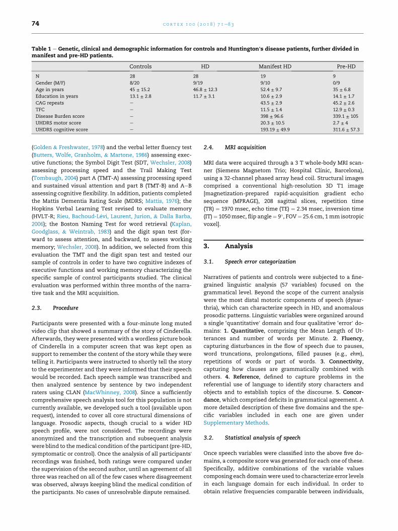
Table 1 e Genetic, clinical and demographic information for controls and Huntington's disease patients, further divided inmanifest and pre-HD patients.
Controls HD Manifest HD Pre-HD
N 28 28 19 9
Gender (M/F) 8/20 9/19 9/10 0/9
Age in years 45 ± 15.2 46.8 ± 12.3 52.4 ± 9.7 35 ± 6.8
Education in years 13.1 ± 2.8 11.7 ± 3.1 10.6 ± 2.9 14.1 ± 1.7
CAG repeats e 43.5 ± 2.9 45.2 ± 2.6
TFC e 11.5 ± 1.4 12.9 ± 0.3
Disease Burden score e 398 ± 96.6 339.1 ± 105
UHDRS motor score e 20.3 ± 10.5 2.7 ± 4
UHDRS cognitive score e 193.19 ± 49.9 311.6 ± 57.3
c o r t e x 1 0 0 ( 2 0 1 8 ) 7 1e8 374
(Golden & Freshwater, 1978) and the verbal letter fluency test
(Butters, Wolfe, Granholm, & Martone, 1986) assessing exec-
utive functions; the Symbol Digit Test (SDT, Wechsler, 2008)
assessing processing speed and the Trail Making Test
(Tombaugh, 2004) part A (TMT-A) assessing processing speed
and sustained visual attention and part B (TMT-B) and AeB
assessing cognitive flexibility. In addition, patients completed
the Mattis Dementia Rating Scale (MDRS; Mattis, 1976); the
Hopkins Verbal Learning Test revised to evaluate memory
(HVLT-R; Rieu, Bachoud-L�evi, Laurent, Jurion, & Dalla Barba,
2006); the Boston Naming Test for word retrieval (Kaplan,
Goodglass, & Weintrab, 1983) and the digit span test (for-
ward to assess attention, and backward, to assess working
memory; Wechsler, 2008). In addition, we selected from this
evaluation the TMT and the digit span test and tested our
sample of controls in order to have two cognitive indexes of
executive functions and working memory characterizing the
specific sample of control participants studied. The clinical
evaluation was performed within three months of the narra-
tive task and the MRI acquisition.
2.3. Procedure
Participants were presented with a four-minute long muted
video clip that showed a summary of the story of Cinderella.
Afterwards, they were presentedwith a wordless picture book
of Cinderella in a computer screen that was kept open as
support to remember the content of the story while they were
telling it. Participants were instructed to shortly tell the story
to the experimenter and they were informed that their speech
would be recorded. Each speech sample was transcribed and
then analyzed sentence by sentence by two independent
raters using CLAN (MacWhinney, 2008). Since a sufficiently
comprehensive speech analysis tool for this population is not
currently available, we developed such a tool (available upon
request), intended to cover all core structural dimensions of
language. Prosodic aspects, though crucial to a wider HD
speech profile, were not considered. The recordings were
anonymized and the transcription and subsequent analysis
were blind to themedical condition of the participant (pre-HD,
symptomatic or control). Once the analysis of all participants'recordings was finished, both ratings were compared under
the supervision of the second author, until an agreement of all
three was reached on all of the few cases where disagreement
was observed, always keeping blind the medical condition of
the participants. No cases of unresolvable dispute remained.
2.4. MRI acquisition
MRI data were acquired through a 3 T whole-body MRI scan-
ner (Siemens Magnetom Trio; Hospital Clınic, Barcelona),
using a 32-channel phased array head coil. Structural images
comprised a conventional high-resolution 3D T1 image
[magnetization-prepared rapid-acquisition gradient echo
sequence (MPRAGE), 208 sagittal slices, repetition time
(TR) ¼ 1970 msec, echo time (TE) ¼ 2.34 msec, inversion time
(IT)¼ 1050msec, flip angle¼ 9�, FOV¼ 25.6 cm, 1mm isotropic
voxel].
3. Analysis
3.1. Speech error categorization
Narratives of patients and controls were subjected to a fine-
grained linguistic analysis (57 variables) focused on the
grammatical level. Beyond the scope of the current analysis
were the most distal motoric components of speech (dysar-
thria), which can characterize speech in HD, and anomalous
prosodic patterns. Linguistic variables were organized around
a single ‘quantitative’ domain and four qualitative ‘error’ do-
mains: 1. Quantitative, comprising the Mean Length of Ut-
terances and number of words per Minute. 2. Fluency,
capturing disturbances in the flow of speech due to pauses,
word truncations, prolongations, filled pauses (e.g., ehm),
repetitions of words or part of words. 3. Connectivity,
capturing how clauses are grammatically combined with
others. 4. Reference, defined to capture problems in the
referential use of language to identify story characters and
objects and to establish topics of the discourse. 5. Concor-
dance, which comprised deficits in grammatical agreement. A
more detailed description of these five domains and the spe-
cific variables included in each one are given under
Supplementary Methods.
3.2. Statistical analysis of speech
Once speech variables were classified into the above five do-
mains, a composite score was generated for each one of these.
Specifically, additive combinations of the variable values
composing each domainwere used to characterize error levels
in each language domain for each individual. In order to
obtain relative frequencies comparable between individuals,

c o r t e x 1 0 0 ( 2 0 1 8 ) 7 1e8 3 75
these composite scores were derived from the variable values
divided by the total number of words in the individuals'speech. Next, to equate the weight of each variable in the
composite score, variable values were divided by their stan-
dard deviation as calculated from the sample of controls.
Composite scores in the three groups (controls, pre-HD and
symptomatic gene-carriers) were compared by means of
ANOVA tests, and in those domains where ANOVAs were
statistically significant (at a p < .01 level), Tukey's Honest
Significant Difference (HSD) post-hoc tests were applied to
know which pairs were different. In three of the domains
(Sentence connectivity, Reference, and Concordance) loga-
rithmic transformations were previously applied to meet the
parametric requirements of the ANOVA test.
Apart from comparing language abnormalities at the
domain level, comparisons were also carried out for each one
of the individual speech variables. Specifically, variables with
less than 50% of null values (zeros) were compared with
KruskaleWallis tests (non-parametric ANOVA), and variables
with more than 50% of zeros were binarized and compared
with Fisher's Exact Tests. A False Discovery Rate (FDR)
correction (Benjamini & Yekutieli, 2001) was applied to the p-
values of all tests to control for the multiple comparisons.
3.3. Correlations of speech domain composite scoreswith clinical tests
In order to study the relationship between each of the speech
domains with different aspects of cognition, a correlation
analysis was carried out between each composite score and
thoseneuropsychological tests inwhich therewere statistically
significant differences between patients and controls, that is,
TMT-A, TMT-B, digit span forward and digit span backward. A
FDR correction was applied to account for multiple compari-
sons in this analysis. Furthermore, a correlation analysis was
also performed between each composite score and two
important clinical scores, the cognitive UHDRS and the motor
UHDRS total scores. In order to have amore specific analysis of
the relation of the linguistic domains with the specific motor
dysfunctions that may affect more directly language produc-
tion, we derived an additional subscore including the items
associated with dysarthria, protrusion and Luria sequencing.
3.4. VBM analysis
Morphometric analysis was carried out using the vbm8
toolbox (http://dbm.neuro.uni-jena.de/vbm/) in the SPM8
software package (Welcome Department of Imaging Neuro-
science Group, London, UK) running on MATLAB (v12.b,
Mathworks, Natick, MA). Specifically, unified segmentation
(Ashburner & Friston, 2005) was applied to the structural T1-
weighted images of each subject to estimate tissue probabil-
ity maps (Gray matter or GMmaps). During this segmentation
step, spatial regularization (regularization: .02, discrete cosine
transform warp frequency cutoff of 22) was adapted to ac-
count for striatal neurodegeneration and ventricle dilatation.
The resulting GM maps were then imported and fed into
DARTEL to achieve spatial normalization into MNI space.
DARTEL normalization alternates between computing an
average template of the GM and white matter (WM)
segmentations from all subjects and warping all subject's GM
and WM segmentations into a better alignment with the
template created (Ashburner, 2009). GM normalized images
were modulated by their Jacobian determinants in order to
identify regional differences in the volume or amount of GM
(Mechelli, Price, Friston, & Ashburner, 2005). These normal-
ized and modulated images were smoothed by using an
isotropic spatial filter (FHWN¼ 8mm) to reduce residual inter-
individual variability. Finally, these images were visually
inspected to ensure good quality in the normalization step.
The individual smoothed GM volume images for both the
HD patients and controls were entered into a second-level
analysis using a two-sample t-test, including intracranial
volume (ICV) as a covariate to create an explicit mask to
differentiate between HD patients and controls. This mask
was used in the following correlation analysis to restrict the
analysis to the areas with significant atrophy in the HD group,
as described in the next subsection. The estimation for the ICV
was taken as the value calculated by FreeSurfer (v. 5.1).
3.5. Correlation analysis
After defining those regions with tissue differences between
HD and the control group, we investigated potential GM in-
dividual differences among the HD group, whichmay relate to
the language deficits observed. A regression analysis within a
linear model was applied individually for each language-
related domains (i.e., Quantitative, Fluency, Concordance,
Connectivity and Reference), including the GM volume images
and the corresponding domain as covariates. ICV was also
included as a nuisance variable in the model in order to cor-
rect for global differences in GM volume (Buckner et al., 2004).
We report two statistical thresholds: p < .05 with FDR
correction for multiple comparisons at cluster level, and an
exploratory threshold of p < .001 uncorrected and taken at a
minimal cluster size of 20 voxels. The maxima of supra-
threshold regions were localized by rendering them onto T1
structural template-image on the MNI reference brain.
4. Results
4.1. Spontaneous speech errors
The ANOVAs for the five composite scores were all significant,
indicating that, at least, one of the groups differed from the
others [Quantitative: F(2,53) ¼ 11.88, p < .001; Fluency:
F(2,53) ¼ 12.61 p < .001; Connectivity: F(2,53) ¼ 15.71, p < .001;
Reference: F(2,53) ¼ 22.09, p < .001; Concordance: F(2,53) ¼8.423, p < .001].
Fig. 1 contains box plots for the five domains, including
results from the Tukey's HSD post hoc tests. As expected from
the ANOVA results, the two most dissimilar groups (controls
and manifest patients) differed significantly in all domains.
The general Quantitative composite score showed no hint of
decline in the pre-HD group but is clearly diminished in the
symptomatic group. In the other remaining domains a gradual
pattern of impairment is observed. For two of the domains
(Connectivity and Reference) significant alterations were
already observed in the pre-HD group. Since the subgroup of
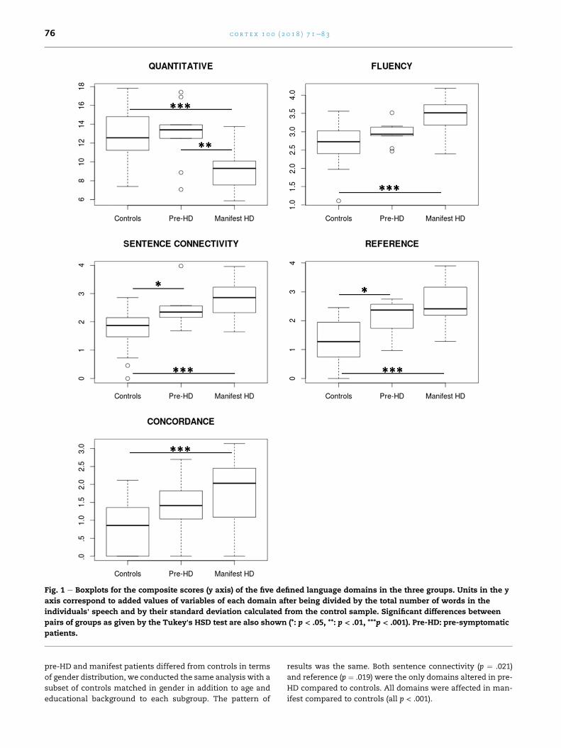
Fig. 1 e Boxplots for the composite scores (y axis) of the five defined language domains in the three groups. Units in the y
axis correspond to added values of variables of each domain after being divided by the total number of words in the
individuals' speech and by their standard deviation calculated from the control sample. Significant differences between
pairs of groups as given by the Tukey's HSD test are also shown (*: p < .05, **: p < .01, ***p < .001). Pre-HD: pre-symptomatic
patients.
c o r t e x 1 0 0 ( 2 0 1 8 ) 7 1e8 376
pre-HD and manifest patients differed from controls in terms
of gender distribution, we conducted the same analysis with a
subset of controls matched in gender in addition to age and
educational background to each subgroup. The pattern of
results was the same. Both sentence connectivity (p ¼ .021)
and reference (p ¼ .019) were the only domains altered in pre-
HD compared to controls. All domains were affected in man-
ifest compared to controls (all p < .001).
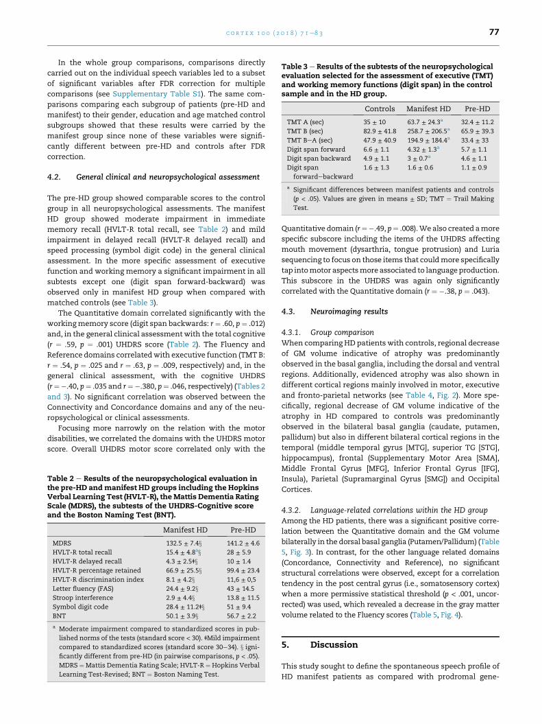
Table 3 e Results of the subtests of the neuropsychologicalevaluation selected for the assessment of executive (TMT)and working memory functions (digit span) in the controlsample and in the HD group.
Controls Manifest HD Pre-HD
TMT A (sec) 35 ± 10 63.7 ± 24.3a 32.4 ± 11.2
TMT B (sec) 82.9 ± 41.8 258.7 ± 206.5a 65.9 ± 39.3
TMT BeA (sec) 47.9 ± 40.9 194.9 ± 184.4a 33.4 ± 33
Digit span forward 6.6 ± 1.1 4.32 ± 1.3a 5.7 ± 1.1
Digit span backward 4.9 ± 1.1 3 ± 0.7a 4.6 ± 1.1
Digit span
forwardebackward
1.6 ± 1.3 1.6 ± 0.6 1.1 ± 0.9
a Significant differences between manifest patients and controls
(p < .05). Values are given in means ± SD; TMT ¼ Trail Making
Test.
c o r t e x 1 0 0 ( 2 0 1 8 ) 7 1e8 3 77
In the whole group comparisons, comparisons directly
carried out on the individual speech variables led to a subset
of significant variables after FDR correction for multiple
comparisons (see Supplementary Table S1). The same com-
parisons comparing each subgroup of patients (pre-HD and
manifest) to their gender, education and age matched control
subgroups showed that these results were carried by the
manifest group since none of these variables were signifi-
cantly different between pre-HD and controls after FDR
correction.
4.2. General clinical and neuropsychological assessment
The pre-HD group showed comparable scores to the control
group in all neuropsychological assessments. The manifest
HD group showed moderate impairment in immediate
memory recall (HVLT-R total recall, see Table 2) and mild
impairment in delayed recall (HVLT-R delayed recall) and
speed processing (symbol digit code) in the general clinical
assessment. In the more specific assessment of executive
function and working memory a significant impairment in all
subtests except one (digit span forward-backward) was
observed only in manifest HD group when compared with
matched controls (see Table 3).
The Quantitative domain correlated significantly with the
workingmemory score (digit span backwards: r¼ .60, p¼ .012)
and, in the general clinical assessmentwith the total cognitive
(r ¼ .59, p ¼ .001) UHDRS score (Table 2). The Fluency and
Reference domains correlatedwith executive function (TMTB:
r ¼ .54, p ¼ .025 and r ¼ .63, p ¼ .009, respectively) and, in the
general clinical assessment, with the cognitive UHDRS
(r¼�.40, p¼ .035 and r¼�.380, p¼ .046, respectively) (Tables 2
and 3). No significant correlation was observed between the
Connectivity and Concordance domains and any of the neu-
ropsychological or clinical assessments.
Focusing more narrowly on the relation with the motor
disabilities, we correlated the domains with the UHDRS motor
score. Overall UHDRS motor score correlated only with the
Table 2 e Results of the neuropsychological evaluation inthe pre-HD andmanifest HD groups including the HopkinsVerbal Learning Test (HVLT-R), theMattis Dementia RatingScale (MDRS), the subtests of the UHDRS-Cognitive scoreand the Boston Naming Test (BNT).
Manifest HD Pre-HD
MDRS 132.5 ± 7.4x 141.2 ± 4.6
HVLT-R total recall 15.4 ± 4.8ax 28 ± 5.9
HVLT-R delayed recall 4.3 ± 2.5ǂx 10 ± 1.4
HVLT-R percentage retained 66.9 ± 25.5x 99.4 ± 23.4
HVLT-R discrimination index 8.1 ± 4.2x 11,6 ± 0,5
Letter fluency (FAS) 24.4 ± 9.2x 43 ± 14.5
Stroop interference 2.9 ± 4.4x 13.8 ± 11.5
Symbol digit code 28.4 ± 11.2ǂx 51 ± 9.4
BNT 50.1 ± 3.9x 56.7 ± 2.2
a Moderate impairment compared to standardized scores in pub-
lished norms of the tests (standard score < 30). ǂMild impairment
compared to standardized scores (standard score 30e34). x igni-
ficantly different from pre-HD (in pairwise comparisons, p < .05).
MDRS ¼Mattis Dementia Rating Scale; HVLT-R ¼ Hopkins Verbal
Learning Test-Revised; BNT ¼ Boston Naming Test.
Quantitative domain (r¼�.49, p¼ .008).We also created amore
specific subscore including the items of the UHDRS affecting
mouth movement (dysarthria, tongue protrusion) and Luria
sequencing to focus on those items that couldmore specifically
tap intomotor aspectsmore associated to languageproduction.
This subscore in the UHDRS was again only significantly
correlated with the Quantitative domain (r ¼ �.38, p ¼ .043).
4.3. Neuroimaging results
4.3.1. Group comparisonWhen comparing HD patients with controls, regional decrease
of GM volume indicative of atrophy was predominantly
observed in the basal ganglia, including the dorsal and ventral
regions. Additionally, evidenced atrophy was also shown in
different cortical regions mainly involved in motor, executive
and fronto-parietal networks (see Table 4, Fig. 2). More spe-
cifically, regional decrease of GM volume indicative of the
atrophy in HD compared to controls was predominantly
observed in the bilateral basal ganglia (caudate, putamen,
pallidum) but also in different bilateral cortical regions in the
temporal (middle temporal gyrus [MTG], superior TG [STG],
hippocampus), frontal (Supplementary Motor Area [SMA],
Middle Frontal Gyrus [MFG], Inferior Frontal Gyrus [IFG],
Insula), Parietal (Supramarginal Gyrus [SMG]) and Occipital
Cortices.
4.3.2. Language-related correlations within the HD groupAmong the HD patients, there was a significant positive corre-
lation between the Quantitative domain and the GM volume
bilaterally in the dorsal basal ganglia (Putamen/Pallidum) (Table
5, Fig. 3). In contrast, for the other language related domains
(Concordance, Connectivity and Reference), no significant
structural correlations were observed, except for a correlation
tendency in the post central gyrus (i.e., somatosensory cortex)
when a more permissive statistical threshold (p < .001, uncor-
rected) was used, which revealed a decrease in the gray matter
volume related to the Fluency scores (Table 5, Fig. 4).
5. Discussion
This study sought to define the spontaneous speech profile of
HD manifest patients as compared with prodromal gene-
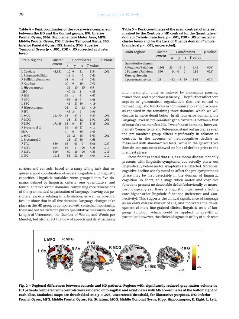
Table 4 e Peak coordinates of the voxel-wise comparisonbetween the HD and the Control groups. IFG: InferiorFrontal Gyrus, SMA: Supplementary Motor Area, MFG:Middle Frontal Gyrus, ITG: Inferior Temporal Gyrus, IPG:Inferior Parietal Gyrus, INS: Insula, STG: SuperiorTemporal Gyrus (p < .001, FDR < .05 corrected at clusterlevel).
Brain regions Clusterextent
Coordinates p-Value
x y z T-value
L Caudate 43,631 �10 0 21 8.54 .001
L Putamen/Pallidum �14 3 �2 7.61
R Pallidum/Putamen 14 4 �5 7.61
R Caudate 10 0 19 7.01
L Hippocampus �31 �10 �15 6.5
LIFG �40 31 1 4.85
R INS 39 1 6 4.67
R STG �54 �19 9 4.48
L STG �46 �27 10 4.19
R Hippocampus 24 �12 �13 4.19
R IFG �45 24 9 3.96
L MOG 24,470 33 �87 6 6.37 .001
R MOG �38 �87 12 5.91 .001
R MFG 17,436 38 0 57 5.85 .001
R Precentral G 39 �19 57 4.11
SMA 1 0 46 3.45
L MFG �39 19 58 5.67 .001
L Precentral G �36 �27 60 4.65
R ITG 659 62 �43 �9 5.06 .037
R MTG 944 56 �1 �20 4.35 .014
R MTG 869 �60 �19 �20 4.35 .016
L IPG 1018 �56 �52 40 4.04 .012
Table 5 e Peak coordinates of the main contrast of interestmasked by the Controls > HD contrast for the Quantitativedomain (*whole brain level p < .001, FDR < .05 corrected atcluster level) and for the Lack of Fluency domain (þwholebrain level p < .001, uncorrected).
Brain regions Clusterextent
Coordinates p-Value
x y z T-value
Quantitative domain
R Putamen/Pallidum 1468 23 �6 1 5.42 .004*
L Putamen/Pallidum 846 �14 0 0 4.92 .022*
Fluency domain
L postcentral gyrus 23 �62 �3 39 3.64 .001þ
c o r t e x 1 0 0 ( 2 0 1 8 ) 7 1e8 378
carriers and controls, based on a story-telling task that re-
quires a good coordination of several cognitive and linguistic
capacities. Linguistic variables were grouped into five do-
mains defined by linguistic criteria, one ‘quantitative’ and
four qualitative ‘error’ domains, comprising core dimensions
of the grammatical organization of language, leaving out pe-
ripheral aspects relating to articulation, as well as prosody.
Results show that in all five domains, language changes take
place in the HD group as comparedwith controls. Importantly,
these are not restricted to purely quantitativemeasures (Mean
Length of Utterances, the Number of Words, and Words per
Minute), but also affect the flow of speech and its structuring
Fig. 2 e Regional differences between controls and HD patients
HD patients compared with controls were rendered onto sagittal
each slice. Statistical maps are thresholded at a p < .005, uncor
Frontal Gyrus, MFG: Middle Frontal Gyrus, Str: Striatum, MOG: M
into meaningful units as indexed by anomalous pausing,
truncations, and repetitions (Fluency). They further affect core
aspects of grammatical organization that are central to
normal linguistic functions in communication and discourse,
as captured in the remaining three domains, which we will
discuss in more detail below. In all four error domains, the
language level in pre-manifest gene-carriers is between that
of controls and manifest HD. Two of these domains, however,
namely Connectivity and Reference, stand out insofar as even
the pre-manifest group differs significantly in relation to
controls, in the absence of neurocognitive decline as
measured with standardized tests, while in the Quantitative
domain our measures showed no hint of decline prior to the
manifest phase.
These findings entail that HD, as a motor disease, not only
presents with linguistic symptoms, but actually starts out
linguistically before motor symptoms are detected. Moreover,
cognitive decline widely noted to affect the pre-symptomatic
phase may be first detectable in the domain of linguistic
cognition. In short, at a stage when motor and cognitive
functions present no detectable deficit behaviorally or neuro-
psychologically yet, there is linguistic impairment affecting
core higher-order linguistic functions (Reference and Con-
nectivity). This suggests the clinical significance of language
as an early disease marker of HD, and motivates the devel-
opment of more fine-grained clinical linguistic tests of lan-
guage function, which could be applied to pre-HD in
particular. However, the clinical diagnostic utility of such tests
. Regions with significantly reduced gray matter volume in
and axial viewswith MNI coordinates at the bottom right of
rected threshold, for illustrative purposes. IFG: Inferior
iddle Occipital Gyrus, Hipp: Hippocampus, R: Right, L: Left.
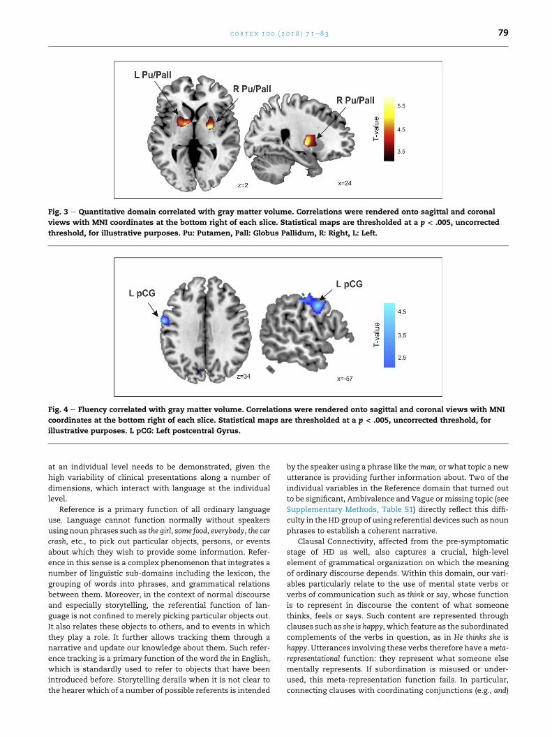
Fig. 3 e Quantitative domain correlated with gray matter volume. Correlations were rendered onto sagittal and coronal
views with MNI coordinates at the bottom right of each slice. Statistical maps are thresholded at a p < .005, uncorrected
threshold, for illustrative purposes. Pu: Putamen, Pall: Globus Pallidum, R: Right, L: Left.
Fig. 4 e Fluency correlated with gray matter volume. Correlations were rendered onto sagittal and coronal views with MNI
coordinates at the bottom right of each slice. Statistical maps are thresholded at a p < .005, uncorrected threshold, for
illustrative purposes. L pCG: Left postcentral Gyrus.
c o r t e x 1 0 0 ( 2 0 1 8 ) 7 1e8 3 79
at an individual level needs to be demonstrated, given the
high variability of clinical presentations along a number of
dimensions, which interact with language at the individual
level.
Reference is a primary function of all ordinary language
use. Language cannot function normally without speakers
using noun phrases such as the girl, some food, everybody, the car
crash, etc., to pick out particular objects, persons, or events
about which they wish to provide some information. Refer-
ence in this sense is a complex phenomenon that integrates a
number of linguistic sub-domains including the lexicon, the
grouping of words into phrases, and grammatical relations
between them. Moreover, in the context of normal discourse
and especially storytelling, the referential function of lan-
guage is not confined to merely picking particular objects out.
It also relates these objects to others, and to events in which
they play a role. It further allows tracking them through a
narrative and update our knowledge about them. Such refer-
ence tracking is a primary function of the word the in English,
which is standardly used to refer to objects that have been
introduced before. Storytelling derails when it is not clear to
the hearer which of a number of possible referents is intended
by the speaker using a phrase like the man, or what topic a new
utterance is providing further information about. Two of the
individual variables in the Reference domain that turned out
to be significant, Ambivalence and Vague ormissing topic (see
Supplementary Methods, Table S1) directly reflect this diffi-
culty in the HD group of using referential devices such as noun
phrases to establish a coherent narrative.
Clausal Connectivity, affected from the pre-symptomatic
stage of HD as well, also captures a crucial, high-level
element of grammatical organization on which the meaning
of ordinary discourse depends. Within this domain, our vari-
ables particularly relate to the use of mental state verbs or
verbs of communication such as think or say, whose function
is to represent in discourse the content of what someone
thinks, feels or says. Such content are represented through
clauses such as she is happy, which feature as the subordinated
complements of the verbs in question, as in He thinks she is
happy. Utterances involving these verbs therefore have ameta-
representational function: they represent what someone else
mentally represents. If subordination is misused or under-
used, this meta-representation function fails. In particular,
connecting clauses with coordinating conjunctions (e.g., and)
c o r t e x 1 0 0 ( 2 0 1 8 ) 7 1e8 380
cannot have this function: The man came and she was unhappy
only states two facts. Individual variables in the domain of
connectivity that were significantly different between pa-
tients and controls (see Supplementary Methods, Table S1)
included use (quantity) of subordinations and both correct
and incorrect coordinations. The HD group used less sub-
ordinations in total with significantly less correct subordina-
tion (i.e., SUB RIGHT, see Table S1). This is in contrast with
coordination, which was significantly higher in both right and
wrong instances of it (i.e., CRD RIGHT, CRDWRONG, see Table
S1). This result strongly suggests that coordination was
replacing subordination in HD patients, which would entail a
loss in meta-representational capacity. A difficulty with
clausal connectivity predicts poor story-telling, since ratio-
nalizing people's actions depends on representing what they
think, say, or desire. This could relate to the noted ‘theory of
mind’ (ToM) deficits in patients with HD (Adenzato & Poletti,
2013; Bora et al., 2016; Brune et al., 2011; Saft et al., 2013).
More direct testing of language-ToM correlations in this pop-
ulation are therefore called for. A link between performance
on sentences with embedded clauses and performance on
false belief tasks has been widely demonstrated, both in pre-
school typically developing children (De Villiers, 2007) and in
older children with Autism Spectrum Disorders (Lind &
Bowler, 2009).
With respect to earlier studies on HD spontaneous speech,
the present study significantly fine-grains results from studies
documenting a reduction in grammatical complexity in HD
speech (Chenery et al., 2002; Gordon & Illes, 1987; Illes, 1989;
Murray & Lenz, 2001; Podoll et al., 1998), by showing that
error patterns can be meaningfully grouped into linguistically
coherent domains made up of linguistically highly specific
variables. Specifically, our findings in the domain of Reference
provide important information with respect to earlier exper-
imental studies manipulating specific aspects of grammatical
complexity. In particular, Sambin et al. (2012) showed, con-
trolling for working memory and executive functioning defi-
cits, that early-stage HD patients face difficulties in
grammatical principles governing the comprehension of the
referential use of noun phrases such as proper names and
pronouns. Here we show that errors in this domain are highly
manifest in spontaneous speech as well, and concern the
referential use of language more generally as based on
grammar. This is clear from linguistic variables included in
the Reference domain, none of which concern lexical-level
errors such as word-finding difficulties, word approxima-
tions, or neologisms. Instead they concern grammatical-level
anomalies, such as the setting (e.g., vagueness or lack) of
topics, the use of determiner phrases, grammatical agreement
between determiner and noun, missing referents for pro-
nouns, or incorrect or inappropriate referents. Therefore,
while error patterns recorded here do not suggest that errors
are due to a defect in any highly specific syntactic principle as
suggested in Sambin et al. (2012), they do clearly indicate that
they concern the role of grammar in reference.
In line with previous VBM studies in HD patients (Kassubek
et al., 2004, 2005) reduced GM was detected in the whole
striatum and in widespread cortical areas including the pari-
etal, temporal and frontal cortices. However, despite the
clearly deviant patterns of spontaneous speech in HD and the
prominent striatal and cortical degeneration observed, neu-
rodegeneration measures showed only significant correla-
tions with the Quantitative domain. Specifically, patients with
poorer speech in terms of length of utterances, number of
words per sentence, etc. had greater neurodegeneration in the
bilateral putamen and pallidum. These striatal structures are
part of the motor loop involving the SMA, premotor and so-
matosensory areas. The deficits observed within this domain
may partially be explained by underlying motor alterations in
this circuit. This is consistent also with the correlations be-
tween the Quantitative and the UHDRSmotor scores and with
the working memory scores that also have a motor compo-
nent for the articulatory rehearsal of phonological informa-
tion to be maintained for the task. The Quantitative score was
also correlated with a subscore derived from the UHDRS items
measuring dysarthria, tongue protrusion and Luria
sequencing indicating that part of the variability in this
domain could only derive fromalteredmouthmovements and
sequencing dysfunction. However, the Quantitative factor
also correlated with the UHDRS cognitive score suggesting
that these errors derive not only from a motor component.
In the domains of Reference and Connectivity, linguistic
impairment occurs pre-symptomatically in the absence of
neurocognitive impairment detectable at least through the
standardized clinical and neuropsychological tests used in
regular clinical assessment of HD. The Fluency and Reference
domains did correlate both with executive function measures
(TMTB)and, in thegeneral clinicalassessment,with thegeneral
cognitiveUHDRS score (r¼�.40, p¼ .035 and r¼�.380, p¼ .046,
respectively) (Tables 2 and 3). These facts, and the absence of
any correlations with clinical or neuropsychological measures
in the domains of Connectivity and Concordance, suggest the
importance of measuring cognitive decline linguistically in HD
and the linguistic specificity of these deficits.
The above VBM results raise the question of why no cor-
relations with specific brain areas of neurodegeneration were
found in any of the other four error domains. One possibility is
that the deficits may be only detectable for a purely quanti-
tative analysis of speech whereas the association between
brain degeneration and decline in the more subtle quality of
speech organization cannot be observed before more
advanced stages of the disease. Indeed, it is noteworthy that,
as mentioned above, it is only in the Quantitative domain that
language levels in pre-HD are essentially at the same level as
in controls. Neuronal dysfunction precedes cell death (Levine,
Cepeda, Hickey, Fleming, & Chesselet, 2004; Tobin & Signer,
2000) and psychiatric, cognitive, and motor symptoms often
appear alongside cellular and synaptic alterations in the
absence of neuronal loss (Vonsattel & DiFiglia, 1998). These
qualitative deficits in speech errors may be more sensitive to
individual differences in brain dysfunction and not sensitive
to brain atrophy measures.
The lack of correlations with neurodegeneration may
also be explainable from the fact that all the five domains
are aspects of language that require the confluence of
multiple linguistic and cognitive mechanisms involved in
ordinary language use and functioning. Therefore a wide-
spread neuronal network may be necessary. For example,
reference is a high-level, integrative linguistic function in
the sense that it comprises multiple systems interacting
c o r t e x 1 0 0 ( 2 0 1 8 ) 7 1e8 3 81
coherently, including different linguistic subcomponents
(lexical organization, phrase structure, agreement, and
grammatical relations) and interfacing working-memory,
and executive functions. It is in line with this that the
Fluency and Reference domains did correlate both with
executive function measures (TMT B) and with the general
cognitive UHDRS scores (Tables 2 and 3). Despite the fact
that distributed networks are likely to be involved, each of
our error domains comprised linguistically highly specific
variables. We avoided linguistically non-specific variables
(e.g., ‘coherence in discourse’). This specificity of the vari-
ables is further attested by the fact that within error do-
mains, several of these variables dissociated from others in
being significant in the individual (non-domain based)
analysis (see Table S1).
Reference is one possible domain where specific brain re-
gions may be expected to be associated with the errors
observed, despite its relation with multiple language levels of
processing and the engagement of different functions. Several
studies have recently identified a critical recruitment of the
parietal lobe (SMG and angular gyrus) in the processing of
reference (Brodbeck & Pylkk€anen, 2017; Egorova, Shtyrov, &
Pulvermuller, 2016; Peeters, Snijders, Hagoort, & €Ozyurek,
2017). Although we did observe decreased GM volume in the
inferior parietal lobe (including SMG), no significant correla-
tion was obtained with neurodegeneration in this brain area
and the score of the reference domain. As noted, this domain
was correlated with executive function, which is related to a
widespread brain network, varying depending on the specific
executive function studied (e.g., dorsolateral prefrontal cor-
tex, anterior cingulate, parietal lobe). It remains to be seen
whether the relation between reference deficits and inferior
parietal lobe function would be visible with functional re-
sponses in this brain area instead of structural measures that
may only be more sensitive with greater progression of the
disease.
6. Conclusions
In sum, our analysis of spontaneous narrative speech
comparing controls and pre- and symptomatic HD patients
reveals that cognitive impairment in HD shows in primary
abnormalities in core domains of linguistic organization and
function. Moreover, it does so prior to motor impairment and
before cognitive decline is detectable at least through the
standardized clinical and neuropsychological tests used here.
This supports the value of using grammatical measures to
track disease progression, which appear to show greater
sensitivity than earlier work focusing on the acoustic pa-
rameters of speech (Vogel et al., 2012).
Funding
This research was supported by the Ministerio de Economıa y
Competitividad (MINECO, Spanish Government), grant
FFI2013-40526P to WH and PSI2011-23624 to RDB, by an ERC-
StG, Grant agreement 313841 TuningLang from the European
Commission to RDB and by the Instituto de Salud Carlos III,
which is an agency of theMINECO, co-funded by the European
Regional Development Fund ‘A way of building Europe’ (CP13/
00225 and PI14/00834, both to EC).
Acknowledgments
We thank the physicians and neuropsychologists Nuria Cab-
allol, Matilde Calopa, Jaime Kulisevsky, Celia Mareca, Saul
Martınez-Horta, Esteban Mu~noz, Jesus P�erez, Nadia Rodri-
guez-Dechich�a, Jesus M. Ruiz, Pilar Santacruz, Susana Subir�a
and Irene Vaquer who provided the clinical evaluations of the
patients. We are grateful to the patients and their families for
their participation.
Supplementary data
Supplementary data related to this article can be found at
http://dx.doi.org/10.1016/j.cortex.2017.07.022.
r e f e r e n c e s
Adenzato, M., & Poletti, M. (2013). Theory of mind abilities inneurodegenerative diseases: An update and a call to introducementalizing tasks in standard neuropsychologicalassessments. Clinical Neuropsychiatry, 10(5), 226e234.
Ashburner, J. (2009). Computational anatomy with the SPMsoftware. Magnetic Resonance Imaging, 27(8), 1163e1174.
Ashburner, J., & Friston, K. J. (2005). Unified segmentation.NeuroImage, 26(3), 839e851. http://dx.doi.org/10.1016/j.neuroimage.2005.02.018.
Banney, R. M., Harper-Hill, K., & Arnott, W. L. (2015). The autismdiagnostic observation schedule and narrative assessment:Evidence for specific narrative impairments in autismspectrum disorders. International Journal of Speech-LanguagePathology, 17(2), 159e171. http://dx.doi.org/10.3109/17549507.2014.977348.
Bedi, G., Carrillo, F., Cecchi, G. A., Slezak, D. F., Sigman, M.,Mota, N. B., et al. (2015). Automated analysis of free speechpredicts psychosis onset in high-risk youths. Npj Schizophrenia,1, 1e7. http://dx.doi.org/10.1038/npjschz.2015.30.
Benjamini, Y., & Yekutieli, D. (2001). The control of the falsediscovery rate in multiple testing under dependency. Annals ofStatistics, 1165e1188.
Bora, E., Velakoulis, D., & Walterfang, M. (2016). Social cognitionin Huntington's disease: A meta-analysis. Behavioural BrainResearch, 297, 131e140.
Bornkessel-Schlesewsky, I., & Schlesewsky, M. (2013). Reconcilingtime, space and function: A new dorsal-ventral stream modelof sentence comprehension. Brain and Language, 125(1), 60e76.http://dx.doi.org/10.1016/j.bandl.2013.01.010.
Brodbeck, C., & Pylkk€anen, L. (2017). Language in context:Characterizing the comprehension of referential expressionswith MEG. NeuroImage, 147, 447e460. http://dx.doi.org/10.1016/j.neuroimage.2016.12.006 (December 2016).
Brune, M., Blank, K., Witthaus, H., & Saft, C. (2011). “Theory ofmind” is impaired in Huntington's disease.Movement Disorders,26(4), 671e678. http://dx.doi.org/10.1002/mds.23494.
Buckner, R. L., Head, D., Parker, J., Fotenos, A. F., Marcus, D.,Morris, J. C., et al. (2004). A unified approach for morphometricand functional data analysis in young, old, and dementedadults using automated atlas-based head size normalization:
c o r t e x 1 0 0 ( 2 0 1 8 ) 7 1e8 382
Reliability and validation against manual measurement oftotal intracranial volume. NeuroImage, 23(2), 724e738.
Butters, N., Wolfe, J., Granholm, E., & Martone, M. (1986). Anassessment of verbal recall, recognition and fluency abilitiesin patients with Huntington's disease. Cortex, 22(1), 11e32.
Caine, E. D., Bamford, K. A., Schiffer, R. B., Shoulson, I., & Levy, S.(1986). A controlled neuropsychological comparison ofHuntington's disease and multiple sclerosis. Archives ofNeurology, 43(3), 249e254.
Caine, E. D., Ebert, M. H., & Weingartner, H. (1977). An outline forthe analysis of dementia. The memory disorder ofHuntington's disease. Neurology, 27(11), 1087e1092.
Chenery, H. J., Copland, D. A., & Murdoch, B. E. (2002). Complexlanguage functions and subcortical mechanisms: Evidencefrom Huntington's disease and patients with non-thalamicsubcortical lesions. International Journal of Language &Communication Disorders, 37(4), 459e474.
De Diego-Balaguer, R., Couette, M., Dolbeau, G., Durr, A.,Youssov, K., & Bachoud-L�evi, A. C. (2008). Striataldegeneration impairs language learning: Evidence fromHuntington's disease. Brain, 131(11), 2870e2881.
De Villiers, J. (2007). The interface of language and theory of mind.Lingua. International Review of General Linguistics. RevueInternationale de Linguistique Generale, 117(11), 1858e1878.http://dx.doi.org/10.1016/j.lingua.2006.11.006.
Eddy, C. M., Sira Mahalingappa, S., & Rickards, H. E. (2012). IsHuntington's disease associated with deficits in theory ofmind? Acta Neurologica Scandinavica, 126, 376e383. http://dx.doi.org/10.1111/j.1600- 0404.2012.01659.
Egorova, N., Shtyrov, Y., & Pulvermuller, F. (2016). Brain basis ofcommunicative actions in language. NeuroImage, 125, 857e867.http://dx.doi.org/10.1016/j.neuroimage.2015.10.055.
Foroud, T., Siemers, E., Kleindorder, D., Bill, D. J., Hodes, M. E.,Norton, J. A., et al. (1995). Cognitive scores in carriers ofHuntington's disease gene compared to noncarriers. Annals ofNeurology, 37(5), 657e664.
Friederici, A. D., & Kotz, S. A. (2003). The brain basis of syntacticprocesses: Functional imaging and lesion studies. NeuroImage,20, S8eS17.
Friederici, A. D., Steinhauer, K., & Frisch, S. (1999). Lexicalintegration: Sequential effects of syntactic and semanticinformation. Memory & Cognition, 27(3), 438e453. http://dx.doi.org/10.3758/BF03211539.
Golden, C. J., & Freshwater, S. M. (1978). Stroop color and word test.Wood Dale, IL: Stoelting.
Gordon, W. P., & Illes, J. (1987). Neurolinguistic characteristics oflanguage production in Huntington's disease: A preliminaryreport. Brain and Language, 31(1), 1e10.
Graybiel, A. M. (1995a). Building action repertoires: Memory andlearning functions of the basal Ganglia. Current Opinion inNeurobiology, 5, 733e741.
Graybiel, A. M. (1995b). The basal Ganglia. Trends in Neurosciences,18, 60e62.
Gusella, J. F., Wexler, N. S., Conneally, P. M., Naylor, S. L.,Anderson, M. A., Tanzi, R. E., et al. (1983). A polymorphic DNAmarker genetically linked to Huntington's disease. Nature,306(5940), 234e238. http://dx.doi.org/10.1038/306234a0.
Hertrich, I., & Ackermann, H. (1994). Acoustic analysis of speechtiming in Huntington' s disease. Brain and Language, 47(2),182e196.
Ho, A. K., Sahakian, B. J., Brown, R. G., Barker, R. A., Hodges, J. R.,An�e, M. N., et al. (2003). Profile of cognitive progression in earlyHuntington's disease. Neurology, 61(12), 1702e1706.
Illes, J. (1989). Neurolinguistic features of spontaneous languageproduction dissociate three forms of neurodegenerativedisease: Alzheimer's, Huntington's, and Parkinson's. Brain andLanguage, 37(4), 628e642.
Jensen, A. M., Chenery, H. J., & Copland, D. A. (2006). Acomparison of picture description abilities in individuals withvascular subcortical lesions and Huntington's disease. Journalof Communication Disorders, 39(1), 62e77. http://dx.doi.org/10.1016/j.jcomdis.2005.07.001.
Kaplan, E., Goodglass, H., & Weintrab, S. (1983). The Boston namingtest. Philadelphia: Lea & Febiger.
Kassubek, J., Juengling, F. D., Kioschies, T., Henkel, K., Karitzky, J.,Kramer, B., et al.Landwehrmeyer, G. B. (2004). Topography ofcerebral atrophy in early Huntington's disease: A voxel basedmorphometric MRI study. Journal of Neurology, Neurosurgery,and Psychiatry, 75(2), 213e220. Retrieved from http://jnnp.bmj.com/cgi/content/long/75/2/213.
Kassubek, J., Unrath, A., Huppertz, H. J., Lul�e, D., Ethofer, T.,Sperfeld, A. D., et al. (2005). Global brain atrophy andcorticospinal tract alterations in ALS, as investigated by voxel-based morphometry of 3-D MRI. Amyotrophic Lateral Sclerosisand Other Motor Neuron Disorders, 6, 213e220.
King, D., Dockrell, J. E., & Stuart, M. (2013). Event narratives in11e14 year olds with autistic spectrum disorder. InternationalJournal of Language & Communication Disorders, 48, 522e533.http://dx.doi.org/10.1111/1460-6984.12025.
Kotz, S. A., & Schwartze, M. (2010). Cortical speech processingunplugged: A timely subcortico-cortical framework. Trends inCognitive Science, 14(9), 392e399.
Kotz, S. A., Schwartze, M., & Schmidt-Kassow, M. (2009). Non-motor basal ganglia functions: A review and proposal for amodel of sensory predictability in auditory languageperception. Cortex, 45(8), 982e990.
Levine, M. S., Cepeda, C., Hickey, M. A., Fleming, S. M., &Chesselet, M.-F. (2004). Genetic mouse models of Huntington'sand Parkinson's diseases: Illuminating but imperfect. Trends inNeurosciences, 27(11), 691e697. http://dx.doi.org/10.1016/j.tins.2004.08.008.
Lieberman, P. (2007). The evolution of human speech; itsAnatomical and neural bases. Current Anthropology, 48, 39e66.
Lind, S. E., & Bowler, D. M. (2009). Language and theory of mind inautism spectrum disorder: The relationship betweencomplement syntax and false belief task performance. Journalof Autism and Developmental Disorders, 39(6), 929e937.
Longworth, C. E., Keenan, S. E., Barker, R. A., Marslen-Wilson, W. D., & Tyler, L. K. (2005). The basal ganglia and rule-governed language use: Evidence from vascular anddegenerative conditions. Brain, 128(3), 584e596.
Ludlow, C. L., Connor, N. P., & Bassich, C. J. (1987). Speech timingin Parkinson's and Huntington's disease. Brain and Language,32, 195e214.
MacWhinney, B. (2008). Enriching CHILDES for morphosyntacticanalysis. In H. Behrens (Ed.), Trends in corpus research: Findingstructure in data. Amsterdam: Benjamins.
Mattis, S. (1976). Mental status examination for organic mentalsyndrome in elderly patients. In L. Bellak, & T. B. Karasu (Eds.),Geriatric psychiatry (pp. 71e121). New York: NY: Grune &Stratton.
Mechelli, A., Price, C. J., Friston, K. J., & Ashburner, J. (2005). Voxel-based morphometry of the human brain: Methods andapplications. Current Medical Imaging Reviews, 1(2), 105e113.
Moro, A., Tettamanti, M., Perani, D., Donati, C., Cappa, S. F., &Fazio, F. (2001). Syntax and the brain: Disentangling grammarby selective anomalies. NeuroImage, 13(1), 110e118.
Murray, L. L., & Lenz, L. P. (2001). Productive syntax abilities inHuntington's and Parkinson's diseases. Brain and Cognition,46(1), 213e219.
Norbury, C. F., & Sparks, A. (2013). Difference of disorder? Culturalissues in understanding neurodevelopmental disorders.Developmental Psychology, 49(1), 45e58. http://dx.doi.org/10.1037/a0027446.
c o r t e x 1 0 0 ( 2 0 1 8 ) 7 1e8 3 83
Papoutsi, M., Labuschagne, I., Tabrizi, S. J., & Stout, J. C. (2014).The cognitive burden in Huntington's disease: Pathology,phenotype, and mechanisms of compensation. MovementDisorders, 29(5), 673e683. http://dx.doi.org/10.1002/mds.25864.
Podoll, K., Caspary, P., Lange, H. W., & Noth, J. (1988). Languagefunctions in Huntington's disease. Brain, 111(6), 1475e1503.
Peeters, D., Snijders, T. M., Hagoort, P., & €Ozyurek, A. (2017).Linking language to the visual world: Neural correlates ofcomprehending verbal reference to objects through pointingand visual cues. Neuropsychologia, 95, 21e29. http://dx.doi.org/10.1016/j.neuropsychologia.2016.12.004.
Rieu, D., Bachoud-L�evi, A. C., Laurent, A., Jurion, E., & DallaBarba, G. (2006). Adaptation francaise du «Hopkins verballearning test». Revue Neurologique, 162(6), 721e728.
Saft, C., Lissek, S., Hoffmann, R., Nicolas, V., Tegenthoff, M.,Juckel, G., et al. (2013). Mentalizing in preclinical Huntington'sdisease: An fMRI study using cartoon picture stories. BrainImaging and Behavior, 7(2), 154e162.
Sambin, S., Teichmann, M., de Diego Balaguer, R., Giavazzi, M.,Sportiche, D., Schlenker, P., et al. (2012). The role of thestriatum in sentence processing: Disentangling syntax fromworking memory in Huntington's disease. Neuropsychologia,50(11), 2625e2635.
Stout, J. C., Glikmann-Johnston, Y., & Andrews, S. C. (2016).Cognitive assessment strategies in Huntington's diseaseresearch. Journal of Neuroscience Methods, 265, 19e24. http://dx.doi.org/10.1016/j.jneumeth.2015.12.007.
Stout, J. C., Paulsen, J. S., Queller, S., Solomon, A. C.,Whitlock, K. B., Campbell, J. C., et al. (2011). Neurocognitivesigns in prodromal Huntington disease. Neuropsychology, 25(1),1e14. http://dx.doi.org/10.1037/a0020937.
Teichmann, M., Dupoux, E., Cesaro, P., & Bachoud-L�evi, A. C.(2008). The role of the striatum in sentence processing:Evidence from a priming study in early stages of Huntington'sdisease. Neuropsychologia, 46(1), 174e185.
Teichmann, M., Dupoux, E., Kouider, S., & Bachoud-L�evi, A. C.(2006). The role of the striatum in processing language rules:
Evidence from word perception in Huntington's disease.Journal of Cognitive Neuroscience, 18(9), 1555e1569.
Teichmann, M., Dupoux, E., Kouider, S., Brugi�eres, P.,Boiss�e, M. F., Baudic, S., et al. (2005). The role of the striatum inrule application: The model of Huntington's disease at earlystage. Brain, 128(5), 1155e1167. http://dx.doi.org/10.1093/brain/awh472.
Teichmann, M., Gaura, V., D�emonet, J. F., Supiot, F., Delliaux, M.,Verny, C., et al. (2008). Language processing within thestriatum: Evidence from a PET correlation study inHuntington's disease. Brain, 131(4), 1046e1056.
Tobin, A. J., & Signer, E. R. (2000). Huntington's disease: Thechallenge for cell biologists. Trends in Cell Biology, 10(12),531e536. http://dx.doi.org/10.1016/S0962-8924(00)01853-5.
Tombaugh, T. N. (2004). Trail making test A and B: Normative datastratified by age and education. Archives of ClinicalNeuropsychology, 19(2), 203e214.
Ullman, M. T., Corkin, S., Coppola, M., Hickok, G., Growdon, J. H.,Koroshetz, W. J., et al. (1997). A neural dissociation withinlanguage: Evidence that the mental dictionary is part ofdeclarative memory, and that grammatical rules areprocessed by the procedural system. Journal of CognitiveNeuroscience, 9, 266e276.
Vogel, A. P., Shirbin, C., Churchyard, A. J., & Stout, J. C. (2012).Speech acoustic markers of early stage and prodromalHuntington's disease: A marker of disease onset?Neuropsychologia, 50(14), 3273e3278. http://dx.doi.org/10.1016/j.neuropsychologia.2012.09.011.
Vonsattel, J. P. G., & DiFiglia, M. (1998). Huntington disease. Journalof Neuropathology & Experimental Neurology, 57(5), 369e384.
Wechsler, D. (2008). Wechsler adult intelligence scale e fourth edition(WAIS-IV). San Antonio, TX: NCS Pearson.
Zinken, J., Blakemore, C., Zinken, K., Butler, L., & Skinner, T. C.(2011). Narrating psychological distress: Associations betweencross-clausal integration and mental health difficulties.Applied Psycholinguistics, 32(2), 263e274. http://dx.doi.org/10.1017/S0142716410000408.