a thin left atrial antral wall around the pulmonary vein ... · 283 junbeom park, et al. plays the...
TRANSCRIPT
282 www.eymj.org
INTRODUCTION
Treatment of atrial fibrillation (AF) has made continuous ad-vancements since the isolation of ectopic foci in pulmonary
veins (PVs) was introduced.1 Eliminating AF triggers in PVs has become a fundamental procedure in patients with AF. How-ever, as AF lasts longer, atrial remodeling progresses, and non-PV atrial substrate plays an important role in arrhythmic foci. Thus, a strategy to modify left atrial (LA) substrate was intro-duced by Nademanee who demonstrated the clinical implica-tions of catheter ablation of complex fractionated atrial elec-trograms (CFAEs).2 Further, it has been reported that catheter ablation of CFAEs with PV isolation (PVI) reduces clinical re-currence and repeat procedures in patients with persistent AF (PeAF).3 We also previously reported an association between CFAEs and LA wall thickness (LAWT).4,5 Meanwhile, however, the mechanism of CFAEs is still controversial and an addition-al line ablation for reducing the critical mass of LA substrate has not shown any additive effects.6 As a consequence, PVI still
A Thin Left Atrial Antral Wall Around the Pulmonary Vein Reflects Structural Remodeling by Atrial Fibrillation and is Associated with Stroke
Junbeom Park1*, Chul Hwan Park2*, Jae-Sun Uhm3, Hui-Nam Pak3, Moon-Hyoung Lee3, and Boyoung Joung3
1Department of Cardiology, School of Medicine, Ewha Womans University, Seoul; 2Department of Radiology, Research Institute of Radiological Science, Yonsei University College of Medicine, Seoul; 3Department of Cardiology, Yonsei University Health System, Seoul, Korea.
Purpose: Circumferential pulmonary (PV) vein isolation (CPVI) is the most important treatment strategy for atrial fibrillation (AF). While understanding left atrial wall thickness around PVs (PVWT) prior to catheter ablation is important, its clinical impli-cations are not known. This study aimed to evaluate PVWT characteristics according to underlying disease and to identify associ-ations between PVWT and reconnections of PV potentials (PVPs) in redo ablation.Materials and Methods: In 28 patients who underwent redo-AF ablation, PVWT and reconnected PVPs were evaluated at 12 sites (1–12 o’clock) around each PV. Clinical characteristics including stroke and CHA2DS2-VASc scores were analyzed according to the PVWT.Results: The PVWT was thicker in males than females (p<0.001) and in those with diabetes (p=0.045) or heart failure (p=0.002) than in those without. Patients with strokes or high CHA2DS2-VASc scores (≥3) had significantly thinner PVWTs than those with-out strokes or low CHA2DS2-VASc scores (p<0.001). In redo-ablation, reconnected PVPs were detected in 60 (53.6%) of 112 PVs, and the PVs were thicker (p<0.001) and had more reconnected PVs (p=0.009) than right PVs. A PVWT of >0.6 mm predicted PV reconnections with a sensitivity of 76.7% and specificity of 52.2% with an area under the curve of 0.695.Conclusion: Thick PVWs were associated with diabetes and heart failure, and also showed significant inverse correlations with stroke and the CHA2DS2-VASc score. Thick PVWs were associated with reconnected PVPs after the CPVI, which were related to AF recurrence.
Key Words: Pulmonary vein, wall thickness, ablation, stroke, reconnection
Yonsei Med J 2017 Mar;58(2):282-289https://doi.org/10.3349/ymj.2017.58.2.282
Original Article
pISSN: 0513-5796 · eISSN: 1976-2437
Received: May 31, 2016 Revised: August 20, 2016Accepted: September 29, 2016Corresponding author: Dr. Boyoung Joung, Department of Cardiology, Yonsei Uni-versity College of Medicine, 50-1 Yonsei-ro, Seodaemun-gu, Seoul 03722, Korea.Tel: 82-2-2228-8460, Fax: 82-2-393-2041, E-mail: [email protected]
*Junbeom Park and Chul Hwan Park contributed equally to this work.•The authors have no financial conflicts of interest.
© Copyright: Yonsei University College of Medicine 2017This is an Open Access article distributed under the terms of the Creative Com-mons Attribution Non-Commercial License (http://creativecommons.org/licenses/by-nc/3.0) which permits unrestricted non-commercial use, distribution, and repro-duction in any medium, provided the original work is properly cited.
283https://doi.org/10.3349/ymj.2017.58.2.282
Junbeom Park, et al.
plays the most important role in preventing AF progression and reconnection of PV potentials (PVPs).
A perfect PVI is affected by anatomical variations in the PVs, exact catheter contact, and whether or not there is an adequate energy delivery to the endocardium. The development of 3-di-mensional cardiac computed tomography (3D-cCT) images made it possible to confirm the LA structure, anatomy, and thickness beforehand. In our previous study, we showed an as-sociation between change in LA substrate and the presence of CFAEs.4,5 Therefore, “tailor-made ablation” according to wall thickness may be possible by confirming LA anatomical varia-tion and antral wall thickness around PVs (PVWT) beforehand, potentially improving catheter ablation outcomes. The pur-pose of this study was to evaluate characteristics of PVWT ac-cording to underlying disease and to identify associations be-tween PVWT and reconnections of PVPs in patients undergoing redo-ablation.
MATERIALS AND METHODS
Patient selectionThe study protocol was approved by the Institutional Review Board of Severance Hospital, Yonsei University Health System. All patients provided written informed consent. The study in-cluded 28 non-valvular paroxysmal AF (PAF) patients (22 males, mean age 53.6±12.4 years) who underwent a redo-ablation pro-cedure due to recurrent AF after radiofrequency catheter abla-tion (RFCA) of drug-refractory AF between January 2009 and December 2012. All patients maintained an optimal anticoag-ulation (target INR 2.0–3.0) before the procedure, and all anti-arrhythmic drugs were discontinued for at least five half-lives of each drug and for at least 4 weeks, especially amiodarone. We examined all patients with transthoracic echocardiography (TTE) and 3D-cCT before the first ablation for AF in order to measure the PVWT and to define LA anatomy before the RFCA.
Cardiac CT and image acquisition4,5 All patients underwent 3D-cCT before RFCA of PAF. All con-trast-enhanced 3D-cCT examinations were performed using a second generation dual-source CT scanner (Somatom Defi-nition Flash, Siemens Healthcare, Forchheim, Germany). A contrast agent (Iopamiro 370; 370 mg iodine/mL, Bracco, Mi-lan, Italy) was injected using a power injector (Envision CT, Me-drad, Warrendale, PA, USA) at a flow rate of 5 mL/s into the right antecubital vein with the triple phase injection method. Follow-ing the administration of 60–80 mL of a contrast agent, 30 mL of a 70:30 saline-to-contrast mixture and 20 mL of pure saline were administered at a flow rate of 5 mL/s through the same venous access. Scanning was performed with the following pa-rameters: prospective electrocardiography (ECG)-gated axial acquisition targeting the end-systolic phase using the absolute
delay method, detector configuration of 2×64×0.6 mm (acqui-sition of 2×128 sections per rotation using a z-axis flying focal spot), gantry rotation time of 0.285 s, tube potential of 80–120 kVp, and tube current-time product of 280–450 mAs, depend-ing on the patient’s body mass index. The scan delay time was determined by the test-bolus technique for optimal contrast enhancement. After injection of a 10-mL bolus of iopamiro, the optimal delay times were determined by automatic detection of contrast enhancement in the ascending aorta. Reconstructed images were transferred to an image server and analyzed using commercially available 3D software (Aquarius iNtuition, Ver 4.4.6, TeraRecon, San Mateo, CA, USA).
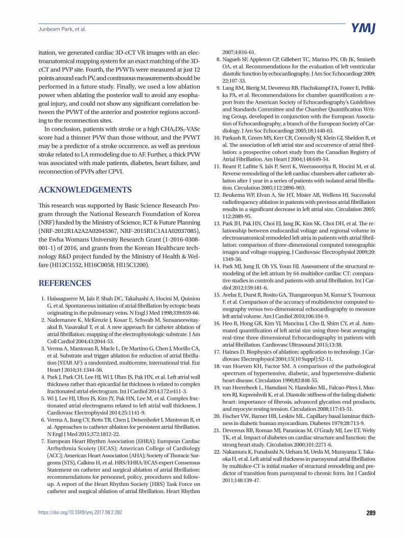
Measurements of the LA antral wall thickness around the PVs (PVWT)A radiologist blinded to the patients’ clinical and electrophysi-ological information evaluated the CT images independently. The intra- and inter-observer correlation thereof was already confirmed in a previous study.4,5 For the measurement of the PVWTs, all CT images were transferred to commercially avail-able 3D reconstruction software (Aquarius iNtuition, Ver 4.4.6, TeraRecon), and then multiplanar (MPR) images were recon-structed. Using axial and coronal images, oblique axial images and oblique coronal images were obtained, which were paral-lel to the target PV passage. Then, a true enface view of the target PV was obtained, which was simultaneously perpen-dicular to the previous oblique axial and oblique coronal im-ages. The lung parenchyma and fat tissue were automatically masked from the PVs using an auto-segmentation technique with a default range from -1024 to -45 Hounsfield unit (HU). On the enface view, the PVWTs were measured carefully at 12 equiangular points (1 o’clock to 12 o’clock sites) using electrical calipers. The PVWT was measured with the Aquarius iNtuition software. First, the oblique axial images (Fig. 1A) and oblique coronal images (Fig. 1B) were obtained, which were parallel to the target PV passage (right superior PV) using axial and coronal images (not shown here). Then, a true enface view of the target PV (Fig. 1C) was obtained, which was simultaneous-ly perpendicular to the previous oblique axial (Fig. 1A) and oblique coronal (Fig. 1B) images. The lung parenchyma and fat tissue were automatically masked from the target PV using an auto-segmentation technique with a default range from -1024 to -45 HU (blue area). On the enface view, the PVWTs were measured carefully at 12 equiangular points (1 o’clock to 12 o’clock sites) using electrical calipers (Fig. 1D). The LA an-trum around the PVs (PVW) was defined as the region consist-ing of a 10-mm space between the PV ostium and left atrium.
Study protocol and assessing the presence of reconnected PV potentials during the redo-ablation All patients underwent circumferential PVI (CPVI) and cavo-tricuspid isthmus block without an additional line or CFAE abla-tion in the first ablation for PAF. An open irrigated tip, 3.5-mm-

https://doi.org/10.3349/ymj.2017.58.2.282284
Pulmonary Vein Wall Thickness and Stroke
tip deflectable catheter (Thermocool, Johnson & Johnson Inc., Diamond Bar, CA, USA; Coolflex, St. Jude Medical Inc., Min-netonka, MN, USA) was used for the RFCA (Stockert genera-tor, Biosense Webster Inc.; Diamond Bar, CA, USA; Irvine Bio-medical, Inc., -1500T11 generator, St. Jude Medical Company, CA, USA). All patients were followed-up in the outpatient clin-ic without the use of any antiarrhythmic drugs after the RFCA. The patients visited an outpatient clinic regularly at 1, 3, 6, and 12 months and then every 6 months or whenever symptoms occurred after the RFCA. All patients underwent an ECG at ev-ery visit and a 24- or 48-hour Holter recording and/or event recording at 3, 6, and every 6 months, following the 2012 HRS/EHRA/ECAS Expert Consensus Statement guidelines. Howev-er, whenever patients reported palpitations, Holter monitor or event monitor recordings were obtained and evaluated for possible recurrences of arrhythmias. We defined a recurrence of AF as any episode of AF or atrial tachycardia lasting at least 30 sec.7 Any ECG documentation of AF recurrences after 3 months was diagnosed as a clinical recurrence,7 and we per-formed a redo-ablation for the patients who had a clinical re-currence of AF. At the time of the redo-ablation procedure, we first evaluated the presence of reconnected PVPs with a vari-able loop circular mapping catheter (LASSO®, Biosense Web-
ster Inc., Diamond Bar, CA, USA) of each PV. The sites of re-connected potentials were marked in a clockwise rotation from 1 to 12 (Figs. 1D and 5), and we generated 3D-cCT VR images with an electroanatomical mapping system (Ensite NavX system, St. Jude Medical Inc., Minneapolis, MN, USA) for exact matching with the 3D-cCT and PVP site.
EchocardiographyAll patients underwent TTE (Sonos 5500, Philips Medical Sys-tem, Andover, MA, USA or Vivid 7, GE Vingmed Ultrasound, Horten, Norway) prior to the RFCA. The chamber size (LA vol-ume index, LA dimension, LV wall thickness, and LV mass in-dex), transmitral flow velocity (E wave, A wave), and tissue Doppler images of the mitral annular septal area (peak diastolic velocity and peak systolic velocity) were acquired according to the American Society of Echocardiography guidelines.8,9 The index was calculated as divided by the body surface area.
Data analysisNormally-distributed continuous variables are expressed as the mean±standard deviation. The statistical significance of com-parisons was assessed using Student’s t-test, χ2 test, and ANO-VA test. A receiver operating characteristic (ROC) curve analy-
Fig. 1. Measurement of the PVWT (left atrial antral wall thickness around the right superior pulmonary vein). The details are described in the Methods section. (A) oblique axial images, (B) oblique coronary images, (C) true enface view of the target pulmonary vein, (D) the PVWTs are measured carefully at 12 equiangular points (1 o’clock to 12 o’clock sites). PVWT, wall thickness around pulmonary veins.
A
B
DC
285https://doi.org/10.3349/ymj.2017.58.2.282
Junbeom Park, et al.
sis was conducted to evaluate the prognostic value of PVWT in relation to reconnections of the PVPs. A p-value<0.05 was considered statistically significant.
RESULTS
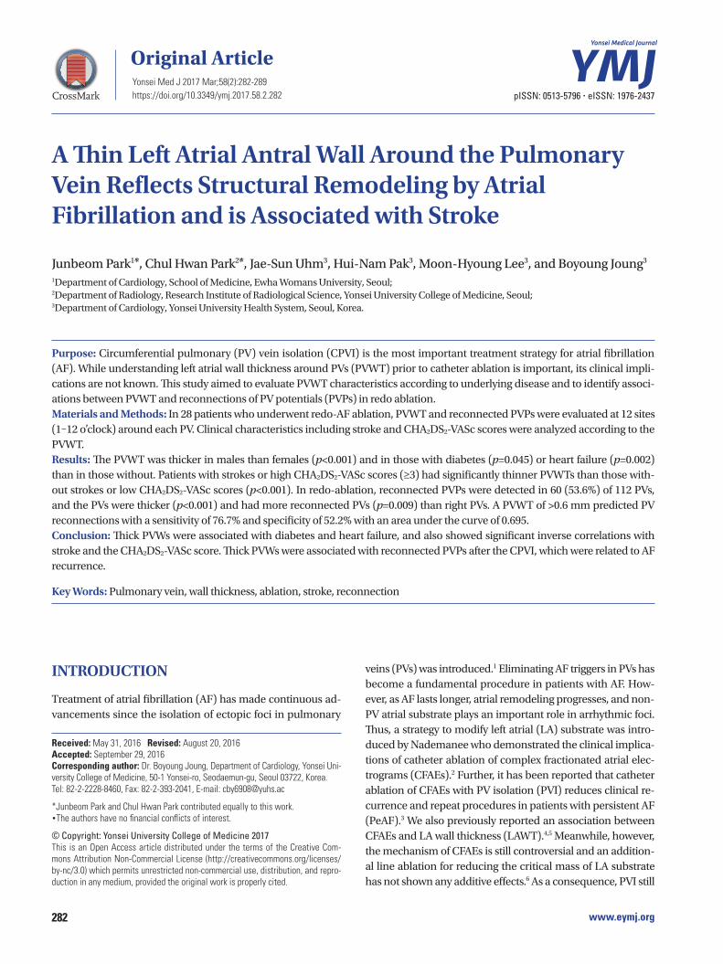
Baseline characteristicsA total of 28 patients (78.6% male, 53.6±12.4 years) were en-rolled. Their mean CHA2DS2-VASc score was 1.4±1.3 with a mean LA dimension of 39.6±6.5 mm. The mean wall thickness of the 112 PVs (4 PVs×28 patients) was 0.64±0.25 mm (range 0.13–2.57 mm). Left PVs were thicker than the right PVs (0.67± 0.24 mm vs. 0.61±0.26 mm, p<0.001) and had more reconnected PVs (57.1% vs. 50.0%, p=0.009). However, there was no signifi-cant difference between the superior and inferior PVs (Table 1).
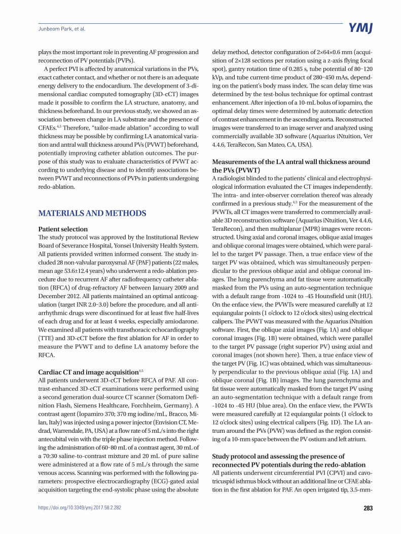
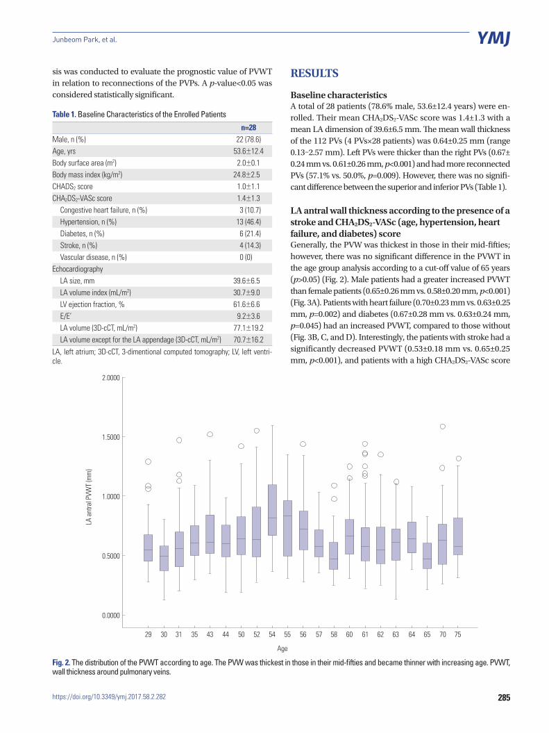
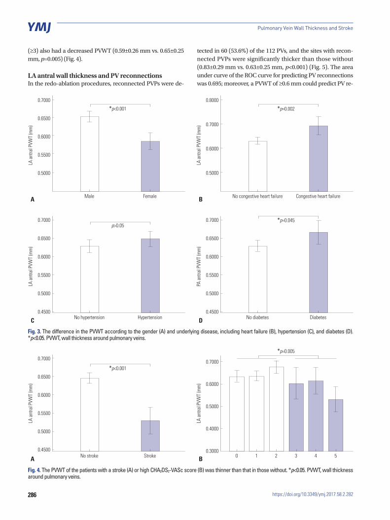
LA antral wall thickness according to the presence of a stroke and CHA2DS2-VASc (age, hypertension, heart failure, and diabetes) scoreGenerally, the PVW was thickest in those in their mid-fifties; however, there was no significant difference in the PVWT in the age group analysis according to a cut-off value of 65 years (p>0.05) (Fig. 2). Male patients had a greater increased PVWT than female patients (0.65±0.26 mm vs. 0.58±0.20 mm, p<0.001) (Fig. 3A). Patients with heart failure (0.70±0.23 mm vs. 0.63±0.25 mm, p=0.002) and diabetes (0.67±0.28 mm vs. 0.63±0.24 mm, p=0.045) had an increased PVWT, compared to those without (Fig. 3B, C, and D). Interestingly, the patients with stroke had a significantly decreased PVWT (0.53±0.18 mm vs. 0.65±0.25 mm, p<0.001), and patients with a high CHA2DS2-VASc score
Table 1. Baseline Characteristics of the Enrolled Patients
n=28Male, n (%) 22 (78.6)Age, yrs 53.6±12.4Body surface area (m2) 2.0±0.1Body mass index (kg/m2) 24.8±2.5CHADS2 score 1.0±1.1CHA2DS2-VASc score 1.4±1.3
Congestive heart failure, n (%) 3 (10.7)Hypertension, n (%) 13 (46.4)Diabetes, n (%) 6 (21.4)Stroke, n (%) 4 (14.3)Vascular disease, n (%) 0 (0)
EchocardiographyLA size, mm 39.6±6.5LA volume index (mL/m2) 30.7±9.0LV ejection fraction, % 61.6±6.6E/E’ 9.2±3.6LA volume (3D-cCT, mL/m2) 77.1±19.2LA volume except for the LA appendage (3D-cCT, mL/m2) 70.7±16.2
LA, left atrium; 3D-cCT, 3-dimentional computed tomography; LV, left ventri-cle.
Fig. 2. The distribution of the PVWT according to age. The PVW was thickest in those in their mid-fifties and became thinner with increasing age. PVWT, wall thickness around pulmonary veins.
2.0000
1.5000
1.0000
0.5000
0.0000
LA a
ntra
l PVW
T (m
m)
29 30 31 35 43 44 50 52 54 55
Age
56 57 58 60 61 62 63 64 65 70 75
https://doi.org/10.3349/ymj.2017.58.2.282286
Pulmonary Vein Wall Thickness and Stroke
(≥3) also had a decreased PVWT (0.59±0.26 mm vs. 0.65±0.25 mm, p=0.005) (Fig. 4).
LA antral wall thickness and PV reconnections In the redo-ablation procedures, reconnected PVPs were de-
tected in 60 (53.6%) of the 112 PVs, and the sites with recon-nected PVPs were significantly thicker than those without (0.83±0.29 mm vs. 0.63±0.25 mm, p<0.001) (Fig. 5). The area under curve of the ROC curve for predicting PV reconnections was 0.695; moreover, a PVWT of ≥0.6 mm could predict PV re-
Fig. 3. The difference in the PVWT according to the gender (A) and underlying disease, including heart failure (B), hypertension (C), and diabetes (D). *p<0.05. PVWT, wall thickness around pulmonary veins.
0.7000
0.6500
0.6000
0.5500
0.5000
LA a
ntra
l PVW
T (m
m)
Male
*p<0.001
FemaleA
0.7000
0.6500
0.6000
0.5500
0.5000
0.4500
LA a
ntra
l PVW
T (m
m)
No hypertension
p>0.05
HypertensionC
0.7000
0.6500
0.6000
0.5500
0.5000
0.4500
PA a
ntra
l PVW
T (m
m)
No diabetes
*p=0.045
DiabetesD
0.8000
0.7000
0.6000
0.5000
LA a
ntra
l PVW
T (m
m)
No congestive heart failure
*p=0.002
Congestive heart failureB
Fig. 4. The PVWT of the patients with a stroke (A) or high CHA2DS2-VASc score (B) was thinner than that in those without. *p<0.05. PVWT, wall thickness around pulmonary veins.
0.7000
0.6500
0.6000
0.5500
0.5000
0.4500
LA a
ntra
l PVW
T (m
m)
No stroke
*p<0.001
StrokeA
0.7000
0.6000
0.5000
0.4000
0.3000
LA a
ntra
l PVW
T (m
m)
B 0 1 2 3 4 5
*p=0.005

287https://doi.org/10.3349/ymj.2017.58.2.282
Junbeom Park, et al.
to LA volume and dimensions.10-12 Measurement of the volume has become more exact with the development of echocardiog-raphy and 3D-cCT imaging, and even a segmental approach for determining the LA volume has recently become possi-ble.13,14 However, many studies on LA volume by 2D echocar-diography have shown consistent underestimation, compared to 3D-cCT, due to ambiguous endocardial borders and geo-metric assumption in the apical view. Because 3D-cCT is tomo-graphic, the chamber orientation and variations of the cardiac size and dimension during the cardiac cycle will not affect the volume assessments.15 This also allows for fewer geometric as-sumptions and has the ability to prescribe any plane across the LA. LA remodeling in patients with AF progression usually oc-curs more in the anteroposterior dimension than LA “elonga-tion”.13 Remodeling of LA dimensions in AF patients might lead to underestimation of LA volume. Although this limitation is inherent to echocardiography, studies have shown that echo-cardiography tends to underestimate LA volume and is strongly correlated with 3D-cCT measurements.16 These results were
connections with a sensitivity of 76.7% and specificity of 52.2% (Fig. 6).
DISCUSSION
In the present study, males exhibited an increased PVWT, com-pared to females, and PVW was thickest in those in their mid-fifties and became thinner with increasing age. Patients with heart failure or diabetes had an increased PVWT, compared to those without. Meanwhile, patients with stroke or a high CHA2DS2-VASc score (≥3) had a lower PVWT than the other pa-tients. PVWT was associated with advanced LA remodeling re-lated to underlying disease in patients with PAF and may have resulted in the occurrence of a stroke.
Previous studies related to the LA wall thickness and data validationMany studies have aimed to predict the outcomes of AF related
Fig. 5. The PVWT and reconnection of the PV potentials (schematic enface view of each PV from 1 to 12 o’clock). The outer circle (blue) indicates the rel-ative PVWT, and inner circle (orange) the relative ratio of the PV reconnections. The numbers in the parentheses denote the number of PV potentials that were detected at each site. RSPV, right superior pulmonary vein; RIPV, right inferior pulmonary vein; LSPV, left superior pulmonary vein; LIPV, left inferior pulmonary vein; PVWT, wall thickness around PVs; PV, pulmonary vein.
RSPV
RS11
RS10 (1)
RS09 (2)
RS08
RS07
RS06 (3)
RS05 (1)
RS04 (1)
RS03 (1)
RS02
RS01 (2)
RS12 (5)
RIPV
RI11 (3)
RI10
RI09 (1)
RI08 (1)
RI07 (1)
RI06 (1)
RI05 (3)
RI04
RI03 (2)
RI02 (1)
RI01 (1)
RI12
LSPV
LS11
LS10
LS09 (2)
LS08 (3)
LS07 (3)
LS06 (1)
LS05 (1)
LS04 (3)
LS03
LS02
LS01
LS12 (3)
LIPV
LI11 (1)
LI10 (2)
LI09 (2)
LI08 (1)
LI07
LI06
LI05 (2)
LI04 (2)
LI03 (1)
LI02 (3)
LI01 (2)
LI12

https://doi.org/10.3349/ymj.2017.58.2.282288
Pulmonary Vein Wall Thickness and Stroke
similar to our findings. In addition, because the LA wall is very thin, its measurement by echocardiography is impossible and the development of the 3D-cCT image resolution was neces-sary for an exact measurement. We already measured the LAWT with 31 segmented sites of the LA and showed an asso-ciation between LAWT and CFAEs.4,5 Further, measurements of the LAWT by two independent observers showed an excellent agreement (inraclass correlation coefficient=0.984, p<0.001).4,5 For the measurement of PVWT in this study, the observers de-termined the points for measurement around the PVs using the three-dimensional volume-rendered images and MPR refor-matted images. If the inner and outer borders of the target wall were determined, the PVWT was measured using electronic calipers with the same method as the previous study.
Clinical implications of the PVWTSince CPVI has been an important milestone of the treatment of AF,1 its clinical importance has increased even in patients with PeAF.6 A complete CPVI and its maintenance is consid-ered to play an important role in the prevention of AF recur-rence regardless of AF type. Thus, it is very important to make a transmural lesion and complete block around all four PVs af-ter evaluating the anatomical variation of each PV. However, most previous studies have focused on geometric variation and exact contact, and there has been no interest regarding the proper amount of energy delivery according to wall thickness: high energy delivery may be needed for a thick wall, in terms of a transmural lesion formation, and a relatively low energy delivery could be adequate for a thin wall, considering the risk of perforation. Generally, the disappearance of PVPs and an increased impedance are considered as endpoints of the abla-
Fig. 6. ROC curve of the PWVT for predicting PV reconnections. ROC, re-ceiver operating characteristic; PVWT, wall thickness around PVs; PV, pulmonary vein.
1.0
0.8
0.6
0.4
0.2
0.00.0 0.2
1-specificity
0.4 0.6 0.8 1.0
Sens
itivit
y
tion at each site.17 With this strategy, the information on the wall thickness enables a tailored ablation and energy delivery at each site. In this study, the reconnection of PVPs was noted more frequently at sites with a thick wall after the same abla-tion strategy, and this finding indicated that tailored ablation according to wall thickness may be necessary. Moreover, with continuous information, not point-by-point information, on the PVWT can be acquired, an exact tailored ablation can be-come possible and could reduce PV reconnections and the risk of ablation.
Another mechanism of strokes: LA remodelingIn this study, males had a thicker wall around PVs than fe-males, although there was no significant difference according to age therein. Further, patients with heart failure or diabetes had a thicker wall around the PVs than those without. Heart failure with a decreased LV function results in a volume over-load and pressure increase in the LA, and this change may cause an increase in the PVWT and LA wall hypertrophy. Fur-ther, pathological changes in the myocardium of patients with diabetes have already been reported, even though it was with-out ventricular systolic decompensation. The reported changes included myocardia fibrosis,18 deposition of periodic acid-Schiff-positive material, advanced glycation endproduct deposition,19 and capillary basement membrane changes.20 As a consequen-ce, those pathological alterations resulted in an increase in the myocardial stiffness, LV mass, and wall thickness.21 Interest-ingly, we found that patients with stroke or a high CHA2DS2-VASc score had a thin wall around the PVs. In previous studies, patients with PAF exhibited a thicker LA wall than those with normal sinus rhythm, although as AF progressed to chronic AF, the LA wall became thin.22 The PVWT reflects LA remodel-ing from AF progression, and as a consequence, a thin PVWT may be associated with the occurrence of a stroke due to a high CHA2DS2-VASc score, as well as a previous stroke. In the begin-ning of AF, risk factors, such as heart failure and diabetes, may increase the wall thickness around the PVs; however, the PVWT will become decreased as the LA remodeling progresses. As a result, a thin PVWT may be associated with the occurrence of a stroke. Therefore, if measurement of LA PVWT is possible, a more exact prediction of a stroke due to AF could potentially be accomplished.
LimitationsThis study had some limitations. First, we focused on recon-nections of the CPVI in patients with PAF and could not ana-lyze the reconnections and bidirectional block of the linear ab-lation in those with PeAF, which are related to wall thickness. Second, this study had a small number of enrolled patients. Third, the reconnection sites of the PVPs during the procedure and the PVWT obtained by 3D-cCT could not be simultane-ously accomplished. Further, measurements around the left PVs with the ridge anteriorly are difficult. To overcome this lim-
289https://doi.org/10.3349/ymj.2017.58.2.282
Junbeom Park, et al.
itation, we generated cardiac 3D-cCT VR images with an elec-troanatomical mapping system for an exact matching of the 3D-cCT and PVP site. Fourth, the PVWTs were measured at just 12 points around each PV, and continuous measurements should be performed in a future study. Finally, we used a low ablation power when ablating the posterior wall to avoid any esopha-geal injury, and could not show any significant correlation be-tween the PVWT of the anterior and posterior regions accord-ing to the reconnection sites.
In conclusion, patients with stroke or a high CHA2DS2-VASc score had a thinner PVW than those without, and the PVWT may be a predictor of a stroke occurrence, as well as previous stroke related to LA remodeling due to AF. Further, a thick PVW was associated with male patients, diabetes, heart failure, and reconnection of PVPs after CPVI.
ACKNOWLEDGEMENTS
This research was supported by Basic Science Research Pro-gram through the National Research Foundation of Korea (NRF) funded by the Ministry of Science, ICT & Future Planning (NRF-2012R1A2A2A02045367, NRF-2015R1C1A1A02037085), the Ewha Womans University Research Grant (1-2016-0308-001-1) of 2016, and grants from the Korean Healthcare tech-nology R&D project funded by the Ministry of Health & Wel-fare (HI12C1552, HI16C0058, HI15C1200).
REFERENCES
1. Haïssaguerre M, Jaïs P, Shah DC, Takahashi A, Hocini M, Quiniou G, et al. Spontaneous initiation of atrial fibrillation by ectopic beats originating in the pulmonary veins. N Engl J Med 1998;339:659-66.
2. Nademanee K, McKenzie J, Kosar E, Schwab M, Sunsaneewitay-akul B, Vasavakul T, et al. A new approach for catheter ablation of atrial fibrillation: mapping of the electrophysiologic substrate. J Am Coll Cardiol 2004;43:2044-53.
3. Verma A, Mantovan R, Macle L, De Martino G, Chen J, Morillo CA, et al. Substrate and trigger ablation for reduction of atrial fibrilla-tion (STAR AF): a randomized, multicentre, international trial. Eur Heart J 2010;31:1344-56.
4. Park J, Park CH, Lee HJ, Wi J, Uhm JS, Pak HN, et al. Left atrial wall thickness rather than epicardial fat thickness is related to complex fractionated atrial electrogram. Int J Cardiol 2014;172:e411-3.
5. Wi J, Lee HJ, Uhm JS, Kim JY, Pak HN, Lee M, et al. Complex frac-tionated atrial electrograms related to left atrial wall thickness. J Cardiovasc Electrophysiol 2014;25:1141-9.
6. Verma A, Jiang CY, Betts TR, Chen J, Deisenhofer I, Mantovan R, et al. Approaches to catheter ablation for persistent atrial fibrillation. N Engl J Med 2015;372:1812-22.
7. European Heart Rhythm Association (EHRA); European Cardiac Arrhythmia Scoiety (ECAS); American College of Cardiology (ACC); American Heart Association (AHA); Society of Thoracic Sur-geons (STS), Calkins H, et al. HRS/EHRA/ECAS expert Consensus Statement on catheter and surgical ablation of atrial fibrillation: recommendations for personnel, policy, procedures and follow-up. A report of the Heart Rhythm Society (HRS) Task Force on catheter and surgical ablation of atrial fibrillation. Heart Rhythm
2007;4:816-61. 8. Nagueh SF, Appleton CP, Gillebert TC, Marino PN, Oh JK, Smiseth
OA, et al. Recommendations for the evaluation of left ventricular diastolic function by echocardiography. J Am Soc Echocardiogr 2009; 22:107-33.
9. Lang RM, Bierig M, Devereux RB, Flachskampf FA, Foster E, Pellik-ka PA, et al. Recommendations for chamber quantification: a re-port from the American Society of Echocardiography’s Guidelines and Standards Committee and the Chamber Quantification Writ-ing Group, developed in conjunction with the European Associa-tion of Echocardiography, a branch of the European Society of Car-diology. J Am Soc Echocardiogr 2005;18:1440-63.
10. Parkash R, Green MS, Kerr CR, Connolly SJ, Klein GJ, Sheldon R, et al. The association of left atrial size and occurrence of atrial fibril-lation: a prospective cohort study from the Canadian Registry of Atrial Fibrillation. Am Heart J 2004;148:649-54.
11. Reant P, Lafitte S, Jaïs P, Serri K, Weerasooriya R, Hocini M, et al. Reverse remodeling of the left cardiac chambers after catheter ab-lation after 1 year in a series of patients with isolated atrial fibrilla-tion. Circulation 2005;112:2896-903.
12. Beukema WP, Elvan A, Sie HT, Misier AR, Wellens HJ. Successful radiofrequency ablation in patients with previous atrial fibrillation results in a significant decrease in left atrial size. Circulation 2005; 112:2089-95.
13. Park JH, Pak HN, Choi EJ, Jang JK, Kim SK, Choi DH, et al. The re-lationship between endocardial voltage and regional volume in electroanatomical remodeled left atria in patients with atrial fibril-lation: comparison of three-dimensional computed tomographic images and voltage mapping. J Cardiovasc Electrophysiol 2009;20: 1349-56.
14. Park MJ, Jung JI, Oh YS, Youn HJ. Assessment of the structural re-modeling of the left atrium by 64-multislice cardiac CT: compara-tive studies in controls and patients with atrial fibrillation. Int J Car-diol 2012;159:181-6.
15. Avelar E, Durst R, Rosito GA, Thangaroopan M, Kumar S, Tournoux F, et al. Comparison of the accuracy of multidetector computed to-mography versus two-dimensional echocardiography to measure left atrial volume. Am J Cardiol 2010;106:104-9.
16. Heo R, Hong GR, Kim YJ, Mancina J, Cho IJ, Shim CY, et al. Auto-mated quantification of left atrial size using three-beat averaging real-time three dimensional Echocardiography in patients with atrial fibrillation. Cardiovasc Ultrasound 2015;13:38.
17. Haines D. Biophysics of ablation: application to technology. J Car-diovasc Electrophysiol 2004;15(10 Suppl):S2-11.
18. van Hoeven KH, Factor SM. A comparison of the pathological spectrum of hypertensive, diabetic, and hypertensive-diabetic heart disease. Circulation 1990;82:848-55.
19. van Heerebeek L, Hamdani N, Handoko ML, Falcao-Pires I, Mus-ters RJ, Kupreishvili K, et al. Diastolic stiffness of the failing diabetic heart: importance of fibrosis, advanced glycation end products, and myocyte resting tension. Circulation 2008;117:43-51.
20. Fischer VW, Barner HB, Leskiw ML. Capillary basal laminar thich-ness in diabetic human myocardium. Diabetes 1979;28:713-9.
21. Devereux RB, Roman MJ, Paranicas M, O’Grady MJ, Lee ET, Welty TK, et al. Impact of diabetes on cardiac structure and function: the strong heart study. Circulation 2000;101:2271-6.
22. Nakamura K, Funabashi N, Uehara M, Ueda M, Murayama T, Taka-oka H, et al. Left atrial wall thickness in paroxysmal atrial fibrillation by multislice-CT is initial marker of structural remodeling and pre-dictor of transition from paroxysmal to chronic form. Int J Cardiol 2011;148:139-47.