a tour of the cell - perry...
TRANSCRIPT

Chapter 6
A Tour of the Cell

Cell Biology or Cytology
• Cyto = cell -
ology = study of
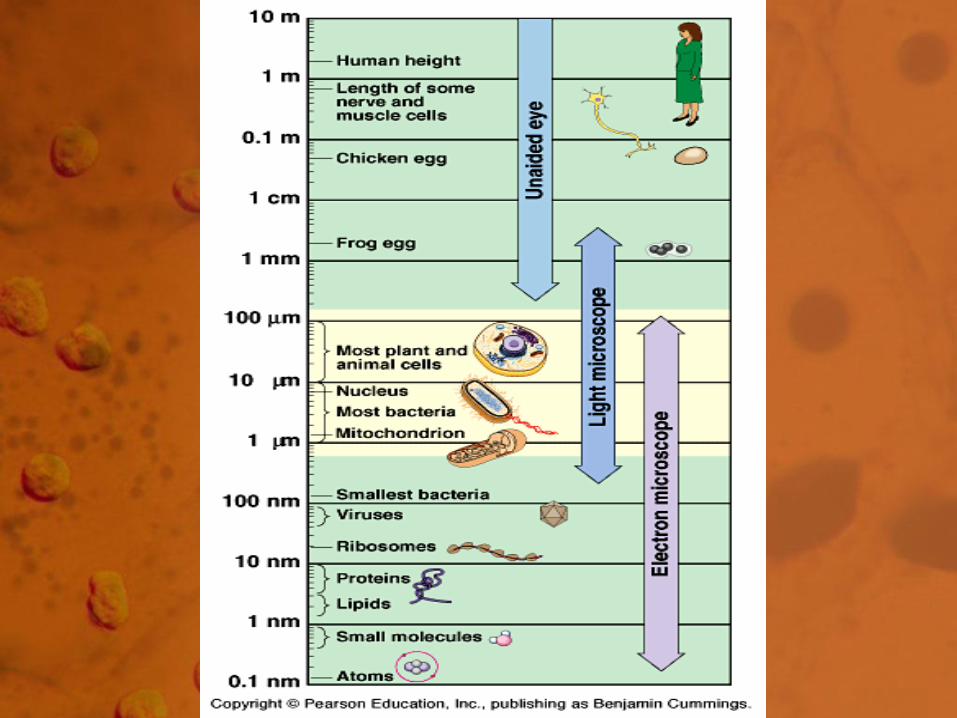
• Should use observations from
several types of microscopes to
make a total picture of how a cell is
put together.

Light Microscope - LM
• Uses visible light to illuminate the
object.
• Relatively inexpensive type of
microscope.
• Can examine live or dead objects.

Light Microscope
Occular Lens
Objective Lens
Stage with specimen
Light Source


Resolution
• Ability to detect two discrete points as separate from each other.
• As Magnification increases, Resolution decreases.
• LM working limits are100 - 1000X.
Limitations - LM
• Miss many cell structures that are
beyond the magnification of the light
microscope.
• Need other ways to make the
observations.

Light Microscope Variations
• Fluorescence: uses dyes to make parts of cells “glow”.
• Phase-contrast: enhances contrasts in density.
• Confocal: uses lasers and special optics to focus only narrow slides of cells.


Electron Microscopes
• Use beams of electrons instead of light.
• Invented in 1939, but not used much
until after WWII.

TEM SEM

Advantages
• Much higher magnifications.
• Magnifications of 50,000X or higher are
possible.
• Can get down to atomic level in some
cases.
Disadvantages
• Need a Vacuum.
• Specimen must stop the electrons.
• High cost of equipment.
• Specimen preparation.

Transmission Electron Microscope
or TEM
• Sends electrons through thinly sliced
and stained specimens.
• Gives high magnification of interior
views. Many cells structures are now
visible.

TEM Limitations
• Specimen dead.
• Specimen preparation uses extreme
chemicals so artifacts are always a
concern.

Scanning Electron Microscope
or SEM
• Excellent views of surfaces.
• Produces 3-D views.
• Live specimens possible.

Other Tools for Cytology
• Cell Fractionation – break the cell apart
and separate out the pieces.
• Chromatography – separates mixtures
based on their solubility.
• Electrophoresis – separates mixtures of
protein or DNA using gels and
electricity.

Cell Fractionation

History of Cells
• Robert Hooke - Observed cells in cork.
• Coined the term "cells” in 1665.
History of Cells
• 1833 - Robert Brown, discovered the nucleus.
• 1838 - M.J. Schleiden, all plants are made of cells.
• 1839 - T. Schwann, all animals are made of cells.

Cell Theory
• All living matter is composed of one or
more cells.
• The cell is the structural and functional
unit of life.
• All cells come from cells.

Types of Cells
• Prokaryotic - lack a nucleus and other
membrane bounded structures.
• Eukaryotic - have a nucleus and other
membrane bounded structures.

Both Have:
• Membrane
• Cytosol
• Ribosomes (but the size is different)
• DNA

Prokaryotic Eukaryotic
Nucleus


Prokaryotic
Eukaryotic

Why Are Cells So Small?
• Cell volume to surface area ratios favor
small size.
• Nucleus to cytoplasm consideration
(control).
• Metabolic requirements.
• Speed of diffusion.


Tuesday, September 22
• The paramecium and euglena are both
single-celled organisms. Identify two
pieces of evidence that make them
eukaryotic as opposed to prokaryotic.
Basic Cell Organization
• Membrane
• Nucleus
• Cytoplasm
• Organelles
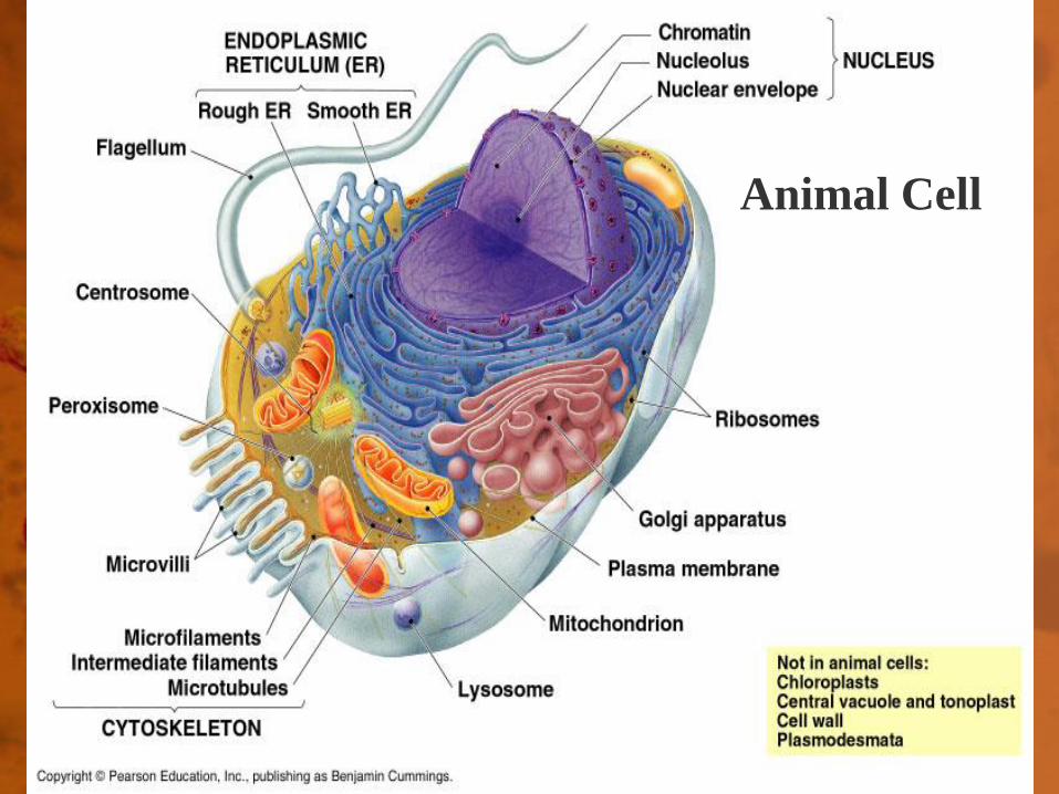
Animal Cell

Plant Cell

Membrane
• Separates the cell from the
environment.
• Boundary layer for regulating the
movement of materials in/out of a cell.

Cytoplasm or Cytosol
• Cell substance between the cell
membrane and the nucleus.
• The “fluid” part of a cell. Exists in two
forms: • gel - thick
• sol - fluid
Organelle
• Term means "small organ”.
Formed body (or compartment) in a cell
with a specialized function.
• Important in organizational structure of
cells.
Organelles - function
• Way to form compartments in cells to
separate chemical reactions.
• Keeps various enzymes separated in
space.

You must be able to:
• Identify the major organelles
• Give their structure
• Give their function
Nucleus
• Most conspicuous organelle.
• Usually spherical, but can be lobed or
irregular in shape.

Structure
• Nuclear membrane
• Nuclear pores
• Nucleolus
• Chromatin


Nuclear Membrane
• Double membrane separated by a 20-
40 nm space.
• Inner membrane supported by a protein
matrix which gives the shape to the
nucleus.

Nuclear Pores
• Regular “holes” through both
membranes.
• 100 nm in diameter.
• Protein complex gives shape.
• Allows materials in/out of nucleus.

Nucleolus
• Dark staining area in the nucleus.
• 0 - 4 per nucleus.
• Storage area for ribosomes.

Chromatin
• Chrom: colored
• - tin: threads
• DNA and Protein in a “loose” format.
Will form the cell’s chromosomes.

Nucleus - Function
• Control center for the cell.
• Contains the genetic instructions.

Ribosomes
• Structure: 2 subunits made of protein
and rRNA. No membrane.
• Function: protein synthesis.


Subunits
• Large: • 45 proteins
• 3 rRNA molecules
• Small: • 23 proteins
• 1 rRNA molecule

Locations
• Free in the cytoplasm - make proteins
for use in cytosol.
• Membrane bound - make proteins that
are exported from the cell.

Endomembrane System
• Membranes that are related through
direct physical continuity or by the
transfer of membrane segments called
vesicles.

Endomembrane System

Endoplasmic Reticulum
• Often referred to as ER.
• Makes up to 1/2 of the total membrane
in cells.
• Often continuous with the nuclear
membrane.


Structure of ER
• Folded sheets or tubes of membranes.
• Very “fluid” in structure with the
membranes constantly changing size
and shape.

Types of ER
• Smooth ER: no ribosomes.
• Used for lipid synthesis,
carbohydrate storage, detoxification
of poisons.
• Rough ER: with ribosomes.
• Makes secretory proteins.

Golgi Apparatus or Dictyosomes
• Structure: parallel array of flattened
cisternae. (looks like a stack of Pita
bread)
• 3 to 20 per cell.
• Likely an outgrowth of the ER system.


Function of Golgi Bodies
• Processing - modification of ER
products.
• Distribution - packaging of ER products
for transport.

Golgi Vesicles
• Small sacs of membranes that bud off
the Golgi Body.
• Transportation vehicle for the modified
ER products.

Lysosome
• Single membrane.
• Made from the Golgi apparatus.

Function
• Breakdown and degradation of cellular
materials.
• Contains enzymes for fats, proteins,
polysaccharides, and nucleic acids.
• Over 40 types known.


Lysosomes
• Important in cell death.
• Missing enzymes may cause various
genetic enzyme diseases.
• Examples: Tay-Sachs, Pompe’s
Disease

Vacuoles
• Structure - single membrane, usually
larger than the Golgi vesicles.
• Function - depends on the organism.
Protists
• Contractile vacuoles - pump out excess
water.
• Food vacuoles - store newly ingested
food until the lysosomes can digest it.


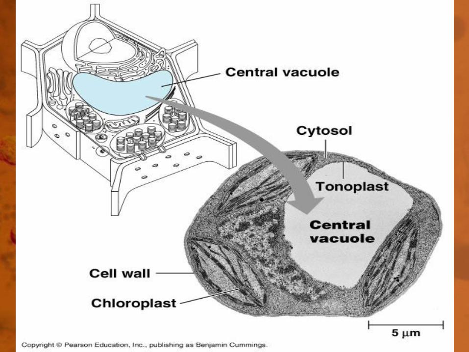
Plants
• Large single vacuole when mature
making up to 90% of the cell's volume.
• Tonoplast - the name for the vacuole
membrane.

Function
• Water regulation.
• Storage of ions.
• Storage of hydrophilic pigments.
(e.g. red and blues in flower petals).
Function: Plant vacuole
• Used to enlarge cells and create turgor
pressure.
• Enzymes (various types).
• Store toxins.
• Coloration.

Microbodies
• Structure: single membrane.
• Often have a granular or crystalline
core of enzymes.

Function
• Specialized enzymes for specific
reactions.
• Peroxisomes: use up hydrogen
peroxide.
• Glyoxysomes: lipid digestion.

Enzymes in a
crystal
Mitochondria
• Structure: 2 membranes. The inner
membrane has more surface area than
the outer membrane.
• Matrix: inner space.
• Intermembrane space: area between
the membranes.
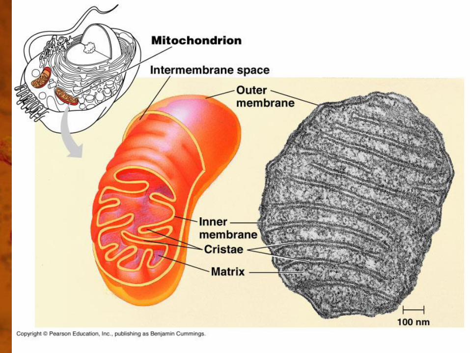

Inner Membrane
• Folded into cristae.
• Amount of folding depends on the level
of cell activity.
• Contains many enzymes.
• ATP generated here.
Function
• Cell Respiration - the release of energy
from food.
• Major location of ATP generation.
• “Powerhouse” of the cell.
Comment – be careful NOT to overuse
this phrase.

Mitochondria
• Have ribosomes (small size).
• Have their own DNA.
• Can reproduce themselves.
• Likely were independent cells at one
time.

Chloroplasts
• Structure - two outer membranes.
• Complex internal membrane.
• Fluid-like stroma is around the internal
membranes.


Inner or Thylakoid Membranes
• Arranged into flattened sacs called
thylakoids.
• Some regions stacked into layers called
grana.
• Contain the green pigment chlorophyll.

Function
• Photosynthesis - the use of light energy
to make food.

Chloroplasts
• Contain ribosomes (small size).
• Contain DNA.
• Can reproduce themselves.
• Often contain starch.
• Likely were independent cells at one
time (cyano-bacteria).

Plastids
• Group of plant organelles.
• Structure - single membrane.
• Function - store various materials.
Examples
• Amyloplasts/ Leucoplasts - store starch.
• Chromoplasts - store hydrophobic plant
pigments such as carotene.
Ergastic Materials
• General term for other substances
produced or stored by plant cells.
• Examples: • Crystals
• Tannins
• Latex
• Resins

Thursday, September 24
• Which of these statements accurately
reflects the relationship between cell size
and surface area? • Larger cells are most efficient at transporting materials across
the membrane since their surface area is increased.
• Smaller cells must have more phospholipids per area in order
to adequately transport materials into the cell.
• Cells must maximize their surface area to volume ratio in
order to maintain homeostasis.
• Cells must minimize their surface area exposure to the
extracellular matrix in order to retain cytosol.

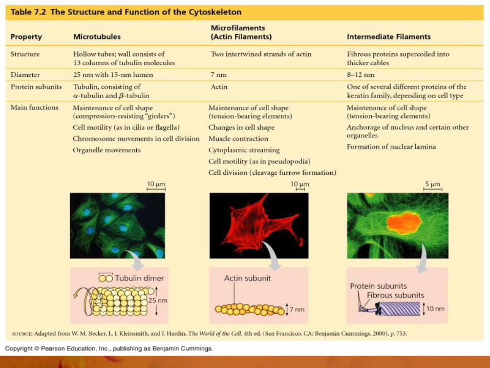
Cytoskeleton
• Network of rods and filaments in the
cytoplasm.


Functions
• Cell structure and shape.
• Cell movement.
• Cell division - helps build cell walls and
move the chromosomes apart.

Cytoskeleton Components
• Microtubules
• Microfilaments
• Intermediate Filaments
Microtubules
• Structure - small hollow tubes made of
repeating units of a protein dimer.
• Size - 25 nm diameter with a 15 nm
lumen. Can be 200 nm to 25 mm in
length.

Tubulin
• Protein in microtubules.
• Dimer - a and b tubulin.
Microtubules
• Regulate cell shape.
• Coordinate direction of cellulose fibers
in cell wall formation.
• Tracks for motor molecules.


Microtubules
• Form cilia and flagella.
• Internal cellular movement.
• Make up centioles, basal bodies and
spindle fibers.

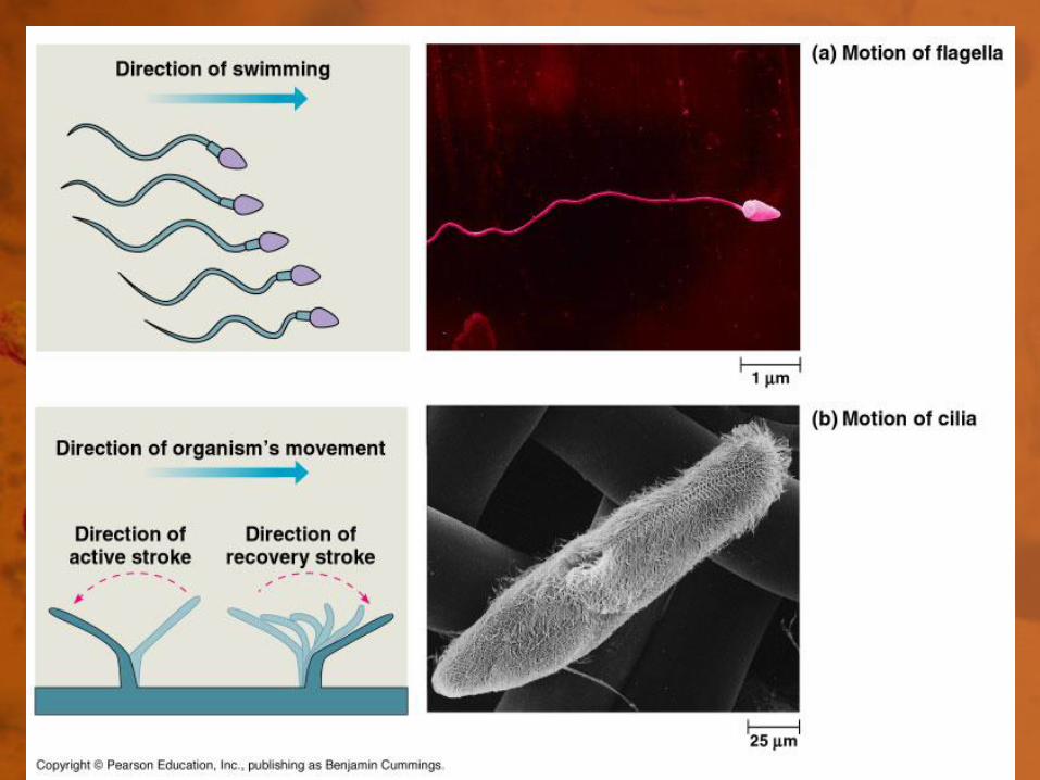
Cilia vs. Flagella
• Cilia - short, but numerous.
• Flagella - long, but few.
• Function - to move cells or to sweep
materials past a cell.

Cilia and Flagella
• Structure - 9+2 arrangement of
microtubules, covered by the cell
membrane.
• Dynein - motor protein that connects
the tubules.


Dynein Protein
• A contractile protein.
• Uses ATP.
• Creates a twisting motion between the
microtubules causing the structure to
bend or move.


Centrioles
• Usually one pair per cell, located close
to the nucleus.
• Found in animal cells.
• 9 sets of triplet microtubules.
• Help in cell division.

Basal Bodies
• Same structure as a centriole.
• Anchor cilia and flagella.
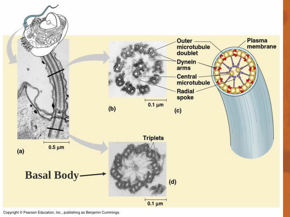
Basal Body

Microfilaments
• 5 to 7 nm in diameter.
• Structure - two intertwined strands of
actin protein.


Microfilaments
are stained green.

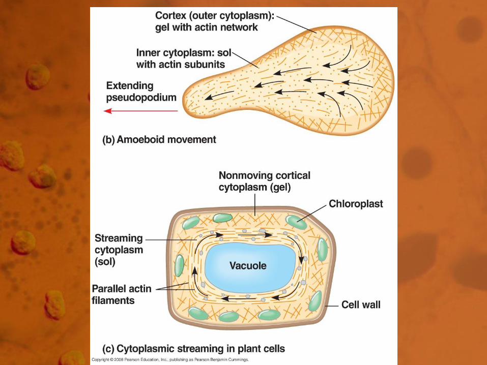
Functions
• Muscle contraction.
• Cytoplasmic streaming.
• Pseudopodia.
• Cleavage furrow formation.
• Maintenance and changes in cell
shape.

Intermediate Filaments
• Fibrous proteins that are super coiled
into thicker cables and filaments
8 - 12 nm in diameter.
• Made from several different types of
protein.

Functions
• Maintenance of cell shape.
• Hold organelles in place.

Cytoskeleton
• Very dynamic; changing in composition
and shape frequently.
• Cell is not just a "bag" of cytoplasm
within a cell membrane.

Cell Wall
• Nonliving jacket that surrounds some
cells.
• Found in: • Plants
• Prokaryotes
• Fungi
• Some Protists

Plant Cell Walls
• All plant cells have a Primary Cell Wall.
• Some cells will develop a Secondary
Cell Wall.


Primary Wall
• Thin and flexible.
• Cellulose fibers placed at right angles to
expansion.
• Placement of fibers guided by
microtubules.

Secondary Wall
• Thick and rigid.
• Added between the cell membrane and the primary cell wall in laminated layers.
• May cover only part of the cell; giving spirals.
• Makes up "wood”.

Middle Lamella
• Thin layer rich in pectin found between
adjacent plant cells.
• Glues cells together.

Cell Walls
• May be made of other types of
polysaccharides and/or silica.
• Function as the cell's exoskeleton for
support and protection.

Extracellular Matrix - ECM
• Fuzzy coat on animal cells.
• Helps glue cells together.
• Made of glycoproteins and collagen.
• Evidence suggests ECM is involved with cell behavior and cell communication.


Intercellular Junctions
• Plants-Plasmodesmata
Plasmodesmata
• Channels between cells through
adjacent cell walls.
• Allows communication between cells.
• Also allows viruses to travel rapidly
between cells.

Intercellular Juctions
• Animals: • Tight junctions
• Desmosomes
• Gap junctions


Tight Junctions
• Very tight fusion of the membranes of
adjacent cells.
• Seals off areas between the cells.
• Prevents movement of materials around
cells.


Desmosomes
• Bundles of filaments which anchor junctions between cells.
• Does not close off the area
between adjacent cells.
• Coordination of movement between
groups of cells.


Gap Junctions
• Open channels between cells, similar to
plasmodesmata.
• Allows “communication” between cells.


Chapter Summary
• Answer: Why is Life cellular and what
are the factors that affect cell size?
• Be able to identify cellular parts, their
structure, and their functions.

Cell Animation Link
• http://multimedia.mcb.harvard.edu/anim
_innerlife_hi.html
• You may need to replay this several
times to catch all of the parts.