a: .. ~uj:pefor. - national oceanic and atmospheric ... nqui ri es about unusual observati ons and...
TRANSCRIPT

A: .. ~UJ:PEFOR. THE RECOGNITION OF SOME DISEASE CONDITIONS AND ABNORMALITIES , " IN MARINE FISH'
MARCH 1978
III

A GUIDE FOR THE RECOGNITION OF SOME DISEASE CONDITIONS
AND ABNORMALITIES IN MARINE FISH
by
Carl J. Sindermann. John J. Ziskowski and Valentine T. Anderson Jr.
Sandy Hook Laboratory Northeast Fisheries Center
National Marine Fisheries Service National Oceanic and Atmospheric Administration
U. S. Department of Commerce Highlands. New Jersey 07732
Technical Series Report No. 14
March. 1978
•

CONTENTS
Page
Introduction ..................................................... 1
Ulcers ........................................................... 4
Fi n eros i on ....................................... ;.............. 7
Tumors ........................................................... 12
Lymphocystis ....................... ... ......... .................. 17
Black spots...................................................... 21
Parasitic copepods .. .................. ........................... 25
Skeletal abnonna1ities ... , ... ........... ............. ............ 29
Reversa 1 in f1 atfi s h ............................................. 36
Abnormal pigmentation in flatfish................................ 41
Key references ................................................... 48
•

I NTRODU eTI ON
Parasites and other disease conditions in marine fishes have been'
recognized and examined sporadically for many decades. The literature
on marine fish parasites and diseases is therefore extensive and highly·
dispersed, although some attempts at summarization have been made.
Realization that world fish production from -natural stocks has
fi nite 1 imits has resulted in expans ion of interest in factors such as
disease that may affect population size. Furthermore, there is great
interest now in the possible effects of increasing coastal/estuarine
pollution on fish stocks. Some of the disease conditions. seen in fish
catches may be associated with environmental degradation by various kinds
of contaminants (summarized in Sindermann, 1977).
Many people -- biologists, sea-going technicians, inspectors, and
processing industry employees -- physically handle great numbers of fish
every day, and may observe some of the 1 arger ectoparasites or gross
disease conditions which occur, but rarely record these. observations.
Thus a large body of potentially valuable data slips away.
Some of tile abnormal ities and disease signs are readily apparent
externally, and recording of observations of these conditions could
provide much more comprehensive knowledge about distribution and pre
valence of fin erosion, ulcers, tumors, skeletal anomalies, and several
other gross abnormalities in fish.
-1-
•

II
This guide has been prepared and distributed so that trawl catches
and other types of landings can be scrutinized for the existence and
abundance of grossly-visible disease conditions. The availability of
identification material may encourage the observation and recording of
such conditions.
The following disease conditions and other abnormalities are
included in this guide:
(1) Ul cers
(2) Fin erosion
(3) Tumors
(4) Lymphocys tis
(5) Black spots
(6) Parasitic copepods
(7) Skeletal anomalies
(8) Reversal in flatfish
(9 ) Abnormal pigmentation
Some representative photographs and drawings are included. Undoubtedly
there will be additional disease conditions that will be recognized and that
should be added to subsequent versions of this field guide. These additions
should be brought to the attention of tile authors as soon as possible. This
document does not consider internal parasites or disease conditions. but they
could logically be added to later revisions.
-2-

Although this guide includes only gross disease signs, some of
the conditions described -- such as tumors and lymphocystis -- should
be confirmed by microscopic examination. Diseased tissue should be
preserved in formalin and sent to appropriate fish disease experts
for a confirmed diagnosis. The authors will attempt to respond to
i nqui ri es about unusual observati ons and to provi de sources of ad
ditional information. For those interested in the scientific
literature on the disease conditions discussed in this document,
relevant references are grouped by disease category at the end of
this report.
-3-
•

I
I. ULCERS
Ulcerations or external sores on fish may have a number of causes.
They may be due to net damage or other mechanical abrasions. to pre
dator attacks. or to infections. Some protozoa (Myxosporida and Micro
sporida) can infect muscle or skin tissue and multiply to produce gross
cysts. These infections, mature to produce many characteristic micro
scopic spores. and in the process the overlying epidermis may be sloughed.
producing ulcers with usually smooth borders (Figure 1).
However. many of the infections that produce grossly visible ulcera
tions are bacterial. and are often due to pathogens of the genera Vibrio.
Pseudomonas or Aeromonas. Vibrio anguillarum has been identified as a
cause of ulcerative lesions in winter flounder. Pseudopleuronectes
ameri can us • and summer flounder. Para 1 i chthys dentatus. from the western
North Atlantic (Levin et al .• 1972; Robohm and Brown. 1977). Ulceration
often begins with scale loss or the formation of small papules, followed
by sloughing of the skin, exposing the underlying muscles. which may also
be destroyed. Bacterial ulcers may have rough or raised irregular margins.
and will often be hemorrhagic (Figure 2).
Recently. ulcers. presumably of bacterial etiology. have been ob
served in several flatfish species, including European flounders.
(Platichthys flesus). dab (Limanda limanda), plaice (Pleuronectes
platessa). and winter flounder (Pseudopleuronectes americanus). Ulcers
may also occur in a number of other fishes. pelagic or demersal. Ulcers
mayor may not be associated with fin erosion.
-4-

Figure 1. Ulcer with smooth margins in sea herring, Clupea harengus, resulting from infection by the Myxosporidan protozoan, Kudoa clupeidae.
...5-

Figure 2. Hemorrhagic ulcers in bluefish (above) and mullet (below), probably a result of Vibrio infection.
-6-

II. FINEROSION
Probably the best known but least understood disease of fish from
polluted waters is a non-specific condition known as "fin rot" or "fin
erosion" -- a syndrome which seems rather clearly associated with de
graded estuarine or coastal environments. The disease has been reported
from the western North Atlantic, the Gulf of Mexico, the eastern and
western Pacific, and the Irish Sea. Twenty-two species of fish from
the New York Bight were reported to be affected to some degree by fin
erosion (Mahoney et al., 1973). In that same area prevalence of the
disease at particular stations reached 35% of individuals in certain
samples, but was usually much lower (Ziskowski and Murche1ano, 1975).
The disease seems to exist in at least two forms: (a) "site
specific" erosion (Figure 3), where the disease is localized in the mid
dorsal and anal fins; and (b) "generalized" erosion where broad areas
of several fins are destroyed (Figures 4a and 4b). Tissues at the base
of eroded areas may become opaque or blackened due to aggregation of pig
ment cells and formation of scar tissue, or they may on rare occasions
be hemorrhagic. Fin rays may be collapsed and resorbed.
Fin erosion in flatfish may be initiated by direct contact of
tissues with contaminated sediments. Toxic substances (sulfides, heavy
metals, chlorinated hydrocarbons, etc.) could remove or modify the
protective mucus coat and expose epithelial tissues to these and pos
sible other chemicals. Fin erosion has been induced experimentally by
-7-
•

•
exposure of normal fish to contaminated sediments, and by exposure to
petroleum hydrocarbons and PCB (Aroclor 1254) (Couch and Nimmo, 1974;
Minchew and Yarbrough, 1977). It should be noted that contact with
contaminated sediments is not necessary in all cases.
It seems quite 1 ikely that the causes of "fin erosion" in fish
includes chemical stress; probably acting on mucus and epithelium;
stress resulting from marginal dissolved oxygen concentrations, pos
sibly enhanced by a sulfide-rich environment (in the case of demersal
fish); and secondary bacterial invasion in at least some instances.
Systemic bacterial infections do not seem to be associated consistently
with fin erosion, although bacterial isolations from superficial fin
tissues may often be made.
In the field, fin erosion is most frequently confused with mechanical
or "net" damage (Figure 5). Net damaged fins, especially those damaged
by trawl nets, are generally characterized by: (a) active hemorrhaging
of fi n ti ssue; (b) many red spots or "petechiae" on the body of the fi sh,
indicating rough handling; (c) exposed fin rays and splitting of fin
membranes; and (d) absence of melanized or darkened tissue at erosion
sites. Other forms of rough handling of catches may also produce super
ficial abrasions that may be confused with fin erosion.
-8-
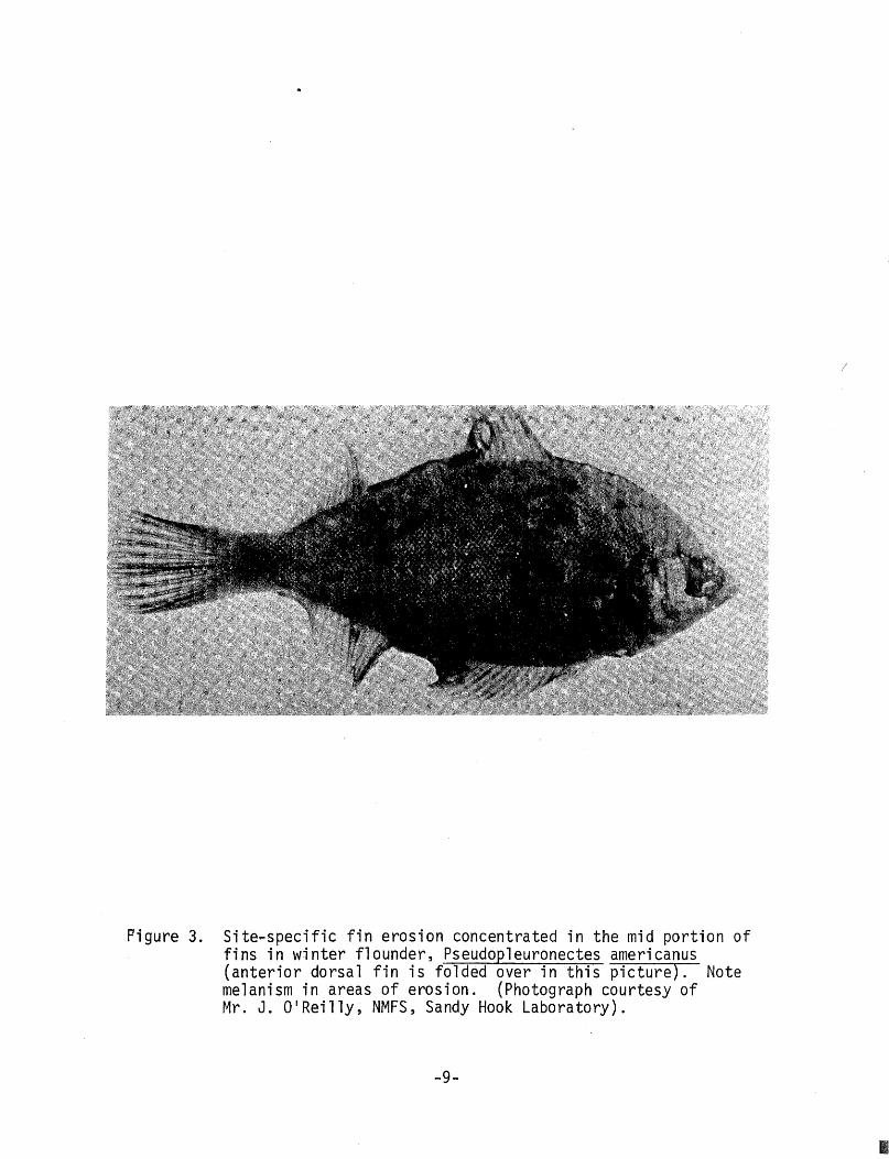
Pigure 3. Site~specific fin erosion concentrated in the mid portion of fins in winter flounder, Pseudopleuronectes americanus (anterior dorsal fin is folded over in this picture). Note melanism in areas of erosion. (Photograph courtesy of ~~r. J. O'Reilly, NMFS, Sandy Hook Laboratory).
-9-
/
III

II
Figure 4. Generalized fin erosion in weakfish, Cynoscion re alis (above) and in four-spot flounder, Paralichthys oblongus below). Note that in the weakfish the anal, caudal, and pelvic fins are eroded, while the dorsal fins are not usually damaged. In contrast the four-spot flounder shows erosion of wide areas of fin fringes.
-10-

FIN EROSION
hemolVr.haging 06 6in w~u~
NET DAMAGE
me1a.lVi.z ed wwe a.t eJto~ion ~.{;t~
/
7 expo~ ed Mn Jtay~
and ~p.e..uung 06 membJtan~
hemoJVr.hagic. ~ pou on body 0 6 6~h
Figure 5. Gross indications of net damage in four-spot flounder, Paralichthys oblongus.
-11-
•

•
III, TUMORS (NEOPLASMS)
Although tumors have been studied principally in humans, the exist
ence of tumors in fish and she11fish.has been recognized for almost a
century (the first oyster tumor, for example, was reported in 1887).
Circumstantial evidence associating environmental contamination with
neoplasms (tumors) in fish and shellfish has accumulated, but a clear
causal relationship has not yet been demonstrated.
Tumors of many kinds have been demonstrated in fish -- in fact
most types of tumors described in humans have their counterparts in fish
(Sch1umberger and Luck~, 1948). Examples of externally obvious fish
tumors are given in Figure 6.
Tumors have been reported in many fishes. Some Pacific flatfish
species are noted for the common occurrence of tumors described as epi
dermal papillomas. These neoplasms begin as small nodules and progress
to the extensive involvement shown in Figure 7. Prevalences are often
highest in young fish. Viral arrays have been seen in affected tissues,
but the etiology of epidermal papillomas is still uncertain -- par
ticularly since an unknown cell type (so-called X-cell) has been seen
consistently. The relationship of this type of tumor with pollution
is inconclusive. Thus far it has not been found in flatfish of the
western North Atlantic.

"Cauliflower disease" (B1umenkoh1krankheit or papillomatosis) of
European eels, Anguilla anguilla, is a remarkable tumorous growth,
found principally in the head region (Figure 8). Prevalences have in
creased in coastal waters and estuaries of northern Europe since World
War II, and a number of authors have suggested that increasing pollution
levels may be responsible. This condition has not been reported from
American eels, Anguilla rostrata.
A number of gadoid fishes have been found to have large tumors within
the gill chamber -- so-called "pseudobranchia1 tumors" since they appear
to arise from the pseudobranchia1 gland. They have been considered to
be neoplasms (adenomas) although they contain large abnormal cells
(X-cells) like those found in flatfish epidermal papillomas (We11ings et
a1.,1977). Pseudobranchia1 tumors have been reported in 7.4% of Pacific
cod (Gadus macrocephalus) from the Bering Sea, and have also been seen in
pollock (Theragra cha1cogramma) from the Pacific (Takahashi, 1929) and
from Atlantic cod (Gadus morhua) (Lange, 1973). The tumors may be
recognized as large convoluted masses in the dorsal pharynx anterior to
the gills (Figure 7).
-13-
•

I'
Figure 6. Wart-like fibrous tumors on mullet (above) and mesenchymal tumor on red snapper (below) from Biscayne Bay, Florida.
-14-

Figure 7. Epidermal papilloma in Dover sole (a) and pseudobranchial tumor in Pacific cod (b). (Photographs courtesy of Ms. M. Sherwood, Southern California Coastal Water Research Project; and Dr. S. Wellings, University of California).
-15-

I
Figure 8. Progressiv~ stages of eel cauliflower disease (Blumenkohl~ krankheit) in European eels, Anguilla anguilla (from Sindermann, 1970).
-16-

IV. LYMPHOCYSTIS
Lymphocystis (sometimes called "fish mange") is caused by a virus
infection which results in enormous enlargement of individual skin cells,
often forming tumor-like nodules on fins and body surfaces (Figure 9).
The disease occurs in many fresh water, estuarine, and marine fishes;
it has been observed in epizootic proportions in certain species, with
prevalences up to 68% of individuals in particular samples (Murche1ano
and Bri dges, 1976).
A number of flatfish n plaice (P1euronectes p1atessa), European
flounder (P1atichthys f1esus), dab (Limanda 1imanda), American plaice
(Hippog10ssoides p1atessoides), summer flounder (Para1ichthys dentatus),
and winter flounder (Pseudop1euronectes americanus) -- are susceptible to
1ymphocystis infections. Epizootic levels of infection have been re
ported in~. p1atessoides, f. p1atessa, f. f1esus, and f. americanus
(Templeman, 1965; Mann, 1970; van Banning, 1971; Perkins et al., 1972;
Shelton and Wilson, 1973).
Additionally, striped bass (Morone saxati1is) which overwinter in
heated effluents of coastal power stations will frequently be infected
with 1ymphocystis (Figure 10). The disease can be common in aquarium
fishes too (Nigrelli and Ruggieri, 1965).
Lymphocystis has been reported recently in Baltic herring (C1upea
harengus var. membras) by Aneer and Ljungberg (1976). Fourteen of 2629
individuals had gross signs of the disease. The authors pointed out
that a number of infections were slight, and might easily have been
overlooked. It is quite likely that this is also the case with other
species.
-17-
•

I
The presence of lymphocystis cells on the viscera of herring was
noted by Aneer and Lj ungberg; there a re several other reports of
systemic lymphocystis infections, particularly that of Dukes and Lawler
(1975) in which lymphocystis cells were found in and behind the eyes
and kidney, spleen, liver, heart, ovaries, and mesenteries of silver
perch (Bardiella chrysura).
Lymphocystis is considered highly infectious but usually non-fatal;
infection may occur at sites of injury or minor abrasions; gross signs
of infection vary seasonally; infectivity seems to have a direct temper
ature relationship; and gross disease signs may disappear from individual
infected fish in a few months' time.
The disease may be readily recognized by a usually gray-colored
tapioca-like enlargement of few or many skin cells; often in severe
infections the enlarged cells become confluent, producing pea-sized
nodules which may sometimes be accompanied by some hemorrhaging. Con
firmation with histological examination is recommended, since the
disease might be confused with early epidermal papillomas.
The "blind" side of flatfish should receive particular attention,
especially the undersides of the pectoral, dorsal, and anal fins.
-18-

Figure 9. Lymphocystis in plaice, Pleuronectes platessa. (Photograph courtesy of Dr. P. van Banning, Rijksinstituut voor Visserijonderzoek).
~19-
I

I
Figure 10. Lymphocystis disease in striped bass from heated effluent of a power plant.
-20-

V. BLACK SPOTS
Fish of many species -- especially those which inhabit estuarine
or nearshore waters for at least part of their lives -- often exhibit
pinhead-size black spots which are the consequence of larval trematode
invasion. The life cycles of the parasites usually involve a mollusk,
then the fish as an intermediate host, and then a predator of the fish
(a larger fish or fish-eating bird or mammal) as the definitive host.
The larval stage (cercaria) which penetrates and encysts in the
integument of the fish elicits the gradual accumulation of host pigment
cells (me1anophores) around the cyst, resulting in a black spot. Size
of the spots and intensity of pigmentation is thus dependent on length
of time after invasion. Numbers of such spots may be few or many
(Figure 11), depending on the availability of infective cercariae in
the immediate habitat of the fish.
One of the best-studied of the trematodes which cause "black spots"
in fish is CrYptocoty1e lingua, which is very common in northern Europe
and North America. Its life cycle involves three hosts in sequence; a
snail (Littorina), coastal fish (especially sea herring, but also in
cluding cod), and the sea gull (Figure,ll). Immature sea herring and
certain other species which inhabit coastal waters near infected Littorina
populations may have literally hundreds of black spots on fins and body
surfaces. Experimentally, heavy larval trematode invasion has been
shown to kill or blind young fish; conspicuous black spots developed in
survivors within a few weeks after experimental exposure.
-21-
•

I
It should be noted that not all larval trematodes which encyst in
the integument of fish will cause black spots. For example. winter
flounder {Pseudop1euronectes americanus) from the western North Atlantic
are often parasitized by larvae of Stephanostomum baccatum. which
matures in the sea raven (Figure 12).· The opaque white unpigmented
metacercarial cysts can be seen easily on the blind side of infected
flounders. frequently in large numbers.
-22-

(I
®.~. I
';
METACERCARIA ENCYST
BENEATH SKIN CAUSING PIGMENT SPOT
ADULT MATURES IN BIRO'S DIGESTIVE TRACT
CERCARIAE DEVELOP IN REDIA AND EMERGE FROM SNAIL
REDIAE DEVELOP IN SNAIL DIGESTIVE GLAND
EGGS SHED IN DROPPINGS AND EATEN BY SNAIL
Figure 11. Immature sea herring (Clupea haren us) with black spots caused by larval trematode invasion above, and the life cycle of the causative organism, the trematode Cryptocotyle lingua (below). From Sindermann and Farrin, 1962.
-23-

I
FLOUNDER SEA RAVEN. MfTACERCARIAE EXCYST AND MATURE IN RECTUM.
WORM EGGS PRESUMABLY EATEN BY SNAILS (BlICcinidl')
LARVAL STAGES IN SNAILS AND FREE-SWIMMING CERCARIAE EMERGE
Figure 12. Life cycle of Stephanostomum baccatum. (Modified from Wolfgang, 1955).
-24-

VI. PARASITIC COPEPODS
Many marine fishes are parasitized by large and often highly
modified copepods. Shapes are often bizarre in the extreme, and the
copepods may cause extensive tissue damage when they anchor or burrow
into the. fish. Some attach to gills, and others to the body surfaces.
Some even attach to the eyes.
Several families (Lernaeoceridae, Sphyridae, and Penellidae) are
particularly injurious to the host. Usually the adult females in these
groups become highly modified and penetrate the flesh, often causing
extensive ulceration (Figure 13), which may persist after the copepod
dies. Usually only a few copepods are found on any single host, and
those attached to body surfaces may be encrusted with algae.
Figure 14 illustrates just a few of the more conspicuous and harm
ful members of this group of parasites. There are many others (espe
cially on the gills) that are less conspicuous and less injurious to
the host, unless they occur in large numbers. There are still others,
often referred to as "fish lice" which can be found loosely attached
to the body surfaces (Figure 13). Many of these smaller copepods are
less-modified than the ones pictured, and many will have a pair of
cylindrical egg cases, which are often opaque white.
-25-
•

I
Figure 13. The parasitic copepods Sphyrion lumpi on redfish, Sebastes marinus (above) and LepeoyhthiriUS salmonis on body surfaces of Atlantic salmon (below. (Photograph courtesy of Dr. E. Egidius, Institute of Marine Research, Bergen).
-26-

Figure 14. Some parasitic copepods: (A) Lernaeocera branchialis from the gill chamber of cod, (B) Sphyrion lumpi from redfish, and (C) Lernaeehicus sprattae from the sardine (from ' Sindermann, 1970).
-27-

I
Note: We feel that the final sections of this guide need a brief intro
duction, because, unlike the disease conditions discussed in previous
sections, the abnormalities described in these final sections are degrees
of morphological divergence from an established form. Therefore, when
dealing with the conditions which follow, it becomes necessary to give
careful consideration to inherent tendencies toward modification of parts.
Also, as a result of differential species or body part vulnerability to
variation, the significance of these conditions may change among popu
lations or indeed within sibling groups.
These final categories can be described loosely as "developmental
abnormalities" .-- morphological features which may depart naturally from
a statistical norm. Although there are many kinds and degrees of such
abnormalities, they will be grouped and discussed generally as (VII.)
skeletal anomalies, (VIII.) reversed laterality in flatfish, and (IX.)
abnormal pigmentation. Each of these will be considered in following
sections.
-28-

VII. SKELETAL ABNORMALITIES
Gross ske1 eta 1 abnonna liti es of fi sh such as pugheadedness, dwarfi sm,
and spinal curvatures (Figure 15), have been observed and recorded for
centuries -: long before significant pollution of the marine environment
existed (Hickey, 1972). Spinal flexures, compressions and fusions have
been observed in many teleosts, as have head and fin abnonna1ities. Early
ichthyologists sometimes mistakenly used these conditions, or accompanying
lesser deformities, to distinguish new species.
Grossly recognizable skeletal anomalies are usually rare in marine
fish populations (Ford, 1937) -- rare enough so that single malformed
individuals have been reported in the scientific literature. Recently,
though, there have been several reports of high prevalences of certain
kinds of gross abnormalities. For example, pugheadedness was found in
more than 10% of young striped bass, Morone saxati1is, and weakfish,
Cynoscion rega1is, collected at two trawl stations in the lower Hudson
River estuary in 1973 (Ziskowski and Anderson, unpub1 ished data).
Increased prevalences of skeletal deformities and anomalies -- con
sidered to be pollution-associated -- have also been recognized in a
few Pacific species from southern California and Japan. Deformed gill
rakers were the most prevalent anomalies observed in southern California
barred sand bass (Para1abrax nebulifer); other abnormalities (pugheaded
ness, cranial asymmetries, deformed vertebrae, and fin anomalies) oc
curred, and were associated directly in frequency and severity with gill
-29-
•

I
raker deformity. Several reports from Japan refer to high and increasing
preva 1 ences of sk.e 1 eta 1 anoma 1 i es in several speci es of fi sh. Malformed
sweetfi sh, Pl ecogl ossus a lti vel is, were observedi n ri vers and culture
farms, and skeletal anomalies were seen in a number of coastal species
(Matsusato, 1973; Komada, 1974; Uekiand Sugiyama, 1976).
In the field, skeletal abnormalities can sometimes be as obvious as
those in Figure 16, but more often they are subtle and not externally
apparent. Therefore, it is usually necessary to collect entire lots
rather than attempting to cull suspect individuals. An exception occurs
among winter flounder· taken from waters adjacent to New York harbor. They
are observed to exhibit what appears to be merely a peculiar condition of
bent or broken fin rays (Figure 17). In severe cases a line of flexure
occurs along whole portions of fins. However, after radiological exami
nation it is found that these fish also display an unusually high inci
dence of skeletal deformities such as vertebral fusions, accessory pro
cesses, or hyperossification. So seemingly minor external evidence of
abnormality can, in some cases, reflect a more extreme internal mal
formation.
Experiments, particularly those concerned with temperature effects
on axial skeletal parts, and field observations, focusing on meristic
characters and their relation to habitat, have shown that skeletal
structure is governed by environmental as well as genetic influences
(Hubbs, 1943, Hubbs and Hubbs, 1945; TlIning, 1952; Bailey and Gosline,
1955; Barlow, 1961; Fowler, 1970; Hickey, 1972; and others). Any
-30.

abnormal fluctuation in these factors, whether genetic or environmental,
can cause morphological variations in the phenotype. The significance
of these developmental abnormalities may vary depending on severity or
prevalence levels. Experjments with the sea trout; Salmo trutta trutta,
have shown that different groups or series of bones and their respective
processes develop, for the most part, independently. A given series is
more vulnerable to environmentally induced change during a specific
"sensitive" period (or periods). (T1Ining, 1952; Barlow, 1961; Fowler,
1970). Further, the sensitive periods of different skeletal groups do
not coincide, and certain skeletal structures are genetically more
flexible than others (Hubbs and Hubbs, 1945; Tanin9, 1952; Fowler, 1970;
and others). Consequently, the interrelation of environmental circum
stances during development and structural susceptibility (as demonstrated
by prevalence and severity) is difficult to interpret. Well planned
sampling and detailed study are necessary before any real ecological
correlations can be made.
•

I
Figure 15. Gross skeletal anomalies: (A) pugheadedness in striped bass, (B) Scoliosis in sea herring, (C) dwarfism in haddock, and (D) abnormal fins in skate. (From Sindermann, 1970).
=32=

Figure 16. X-ray of normal (above) and IIdwarf li (below) summer flounder, Paralichthys gentatus, showing fused vertebrae.
-33-
I

Figure 17. Bent fin rays in winter flounder from the New York Bight. Ex te rn a 1 a p pea ra nee ( a bo v e) and r a d i 0 g rap h ( below) .
-34-

Figure 18. Fusion of last abdominal and a complex first caudal centrum in a winter flounder with bent fin rays (above); Variation within the control sample of winter flounder (accessory haemal and neural arches on caudal vertebrae) (below).
-35-

I
VIII. REVERSAL IN FLATFISH
Reversed laterality.· in which individuals of species which are
nonnally "left-handed" appear as "right-handed", or vice versa •. has
been reported in all four families of flatfishes (Nonnan, 1934; Gudger,
1935; Dawson, 1964, 1966, 1971; Dawson and Heal, 1976.) and may occur
among all species. Reversal is, however, more common in the families
Bothidae and Pleuronectidae than in the families Soleidae and Cynoglos·
sidae (Figure 19).
Reversal is considered to be primarily a genetic aberration .- a
return toward bilateral symmetry,which may have its origin in develop
ment of the central nervous system (Hubbs, in Gudger, 1935). However,
the condition has been associated with environmental stress. High inci
dence of reversal (over 50%) has been reported in populations bordering
on geographic range limits (Nonnan, 1934; Hubbs and Hubbs, 1945).
Sinestral Pleuronectidae found in rearing experiments were attributed to
the culture environment (Houde, 1971). These studies suggest that ad
verse envi ronmenta·l conditions may cause geneti c damage or may affect
morphological development during some early stages of development. How
ever, no real estimates were given in the rearing study as to how many
eggs were actually used, so the ratio -- thus the significance -- of
abnonnal to normal fish may be lower than indicated.
In the field, reversal is commonly found by commercial filleters,
field technicians and sport fishermen. When handling the fish, these
people make a series of precise actions, usually detennined by which side
the eyes are on for a group of individuals of the same species. When a
-36-

reversed individual is encountered, the normal procedure is altered and
usually stopped. The desired body part which is to be counted, measured,
or used as a reference guide for a proper cut, is suddenly on the opposite
side. Figure 21 (below) is a good example. Although not reversed, the
ambico10rate blind side was mistaken for the eyed right-side. After the
first cut behind the head rather than above and behind it, filleting was
stopped with the realization that the fish was, in this case, upside
down. In another instance, while doing routine gill raker counts on
summer flounders, normal handling procedure was halted when an individual
fish was found in which, after the technician reached for its opercular
flap with a pair of forceps, it was immediately apparent that the fish
was upside-down and in fact reversed.
Reversal may be accompanied by a number of other abnormalities:
(1) ambico10ration (Norman, 1934; Hubbs and Hubbs, 1945;
Houde, 1971; Dawson, 1962; Gudger, 1935, 1936);
(2) incomplete eye migration and abnormal dorsal fin development
characterized by an anterior fleshy appendage or "hook"
(Gudger, 1935; Deubler and Fahy, 1958); and Figure 21a;
(3) high frequencies of skeletal abnormalities (Houde, 1971;
and Anderson, unpub1 isheddata);
(4) dwarfism (Anderson, unpublished data);
(5) reversed orientation of scales (Norman, 1934; Hubbs and
Hubbs, 1945);
(6) albinism (Norman, 1934);
-37-
•

•
(7) addition or loss of paired fins (Norman, 1934; Hubbs and
Hubbs, 1945);
(8) overdeveloped musculature on the blind side (Anderson,
unpublished data). The ambicolored specimen in Figure 21b
exhibits this condition.
All of these conditions should be looked for when handling flatfish
and may be helpful in recognizing reversed individuals.
The relative position of the optic nerves (Figure 20) can be used
to determine if a specimen is indeed a reversed bothid or pleuronectid
(Regan, 1910; Kyle, 1923). For example, in the family Bothidae, the
optic nerve of the right eye is dorsal to that of the left regardless of
which eye migrates. For normal bothids (left-eyed), this provides for
a simple or direct upward movement of the dorsal nerve during meta
morphosis, as the right eye revolves over to the left side of the head.
In reversed forms, the dorsal nerve remains stationary, for it is the
left eye which migrates. Thus the ventral nerve of the now migrating
(left) eye must bend around the dorsal nerve of the now stationary
(right) eye when moving to the other side of the head. For the family
Pleuronectidae (right-eyed), the situation is reversed because the nerve
of the left eye is always dorsal to that of the right (Figure 20)
(Anderson, in press).
More investigation is needed to understand reversal as a morpholo
gical phenomenon which can be correlated with external influences.
Sampl ing methods, study procedures, and experimental methods must take
into account differential species or racial prevalences.
-38-

Figure 19. Reversed summer flounder (normal above, reversed below). (Photographs courtesy of Mr. S. Wilk and Mr. M. Silverman, NMFS, Sandy Hook Laboratory).
-39-
I

I
Figure 20. Dissection of summer flounder, showing normal position of the optic nerves (upper photograph, right side) and crossed optic nerves of reversed fish (upper photograph, left side) with enlarged view of crossed nerves (lower photograph). Photographs courtesy.of Mr. M. Silverman, NMFS, Sandy Hook Laboratory).
-40~

IX. ABNORMAL PIGMENTATION IN FLATFISH
Occasionally, flatfish are seen with patches of dark pigmentation
on the normally light-colored blind side (ambicoloration -- a term
coined by Cunningham and McMunn, 1893) -- or with unpigmented patches
on the normally dark-colored eyed side (partial albinism).
Ambicoloration of flatfish is probably the most common anomaly among
members of the Order. Pigmentation on the normally white undersjde of
the fish, of several types and degrees of severity, has been recorded for
many species. These anomalies have been reported in the scientific
literature for two centuries, and it seems that the causation is not
simple -- involving innate as well as environmental factors (deVeen,
1969; Stickney and White, 1975). Norman (1934) found this condition to
have much the same origin(s) as reversal and earlier authors suggested
that ambicoloration merely represents variation in the direction of the
original bilateral symmetry of ancestral forms. Later experimental
studies have shown that susceptibility to environmental changes causes
many of the ambicolor effects, demonstrating that the phenomenon is not
primarily a genetic aberration (deVeen, 1969; Stickney and White, 1975).
In the rearing experiments of Stickney and White, ambicoloration was pro
duced, stopped, and retarded by use of different substrates during early
development. Artificially reared flatfish commonly exhibited abnormal
pigmentation; correlations between pigment anomalies and small size at
metamorphosis existed; and a relationship between pigment anomalies and
abnormal jaw, opercular, and eye development was observed.
-41-
•

I
It should be mentioned here that the head is the portion most
infrequently susceptible to the condition. It is also the most
asynmetrical part of the body. In the majority of cases where
ambicoloratipn is extensive and almost total, the underside of the
head remains white. Also, pigmentation most often occurs on the
portion of the body-which comes in contact with the substrate less
frequently. It has further been observed (Norman, 1934; Gudger,
1945, 1936; Deubler and Fahy, 1958) that individuals in which the
coloration of the blind side extends past the operculum onto the
head, usually have incomplete eye rotation (Figure 21a), although
the specimen in Figure 21b is an exception to that rule.
Different degrees of ambicoloration or albinism exist. Cunning
ham and McMunn (1893), and deVeen (1969) have shown that the con
dition in its varying stages may progress in individual fish.
Also, the prevalence may increase among some groups of fish as the
individuals get older, indicating that the condition may progress
throughout life. This again may be a result of the genetic sus
ceptibility of a given population, some species or races being more
susceptible to environmental change than others. Population dif
ferences in occurrence and frequencies of ambicoloration have been
found, and abnormal behavior of ambicolorate fish has been noted.
-42-

Other reports attempt to link abnormal pigment development in
flatfish with wounds, parasitization by certain larval trematodes,
abnormal light conditions, and pollution of nursery grounds and coastal
waters.
Pigmentation anomalies can be very abundant in certain flatfish
populations. For example, a recent study in the north-east Irish Sea
disclosed a prevalence of 28% ambico10ration in flounders (P1atichthys
f1esus), with much lower prevalences (8% and 0.9% respectively) in plaice
(P1euronectes p1atessa) and dab (Limanda 1imanda) (Shelton and Wilson,
1973). In another study, abnormal. pigmentation was found to be "a fairly
common feature" of plaice populations of the North Sea (deVeen ,1969).
In field work, the degree of ambico10ration can sometimes be obvious,
as in the case of extensive ambico10ration when almost the entire blind
side of the fish is pigmented (Figure 21b). However, only rarely is the
pigmentation complete; usually ambico10red fish exhibit spotting, staining,
or patchiness of pigmentation on the blind side (Figure 22). However,
in some species the extent to which the spots of the eyed side are re
produced on the blind side can also indicate severity. For instance,
summer flounder have very colorful systematically arranged spots on their
eyed side (Figure 19). Their representation on the blind side in terms
of relative positioning and intensity and shape in relation to the upper
color pattern (Figure 22b) can be used to grade or classify the condition.
They will seldom be mirror images of the ones above. Usually the further
-43-

I
back on the body these spots are located, the less severe the degree of
ambicoloration. This may be checked by inserting a pin in the spots of
the eyed side, forcing it straight through the animal's body, and deter
mining its relative position in relation to the spot on the underside.
Also the intensity and shape of the spots, in relation to th~ upper color
pattern, may help to classify specimens. At times only a small section
of the animal's underside has darkened chromatophores; this may some
times be obscured by normal fin pigmentation of the upper side.
Ambicoloration and partial albinism have been used successfully in
attempts to distinguish races or subpopulations of flatfish (deVeen, 1969).
The use of pigmentation anomalies in assessing pollution effects has been
suggested, but not established. As in the case with all the developmental
abnormalities discussed in sections VII, VIII and IX, environmental influences
on established, but plastic, genetic traits are not yet clearly understood.
Only by combining careful field sampling with adequate experimental studies
can we expect to relate cause and effect for ambicoloration or any other
of the abnormaliti es described in these sections to adverse envi ronmental
conditions.
NOTE: Other kinds of abnormal pigmentation are known in fish, for instance
xanthochroism or "golden" fish, associated with reduction or absence
of skin melanophores and increased numbers or maximal expansion of
yellow chromatophores; melanization (blackening) of the flesh, often
visible as a lacy black network throughout the body musculature;
and true albinism -- absence of all pigment (Figure 23) -- character
ized by pink eyes (blood vessels showing through unpigmented tissues).
-44-

figure 21. Reversed surrmer flounder exhibiting incomplete eye migration and dorsal "hook" (above). (Courtesy of the American Museum of Natural History), Extensive ambicoloration in summer flounder with no dorsal fin abnormalities (below). (Photograph courtesy of Mr. T. Azarovitz, NMFS, Woods Hole Laboratory) .
... 45-
II

Figure 22. Patchiness in windowpane flounder, Scophthalmus aguosus (above); and reproduction of spots on normally unpigmented side of fourspot flounder, Paralichthys oblongus (below). (Photographs courtesy of Mr. M. Silverman and Mr. T. Azarovitz, NMFS).
-46-

Figure 23. Normal pigmentation (above) and extensive albinism (below) in winter flounder. (Note: The fish with extensive albinism also had pink eyes). (Courtesy of M. Silverman, NMFS).
-47-
I

•
KEY REFERENCES
GENERAL
Mawdesley-Thomas. L. E. (ed.). 1972. Diseases of fish. Acad. Press.
London. 380 pp.
MlIller. H. 1977a. Indexed bibliography on pa'rasites and diseases of
marine fish from North Sea and Baltic Sea (2nd edition). Ber. Inst.
Meereskde. Kiel 31: 47 pp.
MIIller. H. 1977b. Distribution of some parasites and diseases of fishes
from the North Sea in February. 1977. Internat. Counc. Explor. Sea.
Fish. Imp. Comm. Doc. CM1977/E: 20. 16 pp.
Ribelin, W. E. and G. Migaki (eds.). 1975. The pathology of fishes.
U. Wisconsin Press, Madison.
SchSperclaus, W. 1969. Virusinfectionen bei Fischen. In: Rllhrer, H.
Handbuch der Virusinfektionen bei Tieren. Volume 5, Part 2.
Gustav Fischer Verlag, Jena.
Sindermann, C. J. 1970. Principal diseases of marine fish and shellfish.
Acad. Press. N. Y., 369 pp.
Sindermann, C. J. 1977. Recent data on possible associations of coastal/
estuarine pollution with fish and shellfish diseases. ICES, Fish.
Imp. Comm. Doc. CM1977/E:14, 33 pp.
Snieszko, S. F. (ed.). 1970. A symposium on diseases of fishes and
shellfishes. Amer. Fish. Soc. Spec. Pub. No.5, 526 pp.
-48-

I. ULCERS
Levin, M. A., R. E. Wolke and V. J. Cabe11i. 1972. Vibrio anguillarum
as a cause of disease in winter flounder (Pseudop1euronectes
americanus). Can. J. Microbio1. 18: 1585-1592.
Perkins, E. J., J. R. S. Gilchrist and O. J. Abbott. 1972. Incidence
of epidermal lesions in fish of the northeast Irish Sea, 1971. I
Nature 328: 101-103.
Shelton, R. G. J. and E. W. Wilson. 1973. On the occurrence of lympho
cystis, with notes on other pathological conditions in the flatfish
stocks of the North East Irish Sea. Aquaculture 2: 395-410.
II. FIN EROSION
Couch, J. and Nimmo, D. 1974. Detection of interactions between natural
pathogens and pollutants in aquatic animals. ~: Proc. Symposium on
Diseases of Aquatic Animals, LSU-Sea Grant 75-5, pp. 261-268.
Haastein, T. and G. Holt. 1972. The occurrence of Vibrio disease in
wild Norwegian fish. J. Fish. Bio1. 4: 33-37.
Mahoney, J. B., F. H. Midlige and D. G. Deuel. 1973. A fin rot disease
of marine and euryhaline fishes in the New York Bi9ht. Trans. Am.
Fi sh. Soc. 102: 596-605.
Mearns, A. J. and M. Sherwood. 1974. Environmental aspects of fin
erosion and tumors in Southern California Dover sole. Trans. Am.
Fish. Soc. 103: 799-810.
-49-
•

II
Minchew, C. D. and J. D. Yarbrough. 1977. The occurrence of fin rot
in mullet (Mugi1 cephal us) associated with crude oil contamination
of an estuarine pond ecosystem. J. Fish. Bio1. 10: 319-329.
Murche1ano, R. A. and J. J. Ziskowski. 1976. Fin rot disease studies
in the New York Bight. pp. 329-336. In: Proceedings of the
Symposium on the Middle Atlantic Continental Shelf and the New York
Bight. Amer. Soc. Limno1. Oceanog., Spec. Sympos. Vol. 2,
Allan Press, Lawrence, Kansas.
Perkins, E. J., J. R. S. Gilchrist and O. J. Abbott. 1972. Incidence of
epidermal lesions in fish of the north-east Irish Sea, 1971.
Nature, 32B: 101-103.
Wellings, S. R., C. E. Alpers and B. B. McCain. 1976. Fin erosion disease
of starry flounder (P1atichthys stellatus) and English sole (Parophrys
vetu1us) in the estuary of the Duwamish River, Seattle, Washington.
J. Fish. Res. Bd. Can. 33: 2577-2586.
Ziskowski, J. J. and R. Murche1ano. 1975. Fin erosion in winter flounder
(Pseudop1euronectes americanus) from the New York Bight. Mar. Poll.
Bull. 6: 26-28.
III. TUMORS
Deys, B. F. 1969. Papillomas in Atlantic eels, Anguilla vulgaris.
Nat. Cancer Inst. Monogr. 31: 187-193.
Koops, H., H. Mann, I. Pfitzner, O. J. Schmid and G. Schubert. 1969.
B1umenkoh1krankheit bei Aa1en. Arch. Fischerei-wiss. 20(1): 1-52.
-50-

Lange, E. 1973. Carcinoid-like tumours in the pseudobranch of
Gadus morhua (L.). Compo Biochem. Physiol. 45: 477-48l.
Mawdesley-Thomas, L. E. 1971. Neoplasia in fish: a review.
pp. 88-170. ~: Cheng, T. C. (ed.). Current topics in com
parative pathogiology, Vol. 1. Acad. Press, N. Y.
Schlumberger, H. G. and B. Luck~. 1948. Tumors of fishes,
amphibians and reptiles. Cancer Res. 8: 657-712.
Stich, H. F., A. B. Acton and C. R. Forrester. 1976. Fish tumors
and sublethal effects of pollutants. J. Fish. Res. Bd. Can.
33: 1993-2001.
Takahashi, K. 1929. Studie tiber die Fischgeschwulste. Z. Krebs
forscti. 29: 1-73.
Wellings, S. R. 1969. Environmental aspects of neoplasia in fishes.
pp. 3-22. ~: Neuhaus, O. W. and J. E. Halver (eds.). Fish
in research. Academic Press, N. Y.
Wellings, S. R. and R. G. huinard. 1964. Epidermal papillomas with
virus-like particles in the flathead sole, Hippoglossoides
ellasodon. Science 13: 939-934.
Wellings, S. R., C. E. Alpers, B. B.McCain and M. S. Myers. 1977.
Fish disease in the Bering Sea. Ann. N. Y. Acad. Sci. 298:
290-304.
-51-
•

•
IV. LYMPHOCYSTIS
Aneer, G. and O. Ljungberg. 1976. Lymphocystis disease in Baltic
herring (C1upea harengus var. membras L.). J. Fish. Bio1. 8:
345-350.
Dukes, J. W. and A. R. 'Lawler. 1975. The ocular lesions of naturally
occurring 1ymphocystis in fish. Can. J. Compo Med. 39: 406-409.
" Mann, H. 1970. Uber den Befall der P1attfische der Nordsee mit
Lymphocystis. Ber. Dt. Wiss. komm. Meeresforsch. 21: 219-223.
McArn, G. E. and S. R. Wellings. 1971. A comparison of skin tumors
in three species of flounders. J. Fish. Res. Bd. Can. 28:
1241-1251.
Murche1ano, R. A. and D. W. Bridges. 1976. Lymphocystis disease in
the winter flounder, Pseudop1euronectes americanus. J. Wi1d1.
Dis. 12: 101-103.
Nigrelli, R. F. and G. D. Ruggieri. 1965. .'i!
Spontaneous and experl-
mentally induced 1ymphocystis disease in fishes of the New York
Aquarium, with a report of new cases and an annotated bibliography.
Zoologica, N. Y. 50: 83-95.
Nordenberg, C. 1962. Das Vorkommen der Lymphocystiskrankheit bei
" Scholle und F1under in Oresund. Kg1. Fisiogr. Sa11sk. Lund
Forh. 32: 17-26.
Perkins, E. J., J. R. S. Gilchrist and O. J. Abbott. 1972. Incidence
of epidermal lesions in fish of the northeast Irish Sea.
Nature 238: 101-103.
-52-

Russell, P. H. 1974. Lymphocystis in wild plaice Pleuronectes
platessa (L.) and flounder Platichthys flesus (L.), in British
coastal waters. A histopathological and serological study.
J. Fish. Biol. 6: 771~778.
Shelton, R. G. J. and E. W. Wilson. 1973. On the occurrence of
lymphocystis, with notes on other pathological conditions
in the flatfish stocks of the North East Irish Sea. Aqua-·
culture 2: 395-410.
Templeman, W. F. 1965. Lymphocystis disease in American plaice of
the eastern Grand Bank. J. Fish. Res. Bd. Can. 22: 1345-1356.
van Banning, P. 1971. Wratziekte bij platvis. Visserij. 6:
336-343.
Weissenberg, R. 1965. Fifty years of research on the lymphocystis
disease of fishes (1914-1964). Ann. N. Y. Acad. Sci. 126:
375-385.
V. BLACK SPOTS
Christensen, N. and H. Roth. 1949. Investigations on internal
parasites of dogs. K. Veth~jsk, Aarskr. (Kj~benhavn) 1949: 1~73.
Sindermann, C. J. and A. E. Farrin. 1962. Ecological studies of
Cryptocotyle lingua (Trematoda: Heterophyidae) whose larvae
cause "pigment spots" of marine fish. Ecology 43: 69~75.
-53-
•

I
Sindermann, C. J. and A. Rosenfield. 1954. Diseases of fishes of the
western North Atlantic. Ill. Mortalities of sea herring (C1upea
ha rengus) caused by 1 arva 1 trema tode invas i on. Maine Dept. Sea
Shore Fish., Res. Bull. No. 21:.1-16.
Stunkard, H. W. 1930. The life history of Cryptocoty1e lingua (Crep1in)
with notes on the physiology of the metacercariae. J. Morph. 50:
143-19l.
Wolfgang, R. W. 1954a. Studies on the trematode Stephanostomum
baccatum (Nicoll, 1907). I. The distribution of the metacercaria
in eastern Canadian flounders. J. Fish. Res. Bd. Can. 11: 954-962.
Wolfgang, R. W. 1954b. Studies on the trematode Stephanostomum
baccatum (Nicoll, 1907). II. Biology, with special reference
to the stages affecting the winter flounder. J. Fish. Res. Bd.
Can. 11: 963-987.
Wolfgang, R. W. 1955. Studies on the trematode Stephanostomum
baccatum (Nicoll, 1907). III. Its life cycle. Can. J. Zollo
3: 113-128.
VI. PARASITIC COPE PODS
Grabda, J. 1972. Observations on penetration of Lernaeo10phus
su1tanus (Milne-Edwards, 1840) (Lernaeoceridae) in organs of
Pneumatophorus co1ias (Gme1in, 1788). Acta Ichthyo1. Piscat.
2: 115-125.
-54-

Kabata, Z. 1957. Lernaeocera obtusa n. sp., a hith.erto undescribed
parasite of the haddock (Gadus aeg1efinus L.). J. Mar. Bio1.
Ass. U. K. 36: 569-592.
Kabata, Z. 1970. Crustacea as enemies of fishes. ~: Snieszko, S. F.
and H. R. Axelrod (eds.). Diseases of fishes. Book 1. T.F.H. Pubs.,
Jersey City, N. J. 171 pp.
Kabata, Z. and C. R. Forrester. 1974. Atheresthes stomias (Jordan and
Gilbert, 1880) (Pisces: P1euronectiformes) and its eye parasite
Phrixocepha1us cincinnatus Wilson 1908 (Copepoda: Lernaeoceridae)
in Canadian Pacific waters. J. Fish. Res. Bd. Can. 31: 1589-1595.
Mann, H. 1952. Lernaeocera branchia1is (copepoda parasitical und seine
Schadwirkung bei einigen Gadiden. Arch. Fisch-wiss. 4: 133-144.
Sundnes, G. 1970. Lernaeocera branchia1is (L.) on cod Gadus morhua (L.)
in Norwegian waters. Inst. Mar. Res. Bergen, 48 pp.
Wilson, C. B. 1917. North American parasitic copepods belonging to
the Lernaeidae, with a revision of the entire family. Proc. U. S.
Nat. Mus. 53: 1-150.
Wilson, C. B. 1932 .. The copepods of the Woods Hole region
Massachusetts. Bull. U. S. Nat. Mus. 158, 635 pp.
VII. SKELETAL ABNORMALITIES
Bailey, R. M. and W. A. Gosline. 1955. Variation and systematic sig
nificance of vertebral counts in the American fishes of the family
Percidae. Misc. Pub1. Mus. Zool., Univ. Mich. 93: 1-44.
Barlow, G. W. 1961. Causes and significance of morphological variation
in fishes. Syst. Zool. 10: 105-117.
-55-
•

I
Dawson, C. E. ·1964. A bibliography of anomalies of fishes. Gulf
Res. Rept. 1: 308-399.
Dawson, C. E. 1966. A bibliography of anomalies of fishes.
Supplement 1. Gulf Res. Rept. 2: 169-176.
Dawson, C. E. 1971. A bibliography of anomalies of fishes.
Supplement 2. Gulf Res. Rept. 3: 215-239.
Dawson, C. E. and E. Heal. 1976. A bibliography of anomalies of
fishes. Supplement 3. Gulf Res. Rept. 5: 35~41.
Ford, E. 1937. Vertebral variation in teleostean fishes. J. Mar.
Bio1. Assoc. U. K. 22: 1-58.
Fowler, J, A. 1970. Control of vertebral number in teleosts -- an
embryological problem. Quart. Rev. Biol. 45: 148-167.
Hickey, C. R. Jr. 1972. Common abnormalities in fishes, their causes
and effects. N. Y. Ocean Sci. Lab. Tech. Rept. No. 0013, 20 pp.
Hubbs, C. L. 1943. Criteria for subspecies, species and genera, as
determined by researches on fishes. Ann. N. Y. Acad. Sci. 44:
109-121.
Hubbs, C. L. and L. C. Hubbs. 1945. Bilateral asymmetry and bilateral
variation in fishes. Pap. Mich. Acad. Sci., Arts Lett. 30: 229-310.
Kandler, R. 1932. Unsicherheiten bei Bestimmung der Wirbelzahl infolge
Verwachs ungserschei nungen. J. Cons. Int. Exp 1. Mer 7: 373-385.
Komada, N. 1974. Studies on abnormality of bones in anomalous "Ayu",
Plecoglossus altivelis. Fish. Pathol. 8: 127-135 (in Japanese,
Engl ish abstract).
-56-

Matsusato, T. 1973. On the skeletal abnormalities in marine fishes.
I. The abnormal marine fishes collected along the coast of
Hiroshima. Pref. Bull. Nansei Reg. Fish. Res. Lab. No.6,
pp. 17-43. o
Ta~ing, A. V. 1952. Experimental study of meristic characters in
fishes. Biol. Rev. 27: 169-193.
Ueki, N. and T. Sugiyama. 1976. On abnormality of bones in artificially
produced ayu, P1ecog10ssus a1tive1is (preliminary note). Bull. Fish.
Exp. Sta. Okayama Pref. 50: 360-362 (in Japanese).
Valentine, o. W. 1975. Skeletal anomalies in marine te1eosts. pp. 695-
718. ~: Ribe1in, W. E. and·G. Migaki (eds.). The pathology of
fishes. U. Wisc. Press, 1004 pp.
van de Kamp, G. 1977. Vertebral deformities of herring around the
British Isles and their usefulness for a pollution monitoring
prograrrme. ICES, Fish. Imp.Corrm. Doc. CM1977/E: 5, 4 pp.
Wunder, W.1971. Missbi1dungen beim Kabe1jau (Gadus morhua) verursacht
durch Wirbe1s~u1enverkUrzung. He1g01. wiss. Meeresunters. 22:
201-212.
Ziskowski, J. J., V. Anderson and R. Murche1ano. (unpublished manuscript).
A bent fin disease of winter flounder (Pseudop1euronectes americanus)
from Sandy Hook-Rari tan Bays, New Jersey, and Lower Bay, New York,
-57-
•

I
VIII. REVERSAL IN FLATFISH
Anderson, V. T. Jr. (In press). Reversal in summer flounder,
Paralichthys dentatus (L.) from the Middle Atlantic Bight.
J. N. J. Acad. Sci.
Dawson, C. E. 1962. Notes on anomalous American Heterosomata with
descriptions of five new records. Copeia 1: 138-146.
Dawson, C. E. 1964. A bibliography of anomalies of fishes.
Gulf Res. Rept. 1: 308-399.
Dawson, C. E. 1966. A bibliography of anomalies of fishes.
Supplement 1. Gulf Res. Rept. 2: 169-176.
Dawson, C. E. 1971. A bibliography of anomalies of fishes.
Supplement 2. Gulf Res. Rept. 3: 215-239.
Dawson, C. E. and E. Heal. 1976. A bibliography of anomalies of
fishes. Supplement 3. Gulf Res. Rept. 5: 35-41.
Deubler, E. E. ,Jr. and W. E. Fahy. 1958. A reversed ambicolorate
summer flounder, Paralichthys dentatus. Copeia 1958(1): 55.
Gudger, E. W. 1935. Abnormalities in flatfishes (Heterosomata).
Hour. Morph. 58: 1-39.
Gudger, E. W. 1936. A reversed almost wholly ambicolorate summer
flounder, Paralichthys dentatus. Am. Mus. Nov. 896: 1-5.
Houde, E. D. 1971. Developmental abnormalities of the flatfish,
Achirus lineatus, reared in the laboratory. Fish. Bull. U. S.
69: 537-544.
-58-

Hubbs, C. L. and L. C. Hubbs. 1945. Bilateral asymmetry and bilateral
variation in fishes. Pap. Mich. Acad. Sci., Arts Lett. 30: 299-310.
Kyle, H. M. 1923. The asymmetry, metamorphosis and origin of flatfishes.
Phil. Trans. Roy. Soc. London. Ser. B, 211 (Part II): 75-129.
Norman, J. R. 1934. A systematic monograph of the flatfishes (Heterosomata).
Brit. Mus. Nat. Hist. 1: 1-459.
Regan, C. T. 1910. The origin and evolution of the te1eostean fishes of
the order Heterosomata. Ann. Mag. Nat. Hist. 8: 481-484.
IX. AMBI COLORATI ON
Cunningham, J. T. and C. A. McMunn. 1893. On the coloration of the skins
of fishes, especially of P1euronectidae. Phil. Trans. Roy. Soc.
B184: 765-812.
Deubler, E. E.,Jr. and W. E. Fahy. 1958. A reversed ambico10rate summer
flounder, Para1ichthys dentatus (L.). Copeia 1958(1): 55.
deVeen, J. F. 1969. Abnormal pigmentation as a possible tool in the study
of the populations of the plaice (P1euronectes p1atessa (L.). J. Cons.
Int. Exp1. Mer 32: 344-383.
Gudger, E. W. 1936. A reversed almost wholly ambico10rate summer flounder,
Para1ichthys dentatus (L.). Am. Mus. Nov. 896: 1-5.
Norman, J. R. 1934. A systematic monograph of the flatfishes (Heterosomata).
Brit. Mus. Nat. Hist. 1: 1-459.·
Shelton, R. G. J. and E. W. Wilson. 1973. On the occurrence of 1yrnphocystis,
with notes on other pathological conditions in the flatfish stocks of
the North East Irish Sea. Aquaculture 2: 395-410.
Stickney, R. and B. White. 1975. Ambico10ration in tank cultured flounder,
Pa ra 1 i chthys dentatus. Trans. Amer. Fi sh. Soc. 104: 158-160.
-59-
•

I
ACKNOWLEDGMENTS
The authors wish to thank Drs. C. L. Smith and G. Nelson of the
.American Museum of Natural History for permission to reprint Dr. Gudger's
original photographs; Mr. S. J. Wilko Mr. Myron J. Silverman, Mr. Malcolm
J. Silverman, Mr. J. J. O'Reilly, and Mr. T. Azarovitz of the National
Marine Fisheries Service; Mr. L. Albert, Lincoln University, Dr. S. R.
Wellings, University of California, Ms. M. J. Sherwood, Southern California
Coastal Water Research Project, Dr. P. van Banning, Rijksinstituut voor
Vi sserijonderzoek (Ijmui den), and Dr. E. Egi di us, .Institute of Marine
Research (Bergen), for permission to use a number of the photographs
included in this publication.
~60~