a worm of one's own. how helminths modulate host adipose tissue function and metabolism
DESCRIPTION
Parasitic helminths have coexisted with human beings throughout time. Success in eradicating helminths has limited helminth-induced morbidity and mortality but is also correlated with increasing rates of ‘western’ diseases, including metabolic syndrome and type 2 diabetes. Recent studies in mice describe how type 2 immune cells, traditionally associated with helminth infection, maintain adipose tissue homeostasis and promote adipose tissue beiging, protecting against obesity and metabolic dysfunction. Here, we review these studies and discuss how helminths and helminth-derived molecules may modulate these physiologic pathways to improve metabolic functions in specific tissues, such as adipose and liver, as well as at the whole-organism level.TRANSCRIPT

7/21/2019 A Worm of One's Own. How Helminths Modulate Host Adipose Tissue Function and Metabolism
http://slidepdf.com/reader/full/a-worm-of-ones-own-how-helminths-modulate-host-adipose-tissue-function 1/7
A worm of one’s own: how helminthsmodulate host adipose tissue function
and
metabolismBruno Guigas1,2 and Ari B. Molofsky3,4
1Department
of
Parasitology,
Leiden
University
Medical
Center,
Leiden,
The
Netherlands2Department
of
Molecular
Cellular
Biology,
Leiden
University
Medical
Center,
Leiden,
The
Netherlands3Department
of
Microbiology
&
Immunology,
University
of
California,
San
Francisco,
CA,
USA4Department
of
Laboratory
Medicine,
University
of
California,
San
Francisco,
CA,
USA
Parasitic helminths have coexisted with human beings
throughout time.
Success
in
eradicating
helminths has
limitedhelminth-induced morbidity andmortality but is
also correlated with increasing rates of ‘western’ dis-eases, including
metabolic syndrome and
type
2
diabe-
tes. Recent studies in mice describe howtype 2 immune
cells, traditionally associated with helminth infection,
maintain adipose tissue homeostasis and promote adi-
pose tissue beiging, protecting against obesity and
metabolic dysfunction. Here, we review these studies
and discusshow helminths and helminth-derivedmole-
cules may modulate these physiologic pathways to
improve metabolic functions in specific tissues, such
as adipose and liver, as well as at the whole-organism
level.
Interplay between helminth infection, host metabolism,
and immune response
We are ubiquitously colonized by parasites and have coe-
volved
with
them
over
the
course
of
human
history.
Only
in
the past half century have human beings in high-income
countries
succeeded
in limiting
the
rates
of
parasite
infec-
tion
and
other
infectious
diseases.
Concomitant
to
this
decrease in parasitism, the prevalence of the so-called
western
diseases,
such
as
allergic
and
autoimmune
dis-
eases,
cancer,
cardiovascular
disease,
and
metabolic
syn-
drome, have spectacularly risen [1]. Humans must contain
or
eradicate
invading
pathogens,
activating
distinct
im-
mune
responses
to
appropriately
match
the
pathogen.
Infection with virus or intracellular bacteria promote T
helper 1 (Th1) immune responses, characterized by elevat-ed
interferon
(IFN)-g,
whereas
parasitic
helminth
infection
drives
Th2
allergic
immune
responses,
characterized
by
the type 2 cytokines interleukin (IL)-4, IL-5, and IL-13.
Adaptations
of
host
metabolism
may
be
central
to
the
success
of
each
of
these
distinct
immune
responses.
Indeed,
acute bacterial infections and bacterial sepsis are associ-
ated
with
insulin
resistance,
promoting
abundant
serum
glucose that is believed to support protective effector im-
mune
responses
[2,3].
Parasites
span
a
gamut
of
infectious
routes,
host
responses,
and
pathology,
and
therefore
likely
elicit diverse host metabolic responses. In this review, wefocus
on
the
metabolic
impact
of
helminths,
which
are
endemic in low-to-middle income countries, infecting
one-third
of
the
world
population
[4].
Helminths
are
mul-
ticellular
eukaryotic
worms
that
remain
for
months
to
years in their hosts, eliciting type 2 immune responses
that
often
include
significant
regulatory
elements
that
limit an excessive immune response [5]. Helminths include
three
major
groups:
cestodes
(tapeworms),
nematodes
(roundworms),
and
trematodes
(flukes).
Soil-transmitted
intestinal nematodes are the most ubiquitous and are
comprised
of
hookworms
( Anyclostoma
duodenale
and
Necator
americanus),
roundworms
( Ascaris
lumbricoides),
whipworms
(Trichuris
trichiura), and
threadworms( Stronglyoides
stercoralis). The
outcomes
of
gastrointesti-
nal helminth infection range from asymptomatic persis-
tence
to
significant
host
morbidity,
including
nutritional
and vitamin deficiencies, anemia, growth retardation, and
increased risk of other infectious diseases [4,6–8]. In gen-
eral,
these
effects
have
been
ascribed
to
the
ability
of
helminths to directly use host dietary nutrient-derived
Review
Glossary
AAM: alternatively activated macrophage, involved in tissue repair.
BAT: brown adipose tissue, expresses UCP1, generates heat in response to
cold.
CD4 Th2: type 2 helper cell, adaptive lymphocyte that responds to specific
antigens.
Eosinophil: short-lived granulocyte, potently elicited by helminths.
Hepatic steatosis: excessaccumulation of lipids in hepatocytesthat canleadto
hepatic inflammation and cirrhosis.
ILC2: group2 innatelymphoid cell, responds to cytokinesand damage signals.
IL-33: interleukin-33, a cytokine released with cellular damage that promotes
type 2 immune cells and WAT beiging.
Insulin resistance: impaired insulin action on its target metabolic organs/cells.
Lipogenesis: generation of fatty acids and storage trigylcerides (fat), occurring
primarily in adipose tissue and liver.
SEA: mixture of soluble molecules extracted from S. mansoni eggs.
Type2 diabetes:metabolic disease characterized by reduced insulin sensitivity
and chronic elevated blood glucose.
Treg cell: regulatory T cell, restricts autoimmune and excessive immune
responses.
WAT: white adipose tissue, high-energy triglyceride storage site.
1471-4922/
2015 Elsevier Ltd. All rights reserved. http://dx.doi.org/10.1016/j.pt.2015.04.008
Corresponding author: Molofsky, A.B. ([email protected] ).
Keywords: helminth; metabolism; adipose tissue; type 2 immunity; diabetes.
Trends in Parasitology, September 2015, Vol. 31, No. 9 435

7/21/2019 A Worm of One's Own. How Helminths Modulate Host Adipose Tissue Function and Metabolism
http://slidepdf.com/reader/full/a-worm-of-ones-own-how-helminths-modulate-host-adipose-tissue-function 2/7
energy
and
vitamins
and
promote
intestinal
mobility.
Schistosoma
flukes
( Schistosoma
mansoni,
Schistosoma
japonicum, and
Schistosoma
haematobium)
are
also
ubiq-
uitous worldwide, residing in the mesenteric and urogeni-
tal
circulation,
and
can
cause
human
pathology
via
egg
granuloma
generation
in
the
lungs,
liver,
bladder,
and
central nervous system [9,10]. Although antihelminthic
drugs
have
provided
a
great
service
in
treating
symptom-atic patients, they may have also succeeded in eradicating
the
bulk
of
low-level
commensal-like
helminths,
with
un-
clear
consequences
for
global
human
metabolic
health
[11,12].
Epidemiologic
data
are
emerging
to
support
the
idea
of
an inverse relationship between helminth colonization,
insulin
resistance,
and
type
2
diabetes
[13]. However,
how helminths
modulate
host
metabolism
remains
largely
unknown [14]. Supported by recent landmark studies in
rodents,
we
propose
that
the
‘silent
majority’
of
helminth
infections
have
significant
and
prolonged
metabolic
con-
sequences in the host, notably on white adipose tissue
(WAT, see
Glossary ), but
also
possibly
in
the
liver
and
intestine, via their ability to promote regulated type 2 im-mune
responses.
By
exploring
the
latest
advances
in
the
understanding of type 2 immune cells in the control of
adipose
tissue
homeostasis
and
whole-body
insulin
sensi-
tivity,
we
will
provide
mechanistic
insights
on
how
hel-
minths may affect host metabolism.
Adipose tissue inflammation in metabolic dysfunction
Metabolic syndrome is a cluster of conditions that include
high
blood
pressure
(hypertension),
abnormal
cholesterol
levels
(dyslipidemia),
insulin
resistance,
and
abdominal
obesity [15]. In particular, abdominal obesity is highly
correlated
with
insulin
resistance
and
the
progression
to
type
2
diabetes.
Over
years
to
decades,
type
2
diabetescauses
chronically
elevated
blood
glucose
(hyperglycemia),
resulting in stereotypical damage to the eyes, kidneys,
nerves,
and
peripheral
vascular
system,
and
significantly
increases
the
risks
of
cardiovascular
disease
and
stroke.
The World Health Organization currently estimates type
2 diabetes
affects
9%
of
adults
worldwide
and
is
a
leading
cause of
morbidity
and
mortality
[16], requiring
novel
therapeutic approaches.
An
emerging
paradigm
suggests
that
chronic
low-grade
inflammation associated with obesity is one of the major
contributors to insulin resistance and impaired glucose
and lipid metabolism, leading to increased risk for devel-
oping
type
2
diabetes
[17]. Early
studies
found
that
inflam-
matory
cytokines
such
as
tumor
necrosis
factor
(TNF)-a
[18,19] and IL-6 [20] promoted WAT inflammation and
impaired
both
tissue-specific
and
whole-body
insulin
sen-
sitivity.
Alterations
in
WAT
macrophage
polarization
were
subsequently reported, with an obesity-induced shift in the
balance
of
anti-inflammatory/reparative
alternatively
ac-
tivated
macrophages
(AAMs),
or
M2
macrophages,
and
proinflammatory M1 macrophages [21–23]. Other inflam-
matory
immune
cells,
including
natural
killer
(NK)
cells,
CD8
T
cells,
Th1
CD4
T
cells,
mast
cells,
and
neutrophils
are also
implicated
in
the
obesity-induced
WAT
inflamma-
tion and metabolic dysfunction [24–31]. Ultimately, in-
flammatory
cells
and
cytokines
impair
liver,
adipose,
and skeletal
muscle
tissue
insulin
signaling,
resulting
in
systemic
insulin
resistance
and
further
progression
to
diabetes.
This
model
of
obesity-driven
WAT
inflammation
suggests two possible therapeutic approaches that may
protect
against
metabolic
disorders:
(i)
promoting
loss
of
WAT
mass;
or
(ii)
limiting
WAT
inflammation.
The
metabolic
benefit
of
helminths
infectionLandmark studies using the rodent intestinal nematode
Nippostrongylus
brasiliensis
have
shown
that
transient
helminth
infection
promotes
long-lasting
improvements
in insulin sensitivity and decreased adipose tissue mass
in high-fat-diet-induced
obese
mice
[32,33]. These
effects
correlate with prolonged increases in WAT type 2 immune
cells
[34]. Furthermore,
chronic
infection
with
S.
mansoni
and
treatment
with
a
mixture
of
helminth-derived
mole-
cules ( S. mansoni soluble egg antigens; SEAs) promote
type
2
immune
cells
in gonadal
and
mesenteric
WAT
of
obese
mice
and
improves
insulin
sensitivity
and
glucose
homeostasis [35]. These studies suggest that helminth
infection
or
helminth-derived
products
promote
WAT
type
2 immune responses that may act to limit adipose tissuemass and
inflammation
and
promote
metabolic
benefit.
To
understand how helminths promote these changes and
their
metabolic
impacts,
we
first
review
the
composition
and function
of
type
2
immune
cells
in
normal,
uninfected
WAT.
The
first
clue
to
the
presence
of
type
2
immune
cells
in
healthy
adipose
tissue
was
the
discovery
of
adipose
tissue
AAMs or M2 macrophages [22,23]. These macrophages are
traditionally
supported
via
the
type
2
cytokines
IL-4
and
IL-13,
and
are
associated
with
helminth
infections,
tissue
remodeling, and tissue homeostasis [36]. Subsequently,
eosinophils
were
identified
as
the
primary
IL-4-expressing
cell
inWAT,
necessary
for
optimal
AAM
maintenance
andprotection
against
the
development
of
tissue-specific
and
whole-body insulin resistance [32]. Eosinophils are short-
lived
granulocytes
that
are
normal
residents
in
certain
tissues,
such
as
the
intestine,
blood,
and
adipose;
increased
eosinophils are a hallmark of chronic helminth infection
[37].
In
the
search
for
cells
that
regulate
eosinophils,
WAT
group
2
innate
lymphoid
cells
(ILC2s)
were
found
to be the predominant sources of IL-13 and IL-5, necessary
for
the
maintenance
of
both
eosinophils
and
AAMs
[34,38,39]. ILC2s belong to the recently described family
of ILCs [40], and are systemically distributed in mice and
humans during development [41]. Although similar to CD4
helper
T
cells,
ILC2s
lack
the
ability
to
respond
to
specific
antigens,
instead
responding
to
cytokines,
circadian
cues,
and damage signals to coordinate type 2 immune responses
[41,42]. A
unique
population
of
adipose
tissue
regulatory
T
(Treg)
cells
was
also
identified,
which
express
high
levels
of
ILC2-associated markers, including the transcription fac-
tor GATA
Binding
Protein
3
(GATA3)
and
the
regulated
subunit
of
the
IL-33
receptor
(T1/ST2,
IL1RL1),
and
are
also required for metabolic homeostasis [28,43–45]. Treg
cells
are
the
primary
leukocytes
responsible
for
limiting
excess
immune
responses
and
may
also
contribute
to
tissue
homeostasis
and
repair
[46]. Resting
WAT
supports
an
intriguing combination of regulatory and type 2 immune
cells, including
ILC2s,
Treg
cells,
eosinophils,
and
AAMs,
Review Trends in Parasitology September 2015, Vol. 31, No. 9
436

7/21/2019 A Worm of One's Own. How Helminths Modulate Host Adipose Tissue Function and Metabolism
http://slidepdf.com/reader/full/a-worm-of-ones-own-how-helminths-modulate-host-adipose-tissue-function 3/7
which
strongly
resemble
helminth-induced
immune
responses
in
the
lung
and
intestine
[5,47]. As
sterile
im-
munity
to
helminths
is
rarely
achieved,
the
ultimate
pur-
pose of regulated type 2 immune responses may be to limit
tissue
damage
and
promote
tissue
repair
[48,49], functions
that
are
likely
conserved
in
resting
WAT
(Figure 1). It
is
fascinating that helminths, most of which reside far from
visceral
adipose
tissue,
are
able
to
further
amplify
thisWAT type 2 immune ‘module’ [34,35].
The
influence
of
helminths
on
other
metabolic
tissues,
such
as
the
intestine,
liver,
skeletal
muscle,
or
hypotha-
lamic eating centers, remains largely unknown. An in-
crease
in
eosinophils
and
Th2
cytokine
expression
is
also
observed in the liver of obese mice treated with SEA [35],
suggesting
that
WAT
is
not
an
exclusive
metabolic
target
of
S.
mansoni
and
their
derived
molecules.
Interestingly,
IL-4
and IL-13 are reported to contribute to glucose homeosta-
sis
by
directly
regulating
hepatic
insulin
sensitivity
and
glucose
production,
respectively
[50,51]. The
exact
contri-
bution of the liver to the whole-body healthy metabolic
phenotype
induced
by
helminth
infection
and/or
treatment
with helminth-derived molecules remains to be clarified.Of
note,
both
S.
mansoni
infection
and
SEA
administration
are also reported to reduce circulating total cholesterol and
protect
against
atherosclerosis
in
mice
[52,53]. ILC2s
and
eosinophils
are
also
abundant
in the
intestine
at
rest,
although their metabolic roles in this tissue are poorly
understood.
Helminth
infection
promotes
robust
increases
in
intestinal
ILC2s,
eosinophils,
and
mucus-producing
goblet cells [54], while altering intestinal motility and
absorption
of
nutrients
[33]. A recent
study
found
that
high-fat
diet
induces
low-grade
intestinal
inflammation,
which contributes to WAT inflammation and insulin resis-
tance
[55]; by
extension,
helminth
infection
could
also
promote
intestinal
type
2
immune
responses,
which
coun-teract
these
inflammatory
responses.
The
intestinal
micro-
biota, which is crucial for maintaining immune and
metabolic
homeostasis
in
the
host,
is
also
altered
by
hel-
minth
infection
[56,57]. This
helminth–microbiome
inter-
action
remains
to
be
explored
in
depth,
but
could
be
involved in the beneficial effect of helminths on whole-body
insulin
sensitivity
and
glucose
homeostasis.
Regulation of type 2 immunity in adipose tissue: a role
for
helminths?ILC2s appear to be key organizers of the resting WAT type
2 immune
module,
maintaining
eosinophils,
AAMs,
and
Treg
cells
via
production
of
type
2
cytokines
and
other
signals [34]. After helminth infection, Th2 T cells also
increase
in
WAT
[34,35], likely
cooperating
with
ILC2s
to orchestrate WAT metabolic alterations. One regulator of
this
WAT
immune
unit
is
the
alarmin
IL-33;
a
nuclear
bound
cytokine
that
is
released
with
cell
stress
and
death
[58]. IL-33 potently activates cells that express the IL-33
receptor
T1/ST2,
including
ILC2s,
Th2,
and
subsets
of
Treg
cells
[44,45]. Both
ILC2
and
Th2
subsets
respond
directly
to IL-33 and produce IL-5 [59]. Mice deficient in IL-33
signaling
are
more
susceptible
to
diet-induced
obesity
and
insulin resistance, whereas treating wild-type mice withIL-33
promotes
accumulation
of
adipose
tissue
ILC2s,
Treg
cells, AAMs, and eosinophils, and improves diet-induced
metabolic
dysfunctions
[34,44,60–62].
Both
adipocytes
and
endothelial
cells
are
reported
to
produce
IL-33
in
adipose
tissue [63,64], although the physiologically relevant
sources
and
signals
that
promote
IL-33
production
and
release
are
unknown.
Whether
helminths
promote
signals
in WAT that amplify type 2 immunity, such as IL-33, or
instead
increase
the
trafficking
or
function
of
type
2
im-
mune
cells
requires
additional
studies.
Other
signals
reg-
ulate ILC2s and Th2 cells during helminth infection in the
lung
or
intestine,
including
thymic
stromal
lymphopoietin
(TSLP),
IL-2,
IL-9,
and
IL-25
[49], but
their
function
inWAT
of resting
animals
or
after
helminth
infection
is
unknown.
ILC2
Treg
Eos
AAM
iNKT
CD8 T cells
CD4 Th1
NK cell
PMN
M1 Macrophage
ILC2
Treg
Eos
AAMiNKT
Helminth infeconColdIL-33
Bacteria/Virus infeconObesityOld age
Type 2 immune
dominant
Type 1 immunedominant
CD8 T cells
CD4 Th1
NK cell
PMNM1 Macrophage
TRENDS in Parasitology
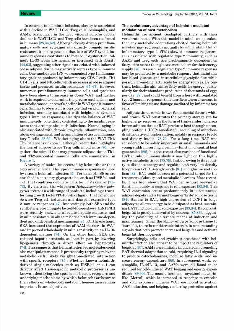
Figure 1. Adipose tissue immunologic balance. In lean adipose tissue (left), immunologic elements associated with a type 2
immune response dominate. These include
ILC2s, Tregs, Eos, and AAMs. IL-33, helminth infection, and possibly exposure to cold can each promote these adipose tissue type 2 associated immune cells. In mice and
humans, obesity is associated with decreases in each of the type 2 immune cells while promoting type 1 immune cells associated with classical inflammatory responses
(right), including PMNs, CD8 T cells, CD4 Th1 T cells, and proinflammatory M1 macrophages. We speculate that type-1-associated bacterial and viral infections, which
promote systemic insulin resistance, may also promote this pathway in white adipose tissue. Similarly, natural age-related alterations in the immune system, including
accumulation of virus-reactive CD8 T cells, may also contribute to this pathway. Abbreviations: AAM, alternatively activated macrophage; Eos, eosinophils; ILC2, group
2 innate lymphoid cell; PMN, neutrophil; Th1, T helper 1; Treg cell, regulatory T cell.
Review Trends in Parasitology September 2015, Vol. 31, No. 9
437
7/21/2019 A Worm of One's Own. How Helminths Modulate Host Adipose Tissue Function and Metabolism
http://slidepdf.com/reader/full/a-worm-of-ones-own-how-helminths-modulate-host-adipose-tissue-function 4/7
In
contrast
to
helminth
infection,
obesity
is
associated
with
a
decline
in WAT
ILC2s,
Treg
cells,
eosinophils,
and
AAMs,
particularly
in
the
deep
visceral
adipose
depots;
declines inWAT ILC2s and Treg cells have been confirmed
in
humans
[28,34,62]. Although
obesity
driven
proinflam-
matory
cells
and
cytokines
can
directly
promote
insulin
resistance, it is also possible that loss of WAT type 2 im-
mune
responses
contributes
to
metabolic
dysfunction.
Ad-ipose IL-33 levels are normal or increased with obesity
[44,63], suggesting
other
signals
associated
with
inflamed
obese
adipose
tissue
may
actively
repress
type
2
immune
cells. One candidate is IFN-g, a canonical type 1 inflamma-
tory
cytokine
produced
by
inflammatory
CD8
T
cells,
Th1
CD4T cells, and NK cells, which increases in obese adipose
tissue
and
promotes
insulin
resistance
[65–67].
However,
numerous
proinflammatory
immune
cells
and
cytokines
have been shown to increase in obese WAT, and further
work
is
required
to
determine
the
precise
mechanisms
and
metabolic
consequences
of
a
decline
in
WAT
type
2
immune
cells. Similar to obesity, it is possible that viral or bacterial
infection,
normally
associated
with
heightened
systemic
type 1 immune responses, also tips the balance of WATimmune
cells,
potentially
contributing
to
the
insulin
resis-
tance that accompanies such infections. Normal aging is
also
associated
with
chronic
low-grade
inflammation,
met-
abolic
derangement,
and
accumulation
of
tissue
inflamma-
tory T cells [68,69]. Whether aging alters the WAT Th1/
Th2
balance
is
unknown,
although
recent
data
highlights
the
loss
of
adipose
tissue
Treg
cells
in
old
mice
[70]. To-
gether, the stimuli that may promote adipose tissue Th1-
and
Th2-associated
immune
cells
are
summarized
in
Figure
1.
A variety of molecules secreted by helminths or their
eggs
are involved
in the type
2
immune responses
induced
by chronic helminth
infection
[5].
For
example, SEAs areenriched
in secretory glycoproteins, such as IPSE/ a1 and
v-1, that condition dendritic cells for Th2 skewing [71–
73].
By contrast,
the whipworm Heligmosomoides
poly-
gyrus
secretes
a
wide range of
products,
including
a
trans-
forming growth factor (TGF)-b-like ligand, that can induce
de novo Treg
cell induction and dampen
excessive
type
2
immune responses
[47].
Interestingly,
both
SEAand
the
synthetic glycoconjugate lacto-N-fucopentaose (LNFP)III
were
recently
shown
to
alleviate hepatic steatosis and
insulin resistance in obese mice via both immune-depen-
dent and -independent mechanisms[74]. Onthe one hand,
SEA increased the expression of AAM markers in WAT
and improved whole-body insulin sensitivity in an IL-10-
dependent
manner
[74].
On the other
hand,
SEA also
reduced hepatic steatosis, at least in part by lowering
lipogenesis through a
direct effect on hepatocytes
[74]. This
suggests
that
helminth-derivedmolecules could
also manipulate metabolic processesby targeting relevant
metabolic
cells, likely
via glycan-mediated
interaction
with specific
receptors
[75].
Whether
known
helminth-
derived single molecules, such as IPSE/ a1 or v-1 can
directly affect tissue-specific metabolic
processes is
un-
known. Identifying the specific molecules,
receptors and
underlying mechanisms by which helminths
orchestrate
their effects on whole-bodymetabolic homeostasis remain
important future
objectives.
The evolutionary advantage of helminth-mediated
modulation
of
host
metabolism
Helminths
are
ancient,
coadapted
partners
with
their
vertebrate hosts. With this model in mind, we speculate
that
any
metabolic
adaptations
elicited
during
helminth
infection
may
represent
a
mutually
beneficial
state.
Unlike
inflammatory type 1 (Th1)-skewed immune responses,
cells associated
with
regulated
type
2
immunity,
such
as AAMs and Treg cells, are predominantly dependent on
fattyacids
rather
than
glucose
metabolism
for
their
energy
supply
[76]. As
such,
regulated
type
2
immune
responses
may be promoted by a metabolic response that maintains
low
blood
glucose
and
intracellular
glycolytic
flux
while
possibly promoting fatty acids for energy sources. By con-
trast,
helminths
also
utilize
fatty
acids
for
energy,
partic-
ularly
for
their
abundant
production
of
thousands
of
eggs
per day [77], and could benefit from a host with regulated
type2
immune
responses
that
sacrifices
worm
clearance
in
favor
of
limiting
tissue
damage
mediated
by
inflammatory
cells.
Adipose tissue
comes
in
two
fundamental
flavors:
white
and brown. WAT constitutes the primary storage site forhigh-energy
reserves
in
the
form
of triglycerides,
whereas
brown adipose tissue (BAT) produces heat through uncou-
pling
protein
1
(UCP1)-mediated
uncoupling
of
mitochon-
drial
oxidative
phosphorylation,
notably
in response
to
cold
and dietary intake [78,79]. For a long time, BAT was
considered
to
be
solely
important
in
small
mammals
and
young
children,
serving
a
primary
function
of
central
heat
generation [80], but the recent observations of functional
BAT in
adult
humans
sheds
a
new
light
on
this
highly
active
metabolic
tissue
[78,79]. Indeed,
owing
to
its
capaci-
ty to dissipate energy and regulate both very low density
lipoprotein
(VLDL)–triglyceride
[81]
and
glucose
metabo-
lism
[82], BAT
could
be
seen
as
a
potential
target
for
thetreatment
of
obesity
and
metabolic
disorders.
More
recent-
ly, it has been shown that WAT can acquire brown-like
function,
notably
in
response
to
cold
exposure
[83,84]. This
WAT
conversion
occurs
predominantly
in
subcutaneous
adipose depots and is termed ‘beige’ or ‘brite’ adipose tissue
[84]. Similar
to
BAT,
high
expression
of
UCP1
in
beige
adipocytes
allows
energy
to
be
dissipated
as
heat,
sustain-
ing BAT function during cold exposure [83,84]. By contrast,
beige fat
is
poorly
innervated
by
neurons
[85,86], suggest-
ing the possibility of alternate means of induction and
maintenance. Given the ability of beige adipose tissue to
‘burn’ fat, there is considerable interest in understanding
signals
that
both
promote
increased
beige
fat
and
activate
beige
fat
thermogenesis.
Surprisingly, cells and cytokines associated with hel-
minth-infection
also
appear
to
be
important
regulators
of
beige
fat
[87]. AAMs
were
initially
implicated
in promoting
BAT thermal adaptation to cold, requiring IL-4 signaling
to produce
catecholamines,
mobilize
fatty
acids,
and
in-
crease
energy
expenditure
[88]. In
subsequent
work,
eo-
sinophils, IL-4/IL-13, and AAMs were all found to be
required
for
cold-induced
WAT
beiging
and
energy
expen-
diture
[89,90]. The
muscle
hormone
(myokine)
meteorin-
like
(Metrnl),
which
is
increased
in
response
to
exercise
and cold exposure, induces WAT eosinophil activation,
AAM
induction,
and
beiging,
conferring
protection
against
Review Trends in Parasitology September 2015, Vol. 31, No. 9
438

7/21/2019 A Worm of One's Own. How Helminths Modulate Host Adipose Tissue Function and Metabolism
http://slidepdf.com/reader/full/a-worm-of-ones-own-how-helminths-modulate-host-adipose-tissue-function 5/7
obesity-induced
metabolic
disorders
[90]. Most
recently,
two studies
have
demonstrated
that
the
cytokine
IL-33
can
also
directly
promote
ILC2
activation
and
bypass
eosino-
phils and AAMs to induce WAT beiging [61,62]. The precise
mechanisms
by
which
ILC2s
mediate
this
phenotype
dif-
fer,
with
one
group
proposing
ILC2-derived
IL-13
promotes
adipose precursors to adopt a beige fate [61], whereas the
other
group
has
suggested
ILC2
production
of
the
endoge-nous opiate methionine–enkephalin mediates the pheno-
type
[62]. Although
IL-33
administration
drives
ILC2s
to
promote
beiging,
the
roles
of
IL-33
and
ILC2s
in
beiging
during normal physiology, cold adaptation, or helminth
infection
are
unclear.
Together,
these
data
suggest
that
signals such as IL-33 and Metrnl regulate WAT ILC2s,
eosinophils,
and
AAMs
to
promote
adipose
beiging
through
various
mechanisms
and
may
confer
protection
against
obesity-induced type 2 diabetes (Figure 2). As helminths
can
promote
WAT
type
2
immune
cells
and
a
loss
of
adiposity,
they
may
be
able
to
induce
significant
adipose
beiging. Helminths and hosts could potentially benefit
from
increased
WAT
beiging
by
promoting
host
survival
in cold climates. Further experimental data carefully
documenting
the
metabolic
consequences
of
helminth
in-
fection,
particularly
in
humans,
are
required
to
clarify
these important points (Box 1).
Concluding remarksBoth helminth infection and helminth-derived products
may be
beneficial
for
maintaining
host
metabolic
homeo-
stasis, in
particular
by
enhancing
type
2
immune
responses
in WAT, thereby limiting excess type 1 inflammation and
possibly promoting increased beige adipocytes. Improved
understanding
of
both
immune-dependent
and
-indepen-
dent
mechanisms
by
which
helminths
modulate
tissue-
specific and whole-body insulin sensitivity, and glucose
and
lipid
homeostasis
may
offer
new
insights
toward
the
development
of
novel
therapeutics
for
the
treatment
of
metabolic disorders and type 2 diabetes.
AcknowledgmentsWe thank Drs. S. Van Dyken, A.V. Molofsky, and L. Hussaarts for
comments on themanuscript. This work was supported byK08DK101604
(ABM) from the NIH, UCSF
REAC
Grant (ABM), an EFSD/Lilly
Research Grant Fellowship from the European Federation for the Study
of Diabetes (BG), and a ZonMW TOPGrant (40-00812-98-14131) from the
Dutch Organization for Scientific Research (BG). The authors have no
conflicting financial interests.
References1 Wiria, A.E. et al. (2012)Helminth infection in populations undergoing
epidemiological transition: a friend or foe? Semin. Immunopathol. 34,
889–901
2 DiAngelo, J.R. et al. (2009) The immune response attenuates growth
andnutrient storage inDrosophilaby reducing insulinsignaling. Proc.
Natl. Acad. Sci. U.S.A. 106, 20853–20858
3 Odegaard, J.I. and Chawla, A. (2011) Alternative macrophageactivation and metabolism. Annu. Rev. Pathol. Mech. Dis. 6, 275–297
4 Hotez, P.J. et al. (2008) Helminth infections: the great neglected
tropical diseases. J. Clin. Invest. 118, 1311–1321
5 Allen, J.E. and Maizels, R.M. (2011) Diversity and dialogue in
immunity to helminths. Nat. Rev. Immunol. 11, 375–388
6 Zaph, C. et al. (2014) Mucosal immune responses following intestinal
nematode infection. Parasit. Immunol. 36, 439–452
7 Saz, H.J. (1981) Energy metabolisms of parasitic helminths:
adaptations to parasitism. Annu. Rev. Physiol. 43, 323–341
8 Stephenson, L.S. et al. (2000) Malnutrition and parasitic helminth
infections. Parasitology 121, S23–S38
9 King, C.H. and Dangerfield-Cha, M. (2008) The unacknowledged
impact of chronic schistosomiasis. Chronic Illn. 4, 65–79
10 Colley, D.G.
et al. (2014) Human schistosomiasis. Lancet 383,
2253–2264
IL-33
Catecholamines
IL-4, others?I
Eosinophils
AAMILC2
IL-5
IL-4
Whiteadipocyte/precursor
Beigeadipocyte
IL-13
Met-Enk
Th2Metrnl
Helminth infecon, cold
IL-13
TRENDS in Parasitology
Figure 2.
Type 2 immunity and WAT beiging.
A proposed model of how type
2 immune cells affectWAT beiging. In this model,helminth infections promote the
accumulation and/or function of ILC2s and Th2 T cells in WAT, possibly via
alterations in IL-33 or Metrnl. ILC2s (and possibly Th2 cells) promote eosinophil
accumulation via IL-5 production and AAM accumulation via both eosinophil-
mediated IL-4 and possibly via direct effects of ILC2-derived IL-13. AAMs can
produce catecholamines to activate beige adipocytes to generate heat, whereas
ILC2s and possible eosinophil-derived IL-4/IL-13 can activate adipocyte precursors
to adopt a beige fate. Further, ILC2-derived compounds, such as enkephalins (Met-
Enk), may further promote these effects. We emphasize how these pathways
overlap with adipose tissue adaptation to cold. Dashed arrows represent more
speculative interactions, solid arrows published interactions. Abbreviations: AAM,
alternatively activatedmacrophage; IL, interleukin; ILC2, group 2 innate lymphoid
cell; Metrnl, meteorin-like; Th2, T helper 2; WAT, white adipose tissue.
Box 1. Outstanding questions
What are the relevant WAT type 2 immune cells andmechanisms
that improve whole-body metabolic homeostasis in obese mice?
Is the helminth-induced type 2 immune response observed in
rodent WAT also present in humans?
What are the exact immune-dependent and -independent me-
chanisms by which helminths and their specific molecules
improve whole-body metabolic homeostasis in obese mice?
What is the contribution of metabolic organs other than WAT tothe beneficial effect of helminth infection or helminth-derived
molecules on whole-body insulin sensitivity and glucose home-
ostasis?
Does helminth nutrient metabolism significantly contribute to
parasitism of the host dietary energy intake?
Does helminthic therapy and/or treatment with helminth-derived
molecules hold promise for treatment of metabolic disorders in
humans?
Review Trends in Parasitology September 2015, Vol. 31, No. 9
439

7/21/2019 A Worm of One's Own. How Helminths Modulate Host Adipose Tissue Function and Metabolism
http://slidepdf.com/reader/full/a-worm-of-ones-own-how-helminths-modulate-host-adipose-tissue-function 6/7
11 Wammes, L.J. et al. (2014) Helminth therapy or elimination:
epidemiological, immunological, and clinical considerations. Lancet
Infect. Dis. 14, 1150–1162
12 Tahapary, D.L. (2015) Helminth infections and type 2 diabetes: a
cluster-randomized placebo controlled SUGARSPIN trial in
Nangapanda, Flores, Indonesia. http://dx.doi.org/10.1186/s12879-
015-0873-4
13 Wiria,A.E. et al. (2014) Helminth infections, type-2 immuneresponse,
and metabolic syndrome. PLoS Pathog. 10, e1004140
14 Hussaarts, L. et al. (2014) Priming dendritic cells for th2 polarization:lessons learned from helminths and implications for metabolic
disorders. Front. Immunol. 5, 499
15 Alberti, K.G.M.M. et al. (2009)Harmonizing themetabolic syndrome a
joint interim statement of the International Diabetes Federation Task
Force on Epidemiology and Prevention; National Heart, Lung, and
BloodInstitute; AmericanHeart Association; WorldHeart Federation;
International Atherosclerosis Society; and International Association
for the Study of Obesity. Circulation 120, 1640–1645
16 Guariguata,L. et al. (2014)Global estimates of diabetes prevalence for
2013 and projections for 2035. Diabetes Res. Clin. Pract. 103, 137–149
17 Donath, M.Y. and Shoelson, S.E. (2011) Type 2 diabetes as an
inflammatory disease. Nat. Rev. Immunol. 11, 98–107
18 Hotamisligil, G.S. et al. (1993) Adipose expression of tumor necrosis
factor-alpha: direct role in obesity-linked insulin resistance. Science
259, 87–91
19 Uysal,
K.T.
et al. (1997) Protection from obesity-induced
insulinresistance in mice lacking TNF-alpha function.
Nature 389,
610–614
20 Pradhan,A.D. et al. (2001)C-reactive protein, interleukin 6, andrisk of
developing type 2 diabetes mellitus. JAMA 286, 327–334
21 Xu, H. et al. (2003) Chronic inflammation in fat plays a crucial role in
the development of obesity-related insulin resistance. J. Clin. Invest.
112, 1821–1830
22 Lumeng, C.N. et al. (2007) Obesity induces a phenotypic switch in
adipose tissue
macrophage polarization. J. Clin. Invest. 117,
175–184
23 Lumeng, C.N. et al. (2007) Increased inflammatory properties of
adipose tissue macrophages recruited during diet-induced obesity.
Diabetes 56, 16–23
24 Nishimura, S. et al. (2009) CD8+ effector T cells contribute to
macrophage recruitment and adipose tissue inflammation in obesity.
Nat. Med. 15, 914–920
25 Mathis, D. (2013) Immunological goings-on in visceral adipose tissue.
Cell Metab. 17, 851–859
26 Osborn, O. and Olefsky, J.M. (2012) The cellular and signaling
networks linking the immune system and metabolism in disease.
Nat. Med. 18, 363–374
27 Winer, S. et al. (2009) Normalization of obesity-associated insulin
resistance through immunotherapy. Nat. Med. 15, 921–929
28 Feuerer,M. et al. (2009)Lean, butnot obese,fat is enriched fora unique
population of regulatory T cells that affectmetabolic parameters. Nat.
Med. 15, 930–939
29 Liu, J. et al. (2009) Genetic deficiency and pharmacological
stabilization of mast cells reduce diet-induced obesity and diabetes
in mice. Nat. Med. 15, 940–945
30 Talukdar, S. et al. (2012) Neutrophils mediate insulin resistance in
mice fed a high-fat diet through secreted elastase. Nat. Med. 18,
1407–1412
31 Wensveen, F.M. et al. (2015) NK cells link obesity-induced adipose
stress to inflammation
and insulin
resistance.
Nat
Immunol. 16,
376–385
32 Wu,D. et al. (2011) Eosinophils sustainadipose alternatively activated
macrophages associated with glucose homeostasis. Science 332,
243–247
33 Yang, Z. et al. (2013) Parasitic nematode-induced modulation of body
weight and associated metabolic dysfunction in mouse models of
obesity. Infect. Immun. 81, 1905–1914
34 Molofsky, A.B. et al. (2013) Innate lymphoid type 2 cells sustain
visceral adipose tissue eosinophils and alternatively activated
macrophages. J. Exp. Med. 210, 535–549
35 Hussaarts, L. et al. (2015) Chronic helminth infection and helminth-
derived egg antigens promote adipose tissue M2 macrophages and
improve insulin sensitivity in obese mice. FASEB J. http://dx.doi.org/
10.1096/fj.14-266239
36 Van Dyken, S.J. and Locksley, R.M. (2013) Interleukin-4- and
interleukin-13-mediated alternatively activated macrophages: roles
in homeostasis and disease. Annu. Rev. Immunol. 31, 317–343
37 Kita, H. (2011) Eosinophils: multifaceted biological properties and
roles in health and disease. Immunol. Rev. 242, 161–177
38 Hams, E. et al. (2013)Cutting edge: IL-25 elicits innate lymphoid type
2 and type II NKT cellsthat regulate obesity inmice. J. Immunol.191,
5349–5353
39 Moro, K. et al. (2010) Innate production of T(H)2 cytokines by adipose
tissue-associated c-Kit(+)Sca-1(+) lymphoid cells. Nature 463, 540–54440 Diefenbach, A. (2013) Innate lymphoid cells in the defense against
infections. Eur. J. Microbiol. Immunol. (Bp) 3, 143–151
41 Nussbaum, J.C. et al. (2013) Type 2 innate lymphoid cells control
eosinophil homeostasis. Nature 502, 245–248
42 Walker, J.A. andMcKenzie, A.N. (2013) Development and function of
group 2 innate lymphoid cells. Curr. Opin. Immunol. 25, 148–155
43 Cipolletta,D. et al. (2012) PPAR-g
is amajor driverof theaccumulation
and phenotype of adipose tissue Treg cells. Nature 486, 549–553
44 Vasanthakumar, A. et al. (2015) The transcriptional regulators IRF4,
BATF and IL-33 orchestrate development andmaintenance of adipose
tissue-resident regulatory T cells. Nat. Immunol. 16, 276–285
45 Schiering,C. et al. (2014) The alarmin IL-33promotes regulatoryT-cell
function in the intestine. Nature 513, 564–568
46 Burzyn,D. et al. (2013) RegulatoryT cells in nonlymphoid tissues. Nat.
Immunol. 14, 1007–1013
47 McSorley, H.J. andMaizels, R.M. (2012)Helminth infections andhostimmune regulation. Clin. Microbiol. Rev. 25, 585–608
48 Gause, W.C. et al. (2013) Type 2 immunity and wound healing:
evolutionary refinement of adaptive immunity by helminths. Nat.
Rev. Immunol. 13, 607–614
49 Moltke et al. (2014) I-L-C-2 it: type 2 immunity and group 2 innate
lymphoid cells in homeostasis. Curr. Opin. Immunol. 31, 58–65
50 Stanya, K.J. et al. (2013)Direct control of hepatic glucoseproduction by
interleukin-13 in mice. J. Clin. Invest. 123, 261–271
51 Ricardo-Gonzalez,R.R. et al. (2010) IL-4/STAT6 immuneaxis regulates
peripheral nutrient metabolism and insulin sensitivity. Proc. Natl.
Acad. Sci. U.S.A. 107, 22617–22622
52 Wolfs, I.M.J. et al. (2014) Reprogramming macrophages to an anti-
inflammatory phenotype by helminth antigens reduces murine
atherosclerosis. FASEB J. 28, 288–299
53 Doenhoff, M.J. et al. (2002) An anti-atherogenic effect of Schistosoma
mansoni infectionsinmiceassociatedwith a parasite-inducedlowering
of blood total cholesterol. Parasitology 125, 415–421
54 Neill, D.R. et al. (2010) Nuocytes represent a new innate effector
leukocyte that mediates type-2 immunity. Nature 464, 1367–1370
55 Luck, H. et al. (2015) Regulation of obesity-related insulin resistance
with gut anti-inflammatory agents. Cell Metab. 21, 527–542
56 Walk, S.T. et al. (2010)Alteration of themurine gutmicrobiota during
infection with the parasitic helminth Heligmosomoides polygyrus.
Inflamm. Bowel Dis. 16, 1841–1849
57 Lee, S.C. et al. (2014) Helminth colonization is associated with
increased diversity of the gut microbiota. PLoS Negl. Trop. Dis. 8,
e2880
58 Cayrol, C. and Girard, J-P. (2014) IL-33: an alarmin cytokine with
crucial roles in innate immunity, inflammation and allergy. Curr.
Opin. Immunol. 31, 31–37
59 Endo, Y. et al. (2015) The Interleukin-33-p38 kinase axis confers
memory T helper 2 cell pathogenicity in the airway. Immunity 42,
294–308
60 Miller, A.M. et al. (2010) Interleukin-33 induces protective effects in
adipose tissue inflammation during obesity in mice. Circ. Res. 107,
650–658
61 Lee, M-W. et al. (2015)Activated type 2 innate lymphoid cells regulate
beige fat biogenesis. Cell 160, 74–87
62 Brestoff, J.R. et al. (2014) Group 2 innate lymphoid cells promote
beiging of white adipose tissue and limit obesity. Nature 519, 242–246
63 Zeyda, M. et al. (2013) Severe obesity increases adipose tissue
expression of interleukin-33 and its receptor ST2, both
predominantly detectable in endothelial cells of human adipose
tissue. Int. J. Obes. (Lond.) 37, 658–665
64 Wood, I.S. et al. (2009) IL-33, a recently identified interleukin-1 gene
family member, is expressed in human adipocytes. Biochem. Biophys.
Res. Commun. 384, 105–109
Review Trends in Parasitology September 2015, Vol. 31, No. 9
440

7/21/2019 A Worm of One's Own. How Helminths Modulate Host Adipose Tissue Function and Metabolism
http://slidepdf.com/reader/full/a-worm-of-ones-own-how-helminths-modulate-host-adipose-tissue-function 7/7
65 Sutherland, A.P.R. et al. (2014) Linking obesity with type 2 diabetes:
the role of T-bet. Diabetes Metab. Syndr. Obes. 7, 331–340
66 Rocha, V.Z. et al. (2008) Interferon-gamma, a Th1 cytokine, regulates
fat inflammation: a role for adaptive immunity in obesity. Circ. Res.
103, 467–476
67 Wong, N. et al. (2011) Deficiency in interferon-gamma results in
reduced body weight and better glucose tolerance in mice.
Endocrinology 152, 3690–3699
68 Franceschi, C. and Campisi, J. (2014) Chronic inflammation
(inflammaging) and its potential contribution to age-associateddiseases. J. Gerontol A: Biol. Sci. Med. Sci. 69, S4–S9
69 Macaulay, R. et al. (2012) The role of the T cell in age-related
inflammation. AGE 35, 563–572
70 Cipolletta,
D.
et al. (2015) Appearance
and disappearance of the
mRNA signature characteristic of Treg cells
in visceral adipose
tissue:
age, diet,
and PPARg effects.
Proc. Natl.
Acad.
Sci.
U.S.A.
112, 482–487
71 Everts, B. et al. (2009) Omega-1, a glycoprotein secreted by
Schistosoma mansoni eggs, drives Th2 responses. J. Exp. Med. 206,
1673–1680
72 Everts, B. et al.
(2012) Schistosome-derived
omega-1 drives
Th2
polarization by suppressing protein synthesis fol lowing
internalization
by the mannose receptor. J. Exp. Med.
209,
1753–1767
73 Schramm, G. et al. (2006) IPSE/alpha-1: a major immunogenic
component secreted from Schistosoma mansoni eggs. Mol. Biochem. Parasitol. 147, 9–19
74 Bhargava, P. et al. (2012) Immunomodulatory glycan LNFPIII
alleviates hepatosteatosis and insulin resistance through direct and
indirect control of metabolic pathways. Nat. Med. 18, 1665–1672
75 Meevissen,M.H.J. et al. (2012) Schistosomamansoni egg glycoproteins
andC-type lectins of host immunecells:molecular partners that shape
immune responses. Exp. Parasitol. 132, 14–21
76 Pearce, E.L. and Pearce, E.J. (2013) Metabolic pathways in immune
cell activation and quiescence. Immunity 38, 633–643
77 Huang, S.C-C. et al. (2012) Fatty acid oxidation is essential for egg
production by the parasitic flatworm Schistosoma mansoni.
PLoS
Pathog. 8, e1002996
78 Tam,C.S. et al. (2012) Brownadipose tissuemechanismsandpotential
therapeutic targets. Circulation 125, 2782–2791
79 Vosselman, M.J. et al. (2013) Energy dissipation in brown adipose
tissue: From mice to men. Mol. Cell. Endocrinol. 379, 43–50
80 Frontini,A. andCinti, S.(2010) Distributionand development ofbrown
adipocytes in the murine and human adipose organ. Cell Metab. 11,
253–25681 Bartelt, A. et al. (2011) Brown adipose tissue activity controls
triglyceride clearance. Nat. Med. 17, 200–205
82 Chondronikola, M. et al. (2014) Brown adipose tissue improves whole-
body glucose homeostasis and insulin sensitivity in humans. Diabetes
63, 4089–4099
83
Bartelt, A. and Heeren, J. (2014) Adipose tissue browning and
metabolic health. Nat. Rev. Endocrinol. 10, 24–36
84 Harms, M. and Seale, P. (2013) Brown and beige fat: development,
function and therapeutic potential. Nat. Med. 19, 1252–1263
85 Daniel, H. and Derry, D.M. (2011) Criteria for differentiation of brown
and white fat in the rat.Can. J. Physiol. Pharmacol. http://dx.doi.org/
10.1139/y69-154
86 Slavin, B.G. and Ballard, K.W. (1978) Morphological studies on the
adrenergic innervationofwhiteadipose tissue. Anat.Rec. 191,377–389
87 Brestoff, J.R. and Artis, D. (2015) Immune regulation of metabolic
homeostasis in health and disease. Cell 161, 146–16088 Nguyen, K.D. et al. (2011) Alternatively activated macrophages
produce catecholamines to sustain adaptive thermogenesis. Nature
480, 104–108
89 Qiu, Y. et al. (2014) Eosinophils and type 2 cytokine signaling in
macrophages orchestrate development of functional beige fat. Cell
157, 1292–1308
90 Rao, R.R. et al. (2014) Meteorin-like is a hormone that regulates
immune-adipose interactions to increase beige fat thermogenesis.
Cell 157, 1279–1291
Review Trends in Parasitology September 2015, Vol. 31, No. 9
441