aaogd secretariatogd secretariat - aogd | 2017aogd.org/aogd bulletin june 2017.pdf · aaogd...
TRANSCRIPT

Volume 17-2; June 2017
1
AOGD SECRETARIATAOGD SECRETARIATRoom No 712, 7th Floor, Private Ward, MCH Block
Department of Obstetrics and GynecologyGuru Teg Bahadur Hospital & University College of Medical Sciences
Dilshad Garden, Delhi-110095, [email protected], [email protected]
www.aogd.org
Volume 17; Issue No.2; June 2017 Price: ` 30 only
AOGD BULLETINAOGD BULLETIN
Issue:Red AlertMaternal Near Miss
AOGD Theme 2017-18AOGD Theme 2017-18‘Optimizing Women’s Health Through ‘Optimizing Women’s Health Through Enhanced Skills and Best Practices’Enhanced Skills and Best Practices’
AOGD BULLETINAOGD BULLETIN

AOGD Bulletin
2

Volume 17-2; June 2017
3
President, AOGDDr Shalini Rajaram (2017-2018)
Vice PresidentDr Kiran Guleria
Hony. SecretaryDr Abha Sharma
Chairperson Skill WorkshopsDr A G Radhika
TreasurerDr Alpana Singh
EditorsDr RashmiDr Bindiya Gupta
Web EditorsDr Rachna AgarwalDr Anshuja Singla
Joint SecretariesDr Himshweta SrivastavaDr Sandhya Jain
Co-TreasurerDr Archana Chaudhary
Co-EditorsDr Richa AggarwalDr Sruthi Bhaskaran
Coordinators Skill WorkshopsDr Richa SharmaDr Sanjeeta BeheraDr Bhanu Priya
Clinical SecretariesDr Vishnu BhartiyaDr Shweta Prasad
Public Relations & HospitalityDr Rashmi GuptaDr Seema Prakash
AOGD Executive Council Members 2017-2018Dr Abha SinghDr Achala BatraDr Amita SaxenaDr Amita SunejaDr Anjali TempeDr B K GoelDr Gita RadhakrishnanDr Harsha KhullarDr Kuldeep JainDr Malvika SabharwalDr Nalini MahajanDr Neerja BhatlaDr Nirmala AgarwalDr Puneeta MahajanDr Pushpa SinghDr Renu MishraDr S N BasuDr Sabhyata GuptaDr Sangeeta GuptaDr Sonia MalikDr Sumita Mehta
AOGD SecretariatRoom No 712, 7th Floor, Private Ward, MCH BlockDepartment of Obstetrics & GynaecologyGuru Teg Bahadur Hospital & University College of Medical Sciences Delhi-110 095, Indiawww.aogd.org
AOGD BULLETINVolume 17-2, June 2017
AOGD Executive Committee 2017-18
DisclaimerThe advertisements in this bulletin are not a warranty, endorsement or approval of the products or services. The statements and opinions contained in the articles of the AOGD Bulletin are solely those of the individual authors and contributors, and do not necessarily refl ect the opinions or recommendations of the publisher. The publisher disclaims responsibility of any injury to persons or property resulting from any ideas or products referred to in the articles or advertisements.
Plagiarism DisclaimerAny plagiarism in the articles will be the sole responsibility of the authors and the editorial board or publisher will not be responsible for this.
Publisher/Printer/EditorDr Rashmi on behalf of Association of Obstetricians & Gynecologists of Delhi.
Printed atProcess & Spot C-112/3, Naraina Industrial Area, Phase-1, New Delhi 110 028
Published fromAOGD Offi ce, Room No 712, 7th Floor, Private Ward, MCH Block, Department of Obstetrics & Gynaecology, Guru Teg Bahadur Hospital & University College of Medical Sciences, Delhi-110 095, India
EditorDr RashmiPh. No. 011-22692505; Email: [email protected]
PatronsDr S N MukherjeeDr S K DasDr Urmil SharmaDr Kamal BucksheeDr Neera AgarwalAdvisorsDr Chitra RaghunandanDr Gauri DeviDr Indrani GanguliDr N B VaidDr Neerja GoelDr S S TrivediDr Shakti Bhan KhannaDr Sharda JainDr Suneeta MittalDr Swaraj BatraScientifi c AdvisorsDr Gita RadhakrishnanDr Amita Suneja
Ex Offi cio ExecutivePast PresidentsDr P K Malkani (1962-66)Dr L V Pathak (1966-72)Dr Anusuya Das (1972-78)Dr S N Mukherjee (1978-81)Dr V Hingorani (1981-88)Dr S K Das (1988-90)Dr P Chadha (1990-94)Dr Neera Agarwal (1994-97)Dr Maya Sood (1997-99)Dr D Takkar (1999-2001)Dr Sudha Salhan (2001-03)Dr Swaraj Batra (2003-05)Dr N B Vaid (2005-06)Dr S S Trivedi (2006-07)Dr Suneeta Mittal (2007-08)Dr I Ganguli (2008-09)Dr Shashi Prateek (2009-10)Dr U Manaktala (2010-11)Dr Neerja Goel (2011-12)Dr C Raghunandan (2012-13)Dr Alka Kriplani (2013-14)Dr U P Jha (2014-15)Dr Pratima Mittal (2015-16)
Immediate Past PresidentDr Sudha Prasad (2016-17)
Immediate Past SecretaryDr Ashok Kumar (2016-17)
President ElectDr Abha Singh (2018-19)
Immediate Past President FOGSIDr Alka Kriplani
ChairpersonsAOGD Sub-CommitteesDr Achla BatraDr Amita JainDr Anjali TempeDr Ashok KumarDr Jyotsna SuriDr K D NayarDr Mala SrivastavaDr Nalini MahajanDr Renu MisraDr Rupinder SekhonDr Shakuntala KumarDr Sunita MalikDr Vatsla Dadhwal
Total number of pages = 68
ContentsNear Miss approach in Maternal Health 9Kiran Guleria, Bhanupriya
Cardiac Events: Early recognition and t reatment 14Taru Gupta, Shweta Singh, Sangeeta Gupta
Managing Septicaemic Shock and Assessment of Postpartum Sepsis
19
Nidhi Gupta, Aruna Nigam
SOP: Basic Life Support and Maternal Resuscitation 23Rashmi Salhotra
Amniotic Fluid Embolism: An obstetrician’s challenge 25Rashmi, Anju Bala
Recent Advances in Management of Post Partum Haemorrhage
39
Esha Gupta, Richa Aggarwal
Thromboprophylaxis in Pregnancy and Puerperium 44Sandhya Jain, Vasudha Gupta
Setting up of an Obstetric Intensive Care Unit and High Dependency Unit
47
Pikee Saxena, Supriya Goyal
Transf usion Guidelines in Obstetric Practice 51Priyanka GogoiMIND, BODY & SOULMystery Surrounding Near Death Experiences
54
Rashmi
Maternal Near Miss: Case snipppets 56Taru Gupta, Shweta Singh, Sangeeta Gupta
Journal Scan 58Sruthi Bhaskaran
Proceedings of AOGD Monthly Clinical Meet 61Quiz Time 63Rashmi, Bindiya Gupta

AOGD Bulletin
4
AOGD Offi ce-Bearers
Editorial Board
Committees
Dr Seema PrakashDr Rashmi GuptaDr Shweta PrasadDr Vishnu BhartiyaPublic Relations & Hospitality
Dr Bindiya GuptaDr RashmiEditors
Dr AnshujaDr RachnaWeb Editors
Dr Richa Aggarwal Dr SruthiCo-Editors
Dr Abha SharmaHon. Secretary
Dr A G Radhika Chairperson
Skill Workshops
Dr Amita SunejaDr Gita RadhakrishnanScientifi c Advisors
Dr Alpana SinghTreasurer
Dr Sandhya JainDr Himshweta Srivastava Dr Archana Chaudhary Co TreasurerJoint Secretaries
Clinical Secretaries
Dr Richa Sharma Dr Bhanu PriyaDr Sanjeeta BeheraCoordinators Skill Workshops
Dr Shalini Rajaram President
Dr Kiran GuleriaVice President

Volume 17-2; June 2017
5
President’s Message
Dear FriendsAt the outset I urge all AOGD members to support Dr. Sudha Prasad as Vice-President FOGSI, (North zone) 2019 and post your ballots in time. You have seen the support and camaraderie she enjoys with members, and her capacity to take on a leadership role nationally. We wish her success in this endeavour.We are now in the second month of holding of ice of AOGD and so much has happened since then. Active AOGD members have invested their time and commitment to bring diverse issues related to women’s health to the table and I applaud them. Our irst ‘Basic life Support Skill workshop’ on 5th May was well attended by practitioners and residents who had hands on experience at the ‘State of the Art Skill lab’ at GTBH with a bonus of BLS certi ication. Dr. AG Radhika, AOGD Skills workshop Chairperson and Dr. Sujata, Director Professor Anesthesiology deserve accolades for the well conducted workshop. ‘ABC’ of CPR has now changed to ‘CAB’ with cardiac compressions of prime importance, done irst, correctly and with adequate depth and number. I suggest all AOGD members get a BLS certi ication and this is non-negotiable!Dr. Ranjana Sharma of Apollo hospital, Chairperson, Urogynecology Commitee (2015-17)Dr. Kuldeep Jain, Chairperson, FOGSI endometriosis committee and Dr. Geeta Mehdiratta, Secretary, IMS Delhi chapter, from Sir Ganga Ram Hospital all held academically and intellectually stimulating CMEs in May. GTB hospital under the leadership of Dr. Kiran and Dr. Abha updated more than 100 delegates on best practices on Antenatal care and culminating May’s activities was Dr. Urvashi Jha and team from Fortis Hospital, Vasant Kunj who held the monthly AOGD clinical meeting.Hope all of you enjoyed the April issue on ‘Adolescent Health’. The feedback from AOGD members was very extremely encouraging and this issue on ‘Maternal Near Miss ’ promises not to disappoint. Every day the obstetrician awakens to the possibility that catastrophes can occur without warning and she/he is always in a state of ‘high alert’. A seemingly normal antenatal woman in the third trimester may go into labor, rupture her membranes, become dyspnoieic and suddenly there’s a ‘Near-Miss’ on hand. Likewise, women with PPH, severe pre-eclampsia, sepsis, cardiac and other events need rational approach and management to avoid mortality. Setting up an obstetric high dependency unit in high volume centres keeping in mind staf ing, equipment and training is a utopian dream waiting to happen.Planning for the forthcoming FOGSI/AOGD ‘BOH – The Triology’ is underway and the organising team under the leadership of Dr. Ranjana Khanna are working hard for a successful conference on 19th and 20th August 2017. Please also save your dates for the 39th Annual Conference of AOGD to be held on the 18th & 19th November, 2017 at the India Habitat Centre and Pre-congress workshops on 17th November. Strategizing for the conference has begun and the irst announcement with highlights will be made soon. This year Dr. Robert Leitao, Head, Robotic Surgery from Memorial Sloan Kettering Hospital will share his expertise and experiences and deliver the Brigadier Khanna oration.Looking forward to exciting year-long interactions with all of you!
Shalini RajaramPresident, AOGD (2017-18)

AOGD Bulletin
6
Dear AOGD members
First of all I would like to congratulate our editorial team from newly elected of ice for doing a fabulous job with the inaugural issue of AOGD bulletin. I hope many members will respond enthusiastically to the monthly quiz. Their constructive criticism and valuable suggestions are also most welcome for quality improvement.
A critically ill pregnant woman who nearly died but survived to tell her tale is an important link in the chain of evidence to enhance best clinical practices, skills and policies governing maternal health. Nearly 118 such events occur for one maternal death. Thus MNM is an important tool to identify the contributory factors and delays in maternal death. GOI has recently released guidelines for MNM with a sharper focus on actions to reduce maternal mortality and morbidity in India. So this is just the right time for AOGD to bring out this issue with focus on Near Miss. I sincerely hope this stirs the obstetrician community to do their bit to save every mother’s life because “She Matters”.
I take this opportunity to invite you all for the forthcoming “FOGSI-BOH: The Triology” conference in partnership with AOGD, Delhi & NCR at The Leela Ambience, Gurugram on 19-20th August. Details are available on our website- www.fogsiboh2017.com Please participate in huge numbers and avail this opportunity to learn & discuss all aspects of BOH.
Cheers & Happy Reading!
Kiran GuleriaVice President AOGD (2017-18)
Vice President’s Message

Volume 17-2; June 2017
7
Dear Friends,
It was heartening to know of your enthusiastic response to our irst bulletin “The Adolescent Issue”. Your comments and suggestions are most welcome and help us in improving the bulletin.
The second edition of bulletin on “Maternal Near Miss” is in your hands and I am sure this will prove to be a valuable aid in managing critical patients. Over the years maternal deaths have reduced because of timely interventions and there are innumerable “near miss” events which have the potential to teach us important lessons.
April was an eventful month with several activities happening. We had an enlightening academic session on “Antenatal care – Best Practices”, in which WHO recommendations for effective antenatal care was highlighted upon.
Our Skills Workshop team conducted a Workshop on fundamentals of Maternal Resuscitation at GTB Hospital with huge response from practitioners as well as residents.
We will have a similar skill enhancing session on Basics of Endoscopy in Gynecology, on 21st of July. Do attend , and enrich your skills!
I exhort you to vote for Dr Sudha Prasad For Vice President FOGSI (North Zone) 2019. Ballot papers will be delivered in irst week of July. Lets keep the Delhi lag lying.
Cheerio & Happy Reading!
Abha SharmaSecretary AOGD (2017-18)
From the Secretary’s Desk.....
Monthly Clinical Meet Monthly Clinical Meet will be held at Army Hospital- Referral and Research
on 30th June, 2017 from 4:00-5:00pm.

AOGD Bulletin
8
Respected Seniors & Dear Friends,
Thanks for the over whelming response and appreciation for the irst bulletin. With appreciation comes the responsibility and we hope that this issue too meets your expectations.
As well said by Hillary Clinton “If you want to know how strong a country’s health system is, Look at the well-being of its mothers”, both maternal mortality and maternal near miss cases re lect on the quality of obstetrical care available to the women. There is always a very thin line between the mortality and near miss. Obstetric patients who almost died but narrowly escaped give an opportunity to the clinicians as well as planners to be better equipped to avert the mortalities. Though our country has seen tremendous fall in the maternal mortality rates during last decade, it still remains a signi icant health problem. Near miss cases share same pathological and circumstantial factors as the mortality cases. Review of these cases help to identify the gaps in the existing care system and to take corrective measures. In this issue, we have tried to cover some important causes of maternal near misses. We start with understanding the maternal near miss approach. Important but complex topics like cardiac events in pregnancy, Sepsis, Amniotic luid embolism are presented in simpli ied manner. Recent advances to tackle postpartum
hemorrhage are as important as thromboprophylaxis when indicated. Also information is provided on setting up of HDU and maternal resuscitation as well as blood transfusions, which are essential for preventing maternal deaths.
In the section on Mind, Body and Soul, an interesting topic of Near Death Experiences in discussed, which is challenging for modern science. We continue with our quiz in the end as it was appreciated well in the irst issue.
Hope this issue will have something for each and everyone of you in terms of information, knowledge. Feedbacks as well as the suggestions are always welcome.
With warm regards,
The Editorial TeamAOGD (2017-18)
From the Editorial Board

Volume 17-2; June 2017
9
IntroductionA maternal near miss (MNM) is an event in which a woman nearly died, but survived a severe complication occurring during pregnancy, childbirth, or within 42 days of its termination. It represents the extreme degree of organ dysfunction/failure in the wide spectrum of morbidity and differs from death only by the outcome. Over 1 in 100 pregnant women suffer a life-threatening event and about 118 such events occur for each maternal death. The World Health Organization (WHO) used organ dysfunction criteria and parameters of extreme severity speci ic to obstetrics to de ine life-threatening conditions associated with pregnancy, standardizing the maternal near miss criteria.1 Signs of organ dysfunction that follow life-threatening conditions are used to identify maternal near misses so that the same classi ication of underlying causes is used for both maternal deaths and near misses. Severe Acute Maternal Morbidity (SAMM) refers to a life-threatening disorder that can end up in near miss with or without residual morbidity. Women who develop SAMM during pregnancy share many pathological and circumstantial factors related to their condition. Although some of these women die, a proportion of them narrowly escape death. Near miss cases and maternal deaths together are referred to as severe maternal outcome (SMO).Several initiatives for maternal and infant health have been implemented worldwide, aimed at achieving the millennium development goals.2 Nevertheless, advances made over the years are far behind those required for effective morbidity and mortality reduction. As maternal deaths are relatively rare events, in order to overcome the dif iculty in estimation and to track quality of service delivery, examining near-miss events has the potential to complement the maternal death reviews. This consistency and a set of near-miss indicators enables assessments of the quality of care provided to pregnant women. Structured health systems are identi ied as fundamental to obtain better results and accelerate progress for achieving these goals.Implementation of this approach in health services will serve to:• Determine the frequency of severe maternal
complications, maternal near-miss cases and maternal deaths
• Evaluate a health-care facility or the health system’s performance in reducing severe maternal outcomes
• Determine the frequency of use of key interventions
for the prevention and management of severe complications related to pregnancy and childbirth; and
• Raise awareness about and promote re lection of quality-of-care issues and foster changes towards the improvement of maternal health care.
The ultimate purpose of the near-miss approach is to improve clinical practice and reduce preventable morbidity and mortality using best evidence-based practices.
Prevalence of Maternal Near Miss (MNM)Due to the wide variation in identi ication of near miss cases, it has been dif icult to make a summary estimate of the prevalence of near miss globally. In a recent review on articles between January 2004 and December 2010 the prevalence rates of maternal near miss varied between 0.6% -14.98% for disease-speci ic criteria, between 0.04% -4.54% for management-based criteria and between 0.14% - 0.92% for organ-based dysfunction. The rates are higher in low-income and middle-income countries of Asia and Africa. Based on meta-analysis, the estimate was 0.42% (95% con idence intervals CI 0.40-0.44%) for the organ dysfunction criteria.3 There are not many studies available from India on maternal near miss. Prevalence was found to be 3.3 – 4.4% for disease speci ic & management –based criteria from three teaching hospitals including ours from India.4,5
Causes of Maternal Near Miss (MNM)Severe morbidity data are vital for policy planners to know the requirements of essential and emergency obstetric care (EmOC) to manage these. It is also assumed to be a better indicator than maternal mortality alone for designing, monitoring, follow up and evaluation of safe motherhood programs.Hemorrhage, hypertensive disorders, sepsis and obstructed labor are the most important causes in the developing countries. Causes of near miss are like causes of maternal deaths prevailing in the area. A systematic review to determine the causes of maternal deaths conducted by the WHO recorded wide regional variation. Hemorrhage was the leading cause of maternal deaths in Africa (33.9%) and in Asia (30.8%) while in Latin America and the Caribbean, hypertensive disorders were responsible for 25% deaths.6,7
Near Miss approach in Maternal HealthKiran Guleria1, Bhanupriya2
1Professor, 2Assistant Professor, University College of Medical Sciences & Guru Teg Bahadur Hospital, Delhi

AOGD Bulletin
10
Anemia was reported as an important cause in 12.8% deaths in Asia, 3.7% in Africa and none in the developed countries. Studies from our country have also reported anemia as an important cause and contributor to maternal mortality and severe maternal morbidity. 8,9
Diagnosis of MNMInclusion criteriaWomen who are pregnant, in labour, or who delivered or aborted up to 42 days ago arriving at the facility with any of the listed conditions or those who develop any of those conditions during their stay at the health-care facility are labelled as MNM. Women who develop those conditions unrelated to pregnancy (i.e. not during pregnancy or 42 days after termination of pregnancy) are not eligible. Women who are already dead or those who die on arrival at health-care facility should be included because they are likely to represent cases involving a major delay in accessing care.
Criteria for identifying and notifying the MNM case:Whenever any pregnant woman comes to the health facility in a critical condition, she needs to be given urgent medical treatment. However, prior to the discharge of such cases, there is a need to identify whether the case falls under the category of Maternal Near Miss.Three major criteria have been mentioned in a review conducted by the WHO, these are described in Table 1. The review has suggested the use of the organ system dysfunction based criteria supplemented with compatible clinical markers of organ system dysfunction that are feasible for collection in the absence of higher-level amenities based criteria for identifying all severe
morbidity and investigating the cause as the most reproducible one across similar areas.10
For identi ication of an MNM case as per MOHFW guidelines, the following criteria (minimum three including one from each category) must be met with:11
1. Clinical indings (either symptoms or signs),2. Investigations3. InterventionsOrAny single criteria, which signi ies cardio respiratory collapse
The clinical criteria have been put under three broad categories:1. Pregnancy speci ic obstetric and medical disorders,2. Pre-existing disorders aggravated during pregnancy,3. Accidental / Incidental disorders in pregnancy.Above mentioned broader categories have further been segregated under different clinical situations like hemorrhage, sepsis, hypertension etc.Inclusion criteria for baseline assessment of quality care:A. Severe maternal complications: Severe postpartum
hemorrhage, severe pre-eclampsia/eclampsia, sepsis or severe systemic infection, rupture uterus and severe complications of abortion
B. Management speci ic criteria: based on critical interventions or intensive care unit use: Admission to intensive care unit, interventional radiology, laparotomy (includes hysterectomy, excludes caesarean section), use of blood products
C. Organ function failure/ Dysfunction based criteria of severity: The various criteria for organ dysfunction are summarized in Table 2.
Table 1: Criteria for near miss cases
Criteria Description Advantages DisadvantagesClinical crteria related to a speci ic disease entitiy
Disease speci ic de initions used for common conditions and clinical criteria de ined for severe morbity. e.g. Pre-eclampsia is a disease and complications such as eclampsia, renal failure and pulmonary edema identify severe cases
Easy to interpret cases can be identi ied retrospectively
Quality-of-care of that disease can be identi ied
All problems may not be covered
Dif icult to de ine and quantify the condition
Management speci ic Management or intervention to disease. e.g. hysterectomy, blood transfusion or admission to ICU
Simple to use in identi ication of cases
Depends on other variables such as availability of ICU beds, indications for hysterectomy
Organ system dysfunction or failure
Based on the concept that there is a sequence of events leading from good health to death. Death is preceded by organ dysfunction and organ failure. Markers for organ system dysfunction or failure are speci ied. e.g. Jaundice in the presence or pre-eclampsia
Allow for identi ication of critically ill women
Keeps focus on severe diseases
Dependent on the existence of a minimum level of care including functioning laboratories and basic critical care monitoring

Volume 17-2; June 2017
11
Table 2: Organ dysfunction criteria for MNM
System involved ParametersCardiovascular dysfunction
ShockLactate > 5, pH < 7.1Use of continuous vasoactive drugsCardiac arrest and Cardiopulmonary resuscitation (CPR)
Respiratory dysfunction
Acute cyanosisRespiratory rate >40 or <6/minOxygen saturation <90% for ≥60 minutesGaspingPaO2/FiO2 < 200mmHgIntubation and ventilation not related to anesthesia
Renal dysfunction Oliguria nonresponsive to luids or diureticsCreatinine ≥300mmol/L or ≥3.5mg/dLDialysis for acute renal failure
Coagulation/hematologicalDysfunction
Clotting failureTransfusion of ≥5 units of blood/red cellsAcute thrombocytopenia (<50 000 platelets)
Hepatic dysfunction
Jaundice in the presence of preeclampsiaBilirubin >100mmol/L or >6.0mg/dL
Neurological dysfunction
Metabolic coma (loss of consciousness AND the presence of glucose and keto acids in urine)StrokeStatus epilepticus/uncontrollable its/total paralysisComa/loss of consciousness lasting 12 hours or more
Uterine dysfunction
Hysterectomy due to infection or hemorrhage
Simple organ dysfunction scores like Sequential Organ Failure Assessment (SOFA) and Modi ied Early Obstetric Warning System (MEOWS) have been used successfully and accurately in Indian obstetric population to predict severity of morbidity and mortality in ICU as well as general patients.9,12
D. Maternal vital status• Maternal death
Maternal Near Miss Indicators1. Severe maternal outcome (SMO) refers to a life-
threatening condition (i.e. organ dysfunction), which includes all maternal deaths and maternal near-miss cases.
2. Women with life-threatening conditions (WLTC) refers to all women who either quali ied as maternal near-miss cases or those who died (i.e. women presenting a severe maternal outcome). It is the sum of maternal near-miss and maternal deaths (WLTC = MNM + MD).
3. Severe maternal outcome ratio (SMOR) refers to the number of women with life-threatening conditions (MNM + MD) per 1000 live births (LB). This indicator gives an estimate of the amount of care and resources that would be needed in an area or facility [SMOR = (MNM +MD)/LB].
4. MNM ratio (MNMR) refers to the number of maternal near-miss cases per 1000 live births (MNMR = MNM/LB). Like SMOR, this indicator gives an estimation of the amount of care and resources that would be needed in an area or facility.
5. Maternal near-miss mortality ratio (MNM: MD) refers to the ratio between maternal near miss cases and maternal deaths. Higher ratios indicate better care.
6. Mortality index refers to the number of maternal deaths divided by the number of women with life-threatening conditions expressed as a percentage [MI = MD/(MNM + MD)]. The higher the index the more women with life-threatening conditions die (low quality of care), whereas the lower the index the fewer women with life-threatening conditions die (better quality of care).
7. Perinatal outcome indicators (e.g. perinatal mortality, neonatal mortality or stillbirth rates) in the context of maternal near-miss could be useful to complement the quality-of-care evaluation.
Maternal Near Miss - Review (MNM-R)The proportion of women arriving at a health-care facility with SMO provide information about the occurrence of the irst delay (in deciding to seek care by the woman and/or her family) or second delay (in reaching an adequate health-care facility) and factors contributing to the delays. In developing countries, about 75% of women with severe obstetric morbidity are in a critical condition upon arrival, underscoring the signi icance of the irst two delays. Availability, accessibility, cost of health-care and behavioral factors play an important role in the utilization of maternal health services. Understanding of these factors by the health personnel, authorities and policy makers and taking appropriate action to address them would improve utilization of maternal health-care services. There are two formats in which the data need to be entered –1. Facility based Maternal Near Miss Review
(FBMNM-R) form. (Available from WHO site)2. MNM-R case register – details of columns to be made
in the register (can be modi ied according to the facility)
Investigating severe maternal morbidity (near-miss) would aim to document the frequency and nature of maternal near-miss at hospital level and to evaluate the level of care at maternal life-saving emergency services.

AOGD Bulletin
12
This will also provide the gaps for corrective actions to be taken at various levels. Figure 1 summarizes the steps for recording MNM-R. A sample data collection form by WHO is shown in Figure 2.
Case satis ies MNM-R Inclusion Criteria
Identify adverse events in each category
The interventions that saved the mother is recorded
For each adverse event elaborate possible disorders/ conditions or Complications
The results of investigations which make women fall under MNM category are identi ied
Criteria for identifying and classifying MNM-RClinical indings / Investigations / Interventions OR any single criteria that indicates cardio respiratory collapse
Categorize based on;1. Pregnancy speci ic obstetric and medical disorders,2. Pre-existing disorders aggravated during pregnancy,
3. Accidental / Incidental disorders in pregnancy.
Figure 1: Diagnosing and notifying MNM11
Implementation Plan of MNM-RIn the initial phase the MNM-R is implemented in selected well performing medical colleges or tertiary centers. Once the implementation of Maternal Near Miss at Medical Colleges is successfully established, then states can decide to extend it to District hospitals/other FRUs.MNM-R is complementary to MDR and purpose of MNM-R is to identify the gaps in service delivery at the earliest which will ultimately help in preventing maternal morbidity and mortality.
ConclusionMaternal near miss has emerged as an adjunct to investigation of maternal deaths as the two represent similar pathological and circumstantial factors leading to severe maternal outcome. As the number of maternal near-miss cases is more than the maternal deaths and
the cases are alive to directly inform on problems and obstacles that had to be overcome during the process of health-care, they provide useful information on quality of health-care at all levels. Thus, there is a need for application of the maternal near-miss concept for assessment of maternal health and quality of maternal care.
References1. World Health Organization. Evaluating the Quality of Care
for Severe Pregnancy Complications: the WHO Near-Miss Approach for Maternal Health. Geneva, Switzerland: WHO, 2011.
2. L. Cochet, R. C. Pattinson, and A. P. Macdonald, “Severe acute maternal morbidity and maternal death audit: a rapid diagnostic tool for evaluating maternal care,” South African Medical Journal, vol. 93, no. 9, pp. 700–702, 2003.
3. Tunçalp O, Hindin MJ, Souza JP, Chou D, Say L. The prevalence of maternal near miss: A systematic review. BJOG. 2012;119:653–61.
4. Pragti Chhabra, Kiran Guleria, Narinder Kumar Saini, Kannan Tulip Anjur, Neelam Bala Vaid. Pattern of severe maternal morbidity in tertiary hospital of Delhi, India, a pilot study. Tropical doctor, 2008; 38(4): 201-204.
5. Jain S, Guleria K, Vaid NB, Suneja A, Ahuja S. Predictors and outcome of obstetric admissions to intensive care unit: A comparative study. Indian J Public Health 2016;60:159-63
6. Khan KS, Wojdyla D, Say L, Gülmezoglu AM, Van Look PF. WHO analysis of causes of maternal death: A systematic review. Lancet. 2006;367:1066–74.
7. Walraven G, Telfer M, Rowley J, Ronsmans C. Maternal mortality in rural Gambia: Levels, causes and contributing factors. Bull World Health Organ. 2000;78:603–13.
8. Pragti Chhabra. Maternal Near Miss: An Indicator for Maternal Health and Maternal Care. Indian J Community Med. 2014; 39(3): 132–7.
9. Jain S, Guleria K, Suneja A, Vaid N. B. and Ahuja, S. Use of the Sequential Organ Failure Assessment score for evaluating outcome among obstetric patients admitted to the intensive care unit. IJGO 2016;132: 332–6.
10. Say L, Pattinson RC, Gülmezoglu AM. WHO systematic review of maternal morbidity and mortality: The prevalence of severe acute maternal morbidity (near miss). Reprod Health 2004;1:3.
11. Maternal near miss review operational guidelines. December 2014. Maternal health division. Ministry of health and family welfare. Government of India.
12. Singh A, Guleria K, Vaid NB, Jain S. Evaluation of maternal early obstetric warning system (MEOWS chart) as a predictor of obstetric morbidity: a prospective observational study. European J Obstet Gynecol Reprod Biol 2016;207: 11 – 17.

Volume 17-2; June 2017
13
Figure 2: Sample data collection form by WHO

AOGD Bulletin
14
IntroductionHeart disease complicates around 0.3-3.5% pregnancies in India; out of which rheumatic heart disease and congenital heart disease are present in 69% and 21% respectively. Mitral stenosis (38.5%) and Atrial septal defect (25%) constitute majority of the congenital cases. In recent times risk of acute MI in pregnancy has also increased 3-4 times due to advanced maternal age, IVF pregnancies and life style factors (obesity, smoking, HT, DM)1.Diagnosing cardiac disease is a challenge due to similar complaints of normal pregnancy and initial stages of heart failure. Table 1 has illustrated the signs and symptoms differentiating heart disease from normal pregnancy.
Table 1: Sign and Symptoms of Normal vs. Heart Disease in Pregnancy
Normal pregnancy Heart Disease1. Fatigue2. Exertional dyspnea3. Palpitation4. Elevated JVP5. Sinus tachycardia6. Third heart sound7. Systolic low murmur8. Pedal edema
1. Chest pain2. Severe breathlessness, orthopnea,
Paroxysmal nocturnal dyspnea, cough
3. Atrial lutter or atrial ibrillation4. Systemic hypotension5. Fourth heart sound6. Pulmonary edema7. Pleural effusion
Acute on Chronic Heart DiseasePatients with congenital heart disease, prior cardiac surgery or cardiac related problems in previous pregnancy may show signs of clinical deterioration during pregnancy which include decreased exercise tolerance, increased palpitations, irregular pulse, change in previous heart murmur, increased blood pressure, decreased oxygen saturation, added sounds on auscultation of the lungs and increasing ankle edema.
Management2,3
Preconception Care1. Baseline cardiac function status (Table no 2) and
calculation of risk prediction according to CARPREG risk scoring (Table no 3).
2. Counseling the patient regarding pregnancy risk to female and fetus.
3. Opinion of cardiologist for optimization of their condition.
4. Review current medications to determine appropriateness of drugs.
5. Co-ordination between cardiologist, obstetrician, physician and anesthesiologist is necessary for successful outcome of pregnancy.
6. Absolute contraindication for conception are as follows:a. Primary pulmonary hypertensionb. Eisenmenger’s syndromec. Coarctation of aorta with valvular involvementd. Marfan syndrome with aortic involvemente. Peripartum cardiomyopathy with persistent left
ventricular dysfunction
Table 2: New York Heart Association functional classi ication of heart failure4
Class I Patients with cardiac disease but without resulting limitations of physical activity.
Class II Patients with cardiac disease resulting in slight limitation of physical activity. They are comfortable at rest.
Class III Patients with cardiac disease resulting in marked limitation of physical activity. They are comfortable at rest.
Class IV Patients with cardiac disease resulting in an inability to carry on any physical activity without discomfort. Symptoms of cardiac insuf iciency may even be present at rest.
Table 3: Risk prediction according to the CARPREG risk score5
Predictors of maternal cardiovascular events1. NYHA Class > II2. Cyanosis3. Prior cardiovascular event4. Systemic ventricular ejection fraction <40%5. Left heart obstructionFor each CARPREG predictor a point is assignedNo of predictors Risk of cardiac events in pregnancy (%)0 51 27>1 75
Antepartum Care• Regular evaluation of patients for any deterioration of
symptoms• Twice weekly follow-up till 28 weeks and weekly
afterwards
Cardiac Events: Early recognition and treatmentTaru Gupta1, Shweta Singh2, Sangeeta Gupta3
1Professor and Academic Head, 2Senior Resident, 3Professor and head, ESIC-PGIMSR Basaidarapur, New Delhi

Volume 17-2; June 2017
15
• ANC checkup should be done by same clinician so that signi icant changes can be detected early, allowing timely intervention.
• ECG and ECHO to evaluate any clinical deterioration.• Anomaly scan and fetal ECHO (in cases of congenital
heart disease) between18-22 week.• Antepartum fetal surveillance at 30-34 weeks in case
of fetal growth restriction.• Admission according to the functional status of patient.• Multidisciplinary team approach involving obstetricians,
cardiologist, pediatrician & anesthetist.
Labor and delivery• Strict input output charting.• Avoid supine position.• Propped up position and O2 supplementation.• Frequent chest auscultation.• Consultant delivery with high dependency care.• Vaginal delivery preferable, LSCS only for obstetric
indication.• Endocarditis prophylaxis:
a. In Indian scenario, prophylaxis given to patients with H/O infective endocarditis, congenital cyanotic heart disease, prosthetic valve and cardiac transplantation with valvulopathy. Drugs used are ampicillin, amoxicillin, gentamicin. Vancomycin is given in resistant cases
b. Inj. Penicillin 1.2 MU 3 weekly – in rheumatic heart disease
Peripartum Cardiomyopathy (PPCM)6
It is de ined as heart failure in last month of pregnancy or within 5 months postpartum in absence of prior heart disease. There is no determinable cause and Echo indings suggest left ventricular dysfunction. The latter includes ejection fraction (EF) <45%, functional shortening <30% and diastolic dimension (diameter/ Body surface area) >2.7 cm/m2. The incidence is 1:1500 to 1:4000 with 90 % occurring in irst 2 months postpartum. Fifty percent deaths occur in irst 6 weeks postpartum. Mortality ranges from 18 to 56%.
ManagementOnce the diagnosis is suspected, other supportive investigations include:1. ECG: Normal sinus rhythm or sinus tachycardia, T
wave inversion, Q wave, Nonspeci ic ST segment2. X ray Chest – Cardiomegaly3. Blood samples – C- Reactive protein is ↑, Brain
natriuretic peptide (BNP) ↑↑, LDL ↑, Interferon gamma ↑
Table 4: Drugs in management of Peripartum Cardiomyopathy
Principle of management
Drugs
Salt restriction
Reduce preload
Diuretics Furosemide 20-40 mg p/o every day
Reduce afterload
Vasodilators 1. Hydralazine 25-100 mg p/o every day and /or
2. Amlodipine 5-10 mg p/o every day
3. Postpartum – Enalapril 5 mg BD
Reduce myocardial oxygen requirement
Maintain HR b/w 80-100 bpm
1. Metoprolol 25-100 mg p/o every day or
2. Carvedilol 3.25-25 mg p/o every day
Reduce in lammation
Pentoxyphylline 400 mg p/o TDS
Inhibit prolactin secretion
Product of Prolactin act as antiangiogenic, proapoptotic and proin lammatory causing myocardial dysfunction, leading to PPCM
Bromocriptine
Anti-coagulation
If cardiomegaly and reduced EF
Heparin/ LMWH/ Oral Anticoagulants
Recent professional society guidelines recommend implantable cardioverter de ibrillators for patients with nonischemic cardiomyopathy and LVEF of ≤40% for optimal medical therapy.Patients presenting with or progressing to decompensated heart failure may exhibit hypoxemia, fulminant pulmonary edema, low cardiac output, and evidence of insuf icient organ perfusion, and these individuals often require specialized care in an intensive care unit. Inotropic support, mechanical ventilation, as well as circulatory support in the form of intraaortic balloon pump counterpulsation, LV assist device, and cardiac transplantation all have been used in patients with PPCMIf the woman develops PPCM in antenatal period, delivery can reduce the hemodynamic stress on heart. Since it is more prevalent in last trimester, delivery is planned since the fetus usually attains lung maturity. Vaginal delivery is preferred and cesarean is reserved only for obstetric indications. Effective pain management like use of epidural analgesia is important to avoid increase in cardiac output from pain and anxiety. Central venous pressure monitoring is recommended for careful monitoring of luid balance. General or regional anesthesia can be used during cesarean delivery. Drugs used in management are outlined in Table-4.
AOGD Bulletin
16
Outcome of PPCM depends on EF and left ventricular end-diastolic volume, response to medical management and normalization of left ventricular function within 6 months.
Acute Myocardial Infarction (MI)5
Incidence and etiology: 1:10,000 deliveries. More common in 3rd trimester or during perpeurium in irst or second pregnancy. Besides high risk factors such as advanced maternal age, hyperlipidemia, diabetes, autoimmune factors and spontaneous coronary dissection, other causes speci ic to pregnancy include use of nifedipine in preterm labour and methyl ergometrine for PPH.Presentation: Ischemic chest pain, abnormal ECG pattern, elevated cardiac enzymes – Troponin I > 0.15ng/ml (more sensitive than CPK-MB)
ManagementReperfusion therapy1. Administer TPA 100 mg over 90 min to lyse
intracoronary thrombus.2. Early coronary angiography3. Coronary stenting4. Emergency coronary artery by-pass grafting
Medical management (MONA; should be completed in < 10 minutes)• M : Morphine sulphate 2 to 4 mg IV• O : oxygen nasal cannula or mask• N : Nitroglycerine sublingual mg every 5 minutes × 3
doses• A : Aspirin 160-325 mg chewed
1. Do a 12 lead ECG; If normal - repeat after 15 min, ST segment elevation or new left bundle branch block (LBBB) – treat as MI.
2. β blocker , IV nitroglycerin.3. Anticoagulation – IV heparin, antiplatelet therapy
(clopidogrel) and anti thrombin therapy4. If fetus is viable- continuous fetal monitoring is
recommended
Congenital Heart DiseaseDue to advancement in cardiothoracic surgery, there has been increase in number of pregnant patients with repaired congenital heart disease. Such patients have good prognosis during pregnancy and postpartum. Patients of Eisenmenger’s Syndrome have worst prognosis.Atrial Septal Defect: It is the most common defect seen in pregnancy and is usually well tolerated. Closure
is done in symptomatic patients or with pulmonary/systemic shunt low ratio >2:1.Ventricular Septal Defect: Smaller lesions (<0.5 cm) have a lower risk of Eisenmenger’s syndrome while larger unrepaired lesions are at high risk. Larger repaired lesions are well tolerated.
Persistent Ductus Arteriosus: This is usually uncommon in pregnancy. Large defects – have signi icant left to right shunts, may develop atrial ibrillation and congestive heart failure. Risk of paradoxical emboli is high.Tetralogy of Fallot (TOF): There is an increased incidence of spontaneous fetal loss. Corrected TOF is well tolerated.
Management of septal defectsEcho should be done to evaluate the size of defect, shunt and measuring pulmonary pressure. Measures should be taken to avoid hypertension, arrhythmias and tachycardia. Medical therapy is recommended in ventricular dysfunction and anticoagulation is not indicated.
OthersCoarctation of aorta: It is rarely seen in pregnancy, mostly are corrected in childhood. Condition may be exacerbated by pregnancy.Marfan Syndrome: It is an autosomal Dominant connective tissue disorder. Due to risk of rupture and dissection of aorta, pregnancy is contraindicated. Aortic root >4.5 cm should be corrected preconceptionally. Preimplantaton genetic diagnosis and selective replacement of unaffected IVF embryos can decrease incidence in offspring.Pulmonary Hypertension7: It is clinically de ined as persistently elevated pressure and mean pressure >25 mmHg at rest. Pulmonary HT is of 2 types. Primary is due to cardiac disease while secondary is due to pulmonary vascular disease. Pregnancy is contraindicated as mortality ranges from 30%-60% in primary and secondary hypertension respectively.If patient conceives and continues pregnancy, treatment includes pulmonary vasodilators like Nifedepin, parenteral Prostacyclin and Nitric Oxide. Presently, Sildafenil and nebulized Iloprost is also recommended. Delivery is planned at 32-34 weeks. Vaginal delivery is planned under epidural analgesia and cesarean is done only in patients with poor cardiac function under GA.
Valvular Disease5
During pregnancy, Valvular incompetence is well tolerated than stenotic lesions. Complications include heart failure, dysrhythmias and pulmonary edema. Risk

Volume 17-2; June 2017
17
of complication depends on speci ic lesion, number of valve involved and degree of obstruction.
Mitral StenosisIt is the most common valvular lesion. Moderate and severe stenosis develop symptoms once cardiac load is increased due to pregnancy. Common complications include pulmonary edema, atrial ibrillation (AF), supraventricular tachycardia and thrombus formation. Tachycardia, luid overload, hypotension and increase pulmonary vascular resistance should be avoided.
Management1. ECHO –
a. to see the severity of stenosis and size of left atrium and ejection fraction
b. Moderate MS = 1-1.5 cm2, Severe MS = <1 cm2 of valve area
2. ECG – to exclude atrial ibrillation3. Medical therapy:
a. Β blocker – to prevent tachycardiab. Pain managementc. Diuretics – to treat pulmonary edemad. Digoxin- for AFe. Anticoagulation – dilated left atrium and chronic
atrial ibrillation4. Labor and Delivery: The woman should be
kept propped up and oxygen saturation should be monitored. Tocolysis is contraindicated and concentrated oxytocin is used for augmentation. Vaginal delivery with epidural analgesia is preferred and LSCS is reserved for obstetric indication. Morphine can also be used for labour analgesia and it also reduces pulmonary edema. Second stage of labor should be cut short by using prophylactic forceps or vaccum. Injection frusemide must be given after delivery of the placenta and methyl ergometrine is contraindicated for active management. Endocarditis prophylaxis should be given in labor.
Aortic Stenosis (AS)Isolated AS is due to congenital bicuspid aortic valve while multiple valve involvement is due to rheumatic heart disease (RHD). Mild disease (Valve area >1.5 cm2, peak gradient <50 mmHg) is well tolerated while severe disease (Valve area <1 cm2; peak gradient >75 mmHg) has signi icant risk and needs preconception correction. Stenosis leads to ixed output leading to complication of under perfusion. Complications include angina, syncope, arrhythmias and pulmonary edema. Hypervolemia, valsalva, hypotension and bradycardia must be avoided. Management is almost like mitral stenotic lesion except that medical therapy is for ventricular arrhythmias and anti-coagulation is not required.
Aortic and mitral valve insuf iciencyIt is well tolerated. Avoid arrhythmias, bradycardia, and increased systemic resistance.
Mitral valve prolapseMost commonly encountered cardiac lesion during pregnancy and is well tolerated.
Mechanical Heart ValveMechanical valves require lifelong anticoagulation while bio-prosthetic valves do not require anti coagulation. Complications include valve failure, thrombosis and mortality (3%). Adequate anticoagulation recommended throughout pregnancy. Anticoagulation therapy is summarized in Table-5.
Table 5: Anti Coagulation Therapy (The 2012 ninth ACCP guidelines)5
Drug Dose Goal1 High dose LMWH
therapy throughout gestation
Enoxaparin 1 mg/kg -every 12 hrs
Anti- Xa levels-4 hr post injection ≈ 1 U/ml
2 High dose UFH throughout gestation
UFH 5000 - 7500 units S/C - every 12 hrs
• Anti- Xa levels 0.35 to 0.7 U/ml
• APTT ≥2 times control
3 Either of 2 regimen till 12 weeks. Change to warfarin. At 36 weeks stop warfarin and change to UFH or LMWH until delivery. Switch back to warfarin post partum
- INR = 2.5-3.5
Cardiac Transplantation4
Well tolerated if cardiac functions are stable prior to pregnancy. Complications include effects of immunosuppressive therapy, hypertension, pre-eclampsia, infections, acute rejection, low birth weight and prematurity. Vaginal delivery is preferred and LSCS is for obstetric indication.
ConclusionEarly detection, appropriate referral and multidisciplinary approach is the key to successful management of cardiac disease in pregnancy. Pre pregnancy counseling, workup and proper antenatal care is important. Low threshold should be kept for ECHO and pulmonary edema and arrhythmias are the most common complications. Intensive monitoring and elective planned vaginal delivery is recommended in tertiary care center and LSCS is reserved only for obstetric indication.

AOGD Bulletin
18
References1. Konar H, Chaudhury S. Pregnancy complicated by maternal
heart disease: A review of 281 women. The Jr. Obstet. Gyn. India 2012; 62 (3): 301-6.
2. Ray P, Murphy GJ, Shutt LE. Recognition and management of maternal cardiac disease in pregnancy. Br J Anaesth 2004; 93: 428-39.
3. Gandhi M, Martin RS. Cardiac Disease in Pregnancy. Obstet Gynecol Clin N Am2015; 42:315-33.
4. The criteria committee of the New York Heart Association. Diseases of the Heart and Blood Vessels: Nomenclature
and Criteria for Diagnosis, 6th edn. Boston: Little, Brown, 1964.
5. Siu SC, Sermer M, Colman JM, Alvarez AN, Mercier LA, Morton BC, et al. Prospective multicentric study of pregnancy outcomes in women with heart disease. Circulation 2001; 104: 515-21.
6. Hibbard JU, Lindheimer M, Lang RM. A modi ied de inition for Peripartum cardiomyopathy and prognosis based on echocardiomyopathy. Obstet Gynecol 1999;94:311-6
7. Gaine SP, Rubin LJ. Primary pulmonary hypertension. Lancet 1998; 352: 719-25.
• President, AOGD 2016-17• Vice President Indian Fertility Society (IFS) for 2016-
2018• Secretary General IFS for 2014-2016• Joint Secretary IFS for 2012-2014• Chairperson Infertility committee AOGD 2009-2012• Member Infertility sub-committee FOGSI 2007-8 and
2014-16, 2017-19• Member endometriosis sub-committee FOGSI 2017-19• Member DGES committee 2017-18• Head of Department, Department OBGY, MAMC 2014-16• Special invitee for Central Supervisory Board Meeting
for PC-PNDT, MoHW, GOI
• Received “State award” by Govt. of Delhi in Jan 2003
• Started “First successful IVF program at Public Sector” at MAMC, Delhi since 2007.
• Awarded Radha Krishnan best Teacher’s Award in 2014.
• Awarded WHO Fellowship for “In-Vitro-Fertilization and Tubal Reconstructive Surgery” at Baylor University, Houston, Texas, USA, 2003
• Associate Dean, Maulana Azad Medical College, New Delhi
• Dean, Faculty of Medical Science, Delhi University 2015-16.
Dir. Prof. Sudha PrasadMD, FICOG, FICMCH
Professor & IVF coordinator, MAMC
Your candidate for
Vice-President FOGSI (North Zone) 2019
AOGD MembershipMembership Form can be downloaded from AOGD website www.aogd.org
Membership Fee:Life Membership: ` 11,000/-New Annual Membership*: ` 2,000/-Renewal of Old Membership+: ` 1,200/- - Enclose/attach two photocopies of all degrees and two photographs * - Annual Membership is for the calendar year January to December.+ - In case of renewal, mention old membership number
Send completed membership form along with cheque (drawn in favour of Association of Obstetricians & Gynaecologists of Delhi) to AOGD Secretariat

Volume 17-2; June 2017
19
“Sepsis is a life threatening condition that arises when the body’s response to an infection injures its own tissues and organs. Sepsis leads to shock, multiple organ failure and death especially if not recognised early and treated promptly. Sepsis remains the primary cause of death from infection despite advances in modern medicine, including vaccines, antibiotics and acute care. Millions of people die of sepsis every year worldwide.”
Merinoff Symposium 2010: Sepsis
IncidenceSepsis may arise in pregnancy at any time: before delivery, during labour or postpartum. In addition, sepsis may arise from many sources and is not limited to infections arising from the genital tract. World wide up to 20 to 30 % of intensive care unit (ICU) admissions of obstetric patients result from sepsis in pregnancy which contributes to maternal mortality between 3 % in developed countries and 12 % in developing countries. Urinary tract infection and chorioamnionitis are common infections associated with septic shock in the pregnant women.1
TerminologySystemic In lammatory Response Syndrome (SIRS): It is de ined as more than one of the following clinical indings: Temperature > 38°C or < 36°C, heart rate > 90 per minute, hyperventilation (respiratory rate > 20 per minute or PCO2 < 32 mm Hg), WBC count >12,000 or < 3,000.Sepsis: It is de ined as presence of both infection (invasion of tissue, luid or a body cavity by pathogenic micro-organisms) and systemic manifestations of in lammatory response syndrome (SIRS).Severe Sepsis: It is de ined as sepsis complicated by sepsis-induced organ dysfunction or developing tissue hypoperfusion.Septic shock: It is de ined as the persistence of hypoperfusion (hypotension) in a patient with sepsis, despite adequate volume resuscitation. Hypotension is de ined as: Systolic blood pressure (SBP) < 90 mmHg or mean arterial blood pressure (MAP) < 60 mmHg, or reduction of SBP > 40 mmHg from baseline.Sepsis-induced tissue hypoperfusion is de ined as hypotension or blood lactate concentration ≥ 4 mmol/L
persisting after initial isotonic crystalloid luid challenge of 30mls/kg.Puerperal sepsis: Infection of the genital tract occurring at any time between the rupture of membranes or onset of labor up to 42nd day postpartum, in which fever (oral temperature 38.5°C or higher on any occasion) and 1 or more of the following signs and symptoms are present which include pelvic pain, abnormal vaginal discharge, e.g. presence of pus, abnormal smell/foul odour of discharge and/or subinvolution, i.e. delay in the rate of reduction of the size of the uterus (<2cm/day during the irst 8 days).
Risk Factors2
1) Maternal: Cesarean section, multiple vaginal exams (>5), prolonged rupture of membranes, prolonged labor, multiple obstetrical maneuvers, retained products of conception, anemia, poor nutrition, existing infection (HIV/AIDS, Malaria), primiparity, multiple pregnancy, obesity
2) Community Based: Low socioeconomic status, unhygienic conditions, lack of adequate healthcare, untrained birth attendant
CausesThe most common sites of infection in pregnancy are urinary tract infection (pyelonephritis), infection of pelvic structures (septic abortion, chorioamnionitis and endometritis), surgical wounds (caesarean section, perineal laceration) and breast (mastitis). Other causes can be infection of intravenous cannula sites, after urological procedures in the presence of urinary tract infection, related to regional anaesthesia e.g. spinal / epidural abscess (rare), pneumonia (viral and bacterial), acute appendicitis, acute cholecystitis, pancreatitis and necrotising fasciitis.1,3
Common PathogensThe most prevalent bacterial organisms responsible for severe infection include Group A beta haemolytic streptococci (GAS) also known as Streptococcus pyogenes, Group B streptococcus, Escherichia coli, Klebsiella, Staphylococcus aureus and anaerobes like peptostreptococci, peptococci, bacteroides. Clostridium
Managing Septicaemic Shock and Assessment of Postpartum SepsisNidhi Gupta1, Aruna Nigam2
1Assistant Professor, 2Professor, Department of Obstetrics & Gynaecology, Hamdard Institute of Medical Science and Research, Jamia Hamdard, New Delhi

AOGD Bulletin
20
species and listeria monocytogenes are less common pathogens involved in septic shock. There can be viral causes e.g. in luenza, varicella, hepatitis and herpes simplex. Malaria and other tropical infections can also rarely cause septicaemia with superadded bacterial infection.
Diagnosis 1,4
Severe sepsis or septic shock can be diagnosed on the basis of clinical as well as laboratory indings or investigations.
Signs and Symptoms• Fever, temperature instability (higher than 38.0°C or
lower than 36.0°C)• Tachycardia (heart rate greater than 110 beats/min)• Tachypnea (respiratory rate greater than 24 beats/min)• Diaphoresis, clammy or mottled skin• Nausea or vomiting• Hypotension or shock• Oliguria or anuria• Pain (location based on site of infection)• Altered mental state (confusion, decreased alertness)
Investigations1) Complete blood
picture• White blood cell (WBC) count > 12
x 109
• Leucopenia - WBC count < 4 x 10⁹
• Normal WBC count with > 10 % immature forms
• Thrombocytopenia2) Plasma C-reactive
protein• > 7 mg/L (usually signi icantly
higher in bacterial sepsis)3) Urea and
electrolytes• Creatinine rise of > 44.2 μmol/L;
sepsis is severe if creatinine level > 176 μmol/L
4) Plasma glucose • Hyperglycaemia in the absence of diabetes (plasma glucose > 7.7 mmol/L
5) Liver function tests (LFTs)
• Hyperbilirubinaemia (plasma total bilirubin > 70 μmol/L)
6) Coagulation pro ile
• Coagulation abnormalities (INR > 1.5 or APTT > 60 seconds
• Disseminated intravascular coagulation
7) Blood gas • Arterial hypoxaemia (PaO2 / FIO2 < 300 mmHg)
• Sepsis is severe if < 250 mmHg in the absence of pneumonia or < 200 mmHg in the presence of pneumonia
• Raised serum lactate ≥ 4 mmol/L• Low arterial pH• Increased base de icit• Metabolic acidosis
8) Positive culture from infection site or blood
Scoring Systems5
There are several scoring systems like Modi ied Early Warning Score (MEWS), REMS score (Rapid Emergency Medicine Score) and Sepsis in Obstetrics Score (S.O.S.) that have been used to identify patients at risk for sepsis and septic shock, morbidity and mortality and need for ICU admission (Table 1). The sensitivity of these scores range from 80-100%, speci icity is 80-99% and positive predictive value is 4.6-16%. All the scores have a high negative predictive value of 99-100%. The Modi ied Early Obstetric Warning Score (MEOWS) is a tool designed speci ically for the obstetric population, and has an 89% sensitivity and 80% speci icity in predicting morbidity. MEOWS cut off of >5 is critical and requires referral to an intensive care unit (Table 2).
ManagementSeptic Shock Management comprises initial resuscitation phase and later maintenance phase.A) Initial Resuscitation Phase ( irst 6 h)Early goal-directed therapy (EGDT) is the mainstay of management of severe sepsis and septic shock that aims to restore perfusion and tissue oxygenation by achieving physiologic targets during the early phases of resuscitation. These include normal or near normal measurements of mean arterial pressure (MAP), central venous pressure (CVP), mixed venous oxygen saturation (SVO2), and clearance of blood lactate. The steps include:• Blood cultures should be obtained and empiric
antibiotics should be initiated preferably within 1 hour• Central line placement should be done and central
venous pressure 8 mm Hg or higher should be achieved• Norepinephrine infusion if indicated (mean arterial
pressure lower than 65 mm Hg after resuscitation)• Transfusion of packed red blood cells if indicated by
hemoglobin less than 7 g/dL• Hemodynamic Management: Fluid resuscitation is
started with the use warm normal saline or lactated Ringer’s. Rapid infusion of 500 mL over 15 min, with a 1-h goal: total 20 mL/kg and 3-h goal: total 30 mL/kg
It is recommended that isotonic crystalloids are used as the initial luid of choice in the resuscitation of severe sepsis and septic shock (Grade 1B, RCOG). The Guideline Development Group recommends AGAINST the use of hydroxyethyl starches for luid resuscitation of severe sepsis and septic shock. (Grade 1B, RCOG) Albumin in the luid resuscitation of severe sepsis and septic shock is suggested when patients require substantial amounts of crystalloids and a colloid is being considered. Physiologic perfusion end points: Central venous pressure 8–12 mm Hg, mean arterial pressure greater than 65 mm Hg, urine output greater than 25 mL/h

Volume 17-2; June 2017
21
• Vasopressor therapy: Vasoactive agents are used if mean arterial pressure is lower than 65 mm Hg after luid resuscitation. Inotropes are started if central venous oxygen saturation remains less than 70%. Vasopressin is added if vasopressor therapy is ineffective.
• Oxygen therapy with nasal cannula or face mask• Intubate, mechanical ventilation, if respiratory failure• Sedation, analgesia, neuromuscular blockade if required• Antimicrobial Therapy6 : Empirical antibiotic therapy
should be started as early as possible. Therapy should not be delayed while awaiting cultures because survival differences are seen in delay of antibiotic therapy of only 1 h. If patient is in shock and blood culture reports are pending, then start Piperacillin-Tazobactam at 4.5 g intravenously every 6 h or Cefoperazone-sulbactam till the sensitivity report is available and modify as per the report. If patient has only fever, with no features of severe sepsis start amoxicillin clavulanate oral 625TDS/IV 1.2 gm TDS Or Ceftriaxone 2gm IV OD+ Metronidazole 500mg IV TDS +/-gentamicin 7mg/kg/day OD if admission needed. MRSA cover may be required if suspected or colonized (Vancomycin/ Teicoplanin)
• Search and Eliminate Source of Sepsis: This includes evacuation of retained products of conception, debridement of infected tissue (incision, episiotomy, fascia), drainage of abscess, pyuria with ureteral obstruction and appendicitis, cholecystitis should be
dealt accordinglyB) Maintenance PhaseThe steps in maintenance phase include:• Insulin protocol initiated, if indicated• Corticosteroid therapy for refractory septic shock:
Hydrocortisone is given at 50 mg intravenously every 6 h• Thromboembolic prophylaxis: This includes
sequential compression device and Enoxaparin at 40 mg subcutaneously once daily or 5,000 units heparin subcutaneously every 8 h if hepatic or renal impairment)
• Reassess antibiotic therapy and narrow spectrum if possible
• Stress ulcer prophylaxis: Famotidine at 20 mg every 12hourly
ConclusionSepsis remains a major cause of maternal morbidity and mortality. Scoring systems like MEOWS and SOS help in early identi ication of sepsis in pregnancy. Management includes initial resuscitation with early goal directed therapy followed by maintenance treatment.
References1. South Australian Perinatal Practice Guidelines. Sepsis in
pregnancy. Available at: http://www.sahealth.sa.gov.au/
Table 1: Sepsis in Obstetrics Score (S.O.S.)
Variable High abnormal range Normal Low abnormal rangeScore +4 +3 +2 +1 0 +1 +2 +3 +4Temperature (OC) >40.9 39–40.9 38.5–38.9 36–38.4 34–35.9 32–33.9 30–31.9 <30Systolic Blood Pressure (mmHg) >90 70–90 <70Heart Rate (beats per minute) >179 150–179 130–149 120–129 ≤119Respiratory Rate (beats per minute) >49 35–49 25–34 12–24 10–11 6–9 ≤5SpO2 (%) ≥92% 90–91% 85–89% <85%White Blood Cell Count (/μL) >39.9 25–39.9 17–24.9 5.7–16.9 3–5.6 1–2.9 <1% Immature Beutrophils ≥10% <10%Lactic Acid (mmol/L) ≥4 <4
Table 2: The Modi ied Early Obstetric Warning Score (MEOWS)
Score 3 2 1 0 1 2 3
Temperature <35o.C 35–37o.C 37.5–39o.C >39o.C
Systolic* BP ≤70 71–79 81–89 90–139 140–149 150–159 ≥160
Diastolic* BP ≤45 46–89 90–99 100–109 ≥110
Pulse ≤40 40–50 51–100 101–110 111–129 ≥130
Respiratory Rate ≤8 9–14 15–20 21–29 ≥30
AVPU Alert Responds to Voice
Responds to Pain
Unconscious
Urine output mLs/hr <10 ≤30 Not MeasuredIf the pulse rate is higher than the systolic blood pressure then score 2 for ‘Pulse’

AOGD Bulletin
22
wps/wcm/connect/public+content/sa+health+internet/c l i n i c a l + r e s o u r c e s / c l i n i c a l + t o p i c s / p e r i n a t a l /perinatal+practice+guidelines.
2. Van Dillen J, Zwart J, Schutte J, van Roosmalen J. Maternal sepsis: epidemiology, etiology and outcome. Curr Opin Infect Dis. 2010 ;23(3):249-54
3. Bacterial sepsis in pregnancy- RCOG. Available at: https://www.rcog.org.uk/globalassets/documents/guidelines/gtg_64a.pdf. Accessed 17.5.2017
4. Barton JR, Sibai BM. Management of severe sepsis and septic shock. In: Sibai BM, editor. Management of acute
obstetric emergencies. 1st ed. Philadelphia (PA): Saunders, an imprint of Elsevier Inc; 2011. p. 93–100. Copyright © Elsevier, 2011.
5. Albright CM, Ali TN, Lopes V, et al. The Sepsis in Obstetrics Score: a model to identify risk of morbidity from sepsis in pregnancy. Am J Obstet Gynecol 2014;211:39.e1–8.
6. National Treatment Guidelines for Antimicrobial Use in Infectious Diseases. NATIONAL CENTRE FOR DISEASE CONTROL, Directorate General of Health Services, Ministry of Health & Family Welfare, Government of India. 2016
I Made a PromiseShe came on a noisy trolley numb lifeless in a pool of bloodHer eyes unresponsive to worldly words and sank furtherAccompanying women crying,” please look what just happened?”As a naive resident in the wee hours shook up a bit, looking at her
Was holding tight, she was on verge of death in full bloom of youthHer pregnancy - a gift of life went totally uncared for!She seemed to be slowly but steadily giving up As I took few quick steps closer, I too wasn’t very sure
With a dead baby inside and what they call as hemorrhagic shock“Take the line sister, start it fast, I need blood, somebody run!”We brisked as life was callously leaving her body, drop by dropBut we would do everything possible, “gear up every one!”
We become nocturnal, we get burned out, we work so hardFor little babies to safely land, promise women their motherhoodThe fi rst loud cry and her everlasting smile are our biggest rewardI prayed hard if we could make her alive, if we really could
I know she was utterly careless, and so were her caretakersThere’s so much more, which these women of my country needBut nothing could deter us from making best of our eff ortsFinally, we could save, and at dawn of hope,saw her breathe!
Cheers.
Dr Akanksha TripathiAssistant prof. (OBGY)Pacifi c Medical College & Hospital,Laparoscopic surgeon & infertilty specialist,Pacifi c IVF centre & Mewar Hospital, Udaipur ( Rajasthan)
Calendar of Monthly Clinical Meetings 2017-2018Months Name of the Institute30th June 2017 Army Hospital- Referral and Research28th July 2017 AIIMS25th August 2017 VMMC & Safdarjung Hospital29th September 2017 Hindu Rao Hospital27th October 2017 ESI Hospital, Basaidarapur24th November 2017 MAMC & LN Hospital29th December 2017 Sir Ganga Ram Hospital19th January 2018 Dr RML Hospital23rd February 2018 Lady Hardinge Medical College23rd March 2018 UCMS & GTB Hospital27th April 2018 Apollo Hospital, Sarita Vihar

Volume 17-2; June 2017
23
SOP: Basic Life Support and Maternal ResuscitationRashmi SalhotraAssociate Professor, Department of Anaesthesia, University College of Medical Sciences & Guru Teg Bahadur Hospital, Delhi
Causes of Cardiac Arrest• All causes as enumerated above (A-H)• General Causes: @ (5H’s and 5T’s)
- Hypoxia
- Hypovolemia- Hypo/hyperkalemia- Hypo/hyperthermia- H+ ion (Acidosis)
*CPR: Cardiopulmonary resuscita on; #AED: Automated external defi brillator; ##PMCD: Perimortem Caesarean Delivery; ^LUD: Le uterine displacement; $ROSC: Return of spontaneous circula on
Collapse in Pregnancy• Activate maternal collapse team• Document time of collapse• Start CPR* with high quality chest
compressions**• Use AED# as soon as available• Give shock if indicated†
Look for Reversible Causes of Cardiac ArrestA Anaesthetic Complications/AccidentsB BleedingC CardiovascularD DrugsE EmbolismF FeverG General causes (5H’s and 5T’s)@H Hypertension
Maternal Collapse team arrives
Maternal Interventions
Obstetric Interventions
1. Secure Airway2. Anticipate Dif icult
Airway3. Expert airway
manager
Secure IV access above diaphragm
1. Administer ACLS drugs when indicated
2. Give luids/blood as indicated
1. Stop Magnesium if being given
2. Give 10 ml Calcium gluconate (10%)
Continuous manual LUD^
PMCD##
Continue CPRNeonatal Team Ready to receive infant
HaemostasisLayered wound closureAntibioticsUterotonics
Remove/detach foetal monitors
Gestational Age
Aesthetic wound closure
StabilizeTransfer to ICU
Prepare for PMCD##
≥24 wk <24 wk
ROSC$ within 4 minNo
No
Yes
YesContinue CPRSuccessful
resuscitation

AOGD Bulletin
24
- Toxins/tablets (poisoning/toxaemia of pregnancy)- Tamponade (Cardiac)- Tension pneumothorax- Thrombosis (Cardiac)- Thromboembolism (pulmonary)
**High Quality Chest Compressions in Pregnancy• Place patient on a irm surface in supine position• Site: Lower half of the sternum in the center of the chest• Push hard and push fast• Rate: 100-120/min• Compression: Ventilation ratio=30:2• Allow complete chest recoil• Depth: 2-2.4 inch• Minimize interruptions between compressions (<10s)• Perform continuous manual Left Uterine Displacement (LUD)• Breathing/ventilation: 2 breaths after every 30 compressions;
each over a period of 1s, allowing 1s for exhalation
†Indications for Shock• AED prompts to deliver shock• Shockable rhythm on the monitor
- Ventricular Fibrillation (VF)- Pulseless Ventricular tachycardia (VT)
• No shock indicated for Asystole ( lat line) or Pulseless Electrical Activity (PEA)
Drugs for ACLSAdrenalineIndications:
1. Cardiac Arrest from VF2. Pulseless VT unresponsive to irst shock3. Asystole4. Pulseless Electrical Activity (PEA)5. Symptomatic Bradycardia
Dose: 1 mg (diluted to 10 ml to make 1:10,000) IV stat followed by 20 ml lush and limb elevation for 10s, every 3 - 5 min
AmiodaroneIndications:
1. Persistent VT or VF after shock and adrenalin
2. Haemodynamically stable VT3. Haemodynamically stable polymorphic VT4. Haemodynamically stable wide-complex tachycardia
of uncertain originDose:- VF / pulseless VT - 300 mg IV bolus followed by 150 mg IV- In stable ventricular and supraventricular
dysrhythmias - administer 150 mg IV over 10 - 15 min (not to exceed 30 mg/min), followed by an infusion of 1 mg/min over 6 hr, followed by 0.5 mg/min IV over next 18 hr
AdenosineIndications:
1. Termination of paroxysmal supraventricular tachycardia (PSVT) (Re-entry type)
2. Supraventricular tachycardia (SVT)Dose:- 6 mg bolus IV over 1 - 3 sec, followed immediately by
20ml saline lush; preferably via antecubital or central vein
- If unsuccessful, give 12 mg bolus (maximum total dose of 30 mg)
AtropineIndications:
1. Symptomatic bradycardiaDose: 0.6 mg IV stat; may be repeated at 3 – 5 min intervals up to a max dose of 3.0 mg
Other DrugsLignocaine, Verapamil, Diltiazem, Magnesium: To be administered under the supervision/advice of physicia/ACLS provider
Suggested Reading1. Jeejeebhoy FM, Zelop CM, Lipman S, Carvalho B, Joglar J,
Mhyre JM, et al. Cardiac Arrest in Pregnancy: A Scienti ic Statement From the American Heart Association. Circulation. 2015;132.
2. Lavonas EJ, Drennan IR, Gabrielli A, Heffner AC, Hoyte CO, Orkin AM, Sawyer KN, Donnino MW. Part 10: special circumstances of resuscitation: 2015 American Heart Association Guidelines Update for Cardiopulmonary Resuscitation and Emergency Cardiovascular Care. Circulation. 2015;132(suppl 2):S501–S518.

Volume 17-2; June 2017
25
IntroductionAmniotic luid embolism (AFE) has been obstetrician nightmare since it was irst described in 1941 by Steiner and Lushbaugh. It is described as one of the ive major direct causes of maternal mortality in developed countries. But to this date the syndrome remains most enigmatic condition in obstetrics as it is still considered an unpredictable and unpreventable event with an unknown cause. To create awareness about AFE even a day i.e. 27th March has been designated as AFE Awareness Day.
IncidenceThe reported incidence varies widely from 2-8 per 1,00,000 in different countries. Case fatality rates ranges from 11 to 43%. Between 5 and 15% of all maternal deaths in developed countries are caused by AFE. More than half of the patients (56%) die in the initial phase (0-23 h after initial clinical manifestations).
EtiopathogenesisThe pathophysiology is incompletely understood. The onset of AFE requires two necessary conditions:1. An influx of fetal components into the maternal
circulation2. A significant pulmonary embolus or maternal
immune/ anaphylactoid reaction against the amniotic fluid or fetal components.
Mechanical Obstruction TheoryAmniotic luid (AF) can enter the maternal circulation via endocervical veins, lesions of the uterus, or the site of placental attachment and was once thought to cause a purely mechanical obstruction of the pulmonary vessels as hypothesized by Steiner and Luschbaugh. Introduction of pulmonary artery catheter into critical care obstetrics in 1980s refuted this hypothesis. Several reports documented fetal cells / adult squamous cells in pulmonary circulation in pregnant women with variety of conditions unrelated to amniotic luid embolism.
Anaphylactoid reaction hypothesisAccording to this hypothesis, fetal antigens entering maternal circulation activate pro-in lammatory mediators similar to seen in systemic in lammatory
response syndrome (SIRS) leading to clinical presentation of AFE. As this is non IgE mediated response, AFE is also known as Anaphylactoid Syndrome of Pregnancy. AF contains vasoactive (bradykinin, histamine, and others) and procoagulant substances that can lead to endothelial activation and cause a massive in lammatory reaction mediated via Mast cell degranulation. The detection of a signi icantly higher number of mast cells and signi icantly higher levels of tryptase at pulmonary level in fatal AFE cases supports this mechanism. The other mechanism suggested is via activation of complement system. Abnormally low levels of C3 and C4 detected in AFE cases are suggestive of complement activation, either through the classical or alternative pathway.Some women may tolerate the transfer of amniotic fluid or its components with no problems if an anaphylactoid reaction is adequately prevented by biological inhibitors such as the C1 inhibitor. The balance between the inflow amount and quality of amniotic fluid and the potential of biological inhibitors may contribute to the occurrence of AFE with variable severities and conditions.
Increased levels of pulmonary
vasoconstrictors (e.g.endothelin) and
mechanical obstruction from cellular and
acellular components of amniotic luid
Disruption of the maternal/fetal interface with potential passage of amniotic luid to maternal circulation
Acute respiratory failure with severe hypoxemia
Acute right ventricular failure
Hemodynamic collapse from right ventricular infarction
and/or inter-ventricular septum displacement to the left and decreased left sided
cardiac outputLate onset left ventricular failure with cardiogenic pulmonary edema and systemic hypotension
Amniotic luid activates Factor VII and platelets with
consequent disseminated intravascular coagulation
(DIC). In lammatory response further activates
clotting cascade
Hemorrhage contributes to hemodynamic instability.
Diffuse intravascular clotting from DIC contributes to
ischemic distal organ dysfunction and multi organ
failure
Figure 1: Proposed pathophysiology of AFE
The initial respiratory reaction possibly begins with a transient pulmonary vasospasm. The irst phase of AFE develops within 30-60 min after the onset of clinical symptoms. Vasospasm may be caused by amniotic micro-emboli that trigger the release of arachidonic acid
Amniotic Fluid Embolism: An obstetrician’s challengeRashmi1, Anju Bala2
1Associate Professsor, 2Senior Resident, Department of Obstetrics & Gynecology, University College of Medical Sciences & Guru Teg Bahadur Hospital, Delhi

AOGD Bulletin
26
metabolites and lead to pulmonary vasoconstriction and acute pulmonary hypertension leading to acute right heart failure with dilatation of the right ventricle and severe tricuspid insuf iciency. Altered pulmonary perfusion and damage to the gas exchange surfaces caused by in lammation result in respiratory failure.In the second phase of AFE, reactive hypovolemia, cardio-depressive humoral factors from the AF and myocardial ischemia may cause acute left heart failure with consequent pulmonary edema (51% to 100% of cases).The third manifestation is a neurological response and multiorgan failure due to subsequent hypotension and hypoxia, which may include seizures, confusion, or coma.In 30% to 45% of patients coagulopathy develops with severe bleeding resulting from disseminated intravascular coagulation (DIC). The procoagulant substances contained in the AF may activate the extrinsic coagulation cascade or urokinase-like plasminogen activator and plasminogen activator 1 contained in the AF may trigger massive hyper ibrinolysis.Proposed mechanism is summarised in igure 1.
Pathophysiology of Uterine Type of AFE (Atypical AFE)This is a condition presenting with PPH of unknown aetiology secondary to uterine atony with evidence of fetal components in the uterine vessels with no evidence of amniotic components in the lung. The local flow of amniotic fluid into uterine tissues may cause ananaphylactoid reaction in the uterus, resulting in an edematous uterus. These features of “Postpartum acute myometritis (PAM)” has been proposed as a histological characteristic in uterine-type AFE.
Predisposing FactorsPredisposing factors have been thought to be conditions where fetal tissue comes in contact with maternal circulation through one of the three routes – uterine trauma sites, endocervical veins or placental attachment sites. Table 1 lists the risk factors associated with amniotic luid embolism. However, no demographic or clinical risk factor has been identi ied to justify alteration of standard obstetrics practice to reduce the risk of amniotic luid embolism.
Table 1: Risk factors associated with amniotic luid embolismAge>35 years Cervical trauma PreeclampsiaMutliparity Uterine rupture EclampsiaMale fetus Uterine hyperstimulation Placenta previaInstrumental delivery
Medical termination of pregnancy
Placental abruption
Caesarean section
Labour induction multiple pregnancies
Clinical PresentationThe symptoms are often sudden. The presenting signs and symptoms of AFE involve many organ systems. The classical triad of symptoms of amniotic luid embolism is hypoxia, circulatory collapse and coagulopathy with onset during labor or immediately after delivery.AFE occurs during labor and delivery/Cesarean section (55% to 76%) or up to 48 h postpartum. It may rarely occur during pregnancy following intrauterine surgery (e.g. abortion), blunt abdominal trauma or amniocentesis. According to recent UKOSS data, AFE presented at or before delivery in 53% of women, at a median gestation of 39 weeks (range 28-42 weeks); 47% presented with AFE a median of 19 min after delivery (range 1 min to 6 h 27 min) having delivered at a median gestation of 39 weeks (range 28-42 weeks).Recently, AFE has been categorized into two types:Typical, (classic) with three phases: Phase 1-respiratory and circulatory disorders, Phase 2-coagulation disturbances of maternal hemostasis, Phase 3-acute renal failure and acute respiratory distress syndrome (ARDS), and leading to cardiopulmonary collapse.Atypical: In contrast to typical embolism, cardiopulmonary collapse does not occur in atypical embolism but the irst symptom is life threatening hemorrhage due to DIC. Atypical embolism was observed during caesarean section or immediately after it, in cases of profound rupture of uterine cervix, as well as in the course of placenta abruption and in association with induced midtrimester abortion.The initial symptoms may be preceded by a non-speci ic prodromal phase or develop suddenly. Acute dyspnea or sudden agitation, sudden chills, shivering, sweating, coughing and anxiety are common premonitory symptoms.The presenting symptoms may include acute dyspnoea (30-40%) and cyanosis (50% to 80%); sudden hypotension (56% to 100%), cardiac arrest (30% to 87%), or fetal distress (20% to 36%). Rarer clinical manifestations are: seizures, acute confusion and, in extreme cases, unconsciousness/coma (15% to 50%) or life-threatening haemorrhage resulting from coagulopathy (≤ 12%). Eventually DIC is present in more than 83% of patients with AFE. The onset can occur as quickly as 10-30 min from the onset of symptoms or may be delayed by as long as 4 h. The coagulopathy of amniotic luid embolism may occur in conjunction with the cardiopulmonary manifestations, be manifest only after initial cardiopulmonary resuscitation has been completed, or in very rare cases may be the only inding in women without cardiorespiratory compromise.Few researchers have proposed that two-thirds of AFE cases present with atonic bleeding, and only one-third with cardiopulmonary collapse.

Volume 17-2; June 2017
27
DiagnosisAmniotic luid embolism is a clinical diagnosis and is primarily a diagnosis of exclusion. Other similar conditions need exclusion before making a diagnosis of amniotic luid embolism Table 2. Detection of fetal cells in pulmonary circulation is no longer considered diagnostic of amniotic luid embolism.
Laboratory InvestigationsHemodynamic parameters, ECG, blood gas analysis, chest X-ray and laboratory tests (including blood count, cardiac enzymes, and coagulation tests) and speci ic tests such as trans-esophageal echocardiography (TEE) and rotational thromboelastometry play a limited role in diagnosis and should be used instead for monitoring and treatment optimization.1. Pulse oximetry and arterial blood gas (ABG)
measurements to determine the degree of hypoxemia. ABG levels will show changes consistent with hypoxia/ hypoxemia (Decreased pH, decreased PO2, Increased PCO2 levels, Base excess increased).
2. Serial complete blood counts and coagulation studies should be sent to follow trends and detect early coagulopathy. Hemoglobin and haematocrit levels should be within reference ranges. Thrombocytopenia is rare. Among coagulation studies PT is prolonged and intervention is indicated when the PT is 1.5 times the control value. aPTT may be within reference range or shortened. If available, ibrinogen levels should be monitored.
3. Chest radiograph posteroanterior and lateral indings are usually nonspeci ic. The chief
radiographic abnormalities in AFE are diffuse bilat eral heterogeneous and homogeneous areas of increased opacity, which are indistinguishable from acute pulmonary edema.
4. A 12-lead electrocardiogram may show tachycardia, ST-segment and T-wave changes, and indings consistent with right ventricle strain.
Diagnostic MarkersThe search for speci ic laboratory markers for diagnosis continues. In fact, zinc coproporphyrin, sialyl-Tn antigen, tryptase or C3 and C4 complement and detection of insulin-like growth factor binding protein-1 appear promising diagnostic markers for AFE, but they have not been established in routine clinical diagnosis. Increased serum tryptase, urinary histamine concentrations and signi icantly lower complement concentrations suggest an anaphylactoid process. Tryptase has proven useful in the diagnosis of anaphylaxis. Decreased serum levels of C3 and C4
complement had sensitivities between 88% and 100% and a speci icity of 100% for detection of in lammatory reaction. More studies are also needed to determine the utility of both monoclonal TKH-2 antibodies and zinc coproporphyrin as rapid diagnostic markers. Few authors reported a signi icant high serum level of STN antigen in the AFE cases.Bedside transesophageal echocardiography may aid early diagnosis by showing acute pulmonary vasoconstriction, right ventricular dilation, and a collapsed left ventricle with leftward deviation of the intraventricular septum, but it is not easily available.Though all these tests appear promising, the use of any speci ic diagnostic laboratory test to either con irm or refute the diagnosis of amniotic luid embolism is not recommended at present. Thus, the diagnosis of AFE is one of exclusion and should be considered in every case of sudden maternal cardiovascular collapse and/or maternal death in childbirth with unexplained aetiology (Grade 1C recommendation).
Table 2: Differential diagnosis of Amniotic Fluid Embolism
Non obstetric causes Obstetric causes
Anesthesia related
Pulmonary embolismPulmonary edemaTension pneumothoraxCardiac- myocardial infarction, heart failure, arrthymias, tamponadeAnaphylaxisSeptic shockAspiration
EclampsiaAbrutio placentaUterine rupturePostpartum hemorrahgePeripartum cardiomyopathy
local anesthesitic toxicityhigh spinal
ManagementPrevention: To prevent AFE, trauma to the uterus must be avoided during manoeuvres such as insertion of a pressure catheter or rupture of membranes. Incision of the placenta during caesarean delivery should also be avoided if possible. Since, one of the most frequent predisposing factors is considered to be tumultuous labor that may occur naturally, excessively strong and frequent uterine contractions should be controlled by administration of intravenous β-adrenergic drugs or magnesium sulfate. Furthermore, oxytocic drugs, which can precipitate excessive tetanic uterine contractions must be used appropriately and judiciously.The key factors in the management of AFE are early recognition, prompt resuscitation, and delivery of the fetus. It is not necessary to diagnose amniotic luid embolism before starting treatment. Management is primarily supportive. Immediate resuscitation is instituted and managed in intensive settings. The mainstay of the treatment includes the following procedures:

AOGD Bulletin
28
Maintaining vital signs. The initial goal is the rapid correction of maternal hemodynamic instability, which includes a correction of hypoxia and hypotension, for preventing the additional hypoxia and subsequent endorgan failure.1. Safeguard the airways: endotracheal intubation
and early suf icient oxygenation should be performed using an optimized Fi02: PEEP (positive end-expiratory pressure) ratio.
2. Fluid based resuscitation is imperative to counteract hypotension and hemodynamic instability. Treatment of hypotension includes optimization of preload, with rapid volume infusion of isotonic crystalloid and colloids solutions.
3. Pharmacological Vasopressors and inotropic support are generally needed to varying degrees in AFE. Central venous access should be established for vasopressor infusion and monitoring. Choice of drug depends on the clinical scenario. Epinephrine, Phenylephrine, Dopamine, Noradrenaline, Vasopressin may be used. Other speci ic interventions aimed at decreasing the pulmonary vascular resistance include sildena il, inhaled or intravenous prostacyclin, and inhaled nitric oxide. Other drugs that may be used are Milrionone, Digoxin, Hydrocortisone.
Correction of Coagulopathy—Blood and blood products, including fresh frozen plasma (FFP), platelets and cryoprecipitate, must be administered early in the resuscitation phase of AFE.a. If platelets are <20,000/μL, or if bleeding occurs
and platelets are 20,000-50,000/μL, transfuse platelets at 1-3 U/10 kg/day.
b. Administer FFP to normalize the PT if > 1.5.c. If ibrinogen level is <100 mg/dL, administer
cryoprecipitate. Each unit of cryoprecipitate raises the ibrinogen level 10 mg/dL.
d. In the setting of massive hemorrhage, blood product administration should not be delayed while awaiting the results of laboratory tests. Instead, early aggressive resuscitation with packed red blood cells, fresh-frozen plasma, and platelets at a ratio of 1:1:1 (hemostatic resuscitation) results in improved outcomes.
e. PPH is managed with Oxytocin, Methergin & Prostaglandins. Other anti ibrinolytic drugs, such as aminocaproic acid and tranexamic acid can also be used. Aprotinin & Recombinant Factor VIIa have also been used in AFE associated hemorrhage. Due to risk of diffuse thrombosis and multiorgan failure, Factor VIIa may be considered as a last resort option in cases of uncontrollable hemorrhage.
Urgent delivery: If the patient is undelivered at the time of cardiac arrest, expeditious delivery is indicated if the fetus has reached an age of potential viability (≥23 weeks). Not only may this be life saving for the fetus but in theory may assist in maternal resuscitation by removing venacaval compression. Perimortem cesarean delivery should be performed if there is failure to obtain spontaneous circulation after 4 minutes of cardiopulmonary resuscitation.
Novel treatment strategies for AFE based on the pathophysiology: Antithrombin concentrates may improve outcomes in patients with AFE that develop coagulopathy Invasive hemodynamic support may be considered when institutionally available in patients unresponsive to initial resuscitative interventions. Extracorporeal membrane oxygenation, cardiopulmonary bypass, intra-aortic balloon pump, pulmonary artery thromboembolectomy, hemo iltration and plasma exchange transfusions have been the object of several case reports, but their safety and ef icacy needs to be further tested. Plasma exchange may remove chemical mediators and cytokines responsible for the anaphylactoid response. High-dose corticosteroid treatment is also supposed to counteract the in lammatory reaction. Administration of C1 esterase inhibitor (C1INH) has also been proposed as a therapeutic option. Use of Sodium bicarbonate is suggested in suspected cases of AFE with Right heart failure not responding to advanced life support measures.
AFE suspected
Start immediate high quality CPR-ACLS and
call for help
Consider immediate delivery in viable pregnancies either by operative vaginal delivery or emergent cesarean section
Early phase commonly characterized by right ventricular failure. May con irm with bedside transthoracic echocardiography
Second phase characterized by left ventricular failure and cardiogenic pulmonary edema.Maintain hemodynamics with use of norepinephrine and inotropes, such as dobutamine or milrinone, Limit excessive luid administration.
Avoid excessive luid resuscitation.
Consider norepinephrine to maintain blood pressure. Right ventricular failure addressed with inotropes, such as dobutamine or milrinone. Decrease pulmonary afterload with inhaled nitric oxide or inhaled/intravenous prostacylin if indicated.
Coagulopathy may have immediate or delayed onset following cardiovascular collapse. Activate massive transfusion protocols where available. Aggressive treatement on uterine atony. Search for anatomic etiologies of bleeding such as pelvic lacerations.
Immediate noti ication to neonatology, maternal-fetal medicine and/or obstetric
care provider, anesthesiology, intensive care
Figure 2: Immediate Supportive treatment in Cases of suspected AFE

Volume 17-2; June 2017
29
ConclusionsAmniotic luid embolism is a rare but often lethal condition. Maternal and perinatal mortalities appear to have decreased during the last decades likely because of improvements in the delivery of critical care, recognition of atypical or milder cases with no cardiorespiratory collapse, and the likely inclusion of patients with conditions other than amniotic luid embolism.The diagnosis remains clinical and is often one of exclusion because no single speci ic diagnostic test is currently available. Treatment is mainly supportive and involves the delivery of the fetus when indicated, respiratory support (usually in the form of endotracheal intubation and mechanical ventilation), and hemodynamic support with the judicious use of luids, vasopressors, inotropes, and pulmonary vasodilators. Rapid initiation of treatment, aided by a high index of clinical suspicion, is essential. Uniform diagnostic criteria for amniotic luid embolism cases reported in research publications are badly needed and may accelerate our understanding of this condition.
Table 3: Summary of RecommendationsNumber Recommendation Grade1 Consideration of AFE in the differential diagnosis of sudden cardiorespiratory
collapse in the laboring or recently delivered woman.1CStrong recommendationWeak-quality evidence
2 Use of any speci ic diagnostic laboratory test to either con irm or refute the diagnosis of AFE is not recommended; at the present time, AFE remains a clinical diagnosis
1CStrong recommendationWeak-quality evidence
3 Immediate high-quality cardiopulmonary resuscitation with standard BCLS and ACLS protocols in patients who develop cardiac arrest associated with AFE.
1CStrong recommendationWeak-quality evidence
4 Multidisciplinary team including anesthesia, respiratory therapy, critical care, and maternal-fetal medicine should be involved in ongoing care of women with AFE.
Best practice
5 Following cardiac arrest with AFE immediate delivery in the presence of a fetus >23 weeks of gestation.
2CWeak recommendationWeak-quality evidence
6 Provision of adequate oxygenation and ventilation and, when indicated by hemodynamic status, the use of vasopressors and inotropic agents in the initial management of AFE. Excessive luid administrationshould be avoided.
1 CStrong recommendation,Weak-quality evidence
7 Because coagulopathy may follow cardiovascular collapse with AFE, early assessment of clotting status and early aggressive management of clinical bleeding with standard massive transfusion protocols.
1CStrong recommendation,Weak-quality evidence
Suggested Reading1. Naoaki T, Mustari F, Tomoaki O, et al. Amniotic fluid
embolism: Pathophysiology from the perspective of pathology. J. Obstet. Gynaecol. Res. 2017; 43(4): 627–632.
2. Society for Maternal-Fetal Medicine (SMFM) with the assistance of Pacheco LD, Saade G,et al. Amniotic fluid embolism: diagnosis and management. Am J Obstet Gynecol 2016; 215:B16-24.
3. Piva I, Scutiero G, Greco P. Amniotic Fluid Embolism: An Update of the Evidence. Med Toxicol Clin Forens Med. 2016, 2:2.
4. Kramer MS, Abenhaim H, Dahhou M, Rouleau J, Berg C. Incidence, risk factors, and consequences of amniotic luid embolism. Paediatr Perinat Epidemiol 2013;27:436-41.
5. Clark SL. Amniotic luid embolism. Obstet Gynecol 2014; 123:337-48.
6. Johnston TA, Grady K. Maternal Collapse in Pregnancy and the Puerperium. Green-top Guideline No. 56. London (UK): Royal College of Obstetricians and Gynaecologists; 2011. Available from: https://www.rcog.org.uk/globalassets/documents/guidelines/gtg_56.pdf.
According to World Health Organization, India has reported three con irmed cases of Zika virus. from Gujarat. Cases were detected during testing in February and November last year, while one was detected in January this year.
WHO in its statement further added that “Zika virus is known to be circulating in South-East Asia Region and these indings do not change the global risk assessment. These indings suggest low level transmission of Zika virus and
new cases may occur in the future and efforts should be made to strengthen surveillance.”
Obstetrician Alert!!!Zika virus leads to birth defects like microcephaly and other neurological problems in newborns. It is of great worry in our country as India provides fertile climate for the aedes egypti mosquito to grow and multiply. With dengue and chikungunya viruses carried by same mosquito claiming 100s of lives every year, there is a potential of an outbreak situation of Zika in the country. Also, birth rate in India is high and special care needs to be taken.

AOGD Bulletin
30
Forthcoming Events• “IUI Workshop” under the aegis of AOGD on 2nd June, 2017 by Arogya Hospital at Hotel Woodapple, Delhi.
• “AOGD Monthly Clinical Meeting”, 30th June, 2017, Army Hospital (R&R) Delhi Cant, Sushruta Hall, 2nd Floor (above Accident & Emergency), contact Brig BK Goyal, 9831423985
• Skill Workshop on “Basics of Endoscopy in Gynecology” on 21st July, 2017, 11:00am-5:00pm, 7th Floor MCH Block, GTB Hospital.
• “Challenges in Management of Preterm Labour” on 23rd July, 2017 in Army Hospital by Dr BK Goyal and Team.
• “CME on Women’s Reproductive Health”, 12th August, 2017, 2.00pm-5.30pm, contact Dr. Nalini Mahajan, [email protected] for further details
• “BOH- The Trilogy 2017” on 19th & 20th August, 2017 focused on current practices, breakthrough and current dilemmas on BOH patients by FOGSI - AOGD, Leela Ambience, Gurugram.
• DGES (Delhi Gynecological Endoscopists Society) Annual Conference & IAGE (Indian Association of Gynecological Endoscopists- NZ) in association with AOGD on 25th-27th August, 2017 at India Habitat Centre, Lodhi Road; PreCongress workshop on 25th August.
• “39th AOGD Annual Conference” on 18th and 19th November, 2017 at India Habitat Centre; Pre- conference workshops on 17th November 2017.
AOGD Sub-Committee ChairpersonsCongratulations to the newly elected chairpersons of AOGD sub- committees for the period 2017-19. All interested AOGD members working in the ield may contact the concerned chairperson to become members of respective sub-committees.
Sub - Committee Chairperson Contact No. E-mail
Urogynaecology Committee Dr Amita Jain 9871136110 [email protected]
Adolescent Committee Dr Shakuntala Kumar 9811445853 [email protected]
Safe Motherhood Committee Dr Ashok Kumar 9968604346 [email protected]
Fetal Medicine & Genetics Committee Dr Vatsla Dadhwal 9868397308 [email protected]
Oncology Committee Dr Rupinder Sekhon 9810163076 [email protected]
Endoscopy Committee Dr Anjali Tempe 9968604343 [email protected]
Endometriosis Committee Dr Renu Misra 9811147217 [email protected]
Reproductive Endocrinology Committee Dr Nalini Mahajan 9810087666 [email protected]
All AOGD members are directed to become members of only one sub-committee and not several sub-committees, as has been observed in the past, so that they can contribute meaningfully to the committee concerned.
Existing AOGD Subcommittee Chairpersons 2016 - 2018
Sub - Committee Chairperson Contact No. E-mail
Breast Cancer Prevention Dr Sunita Malik 9818914579 [email protected]
Cervical Cancer Awareness and Prevention Dr Mala Srivastava 9811228336 [email protected]
Infertility Dr K D Nayar 9810398765 [email protected]
Rural Health Dr Achla Batra 9811105560 [email protected]
Multidisciplinary Patient Sub-committee Dr Jyotsna Suri 9810858358 [email protected]

Volume 17-2; June 2017
31
Events Held in May 2017• AOGD 1st Skill workshop on
Basic Life Support and Maternal Resuscitation was organized by the Department of Obstetrics and Gynaecology, GTB Hospital on 6th May 2017. Each lecture was followed by hands on training on mannequins. A total of 51 delegates attended the programme. It was well appreciated by the participants.
• CME on Interstitial cystitis and bladder pain syndrome organized by Global Interstitial cystitis/ Bladder pain syndrome society (GIBS) and endorsed by ICOG under the aegis of Urogynae subcommittee of AOGD on 6th May at Apollo Hospital, Sarita Vihar. It was attended by 85 delegates and was greatly appreciated.
• CME on Menopause Management - An Executive Guide on 12th May 2017 (Friday), 12:00-5:00pm at Auditorium, Sir Ganga Ram Hospital, New Delhi.
CME on Menopause Management at Sir Ganga Ram Hospital
Workshop on Maternal Resuscitation at GTB Hospital
CME on Interstitial cystitis and bladder pain syndrome at Apollo Hospital, Delhi

AOGD Bulletin
32
• 1st Endometriosis Summit Organized by Endometriosis Committee, FOGSI in association with AOGD and SIG-Endometriosis, IFS on 14th May 2017 at Silver Oak Hall IHC.
• CME on ‘Antenatal care: Best practices’ by Dept. of Obs and Gynae UCMS and GTB Hospital on 19th May, 2017 attended by 101 delegates.
• AOGD Monthly Clinical Meeting at Fortis Vasant Kunj on 26th May, 2017. Very interesting cases including audit on thromboprophylaxis in gynecologic surgery presented.
1st Endometriosis summit organized by Endometriosis Committee
CME on ‘Antenatal Care: Best Practices’ at GTB Hospital
AOGD Monthly Clinical Meeting at Fortis Vasant Kunj

Volume 17-2; June 2017
33
AOGD Bulletin
34
Volume 17-2; June 2017
35

AOGD Bulletin
36
Cold CoagulationAblation of cervical lesions by „boiling“ of tissue
TREATMENT THERAPY PROBES with 8 different versionsOF CIN 1-3
EXACT DEPTH-EFFECT and wide range of applications
Large-area Erythro- plakia and Leuko- plakia
For ectopies and dehiscent cervical os
Endocervical / endo- uterine therapy
For small circular ectopies at narrow cervical os
Haemostasis after conisation
APPLICATIONHPV (CIN 1-3)
tation of knife conization
TREATMENT DEPTHat 80 -100°C
5“ 1mm 10“ 2mm15“ 3mm
6002 6007
6003 6008
6005 6006
6004
6009
Refer to manual!
CERVIX COAGULATOR 6001focused & dosed thermal treatment
THERAPY PROBESwith 8 different versions
Exclusively available at
KRITICARE SOLUTIONSDwarka, New Delhi-110075 Email: [email protected] Mobile: 9958366200, 9810504326

Volume 17-2; June 2017
37

AOGD Bulletin
38

Volume 17-2; June 2017
39
IntroductionPostpartum hemorrhage (PPH) is the leading cause of maternal mortality worldwide, with an estimated mortality rate of 140,000 per year or one maternal death every four minutes. It is also a major contributor to maternal morbidity, such as anemia. Its reported incidence in India is 2% - 4% after vaginal delivery and 6% after caesarean section with uterine atony being the most common cause (50%). It contributes to 19.9% of maternal mortality in India.Etiology of PPH is traditionally referred to as the ‘4 Ts’: tone, trauma, tissue and thrombin. ‘Tone’ describes uterine atony, responsible for approximately 70% of cases. Genital tract ‘trauma’ is responsible for about 20% of PPH. ‘Tissue’ etiologies including retained placenta and abnormal placentation are responsible for 10% of cases. ‘Thrombin’ refers to inherited or acquired coagulation disorders including dysfunctions of the clotting cascade or platelets, and disseminated intravascular coagulopathy (DIC), which cause approximately 1% of PPH.
DiagnosisEarly diagnosis is the key to proper management of PPH. Majority of PPH occurs without warning. Frequent monitoring of vital signs and palpation of the uterine fundus after delivery is recommended to identify PPH development. The gold standard for blood loss estimation is photospectrometry or colorimetric measurement of alkaline hematin but is impractical in clinical settings. Visual estimation is the most common method of quantifying blood loss, however, it underestimates blood loss to an extent of 30 to50%. A calibrated, plastic, closed-ended under buttock blood-collection drape improves valid estimation Figure 1 .
Figure 1: Blood Collection Drape
Trigger systems have been designed for early diagnosis of PPH. The California Maternity Quality Care Collaborative (CMQCC) has proposed designated values for alert and action lines (e.g., heart rate±110 bpm, blood pressure (BP) 85/45 mmHg and oxygen saturation <95%), and the UK Con idential Enquiry into Maternal and Child Health (CEMACH) developed an ‘Obstetric Early Warning Chart’ to alert providers when either one markedly abnormal observation or a combination of two mildly abnormal observations for the vital signs are being found (e.g., respiratory rate, O2 saturation, temperature, heart rate, BP). The shock index, a combined measure of pulse and systolic blood pressure (pulse/systolic BP) has been found to have clinical utility for early diagnosis of hemorrhage in a recent systematic review. A low cost application for mobile phones is being developed to estimate blood loss using the camera of the phone and a built in algorithm to provide real-time blood loss monitoring.
PreventionPPH is preventable entity. Active management of third stage of labour (AMTSL) is the cornerstone in PPH prevention. WHO recommends prophylactic uterotonic administration during the third stage of labor, with oxytocin (IM/IV, 10 IU) being the preferred drug. Where oxytocin is unavailable ergometrine/methlergometrine or oxytocin/ergometrine or oral misoprostol (600 mcg) can be used. In low resource settings in the absence of skilled workers, oxytocin in a Uniject system, an easy-to-use, pre illed, single-dose injection with a ixed needle has been found to be safe and feasible for AMTSL. Pharmaceutical development of powdered, heat-stable oxytocin that can be inhaled is also being developed for an aerosol delivery system to remove the need for cold supply chain, sterile conditions and trained health workers. Recent literature suggests that carbetocin, a long acting oxytocin analogue is equally ef icacious as oxytocin with decreased need for subsequent uterotonic administration, less blood loss, fewer adverse effects and greater cost effectiveness than syntometrine.
Medical ManagementPharmacologic management of atonic PPH includes the use of oxytocin, ergometrine and prostaglandins. If
Recent Advances in Management of Post Partum HaemorrhageEsha Gupta1, Richa Aggarwal2
1Senior Resident, 2Assistant Professor, Department of Obstetrics and Gynaecology, University College of Medical Sciences and Guru Teg Bahadur Hospital, Delhi

AOGD Bulletin
40
bleeding is unresponsive to uterotonics, consideration may be given to tranexamic acid (TXA), a synthetic derivative of lysine with anti ibrinolytic properties, or recombinant activated factor VII (rvFIIa).A 2010 Cochrane Review of TXA reported decreased blood loss after vaginal and cesarean birth but called for further investigation around ef icacy and safety. The recently concluded WOMAN (World Maternal Anti ibrinolytic) Trial has supported the use of TXA for PPH treatment.The administration of tranexamic acid to women with PPH reduces deaths due to bleeding and laparotomy to control bleeding with no evidence of any adverse effects. When given soon after delivery, mortality reduction is by 30%.Recombinant Activated Factor VII (rFVIIa) is an effective, yet expensive, synthetic agent used to control bleeding among patients with hemophilia and factor VII de iciency but is now used in refractory PPH also. The effectiveness of rFVIIa is dependent upon adequate ibrinogen and platelets. rFVIIa assists hemostasis in
PPH patients with bleeding refractory to pharmacologic management and uterus sparing surgical techniques (e.g., uterine and hypogastric artery ligation). It should be given only when hematocrit is adequate, platelet count is >50x109/l, ibrinogen >1 gm/l, pH>7.2 and temperature >34°C. Dose is 90 μg/Kg IV over 3-5 minutes, repeated only if necessary. Its use may lead to thrombotic complications.
Mechanical ProceduresThese include uterine massage, uterine packing and tamponade. The use of uterine massage for treatment of PPH is strongly recommended by WHO and FIGO. Uterine packing is no longer recommended. However, WHO recommends the use of intrauterine balloon tamponade (IUB) for atonic PPH unresponsive to uterotonics or when uterotonics are unavailable. Uterine balloons such as the Sengstaken tube, Bakri and Rusch balloons are available but are expensive for use in low-resource areas. The commercially available uterine-speci ic devices are designed with an intrauterine drainage port but have a prohibitively high cost. Low resource settings have to rely on lower cost adaptations like condom balloon tamponade which is the most cost-effective option.The condom balloon tamponade has two main disadvantages. First, it does not having a drainage port and therefore the clinician cannot assess the actual blood loss and secondly, the thread or suture is used to tie the condom to the catheter which often causes leakage of saline. An innovative variation of condom balloon catheter developed in India is ‘‘CG Balloon’’ (CG is our state of Chhattisgarh in India).
Figure 2: Preparation of CG balloon
It is prepared manually with all aseptic precautions as follows (Fig. 2):1. Collect a Foley’s catheter of size 20–22, a packed
condom, scissors, two 20-ml syringes and 500-ml bottle of saline in a tray.
2. From the drainage tube of the catheter, cut two rings of approximately 1–2 mm width (Fig. 2a).
3. Excise (not merely incise) the bulb of the catheter after in lating it with air (Fig. 2b).
4. Unfold the condom over distal one-third of the catheter (Fig. 2c).
5. Use these rings encircling twice only (like a rubber band in a ponytail) to secure the condom over catheter leaving 1.5–2 cm from both the ends of condom (Fig. 2d).
6. Excise the tip of the Foley’s catheter and condom together to facilitate drainage of blood (Fig. 2e). Wash the device with antiseptic solution.
It overcomes the major disadvantages of conventional condom balloon tamponade by having a drainage channel for assessing the uterine bleeding. It uses rings cut out of catheter only and not the thread to tie the condom to catheter which avoids loose/too tight knots, saves time and is simple to use. CG Balloon is successful in 92.3 % cases. Larger trials are needed to con irm the indings.
The newly developed Belfort-Dildy Obstetrical Tamponade System Figure-3 has an upper uterine balloon approved for illing to 750 ml and a lower vaginal balloon for illing to 300 ml. It prevents displacement of the uterine balloon and obviates the need for vaginal packing.
Figure 3: Belfort-Dildy System

Volume 17-2; June 2017
41
Various other modi ications of condom catheter have been used including the use of surgical gloves in place of condom.
Surgical Management of PPHIt includes uterine compression sutures, vascular ligations and Peripartum Hysterectomy.Uterine compression sutures: These compression sutures cause mechanical compression of the uterine vascular sinuses without occluding uterine arteries or uterine cavity. Several techniques like B-lynch, Cho-square sutures, Hayman sutures, Pereira suture, Cervico Isthmic Compression sutures etc. have been used. Complications like pyometra, endometritis, sepsis, ischemic uterine necrosis, uterine suture erosion, uterine synechiae have been reported. Uterine synechia has been reported with 18-24% frequency, which surely compromises fertility. The risk of potential complication appears to be higher when non absorbable sutures are used.
Recently, a removable uterine brace suture has been used which is supposed to be free from above said complications. It compresses uterus against the pubis. After uterine exteriorization and pelvic exploration, tamponade test is carried out to assess the effectiveness. The bladder peritoneum is reflected inferiorly. Using a number 2 sliding non-reabsorbable suture wire with 70mm round-bodied hand needle or with wire guide, the first stitch is applied from outside, running through the full thickness of anterior abdominal wall immediately above the pubis and 2cm laterally from the median line. The needle is then passed through the inferior uterine segment from the anterior to posterior wall as low as possible, under sutured hysterotomy, and 2cm inside from uterine artery cross. The wire is then passed over as abrace to compress the uterine fundus by approximately 3 or 4cm inside the cornua. Finally, the last stitch is applied from inside to outside through the abdominal wall 2cm above the first parietal stitch but 4cm laterally
from the median line. The same procedure is performed on the other side. Finally, the right and the left lower suture extremities are tied anteriorly, followed by the upper extremities with added curves and compression of the uterus against the pubis Figure 4. Twenty four to forty eight hours later, the throws are cut, and sutures are removed by simple wire traction without any anaesthesia.
Vascular LigationsSystematic devascularisation starting from bilateral uterine artery ligation followed by ovarian artery ligation followed by internal iliac artery ligation is performed in refractory PPH and prevents the need for hysterectomy. Peripartum hysterectomy is done as a last resort when all other methods to control PPH fail.Pelvic packing Severe haemorrhage and emergency hysterectomy are often accompanied by secondary coagulopathy when bleeding may continue despite securing surgical pedicles. Abdominal and pelvic postsurgical packing is an old concept where a number of gauze bandages are tied end-to-end to pack the pelvis tightly and tamponade the hemorrhage. The free end of the gauze is extracorporealized through one end of the main incision and the abdomen is closed in the usual fashion. This procedure however, requires relaparotomy after initial stabilization to remove the packing materials.A variation to this is to ill a sterile plastic bag (eg, drawstring bag used to cover x-ray ilm) or cloth container with gauze and place it against the pelvic bleeders. The drawstrings are pulled through the vagina and attached to a weight, which provides traction so that the pack exerts pressure against the pelvic loor Figure 5. This is known as umbrella, parachute,
mushroom, pelvic pressure, or logothetopoulos pack. Re-laparotomy is usually not required and the pack can be removed transvaginally without anaesthesia. The pelvic pressure pack controls hemorrhage from large raw surfaces, venous plexuses and inaccessible areas by exerting well distributed pressure, compressing bleeding areas against the bony and fascial resistance of the pelvis.
Figure 5: Umbrella PackFigure 4: Removable Uterine Brace Suture
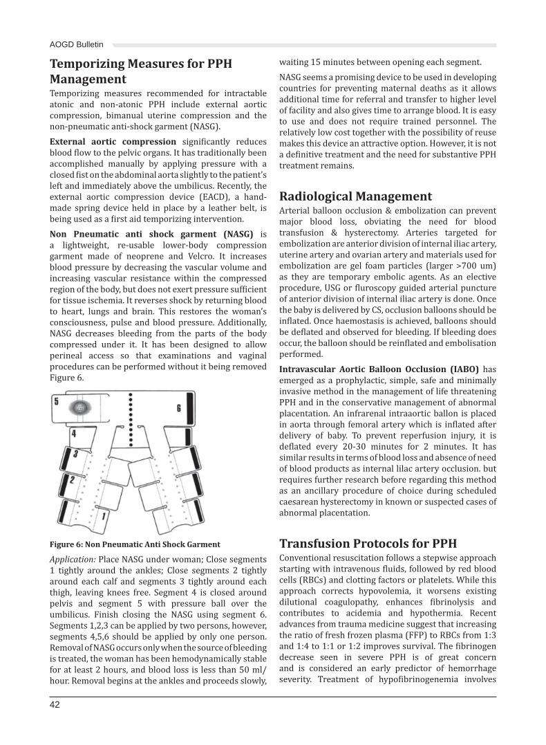
AOGD Bulletin
42
Temporizing Measures for PPH ManagementTemporizing measures recommended for intractable atonic and non-atonic PPH include external aortic compression, bimanual uterine compression and the non-pneumatic anti-shock garment (NASG).External aortic compression signi icantly reduces blood low to the pelvic organs. It has traditionally been accomplished manually by applying pressure with a closed ist on the abdominal aorta slightly to the patient’s left and immediately above the umbilicus. Recently, the external aortic compression device (EACD), a hand-made spring device held in place by a leather belt, is being used as a irst aid temporizing intervention.Non Pneumatic anti shock garment (NASG) is a lightweight, re-usable lower-body compression garment made of neoprene and Velcro. It increases blood pressure by decreasing the vascular volume and increasing vascular resistance within the compressed region of the body, but does not exert pressure suf icient for tissue ischemia. It reverses shock by returning blood to heart, lungs and brain. This restores the woman’s consciousness, pulse and blood pressure. Additionally, NASG decreases bleeding from the parts of the body compressed under it. It has been designed to allow perineal access so that examinations and vaginal procedures can be performed without it being removed Figure 6.
Figure 6: Non Pneumatic Anti Shock Garment
Application: Place NASG under woman; Close segments 1 tightly around the ankles; Close segments 2 tightly around each calf and segments 3 tightly around each thigh, leaving knees free. Segment 4 is closed around pelvis and segment 5 with pressure ball over the umbilicus. Finish closing the NASG using segment 6. Segments 1,2,3 can be applied by two persons, however, segments 4,5,6 should be applied by only one person. Removal of NASG occurs only when the source of bleeding is treated, the woman has been hemodynamically stable for at least 2 hours, and blood loss is less than 50 ml/hour. Removal begins at the ankles and proceeds slowly,
waiting 15 minutes between opening each segment.NASG seems a promising device to be used in developing countries for preventing maternal deaths as it allows additional time for referral and transfer to higher level of facility and also gives time to arrange blood. It is easy to use and does not require trained personnel. The relatively low cost together with the possibility of reuse makes this device an attractive option. However, it is not a de initive treatment and the need for substantive PPH treatment remains.
Radiological ManagementArterial balloon occlusion & embolization can prevent major blood loss, obviating the need for blood transfusion & hysterectomy. Arteries targeted for embolization are anterior division of internal iliac artery, uterine artery and ovarian artery and materials used for embolization are gel foam particles (larger >700 um) as they are temporary embolic agents. As an elective procedure, USG or luroscopy guided arterial puncture of anterior division of internal iliac artery is done. Once the baby is delivered by CS, occlusion balloons should be in lated. Once haemostasis is achieved, balloons should be de lated and observed for bleeding. If bleeding does occur, the balloon should be rein lated and embolisation performed.Intravascular Aortic Balloon Occlusion (IABO) has emerged as a prophylactic, simple, safe and minimally invasive method in the management of life threatening PPH and in the conservative management of abnormal placentation. An infrarenal intraaortic ballon is placed in aorta through femoral artery which is in lated after delivery of baby. To prevent reperfusion injury, it is de lated every 20-30 minutes for 2 minutes. It has similar results in terms of blood loss and absence of need of blood products as internal lilac artery occlusion. but requires further research before regarding this method as an ancillary procedure of choice during scheduled caesarean hysterectomy in known or suspected cases of abnormal placentation.
Transfusion Protocols for PPHConventional resuscitation follows a stepwise approach starting with intravenous luids, followed by red blood cells (RBCs) and clotting factors or platelets. While this approach corrects hypovolemia, it worsens existing dilutional coagulopathy, enhances ibrinolysis and contributes to acidemia and hypothermia. Recent advances from trauma medicine suggest that increasing the ratio of fresh frozen plasma (FFP) to RBCs from 1:3 and 1:4 to 1:1 or 1:2 improves survival. The ibrinogen decrease seen in severe PPH is of great concern and is considered an early predictor of hemorrhage severity. Treatment of hypo ibrinogenemia involves

Volume 17-2; June 2017
43
cryoprecipitate transfusion to maintain ibrinogen levels (100–200 mg/dl). Fibrinogen concentrate contains a greater concentration of ibrinogen and is stable at room temperature and can be rapidly administered, unlike cryoprecipitate, which must be kept frozen and then thawed prior to administration. Rapid blood product selection may bene it from the use of a thromboelastograph, a point-of-care device that examines clot formation and dissolution in whole blood, and provides faster results than laboratory testing and its availability in labour rooms can signi icantly improve PPH management.
ConclusionTimely diagnosis and treatment and prevention are the cornerstone in PPH management. Better training in the form of PPH teaching and drills can improve team preparedness for managing PPH. Greater use of obstetric warning systems and more precise identi ication of warning thresholds such as the shock index to trigger
focused medical attention should expand across facilities.
Suggested Readings1. Shakur H, Elbourne D, Gulmezoglu M, Al irevic Z, Ronsmans
C, Allen E, et al. [33] The Woma Trial (World Maternal Anti ibrinolytic Trial): Tranexamic acid for the treatment of postpartum haemorrhage: An international randomized double blind placebo controlled trial. Trials 2010; 11: 40.
2. World Health Organization. WHO recommendations for the prevention and treatment of postpartum hemorrhage. (2012).
3. Usman N, Nobelet J, Low D, Thangaratinam S. Intra aortic balloon occlusion [12] without luoroscopy for severe postpartum haemorrhage secondary to placenta percreta. Int J Obstet Anesth. 2014;23:91-93.
4. Effect of early tranexamic acid administration on mortality, hysterectomy, and other morbidities in women with post-partum haemorrhage (WOMAN): an international, randomised, double-blind, placebo-controlled trial. Lancet 2017;389(10084):2105-16.

AOGD Bulletin
44
Venous thromboembolism (VTE) is a major cause of maternal morbidity and mortality. The incidence has been reported to range from 0.5-3/1,000 pregnancies occurring with approximately equal frequency in all three trimesters and postpartum1,2. Physiological changes in pregnancy increase the risk of VTE due to all the three
factors described by Rudolf Virchow as Virchow’s triad, i.e. hypercoagulability, stasis and endothelial damage
Obstetric Risk Assessment and Management (Figure 1&2)All women should undergo a documented assessment
of risk factors for VTE in early pregnancy. Risk assessment should be repeated when a woman is admitted to hospital for any reason or develops other intercurrent problems.
Single previous VTE, previous VTE associated with antithrombin de iciency, VTE associated with APLAWomen with previous thromboembolism whether single previous VTE (except those with a single previous VTE related to major surgery and no other risk factors), previous VTE associated with antithrombin de iciency, VTE associated with the antiphospholipid antibody syndrome should be given LMWH antenatally and for at least 6 weeks postpartum or until returned to oral anticoagulant therapy after delivery.The management of these patients should have a multidisciplinary approach involving obstetrician, hematologist and rheumatologist. Proper counseling of patient and the family members should be done regarding the risks and bene its of anticoagulant therapy to the patient and the baby.
Recurrent VTEWomen with recurrent VTE and on long-term warfarin or other oral anticoagulants should be
Thromboprophylaxis in Pregnancy and PuerperiumSandhya Jain1, Vasudha Gupta2
1Associate Professor, 2Senior Resident, Department of Obstetrics & Gynecology, University College of Medical Sciences & Guru Teg Bahadur Hospital, Delhi
Figure 1: Obstetric thromboprophylaxis (Antenatal)

Volume 17-2; June 2017
45
Figure 2: Obstetric thromboprophylaxis (Postnatal)
counseled about the risks of these agents to the fetus. Fetal warfarin syndrome is a condition associated with administration of warfarin during pregnancy. The symptoms are nasal hypoplasia, stippling of uncalci ied epiphyses during irst year, shortened ingers, low birth weight etc. Patients are advised to stop their oral anticoagulant therapy and change to LMWH as soon as pregnancy is con irmed, ideally within 2 weeks of the missed period and before the sixth week of pregnancy. Women not on warfarin or other oral anticoagulants should be advised to start LMWH as soon as they have a positive pregnancy test.3
Other Risk FactorsAccording to RCOG 2015 guidelines women with hyperemesis gravidarum who are admitted to the
hospital for this condition should be given LMWH till the condition resolves. LMWH should also be considered for women with ovarian hyperstimulation syndrome.
Screening for ThrombophiliasThrombophilia screening is not recommended for women who have experienced recurrent fetal loss, placental abruption, FGR, preeclampsia because it is unclear if anticoagulation therapy reduces recurrence. Management decisions regarding thromboprophylaxis or antepartum surveillance should be made through an individual risk assessment including prior history of VTE, inherited thrombophilia, cesarean delivery, immobility, obesity, and family history of VTE as mentioned earlier.
Recommendations for screening include:• Women with a personal history of VTE.• Women with a irst-degree relative (parent or sibling) with a history of thrombophilia.Ideally, screening tests for thrombophilias should be done
at least 6 weeks after a thrombotic event and while the patient is not pregnant or on anticoagulation therapy.
If a patient has these clinical indications, recommended screening tests include4:• Factor V Leiden mutation• ProthrombinG20210A mutation• Protein C de iciency• Protein S de iciency• Antithrombin de iciency
Thromboprophylaxis during labour and delivery, including the use of regional analgesia
LMWH, UFHRegional anaesthesia should be avoided if possible until at least 12 hours after the previous prophylactic dose of LMWH and 24 hours after last therapeutic dose of

AOGD Bulletin
46
LMWH. LMWH should not be given for 4 hours after use of spinal anesthesia or after the epidural catheter has been removed and the catheter should not be removed within 12 hours of the most recent injection.Women receiving antenatal LMWH having an elective caesarean section should receive a thromboprophylactic dose of LMWH on the day prior to delivery and, on the day of delivery, any morning dose should be omitted. Warfarin, LMWH, and UFH do not accumulate in breast milk thus they can be used in women who breastfeed.The irst thromboprophylactic dose of LMWH should be given as soon as possible after delivery provided there is no postpartum haemorrhage and regional analgesia has not been used. The risk of postpartum VTE after an emergency caesarean section is twice that after an elective caesarean section and four times that after a vaginal delivery. Therefore women who have had emergency caesarean section in labour should be considered for thromboprophylaxis with LMWH for 10 days after delivery apart from those having an elective caesarean section who should be considered for thromboprophylaxis with LMWH for 10 days after delivery if they have any additional risk factors.3
Women at high risk of haemorrhage including major antepartum haemorrhage, coagulopathy, progressive wound haematoma, suspected intra-abdominal bleeding and postpartum haemorrhage may be managed with anti embolism stockings (AES), foot impulse devices or intermittent pneumatic compression devices. Unfractionated heparin (UFT) may also be considered.
Agents Used for ThromboprophylaxisLMWHs are the agents of choice for antenatal and postnatal thromboprophylaxis and the dose is weight based.3
Table 1: Suggested thromboprophylactic doses for antenatal and postnatal LMWHWeight Enoxaparin Dalteparin Tinzaparin
(75 u/kg/day)<50 kg 20 mg daily 2500 units
daily3500 units daily
50-90 kg 40 mg daily 5000 units daily
4500 units daily
91-130 kg 60 mg daily* 7500 units daily
7000 units daily*
131-170 kg 80 mg daily* 10 000 units daily
9000 units daily*
>170 kg 0.6 mg/kg/day*
75 u/kg/day 75 u/kg/day*
High prophylactic dose for women weighing 50–90 kg
40 mg 12 hourly
5000 units 12 hourly
4500 units 12 hourly
*may be given in 2 divided doses
Monitoring of anti-Xa levels is not required when LMWH is used for thromboprophylaxis. In women at very high risk of thrombosis UFH may be used peripartum period
in preference to LMWH where there is an increased risk of haemorrhage or where regional anaesthetic techniques may be required. The precautions for LMWH use are summarised in Table 2.If UFH is used after caesarean section (or other surgery), the platelet count should be monitored every 2–3 days from days 4–14 or until heparin is stopped. The risk of heparin-induced thrombocytopenia (HIT) is substantially lower with LMWH.Warfarin use in pregnancy is restricted to the few situations where heparin is considered unsuitable, e.g. women with mechanical heart valves. Women receiving long-term anticoagulation with warfarin can be converted from LMWH to warfarin postpartum when the risk of haemorrhage is reduced, usually 5–7 days after delivery. Warfarin is safe in breastfeeding.The use of properly applied anti-embolism stockings (AES) of appropriate size and providing graduated compression with a calf pressure of 14–15 mmHg is recommended in pregnancy and the puerperium for women who are hospitalized and have a contraindication to LMWH. These include women who are hospitalized post-caesarean section (combined with LMWH) and considered to be at particularly high risk of VTE (e.g. previous VTE, more than four risk factors antenatally or more than two risk factors postnatally) and women travelling long distance for more than 4 hours.3 Aspirin is not recommended for thromboprophylaxis in obstetric patients.
Table 2 : Contraindications/cautions to LMWH useKnown bleeding disorder (e.g. haemophilia, von Willebrand’s disease or acquired coagulopathy)Active antenatal or postpartum bleedingWomen considered at increased risk of major haemorrhage (e.g. placenta praevia)Thrombocytopenia (platelet count < 75 × 109/l)Acute stroke in previous 4 weeks (haemorrhagic or ischaemic)Severe renal disease (glomerular iltration rate [GFR] < 30 ml/minute/1.73m2)Severe liver disease (prothrombin time above normal range or known varices)Uncontrolled hypertension (blood pressure > 200 mmHg systolic or > 120 mmHg diastolic)
References1. Walker MC, Garner PR, Keely EJ. Thrombosis in pregnancy:
A review. J Soc Obstet Gynaecol Can 1998;20(10):943-52.2. Toglia MR, Nolan TE. Venous thromboembolism during
pregnancy: A current review of diagnosis and management. Obstet Gynecol Survey 1996;52(1):60-72.
3. Reducing the Risk of Venous Thromboembolism during Pregnancy and the Puerperium Green-top Guideline No. 37a April 2015. Available from: https://www.rcog.org.uk/globalassets/documents/guidelines/gtg-37a.pdf. Accessed on 20th May 2017.
4. ACOG. Inherited Thrombophilias in Pregnancy Practice Bulletin #138. The American College of Obstetricians and Gynecologists, 2013.

Volume 17-2; June 2017
47
India has made tremendous strides in healthcare and the maternal mortality ratio (MMR) has reduced from 600 in 1990 to 167 per 100,000 live births in 2011-2013. Though maternal mortality since 1990 has reduced by 69 % in India, as compared to a global reduction of 44%, India still accounted for 15% of global maternal deaths in 2015. While India’s MMR is reducing at an average of 4.5% annually, this rate has been increased to 5.5% to meet the Millennium Development Goal of 140 deaths per 100,000 live births.National Family Health Survey 2015-16 (NFHS-4) data shows signi icant improvement in several maternal and child health indicators due to initiatives started by the government under National Rural Health Mission. The proportion of institutional deliveries has increased from 38.7% in 2005-06 to 78.9% in 2015-16. Birth assisted by trained personnel has improved from 46.6% in 2005-06 to 81.4% in 2015-16. Although these interventions have helped to bring down MMR, for further reduction of MMR, there is a need to understand the relation between morbidity and mortality. Any pregnant woman can develop life threatening complications with little or no warning. A continuum of adverse pregnancy events may be recognized as follows: Normal healthy pregnancy→ high risk pregnancy→ morbidity→ severe morbidity→ near miss → mortality. Therefore, there is a need to target high risk mothers who constitute approximately 7-8%, pick up early warning signs and step up their management to provide high quality, dedicated critical maternal care to alter the future outcome/prevent progression to death.
Critical Care: GradingCritical care has been graded into 4 levelsGrade 0- Patients who can be treated in the general wardGrade 1- Patients at risk of deterioration of clinical condition or those who have been shifted out from higher level of care, who need to be cared for in the general ward with additional advice and support of critical care specialists/ multidisciplinary team.Grade 2- Patients who require detailed monitoring and intervention including support for a single failing organ system or post operative care or those who have been shifted out of higher level of care.Grade 3- Patients who require advanced respiratory
support or respiratory support along with support of at least 2 organ systems including patients with multi organ failure.
De inition and IndicationsIntensive care unit (ICU) is a specialized area of the hospital which is speci ically designed, staffed, located, furnished and equipped, dedicated to management of critically sick patient, injuries or complications. ICU provides highest grade or grade 3 critical care. Obstetric ICU is an ICU which is dedicated to pregnant women before, during and after delivery. High Dependency Unit (HDU) or intermediate care unit or critical care unit is an area in a hospital where patients can be cared
Setting up of an Obstetric Intensive Care Unit and High Dependency UnitPikee Saxena1, Supriya Goyal2
1Professor, 2Senior Resident, Department of Obstetrics & Gynecology, Lady Hardinge Medical College & Smt. Sucheta Kriplani Hospital, New Delhi
Table 1: The Scope of Obstetric HDU: Following conditions may require admission in Obstetric HDU
Obstetric complications• Pregnancy/ labor pain with sever anaemia (< 7 gm%) and
its complications• Accidential hemorrhage• Multiple gestation with complications.• Post Partum Hemorrhage• Placenta previa, morbidly adherent placenta• Severe pre-eclampsia/hypertensive crisis, ecclampsia,
HELLP syndrome• Obstetric hysterectomy, broad ligament hematoma• Perforation during abortion• Sepsis & Systemic in lammatory Syndrome(SIRS).Pregnancy with Medical complications• Diabetes and diabetic ketoacidosis• Cardiac Disease• Jaundice• Thyroid storm, thyrotoxicosis• Pheocromocytoma, other endocrinal crisis like Addison’s
disease• Renal disease like ARF• Leukemia and other hemolytic disorders• Infections: Dengue, complicated malaria, tuberculosis• Asthma and other respiratory problems• Thrombophillia, DIC• Pulmonary Edema due to perioperative luid overload,
CCF, complication of severe pre-eclampsia or tocolytic therapy with beta-agonists etc
Others: Ruptured ectopic, Hydatidiform mole, Complications due to uterine anomaly and pathologies, Postoperative patients requirig hemodynamic monitoring or intensive nursing care, Appendectomy or any other surgical emergency, pregnancy with trauma, poisoning, burns, cancer, OHSS and pregnancy

AOGD Bulletin
48
more comprehensively than in a normal ward, but not to the point of intensive care. Patients in HDU may require ICU admission later (step up) or may be shifted from ICU to HDU (step down), before shifting them to the general ward. HDU provides grade 2 critical care.Obstetric HDU is a part of the obstetrics unit located near the labor room and operation theatre, for easy and prompt shifting of the patient whenever required. It is recommended that all pregnancies with complications be managed in obstetric HDU. The HDU should be headed by an experienced obstetrician with signi icant input from anesthetists and other multispecialty physicians. HDU is more patient friendly where breastfeeding can be continued and has lower rates of infection than in the ICU. The indications are summarized in Table 1.
Process of Shifting Patient from Ward/ Triage Room to HDU or ICU/ Obstetric ICUThe key steps to be taken are:1. Inform the family/companion of pregnant woman of
the decision, and take a written informed consent.2. Case sheets containing history, examination,
investigations and management should be maintained.3. Obstetric HDU staff should be appropriately informed.4. The patient should be escorted by doctor/staff with
all existing treatment including continuation of patent IV line.
5. Keep monitoring the vitals of the patient, temperature should be maintained.
6. Adequate follow-up by the treating doctor.7. Supine hypotension prevention (by performing
lateral tilt to 15-20 degree), if required.8. Ensure patent airway.9. Baby should be shifted along with the mother, if
delivered already.
Obstetric ICU/ HDU: The set upWhere to set up Obstetric HDU & Obstetric ICU?It has been suggested that all District Hospitals should have an Obstetric HDU and all the Medical Colleges should have both- an obstetric HDU and an obstetric ICU (or ICU with dedicated obstetric beds). It should have an isolation room for mothers with infections requiring isolation (H1N1, Chicken pox etc.).
Table 2: Number of beds in obstetric HDU
Number of deliveries per month
Number of beds required
Up to 250 4250-500 8>500 Can be increased
LocationIt should be located near the labor room and operation theatre and should have proximity to other areas and essential support services such as the main wards, ICU, radiology, laboratory, blood bank etc. There should be single entry/ exit point to obstetric HDU and obstetric ICU. The isolation room should have a separate entry.There should be provision for emergency exit points for ease of evacuation in cases of disasters.
SpaceThe space should be 120-130 sq. ft. per bed in obstetric HDU and 130-150 sq. ft. per bed in obstetric ICU. Apart from the patient care area, there should be an ancillary area (of 100% to 120% extra space) for accommodating the nursing station/ storage/ patient movement area/ equipment area, patient toilet and to maneuver equipment, beds and trolleys etc. The beds should be at least 2 ft. from the back wall to give caregivers an easy access to the head in case of any emergency. There should be space for bed-side movable lockers with facility of trolleys/ drawers and for keeping medicines, consumables and personal belongings of the patient.
PrivacyThere should be a partition between the beds for ensuring privacy of the patients.If the space is more, each bed can be separated by ixed partitions or curtains. The curtain fabric should
be ire and water proof, washable, clean, light colored, inherent stain resistance and non- allergic. The curtain height is determined by the loor-to-ceiling height, and curtains usually should inish approximately 8-10” above the inished loor (Joint Commission’s Patient Privacy Standards). The curtains should have mesh at the top which will allow both light and ventilation in the patient room. The curtains should be hanging from the overhanging rails.
FlooringThe loor should be made of large vitri ied, antiskid, stain proof and easy to clean tiles with seamless joints. The tiles should be of light color (preferably white or off-white).
WallsThe walls should be of durable glazed tiles which are easy to clean, stain resistant, lame resistant and have a visual appeal. It will be preferable to have a inishing of wall height of up to 6-7 ft. with the tiles like loor tiles. Colors should be chosen carefully to avoid an adverse impact on the skin color of patients and neonates, light color (white or off-white) being the preferable choice.
CeilingThe ceiling should be leak proof. It is suggested that no lines or wires be kept or run over the ceiling or underground. It should be easy to explore in case repair

Volume 17-2; June 2017
49
is required, as damages are common and may occur any time.
Nursing StationThe nursing station should be with the following facilities and seating capacity:
Table 3: Staff requirement for Obstetric HDU/ ICU
Staff Requirements (per shift)8 bedded HDU 4 dedicated nurses
and 2 EmOC/ MOs4 bedded HDU 2 dedicated nurses
and 1 EmOC/ MOFor 4 bedded obstetric ICU (no separate guidance is being given for existing ICU in medical colleges)
4 dedicated nurses and 1 intensivist
Space requirements for nursing station:a. Adequate space for central monitoring and 2
computers (for 8 bedded HDU/ ICU)b. Adequate space for central monitoring and 1
computer (for 4 bedded HDU).c. Scrub area and wall clock behind the nursing station.d. Facility for keeping records and emergency medicines.
Health Management and Information System (HMIS): Facility of HMIS entry should be provided for data collection, data transmission, data storage, data processing, data analysis and presentation of the data. This will help in decision making by the hospital management for improved health care services.Store Room: A separate room should be made to keep the equipment such as the sonography machine, portable X-ray machine, transport ventilator, nebulizer, radiant warmer, blood warmer, crash cart(s), BIPAP/ CPAP machine, etc. A general store room should be made separately to keep bed linen, disposables and consumables, personal protective attire like caps and masks, slippers, etc.Buffer Zone: A protective clean zone should be made available located before the entry in obstetric HDU/ ICU. This zone would include area for changing shoes, patient’s changing room with lockers, janitor’s closet, two changing/ overnight stay rooms for doctors (1 each for male and female with lockers, beds, book shelves etc.), dining area, space to keep wheel chairs and trolleys, changing room for staff, toilets (1 each for male and female with urinal in the male toilet with lush facility).Autoclaving and Sterilized Supply: From the Central Sterilized Supply Department (CSSD).Waiting Area: Waiting area for the patients’ attendants should be provided, with facility for seating capacity of at least 2 relatives per patient, facility of drinking water, a large TV with LCD display, toilets, newspapers and educational material etc. The waiting area can be shared with waiting area for the LR and/or OT.
Facilities:a) All cots in the obstetric HDU/ ICU must be birthing
cum patient bed with the following features: Safe working load of 180 kg (392 lbs). Size 1800x620x800 mm. Electrically/manually operated with battery back-up in the event of power failure. Dual-sided manual CPR levers. Easy-to-operate Trendelenburg, reverse Trendelenburg, with both side tilts with compact size to it into the small delivery room. Retractable platform should be present, optimizing access to perineal area, mother and baby during the delivery procedure. Mattress should be in 3-parts of minimum 6 inch, with perineal cut-out seat section. Mattress should be washable with seamless molding. Adjustable safety rails should be present on both the sides. Good quality castors with brake system for optimum mobility/stability. Comprehensive range of easy-to- it accessories, including adjustable leg rests, IV rod, handgrips, high and low foot supports, a removable step and a luid collection bowl. Removable, moulded head and foot panels should be present.
b) Each bed will require at least 2 oxygen, 1 air and 2 suction outlets, and at least 10 central voltage stabilized power points (6 power-points of 5 amps and 4 points of 15amp) preferably 5 on each side of the bed. Adapters should be discouraged as they tend to become loose.
c) Heating, Ventilation and Air-conditioning (HVAC) system along-with ceiling fans & power backup exclusive for HDU for uninterrupted power supply should be installed. Voltage stabilization is mandatory. Suitable and safe air quality must be maintained always. Temperature should be adjustable within each cubicle/ room as per the patient comfort and choice.
d) General Lighting: Access to outside natural light is recommended. The colorless concealed LED Lights with suf icient high illumination should be provided. It should be bright enough to ensure adequate vision without eye strain.
e) Hand Hygiene and Prevention of Infection: Sink should be of the operation room style with water supply through elbow operated taps. Every bed should have alcohol based anti-microbial instant hand wash solution source. All entrants should don a mask and cap, and ideally an apron which should be replaced daily.
f) Waste Disposal and Pollution Control: This is mandatory and a huge safety issue both for the patient and the staff/doctors of the hospital, and society at large. It is important that all government regulations (State Pollution Control Board) should strictly be complied with.
g) Laboratory backup facility for 24 hours.h) In-house or nearby blood bank.i) In- house or nearby neonatal intensive care unit.j) Lactation Support: Facilities for breast feeding (or

AOGD Bulletin
50
use of breast pump) should be available to all post-natal mothers in obstetric HDU. Babies are usually not allowed in obstetric ICU, but breast pump facility should be made available.
k) Provision for ire safety, preferably through automatic water sprinkling system or normal ire extinguishers.
l) For uninterrupted power supply, noiseless generator or inverter with instant switchover, search protector and voltage stabilizer. An earthing pit for proper earthing should be ensured, with monitoring once in 2 years.
m) Nurse calling system with a central display and an audio-visual alarm.
n) Facility for keeping records and registers at the nursing station.
o) Ambulance: One hi-tech ambulance with all advanced life support measures for critically ill mother, and with a transport incubator and ventilator support for critical new born.
Equipment required in HDUIt includes vital monitors depicting Pulse, BP, RR, ECG, SPO2 and with transducer facility for invasive monitoring, equipment for insertion and management of invasive monitoring (arterial line and central venous pressure (CVP)), piped oxygen and suction, anesthesia trolley, IV luid warmer, forced air warming device, electric blanket, blood gas analyzer, infusion pumps, syringe pumps, uro lowmeters, delivery tray, emergency hemorrhage/eclampsia trolley, intubation tray, neonatal resuscitation equipment, mobile X ray, USG machine, CTG machine, transfer equipment – monitor and ventilator, computer terminal to facilitate access to blood results, a crash cart fully loaded with BCLS (basic cardiac life support) medications, copy of hospital obstetric guidelines, resuscitation trolley with de ibrillator and airway management equipments, CNS tray with torch and hammer. Availability of all essential and emergency drugs should be ensured after checking the date of expiry on a regular basis. Intermittent compression device for DVT prophylaxis, color coded bins for biomedical waste, wall clock should be available.
Human ResourcesObstetrician is the head of the obstetric HDU. He/ she will decide when and whom to call from the list of multidisciplinary teams (wherever available). For management of the obstetric patientsEmergency obstetric care/ Medical Of icers should be posted round the clock. Obstetric nursing staff (24x7) with nursing staff to patient ratio should be 1:2; there should be 1 extra for lay off or covering leave/day off. Support Staff includes pharmacist, dietician, counsellor, housekeeping and cleaning, security, data entry operator and electrical technician.
Training of Staff of HDUTraining should be given to staff, medical of icer, residents and consultants by posting them in the ICU and labor room for 3 months for basic resuscitation, intubation, IV luid management, blood component transfusion, ICU procedures, obstetric drills, record keeping and management of equipment used in the HDU. Refresher training should also be arranged at regular intervals.Monitoring and Record keeping in a HDU is of utmost importance and should include history, thorough examination, details of investigations and management, probable or inal diagnosis, details of any operative management, nursing charts including one hourly temperature, pulse, respiratory rate, blood pressure, pulse oxymetry, strict input and output, luid balance and details of luid administered, abdominal girth, CVP charting whenever required, anesthesia charts and detailed drug charts. Antenatal patients should be nursed in left lateral position and fetal heart sound should be monitored by CTG. Adequate pain management should be done. All teams looking after the patient should have appropriate coordination with each other and should be informed about any change in treatment. Timing, date and signatures should be put on case records.The patient and her relatives should be treated with kindness, respect and dignity always. The relatives need to be updated regularly regarding the patient’s progress and a written informed consent should be obtained for any procedure. Ideally protocols and standard operating procedures should be developed to improve care for patients at a local, national and international level.Suggested discharge/transfer criteria for the HDU is that the patient is conscious and alert, has stable and normal respiratory status and hemodynamic parameters with no evidence of hemorrhage, intensive/invasive monitoring is no longer required and 4-hourly recording of vital signs is considered appropriate. In case the patient develops pulmonary edema / ARDS/ systemic in lammatory response syndrome/ DIC /multiorgan failure with poor cardiac output despite luid resuscitation/septic shock, patient should be shifted to Obstetric ICU.
Suggested Readings1. Guidelines for Obstetric HDU & ICU. Maternal Health
Division, Ministry of Health and Family Welfare, Government of India, March 2016
2. rchiips.org/NFHS/factsheet_NFHS-4.shtml. Accessed on 20/5/17.
3. Handbook of Obstetric High Dependency Care, 1st edition. By © D. Vaughan, N. Robinson, N. Lucas and S. Arulkumaran. Published 2010 by Blackwell Publishing Ltd
4. ICU planning and designing in India-guidelines 2010, Guidelines Committee (ISCCM).

Volume 17-2; June 2017
51
Obstetric hemorrhage and anemia remain a major cause of maternal morbidity and mortality worldwide. Thrombotic and vascular complications of several maternal diseases like diabetes, renal failure, hemoglobinopathies, systemic lupus, and antiphospholipid syndrome do not cause primary bleeding per se but increase maternal and fetal morbidity by inducing preeclampsia, thrombo-embolism, and placental insuf iciency. These maternal diseases associated with maternal thrombocytopenia and coagulopathies can pose primary bleeding problems requiring transfusion.
Indications for Maternal TransfusionsMajor obstetric bleedingThe goals are to establish rapid control of bleeding and restore systemic oxygen delivery. Recent complex trauma protocols advocate that when massive bleeding is anticipated, the patient should be rapidly transfused with an optimal ratio of RBC, plasma, cryoprecipitate and platelets, without waiting for consumption and dilutional coagulopathies. Restoration of the components of the blood is also essential to ensure adequate tissue perfusion and to prevent acidosis and hypothermia.The therapeutic components are sequentially administered, beginning with crystalloid- colloid solutions infused to replace lost intravascular volume. Second, red blood cells (RBCs) are transfused to restore oxygen carrying capacity. Most protocols recommend six units of packed red cells (PRBCs) be prepared and available and hematocrit be maintained minimally at 21–24%. WHO recommends transfusing Group O negative antibody-screened blood, and/or uncrossmatched group speci ic blood until fully cross-matched blood is available.
Component therapyFresh frozen plasma (FFP) is plasma that has been separated from a unit of whole blood within 6–8 hours of donation and is rich in all intrinsic clotting factors. Cryoprecipitate is the cold-insoluble portion of plasma remaining after FFP has been thawed. It contains approximately 50% of the Factor VIII and von Willebrand Factor, 20–40% of the ibrinogen and some of the Factor XIII originally present in the fresh plasma. Coagulation screens should be performed after 500ml
blood loss, in case of active ongoing bleeding. The tests include prothrombin time (PT), partial thromboplastin time (PTT), ibrinogen, D-dimer, and a complete blood count. Clotting factors and platelets (PLTs) are delivered to restore physiologic hemostasis.Fresh frozen plasma (12-15 ml/kg), 1 unit for every 4–6 units of blood is given to prevent coagulation defects resulting from use of stored red cell concentrates. At least 10 packs of cryoprecipitate, prepared from single donor units, containing 3–4 gm ibrinogen in total should be transfused in massive hemorrhage. Some studies have shown that using a high ratio of PRBCs to FFP (1.5:1 or 1:1) has signi icantly improve survival from hemorrhage after trauma and similar recommendations have been established for massive obstetric hemorrhage protocols with the goal of maintaining the INR at <1.5–1.7, ibrinogen level above 1.5 gm/L and platelet count at >50×109/L. Blood products should not be continued on basis of clinical suspicion alone and their use should be guided by the results of coagulation tests only. The FFP and cryoprecipitate should ideally be of the same group as the recipient. If unavailable, FFP of a different ABO group is acceptable, if it does not have a high titer anti-A or anti-B activity. If these blood components are not available, the freshest whole blood available (ideally no more than 36 hours old) is transfused.
Platelet transfusions during pregnancy and for deliveryWhatever be the cause of thrombocytopenia, most guidelines recommend a platelet count above 50×109/L for vaginal delivery. Guidelines differ in their threshold for giving local anesthesia, most guidelines advise 50 or 80×109/L. According to RCOG, platelet counts should not fall below 50×109/L in a woman with ongoing hemorrhage, and 75×109/L should be the threshold for platelet transfusion in ongoing bleeding to keep the safety margin. The recommendation of Indian Society of Hematology and Transfusion (ISHBT) for platelet threshold for vaginal delivery is 30×109/L, for cesarean section is 50×109/L and for epidural anesthesia is 80×109/L. Because of different pathophysiology and bleeding history, an individual treatment plan for every pregnant patient with thrombocytopenia is required. Except in case of TTP which is a relative contraindication for platelet transfusions, platelet transfusions are not contraindicated in ITP or DIC. The platelets should ideally
Transfusion Guidelines in Obstetric PracticePriyanka GogoiAssociate Professor, Department of Pathology and Blood Bank, University College of Medical Sciences& Guru Teg Bahadur Hospital, Delhi

AOGD Bulletin
52
also be group compatible and if RhD-negative woman receives RhD-positive platelets, anti-D immunoglobulin (dose of 250 IU) should be administered.If a patient continues to bleed or future hemorrhage is anticipated, additional components either in the original ratio or customized as per the patient’s need, can be requested from the transfusion service. Additional samples for coagulation pro ile are sent with each request and abnormal laboratory values such as prolonged PT and/or aPTT, low PLT count, and low ibrinogen levels can be addressed individually.
Intraoperative cell salvage (IOCS)It may have a role in obstetric practice in reducing the exposure of obstetric patients to the risks of allogenic blood transfusion. It is the process by which blood shed within the surgical ield is retrieved by an anticoagulated suction apparatus and collected within a reservoir from where it is centrifuged, washed and pumped into an infusion bag. This salvaged blood can then be returned to the patient. The process has been widely used in adult orthopedic and cardiac surgery without complication. However, its use in obstetric practice has been limited, owing to concerns about contamination by amniotic luid, speci ically the risks of amniotic luid embolism,
and by fetal blood cells. In the present scenario, the use of cell salvage in obstetrics remains controversial and the experience of its use remains limited.In a similar vein, the ef icacy and safety of the use of recombinant factor VIIa (rFVIIa) for cases of obstetric hemorrhage remains to be established.
Chronic anemia in pregnancyTransfusion protocols should not be based on the patient’s hemoglobin concentration alone, but also on her clinical needs, including stage of pregnancy and existing clinical conditions like established or incipient cardiac failure or clinical evidence of hypoxia, co-existing infections and morbidities including pre-existing heart disease. The need for transfusion is also determined by the mode of delivery and history of hemorrhage in the present or previous pregnancies. The speci ic indications for transfusion for chronic anemia in pregnancy should be based on national guidelines, modi ied as appropriate to the local situation. Usually hemoglobin levels of < 7gm% require transfusion.
Postpartum anemiaAlthough guidelines suggest a transfusion threshold at a hemoglobin concentration of 7.0-8.0gm/l, results of various studies underscore that in case where there is no continuing or threat of bleeding, transfusion is only required for symptomatic anemia.
Storage of Blood and Transfusion PracticesWhole blood and red cells must be stored at a temperature of +2°C to +6°C and red cells must never be allowed to freeze. This is important to maintain ATP and prevent clotting, hemolysis and infection. The maximum storage time is 35 days. Fresh frozen plasma (FFP) and cryoprecipitate are rapidly frozen, and maintained at, a temperature of –20°C or colder. Once thawed they must be kept at +2°C to +8°C and transfused within 6 hours. To maintain platelet function, platelet concentrates should be stored at a temperature of +20°C to +24°C with continuous agitation on a special platelet agitator. They should never be placed in a refrigerator or freezer. They can be stored for up to 72 hours and upto ive days in specialized platelet packs. If no platelet agitator is available, platelet concentrates must be transfused within 30 minutes of completion of testing, else within 4 hours.
Special Precautions and ComplicationsEnsure blood group cross matched transfusion of packed RBC and platelets to minimize risks of isoimmunization. A written informed consent with complete identity check is advisable. Always match the details on the bag with patient details and check compatibility report.Besides enhanced induction of alloantibodies, blood products can potentially transmit infections {including HIV, Hepatitis B, Hepatitis C, malaria, syphilis, cytomegalovirus (CMV)} which can be reduced by proper selection and screening of donors. Voluntary, non-remunerated donors have been proven to be safer donors than relatives. Improvement of a system of repeat replacement donors is feasible and is worth giving more attention to avoid irst-time relatives who have high rates of hepatitis and HIV infections.Transfusion reactions (haemolytic and nonhaemolytic) causing signs of fever, rash, urticaria, tachycardia and hypotension are still common, occurring in up to 13% of blood recipient. Most of these are minor reactions but immune hemolysis which can be fatal is estimated to occur in about 1 in 6000 units transfused. Rapid infusion of cold blood and stored red cell may give rise to hypothermia, hyperkalemia and hypocalcemia.
ConclusionStrategies aimed at diagnosing and treating chronic anemia during pregnancy and active management of the third stage of labor to minimize blood loss may reduce the need for blood transfusion. This is important as unnecessary blood transfusion is not only wastage of a precious resource but also increase exposure to transfusion-related risks and infections. Protocol to

Volume 17-2; June 2017
53
manage major obstetric hemorrhage should be in place with involvement of a consultant obstetrician, anesthetist and hematologist and the blood bank.
Suggested Reading1. Carrolli G, Cuesta C, Abalos E, Gulmezoglu AM. Epidemiology
of post partum haemorrhage: a systematic review. Best Pract Res Clin Obstet Gynaecol 2008;22:999–1012.
2. Royal Australian and New Zealand College of Obstetricians and Gynaecologists. Management of Postpartum Haemorrhage (C-Obs 43). Australia, 2011.
3. University of Washington. Obstetric Emergency Hemorrhage Protocol. CMQCC Obstetric, Hemorrhage Toolkit, Obstetric Hemorrage Care Guidelines and Compendium of Best Practices. Reviewed by CADPH-MCAH: 11/24/09.
4. Burtelow M, Riley E, Druzin M, Fontaine M, Viele M, Goodnough LT. How we treat: management of life-threatening primary postpartum hemorrhage with a standardized massive transfusion protocol. Transfusion. 2007; 47(9):1564-72.
5. Shields LE, Wiesner S, Fulton J, Pelletreau B. Comprehensive maternal hemorrhage protocols reduce the use of blood products and improve patient safety. AmJ Obstet Gynecol 2015;212(3):272–80.
World Blood Donor Day 2017 will be celebrated by people all around the world on 14th of June 2017 to mark the birth anniversary of Karl Landsteiner on 14th of June in 1868. This event celebration was irst started in the year 2004 by “the World Health Organization, the International Federation of Red Cross and Red Crescent Societies” to raise public awareness about the need for safe blood donation (including its products) voluntarily and unpaid by a healthy person. The slogan of the year 2017 celebration is “Give blood, Give now, Give often” . World Blood Donor Day is celebrated to ful ill the need of blood transfusion, save more than millions of lives annually, help patients suffering from variety of life-threatening health conditions and caring for the women during pre-and post pregnancy. Some of the objectives of the World Blood Donor Day celebration are mentioned below:• World Health Organization has aimed to obtain the
suf icient blood supplies from the voluntary and unpaid blood donors all over the world by 2020.
• To motivate more voluntary blood donors through educational programs and campaigns worldwide and acknowledge the people who are already donors.
• WHO runs this campaign by organizing many activities in all countries highlighting people’s stories who need immediate blood donation to continue their heart beat.
• It is celebrated to reduce the mortality rate because of insuf icient blood supply.
WHO Theme poster 2017
Day of the Month: World Blood Donor DayBindiya GuptaAssistant Professor, University College of Medical Sciences & Guru Teg Bahadur Hospital, Delhi

AOGD Bulletin
54
“It is worth dying to ind out what life is.” - T.S. EliotNothing fascinates humans more than the mystery of death or especially what lies beyond death. Death is de ined as the cessation of all vital functions of the body. Death is basically the moment at which life ends and life may be de ined in terms of consciousness. A near-death experience (NDE) is a personal experience associated with death or impending death. They have also been described as Out of Body Experiences. When someone almost died only to return back and live further, their experiences arouse curiosity and perhaps an insight into the moment of death. The question one wants to know is as to what happens at that moment, Do we just die or part of us, the Soul or Consciousness, (as believed in many religions and spiritual systems) moves to another realm.The irst recorded NDE was in the time of Plato. NDEs have been recorded over the centuries from all around the world and from countries with widely diverse cultures. Reported incidence of NDEs varies from 4-15%. NDEs are not dreams, hallucinations, or the random memories of a dying brain. In 1968 Celia Green published an analysis of 400 irst-hand accounts of out-of-body experiences. These experiences were popularized by the work of psychiatrist Raymond Moody in 1975 as the near-death experience (NDE).
ND ExperiencesAbout 10-20% of people experiencing a life-threatening event will have a NDE. Although the features of NDEs vary from one case to the next, a striking feature is their remarkable similarity of content like:• A sense of peace, well-being and painlessness. A sense
of removal from the world.• An out-of-body experience. A perception of one’s
body from an outside position. Sometimes observing medical professionals performing resuscitation efforts.
• A “tunnel experience” or entering a darkness. A sense of moving up, or through, a passageway or staircase.
• A rapid movement toward and/or sudden immersion in a powerful light (or “Being of Light”) which communicates with the person.
• An intense feeling of unconditional love and acceptance.• Encountering “Beings of Light”, “Beings dressed
in white”, or similar. Also, the possibility of being reunited with deceased loved ones.
• Receiving a life review, commonly referred to as “seeing one’s life lash before one’s eyes”.
• Receiving knowledge about one’s life and the nature of the universe.
• Approaching a border, or a decision by oneself or others to return to one’s body, often accompanied by a reluctance to return.
• Suddenly inding oneself back inside one’s body.• Connection to the cultural beliefs held by the
individual, which seem to dictate some of the phenomena experienced in the NDE and particularly the later interpretation thereof.13
Kenneth Ring (1980) subdivided the NDE on a ive-stage continuum of Peace, Body separation, Entering darkness, Seeing the light & Entering the light. He stated that 60% experienced stage 1 but only 10% experienced stage 5.
After-effectsNDEs are associated with changes in personality and outlook on life like greater appreciation for life, higher self-esteem, greater compassion for others, less concern for acquiring material wealth, a heightened sense of purpose and self-understanding, desire to learn, elevated spirituality, greater ecological sensitivity and planetary concern, and a feeling of being more intuitive.The NDE literature contains other accounts of remarkable and even apparently medically inexplicable healing following a NDE. Recently lot of interest & curiosity about NDEs has been generated on social media by videos and talks by Anita Moorjani. She was diagnosed with terminal cancer. It was at this point that she had Near Death Experience which was described as crossing over to other dimension and then returning again into this world with a clearer understanding of her life and purpose on earth. This understanding subsequently led to a total miraculous recovery of her health.
Near-death StudiesRaymond Moody’s book Life After Life, which was released in 1975, brought public attention to the topic of NDEs. This was soon to be followed by the establishment
“Mind, Body & Soul”“Mind, Body & Soul”Mystery Surrounding Near Death ExperiencesRashmiAssistant Professor, Deptt of Obstetrics & Gynecology, University College of Medical Sciences& Guru Teg Bahadur Hospital, Delhi

Volume 17-2; June 2017
55
of the International Association for Near-Death Studies (IANDS) in 1981 which encourages scienti ic research and education on the physical, psychological, social, and spiritual nature and rami ications of near-death experiences.In 1998 Dr. Long (a radiation oncologist) and his wife, Jody, began the Near Death Experience Research Foundation with the goal of creating a forum for near death “experiencers” to share their stories. Awareness during Resuscitation (AWARE) study was a multicenteric study in UK & USA which tested consciousness, memories and awareness during cardiac arrest. 9% could be described as real NDEs. Results from AWARE II study which was completed in March, 2017, are still awaited.Approaches to explain NDEs fall into three broad groups i.e. Spiritual theories (also called transcendental), Psychological theories and Physiological theories that provide a physical explanation for NDEs.
Psychological ExplanationsThese explain NDEs as psychological responses resulting in hallucinations or withdrawal from reality and events being constructed in the mind, to extreme stressful situations like near death events. But the main shortcoming of this theory is that people with NDE never lose the feeling of their identity.
Physiological ExplanationsPhysiological theories explain NDEs to be due to cerebral hypoxia, anoxia, and hypercarbia; endorphins and
other neurotransmitters; and abnormal activity in the temporal lobes. Further studies are underway trying to ind the functional neuroanatomy of near-death experiences. Some theories hypothesize that drugs used during resuscitation may be responsible for NDEs. But these theories fail to explain all features of NDEs and the after effects.
Spiritual Explanations - After life claims and skeptical responsesMany individuals who experience an NDE see it as a veri ication of the existence of an afterlife, and as evidence that human consciousness may continue to exist after death. According to this interpretation, consciousness can become separated from the brain under certain conditions and glimpse the spiritual realm to which souls travel after death. The transcendental model is in some friction with the dominant view from mainstream neuroscience; that consciousness is a product of, and dependent on, the brain. According to the mainstream neuroscienti ic view, once the brain stops functioning at brain death, consciousness fails to survive and ceases to exist.So the Near Death Experiences remain challenging to the modern sciences. Perhaps this is the proof that human understanding is still limited and there is a whole dimension about consciousness that is beyond understanding of scienti ic methodology. These experiences are bridging the gap between science and spirituality and perfect example where the boundaries between Mind, Body and Soul are getting blurred.
Announcement39th Annual Conference of AOGD
Block your Dates: 18th & 19th November, 2017 at India Habitat Centre, Lodhi Road, New Delhi
Conference Theme: Bridging the Gap: Taking Evidence & Innovation to Clinical Practice
Theme Topics: Improvising Surgical Techniques: Old & New High Dependency Obstetrics Gynecological Emergencies Rational use of Hormones in Obstetrics & Gynecology
Highlight: Eminent International Faculty, Dr Robert Leitao from Memorial Sloan Kettering Hospital, New York to Deliver “Brigadier Khanna Oration”
Pre - Conference 17th November, 2017Workshops: Infertility, Endoscopy, Fetal Medicine, Intrapartum Skills, Gynae Oncology
Early Bird Registration: Conference - Rs. 4,500/- Workshop - Rs 2,000/-
For Queries Contact: Mr. Ashish > 011- 22692505, 09136708721

AOGD Bulletin
56
Case 1Repeated Maternal Near Miss in a case of Recurrent Rupture Uterus: A reportTaru Gupta1, Shweta Singh2, Sangeeta Gupta3
1Professor, 2Senior Resident, 3Senior Consultant & HOD, Department of Obstetrics & Gynecology, ESI PGIMSR, Basaidarapur, Delhi
A 25 years female, G3P1+0+1+0, married for 10 years at 25 weeks 5 days of gestation presented to Gynae casualty of ESI- PGIMSR, Basaidarapur New Delhi, with complaints of pain abdomen which was initially localised in the pelvis but later on became generalized. She had a history of infertility and D&C done two years ago. Her irst pregnancy was unsupervised and she had gone to a dai for delivery. After trial of labour at home for two days, she was referred to a private hospital where she underwent emergency laparotomy for Rupture uterus and a star shaped fundal rupture was found. Rent was repaired and 6 units of PCV and 4 FFP were transfused. After 2 year she conceived spontaneously. Her pregnancy was uneventful till 26 week of gestation when she had sudden pain abdomen. On USG spontaneous rupture uterus was found and on laparotomy again a fundal rupture was found and repair of rent was done in Pvt. Hospital. She received 7 units of RCC and was kept in ICU for 2 days.When she came to our casualty on examination, her general condition was poor, she was in shock. She was pale (clinically 4-5 gm%), with PR of 110/min , low volume, and B.P of 77/54 mg hg. Abdomen was mildly distended; tense, and tender, uterus was 26 weeks palpable and tender and dullness was present in bilateral lanks. On per vaginal examination the cervix was closed. USG showed a posterior uterine rupture and fetus was lying in Pouch of Douglas, and cardiac activity was absent, moderate amount free luid was present in POD and abdominal cavity. On laparotomy an intact sac of amniotic luid containing a fetus of 600 gms and the placenta in the abdominal cavity was found and there was rent of 6×7 cm on fundo-posterior part of uterus and 1100cc of hemoperitoneum was drained. The uterus was sutured in 3 layers with Vicryl® n°1.Bilateral tubal ligation was done using the Modi ied Pomeroy technique. She was resuscitated with 6 unit of PRBC + 4 FFP. Post-op period was uneventful. Her post op Hb was 9.6 gm. Pt. was discharged after counseling as she has no live issue.
Figure 1: Fundal rupture of the rent from previous rupture site
DiscussionIncidence of rupture of an upper segment scar and spontaneously healed or repaired iatrogenic uterine perforation are respectively 4% to 19% and 0.16% to 0.9% (3-4).The patient survived thrice due to recurrent rupture of uterus at the same fundal site. We propose our patient had 3 predisposing factors. Firstly, a suspected uterine perforation during D&C 8 years ago, leading to inadequate healing of scar by ibrosis. Secondly, prolonged trial of labor during irst pregnancy that lead to rupture at the potential weak area. Lastly, in successive pregnancies she had scarred uterus. The repeat rupture occurred spontaneously at 26 weeks and 25 weeks respectively. And every time she needed multiple transfusions for resuscitation, which led here to fall in near miss criteria.
ConclusionUterine rupture must be viewed now a days as a potentially preventable complication. Great caution during trial of labor in patient with scarred uterus and detailed postnatal discussions with uterine rupture patients about subsequent fertility and contraceptive advice is also a good practice, in view of its possible recurrence and prevent subsequent near miss.
References1. Hofmeyr GJ, Say L , Gulmezoglu AM. WHO systematic review
of maternal mortality and morbidity: the prevalence of uterine rupture. BJOG. 2005; 112: 1221–8
2. Amarin ZO, Badria LF. A survey of uterine perforation following dilatation and curettage or evacuation of retained products of conception. Arch Gynecol Obstet 2005;271(3):203-6.
Case 2A Case of Sudden Cardio Respiratory CollapseVishnu BhartiyaAssistant Professor, Department of Obstetrics & Gynecology, UCMS & GTB Hospital, Delhi
A 35- yr Old woman, G5P4L4 at 26 weeks gestation was admitted to gynae casuality with complaints of leaking per vaginum from 2 days and bleeding per vaginum and pain abdomen for 2 hrs. There was no h/o fever, foul smelling discharge and trauma. On examination, her GC - ine, temperature normal, no pallor, icterus and pedal edema. The vitals were stable (pulse 86/min, BP-110/60 mmHg) and chest and CVS examination were normal. On per abdomen examination uterus was 26 wk soft, relax, non tense, non tender, and fetal heart was not localised. Perspeculum examination revealed blood mixed discharge and or was closed. Her obstetric scan revealed a single live intrauterine
Maternal Near Miss: Case snippets

Volume 17-2; June 2017
57
fetus, anterior low lying placenta 3cm from OS with severe oligohydramnios. With a provisional diagnosis of preterm PROM she was put on conservative management including temperature and pulse charting and antibiotics. Her Hb-10.5 gm%, TLC -12,000 /cumm, P/C-255 lacs, BU-18 mg/dl, s.cret-1.1 with normal LFT and coagulation pro ile.On third day of admission patient had high grade fever, tachycardia, raised TLC (26,000 /cumm), and deranged coagulation pro ile (PT-17 sec, PTTK-72 sec, INR1.9). Abdominal tone was raised, cardiac activity was absent and there was dark altered blood per vaginum. After transfusing 4unit FFP she was induced in view of chorioamnionitis and abruption with septicaemia and DIC. Immediately after induction the patient rapidly deteriorated with peripheral oxygen saturation - 80%, BP- 80/60 mmHg, PR-126 beats/min, RR-30/min, urine output-20ml/hr. She was put on ventilatory support, inotropes and metabolic acidosis was corrected. She delivered uneventfully within four hours of induction and one packed cell and four FFP were further transfused to correct anaemia and DIC. Her condition kept on worsening with persisting tachycardia and tachypnea, and inotrope support was increased. Chest x ray revealed reticulonodular opacity in B/L lung,CP angles clear and Cardiomegaly. ECG also showed some non speci ic changes in ST segment andT waves. USG doppler of B/L leg veins and abdomen and pelvis were normal.
2D ECHO revealed mild MR with trivial TR with normal ejection fraction excluding postpartum cardiomyopathy. D- dimer levels and ibrin degradation products levels were raised (7256 ng/ml and 40ug/ml respectively). Provisional diagnosis of ARDS, sepsis and DIC secondary to amniotic luid embolism was made. Low molecular weight heparin (clexane 0.6 ml S/C 12 hourly) was started on 3rd postpartum day after correction of coagulation pro ile. She responded to the treatment dramatically. The vitals became stable, (BP norm100/60 mmHg, pulse: 82-90/min), saturation improved and ventilatory support was discontinued on day 3 and inotrope support was tapered and stopped on day 6. Injection clexane was continued for 1 week and patient was discharged on day postpartum in a stable condition.
DiscussionAmniotic luid embolism (AFE), a rare but potentially catastrophic obstetric emergency presents with variable presentation, ranging from cardiac arrest and death through to mild degrees of organ system dysfunction with or without coagulopathy. As there is no diagnostic test for AFE, diagnosis is purely clinical based on high suspicion and combination of left ventricular dysfunction and acute lung injury occur, with activation of several clotting factors. In the present case,the patient deteoriated rapidly, immediately after induction. The risk of amniotic luid embolism is further increased in abruption. However, this case was further complicated due to chorioamnionitis and sepsis. The role of Heparin in AFE is controversial.

AOGD Bulletin
58
Journal ScanSruthi BhaskaranAssistant Professor, Department of Obstetrics and Gynecology, University College of Medical Sciences & Guru Teg Bahadur Hospital, Delhi
1. Lancet. 2017 Apr 26. pii: S0140-6736(17)30638-4. doi: 10.1016/S0140-6736(17)30638-4
Effect of early tranexamic acid administration on mortality, hysterectomy, and other morbidities in women with post-partum haemorrhage (WOMAN): an international, randomised, double-blind, placebo-controlled trialWOMAN Trial Collaborators
BackgroundPost-partum haemorrhage is the leading cause of maternal death worldwide. Early administration of tranexamic acid reduces deaths due to bleeding in trauma patients. We aimed to assess the effects of early administration of tranexamic acid on death, hysterectomy, and other relevant outcomes in women with post-partum haemorrhage.
MethodsIn this randomised, double-blind, placebo-controlled trial, we recruited women aged 16 years and older with a clinical diagnosis of post-partum haemorrhage after a vaginal birth or caesarean section from 193 hospitals in 21 countries. We randomly assigned women to receive either 1 g intravenous tranexamic acid or matching placebo in addition to usual care. If bleeding continued after 30 min, or stopped and restarted within 24 h of the irst dose, a second dose of 1 g of tranexamic acid or placebo could be given. Patients were assigned by selection of a numbered treatment pack from a box containing eight numbered packs that were identical apart from the pack number. Participants, care givers, and those assessing outcomes were masked to allocation. We originally planned to enrol 15 000 women with a composite primary endpoint of death from all-causes or hysterectomy within 42 days of giving birth. However, during the trial it became apparent that the decision to conduct a hysterectomy was often made at the same time as randomisation. Although tranexamic acid could in luence the risk of death in these cases, it could not affect the risk of hysterectomy. We therefore increased the sample size from 15 000 to 20 000 women in order to estimate the effect of tranexamic acid on the risk of death from post-partum haemorrhage. All analyses were done on an intention-to-treat basis.
FindingsBetween March, 2010, and April, 2016, 20 060 women were enrolled and randomly assigned to receive tranexamic acid (n=10 051) or placebo (n=10 009), of whom 10 036 and 9985, respectively, were included in the analysis. Death due to bleeding was signi icantly reduced in women given tranexamic acid (155 [1·5%] of 10 036 patients vs 191 [1·9%] of 9985 in the placebo group, risk ratio [RR] 0·81, 95% CI 0·65–1·00; p=0·045), especially in women given treatment within 3 h of giving birth (89
[1·2%] in the tranexamic acid group vs 127 [1·7%] in the placebo group, RR 0·69, 95% CI 0·52–0·91; p=0·008). All other causes of death did not differ signi icantly by group. Hysterectomy was not reduced with tranexamic acid (358 [3·6%] patients in the tranexamic acid group vs 351 [3·5%] in the placebo group, RR 1·02, 95% CI 0·88–1·07; p=0·84). The composite primary endpoint of death from all causes or hysterectomy was not reduced with tranexamic acid (534 [5·3%] deaths or hysterectomies in the tranexamic acid group vs 546 [5·5%] in the placebo group, RR 0·97, 95% CI 0·87-1·09; p=0·65). Adverse events (including thromboembolic events) did not differ signi icantly in the tranexamic acid versus placebo group.
InterpretationTranexamic acid reduces death due to bleeding in women with post-partum haemorrhage with no adverse effects. When used as a treatment for postpartum haemorrhage, tranexamic acid should be given as soon as possible after bleeding onset.
Editor’s CommentsWHO statistics suggest that 25% of maternal deaths are due to PPH. Tranexamic acid reduces bleeding by inhibiting the enzymatic breakdown of ibrinogen and ibrin by plasmin. Based on results of clinical trials in
surgery and trauma, tranexamic acid is recommended by WHO (2012) for the treatment of primary post-partum haemorrhage if uterotonics fail to control the bleeding or if the bleeding is thought to be due to trauma. This study suggests that if tranexamic acid is used in the treatment of post-partum hemorrhage it should be given soon after the onset of post-partum hemorrhage alongside uterotonics preferably within 3 h of birth. In contrast, a recent systematic review of 26 studies concluded that existing trials are unreliable, with serious laws and there is no evidence that TXA prevents postpartum hemorrhage.
Suggested Reading1. Ker K, Shakur H, Roberts I. Does tranexamic acid prevent
postpartum haemorrhage? A systematic review of randomized controlled trials. BJOG. 2016;123(11):1745-52.
2. Novikova N, Hofmeyr GJ, Cluver C. Tranexamic acid for preventing postpartum haemorrhage. Cochrane Database Syst Rev. 2015;16(6):CD007872. doi: 10.1002/14651858.CD007872.

Volume 17-2; June 2017
59
IntroductionThe current approach to, cardiopulmonary resuscitation of pregnant women in the third trimester has been to adhere to the “four-minute rule”: If pulses have not returned within 4 min of the start of resuscitation, perform a cesarean birth so that birth occurs in the next minute. This investigation sought to re-examine the evidence for the four-minute rule.
MethodsA literature review focused on perimortem cesarean birth was performed using the same key words that were used in formulating the “four-minute rule.” Maternal and neonatal injury free survival rates as a function of arrest to birth intervals were determined, as well as actual incision to birth intervals.
ResultsBoth maternal and neonatal injury free survival rates diminished steadily as the time interval from maternal arrest to birth increased. There was no evidence for any speci ic survival threshold at 4min. Skin incision to birth intervals of 1min occurred in only 10% of women.
ConclusionOnce a decision to deliver is made, care providers should
proceed directly to Cesarean birth during maternal cardiac arrest in the third trimester rather than waiting for 4min for restoration of the maternal pulse. Birth within 1min from the start of the incision is uncommon in these circumstances.
Editor’s CommentsIf resuscitative efforts following maternal circulatory arrest are unsuccessful, cesarean delivery should be commenced at 4 minutes and completed by 5 minutes to optimize fetal outcome- A landmark 1986 review of all case reports led to the adoption of this 4-5 rule for viable pregnancies. An article published in AJOG 2015 suggested modi ications based primarily on maternal status. They suggested to change its terminology from peri mortem cesarean delivery (PMCD) to resuscitative hysterotomy and immediate delivery of the baby for Maternal cardio pulmonary arrest (MCPA) with non-shockable rhythm as this will enhance success of other interventions and procedures while optimizing fetal outcome.
Suggested Reading1. Rose C, Faksh A, Traynor K etal. Challenging the 4- to 5-minute
rule: from perimortem cesarean to resuscitative hysterotomy. Am J Obs & Gyn, Nov 2015; 653.e1
2. EBioMedicine 2016;6:253-7.
Maternal collapse: Challenging the four-minute ruleBenson MD, Padovano A, Bourjeily G, Zhou Y
ObjectiveTo determine the normal ranges of vital signs, including blood pressure (BP), mean arterial pressure (MAP), heart rate (HR) and shock index (SI) (HR/systolic BP), in the immediate postpartum period to inform the development of robust obstetric early warning scores.
Study DesignWe conducted a secondary analysis of a prospective observational cohort study evaluating vital signs collected within one hour following delivery in women with estimated blood loss (EBL) <500ml (316 women) delivering at a UK tertiary centre over a one-year period. Simple and multiple linear regression were used to explore associations of demographic and obstetric factors with SI.
ResultsMedian (90% reference range) was 120 (100-145) for systolic BP, 75 (58-90) for diastolic BP, 90 (73-108)
3) PLoS One. 2016 Dec 20;11(12):e0168535. doi: 10.1371/journal.pone.0168535.
Determination of normal ranges of shock index and other haemodynamic variables in the immediate postpartum period: A Cohort studyNathan HL, Cottam K, Hezelgrave NL, Seed PT, Briley A, Bewley S, Chappell LC, Shennan AH
for MAP, 81 (61-102) for HR, and 0.66 (0.52-0.89) for SI. Third stage Syntometrine® administration was associated with a 0.03 decrease in SI (p = 0.035) and epidural use with a 0.05 increase (p = 0.003). No other demographic or obstetric factors were associated with a change in shock index in this cohort.
ConclusionThis is the irst study to determine normal ranges of maternal BP, MAP, HR and SI within one hour of birth, a time of considerable haemodynamic adjustment, with minimal effect of demographic and obstetric factors demonstrated. The lower 90% reference point for systolic BP and upper 90% reference point for HR correspond to triggers used to recognise shock in obstetric practice, as do the upper 90% reference points for systolic and diastolic BP for obstetric hypertensive triggers. The SI upper limit of 0.89 in well postpartum women supports current literature suggesting a threshold of 0.9 as indicating increased risk of adverse outcomes.

AOGD Bulletin
60
Editor’s CommentsPrompt identi ication and treatment are crucial to reduce obstetric haemorrhage related maternal mortality and morbidity. However, blood loss is often underestimated and hence, the importance of routine measurement of vital signs and the use of modi ied early obstetric warning score (MEOWS) charts in all pregnant and postpartum women, to aid more timely recognition of compromise, cannot be over emphasized. Impending shock may be masked by the hemodynamic changes of pregnancy, making conventional vital signs less useful and signs
taken in isolation may miss impending deterioration. Shock index (SI), the ratio of pulse to SBP, is proposed as an earlier marker of hemodynamic compromise in few studies. Its incorporation in early warning system needs exploration.
Suggested Reading• Nathan H, Cottam K, Natasha L etal. Vital Sign Prediction of
Adverse Maternal Outcomes in Women with Hypovolemic Shock: The Role of Shock Index. PLoS One. 2016 Feb 22;11(2):e0148729. doi: 10.1371/journal.pone.0148729.
Calendar of AOGD Skill Workshops (2017-2018)AOGD & The Department of Obstetrics and Gynecology, UCMS & GTBH plan to hold skill workshop series in the year April 2017 to March 2018.
Basics of Endoscopy in Gynaecology 21st July Venue7th Floor Seminar RoomDepartment of Obstetrics & GynaecologyMCH Block, GTB Hospital, Delhi
ContactsMr Ashish -9136708721Dr A G Radhika -9868399726Dr Richa Sharma -9868399747
Interpreting the CTG: practical aspects 1st Week of October
Techniques for control of PPH (including Bakri Balloon demonstration & internal iliac artery ligation)
1st Week of December
Basics of Evidence Based Health Care 1st Week of February
AOGD Skill Workshop
Basics of Endoscopy in Gynaecology21st July, 2017, 11:00am - 05:00pm
Registration Free
Only 30 spots available. Confi rm attendance through Registration Form
Content overview
Lectures followed by skills training• Laparoscopy: Instruments & safe entry into pelvis• Hysteroscopy: What every Gynecologist should know• Principles of energy sources in endoscopic surgery• Knot tying & suturing techniques• Sterilization and maintenance of instruments• Troubleshooting• Basic Endotrainer session
Registration Form
Name: ........................................................................................................................... AOGD Membership No.: .......................................
Mailing Address: ...............................................................................................................................................................................................
City: ........................................................ State: ................................................................ Pin Code: .............................................................
Affi liated Institution: ..................................................................................................... Designation: .......................................................
Email id: ........................................................................... Mobile no.: ................................................ Tel no.: .............................................
Filled Form to be sent to: AOGD Offi ce,7th Floor MCH Block, Department of Obstetrics & Gynecology
UCMS & & Guru Teg Bahadur Hospital, Shahdara, [email protected]

Volume 17-2; June 2017
61
Proceedings of AOGD Monthly Clinical Meet
AOGD Monthly Clinical Meet was held at Fortis Hospital, Vasant Kunj on 26th May, 2017, 4:00pm-5:00pm. Three interesting presentations were made including a review, a case report and a clinical audit.
Hysteroscopy -2yrs experience in a private urban Hospital in IndiaNeema Sharma, UP Jha, Ramandeep
IntroductionHysteroscopy represents standard diagnostic and therapeutic method in the treatment of endometrial pathology. In majority, patient selection for this procedure depends on the preoperative uterine ultrasound scan. Hysteroscopy can be used for removal of polyps or myomas, endometrial tumor resection, synechiolysis, sterilisation or removal of remnants from pregnancy. Hysteroscopic surgery can be also an option for patients who wish to preserve the uterus for the treatment of recurrent bleeding.
AimIn this study, we present a review of hysteroscopic procedures performed over a 2-year period analyzing the indications,diagnosis and complications associated with it.
Material & Methods363 hysteroscopic procedures performed over a period of 2 years were reviewed retrospectively. Indications, intraoperative diagnoses, and complications were particularly highlighted.
Results During study period a total of 363 hysteroscopies were performed. 190 cases were diagnostic, in 170 cases some operative intervention was required, in 2 cases procedure had to be abandoned and in 1 case procedure could not be done.The most frequent indication for hysteroscopy was menorrhagia (230cases),followed by postmenopausal bleeding (70 cases) , Infertility (40cases) and recurrent miscarriage (10cases).There were normal indings in 190 cases, endometrial polyp in 70cases, Uterine ibroid in 40 cases, Endometrial hyperplasia in 20 cases, Uterine malignancy in 12 cases, 10 cases each of uterine tuberculosis and Asherman’s syndrome, 5 cases of uterine septum, 3 cases of cornual block and 2 cases of retained bone were there. Complication rate was 4.1%. The most frequent surgical complication was intraoperative bleeding in 4 cases, 3 cases each of false passage and post operative infection, 2 cases each of luid overload and air emboloism, 1case of uterine perforation.
ConclusionHysteroscopy is a safe, minimally invasive procedure with a very low rate of complications.
Reproductive outcome in a woman with bilateral ovarian agenesis Mamta Mishra
Case historyA 25 year old,married woman presented with vague menstrual history. She complained of irregular bleeding only on medication. On examination she was 158cm tall, thin built, breast –tanner stage 2, pubic and axillary hair were absent. External genitalia, vagina and were well developed but uterus was small. Ultrasound suggested infantile uterus and ?hypoplastic or absent ovaries .S FSH was 60.4mIu/ml, LH 28.6mIu/ml and oestradiol was 6pg/ml . S.TSH and prolactin was normal. Karyotype was XX. Diagnosis of hypergonadotropic hypogonadism due to ovarian agenesis was made.She was started on cyclical progesterone and oestrogen and after one and half year ET increased from 2mm to 7.5mm.IVF with donor ovum and husband sperm was done. She conceived and had dichorionic diamniotic twin pregnancy. She developed morbid oedema at 33 weeks and preeclampsia at 35 weeks.LSCS was done at 35 weeks and two baby of weight 1.9 kg and 2.3 kg were delivered. She had moderate PPH which responded to conservative management.
DiscussionIncidence of XX gonadal agenesis is 0.0089%. It is a heterogeneous condition. In some forms, the defect is restricted to the gonads, whereas other affected females show neurosensory hearing loss (Perrault syndrome). In another form, brothers may have germ cell aplasia.It is an an autosomal recessive condition. Only two cases of pregnancy in woman with bilateral ovarian agenesis have been reported.
A clinical audit on adequacy of venous thromboembolism prophylaxis in Patients undergoing hysterectomy in Fortis Flt. Lt. Rajan Dhall Hospital, Vasant Kunj.Maria, UP Jha
Background and AimSurveillance studies have found the absolute risk of DVT ranges up to 15-40% in patients hospitalized for gynecologic surgery in U.S. Our aim was to study current status of the following in hysterectomy patients:

AOGD Bulletin
62
a. VTE risk assessment; b. Use of perioperative VTE prophylaxis; c. Adequacy of VTE prophylaxis given.To compare the above with the standard recommendations by NICE guidelines and make appropriate recommen-dations regarding VTE prophylaxis protocol in hysterectomy patients in hospital.
Materials and MethodsRetrospective review of 86 case iles of women who have undergone hysterectomy( by any route) between 01 Jan 2016 to 31st Dec 2016.
Factors assessed:1. If patients were assessed on admission and after
24hrs to identify those who are at increased risk of VTE and bleeding.
2. If procedure took more than 60 minutes (surgery+ anaesthesia)
3. Any associated in lammatory condition.4. Any expected signi icant reduction in mobility.5. Other Risk factors (cancer, >60 yrs, dehydration,
BMI>30, one or medical co morbidities, personal or family history of VTE, HRT, OC use, Varicose veins)
6. Mechanical prophylaxis (midthigh stockings and ICD pump) given and any contraindications.
7. Pharmacological prophylaxis given and any contraindications.
Inj clexane 40 mg SC starting 12 hours before major surgery and continued postop and extended for one week postop in all major surgeries and 4 weeks in
cancer patients as per NICE guidelines.8. Was thromboprophylaxis adequate to the level of
risk assessed in accordance with NICE guidelines?
Exclusion criteria- Patients under the age of 18 and who are admitted to hospital with suspected or diagnosis of DVT or pulmonary embolism.
Results• None of the patient were assessed for risk factors for
VTE.• 72.61% of the total patient under evaluation did not
receive adequate prophylaxis for VTE .• 20 % of the total patient under evaluation received
only preop LMWH.• 15 .11% of the total patient received only mechanical
prophylaxis.• DVT/PE at the time of admission,as result of surgical
complication during same admission : none• Patient readmitted for DVT/PE within 30 days of
surgical procedure: none
RecommendationsEvery patient should be assessed preoperatively / at admission for individual risk factor for developing VTE. Preoperative explanation of VTE, importance of stockings, hydration, early ambulation to patient and relatives. Risk factor strati ication CAPRINI SCORE needed to start standardized regimen. Postoperatively early ambulation, hydration, chest and leg physiotherapy is recommended.

Volume 17-2; June 2017
63
1. All are the components of CAPREG score except: a. NYHA Class > IIb. Cyanosisc. Atrial ibrillationd. Left heart obstructione. Systemic ventricular ejection fraction <40%
2. All is true for peripartum cardiomyopathy except:a. Heart failure in last month of pregnancy or
within 12 weeks postpartumb. Digitalization is not the treatment of choicec. Ejection fraction (EF) <45%, functional
shortening <30%d. Bromocriptine has a role in management
3. Sepsis-induced tissue hypoperfusion is de ined as ……………..........................…………
4. Which statement is true regarding luid management in Septic shock:a. Use of hydroxyethyl starch is recommended
for initial luid resuscitation b. Physiologic perfusion end points: Central
venous pressure 8–12 mm Hg, mean arterial pressure greater than 80 mm Hg
c. Fluid resuscitation is started with the use of warm normal saline or lactated Ringer’s
d. Albumin should not be used for luid management
5. Name any two scoring systems used in management of septic shock: ….......………………….
6. HDU provides grade …………...........….. critical care
7. Indications of shock therapy in maternal resuscitation includes all except:a. Ventricular Fibrillation (VF)b. Pulseless Ventricular tachycardia (VT)c. Asystole ( lat line)
8. In typical Amniotic luid embolism the sequence of events is as follows:a. Left heart failure, Respiratory Failure,
Seizure, Right heart failure
b. Respiratory failure, right heart failure, left heart failure, Seizure
c. Right heart failure, Left heart failure, Respiratory failure, Seizure
d. Right heart failure, Respiratory failure, Left heart failure, Seizure
9. All these bio markers may be increased in Amniotic Fluid Embolism excepta. Serum Tryptaseb. Urinary Histaminec. Serum C2 & C3 complementsd. Serum STN Antigen
10. No of HDU beds required if number of deliveries per month are 400-500a. 2b. 4c. 8d. 12
11. Near Miss cases and maternal mortality are together referred to as
…………………..................................……............……………….
12. As per MOHFW, for diagnosis of a case of MNM how many criteria must be met a. Three including one each from clinical
indings, investigations and interventionsb. Any three from clinical indings, investigations
and interventionc. Three criteria including one signifying
cardiovascular collapse
13. Name this system:
…………………………….......…………………………………….
Quiz Time: Tick it, Fill it, Click it, Whatsapp/ Email itRashmi, Bindiya GuptaAsstt Professor, Department of Obstetrics & Gynecology, University College of Medical Sciences & Guru Teg Bahadur Hospital, Delhi
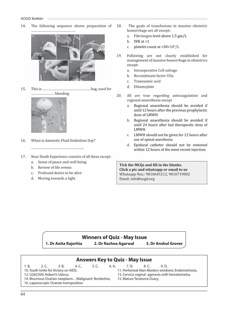
AOGD Bulletin
64
14. The following sequence shows preparation of ……………………………………….
15. This is ………………………………………….. bag, used for …………………… bleeding
16. When is Amniotic Fluid Embolism Day?
……………………………………………….
17. Near Death Experience consists of all these except: a. Sense of peace and well being b. Review of life eventsc. Profound desire to be alived. Moving towards a light
18. The goals of transfusions in massive obstetric hemorrhage are all except:a. Fibrinogen level above 1.5 gm/Lb. INR at <1c. platelet count at >50×109/L
19. Following are not clearly established for management of massive hemorrhage in obstetrics except:a. Intraoperative Cell salvageb. Recombinant factor VIIac. Tranexamic acidd. Ethamsylate
20. All are true regarding anticoagulation and regional anaesthesia excepta. Regional anaesthesia should be avoided if
until 12 hours after the previous prophylactic dose of LMWH
b. Regional anaesthesia should be avoided if until 24 hours after last therapeutic dose of LMWH.
c. LMWH should not be given for 12 hours after use of spinal anesthesia
d. Epidural catheter should not be removed within 12 hours of the most recent injection
Answers Key to Quiz - May Issue1. B, 2. C, 3. B, 4. C, 5. C, 6. A, 7. D, 8. C, 9. D,10. Youth Unite for Victory on AIDS, 11. Peritoneal Alan-Masters windows; Endometriosis,12. U2bC0V0, Robert’s Uterus, 13. Cervico vaginal agenesis with hematometra,14. Mucinous Ovarian neoplasm…Malignant/ Borderline, 15. Mature Teratoma Ovary,16. Laparoscopic Ovarian transposition
Winners of Quiz - May Issue1. Dr Anita Rajorhia 2. Dr Rachna Agarwal 3. Dr Anshul Grover
Tick the MCQs and ill in the blanks.Click a pic and whatsapp or email to usWhatsapp Nos.: 9810645212, 9810719002Email: [email protected]

Volume 17-2; June 2017
65
AOGD Bulletin
66
Volume 17-2; June 2017
67
Skill Building
Workshops
Early Bird Registration
11th World Congress on Adolescent HealthInvesting in Adolescent Health - the Future is Now!
27-29 October, 2017Hotel Pullman, Aerocity, New Delhi
REGISTER TODAYwww.iaah2017congress.org
Professional Association Partners
Federation of Obstetrics and
Gynecological Societies of IndiaThe Trained Nurses’
Association of India
In partnership with
Ministry of Health and Family Welfare, Government of India

AOGD Bulletin
68
Postal Registration No. DL(E)-20/5525/2017-19June 14-15, Date of Publication, June 7-8
Registered with Registrar of Newspapers for IndiaDELENG/2001/04547/8.25” x 11.25”
Effective& Mild
care forbabies
Proven,
DEMONSTRATION OFTHE MILDNESS OF TOP-TO-TOE
Another BabyCare Brand
Signs of Damage No Damage
Johnson’stop-to-toeOnly Water
Dirt Remains
Clinicallyproven forextra
delicateskin
pH balanced
Hypoallergenic &dermatologically tested
100% soap free
RA
/JB
/13J
an20
17/0
2