aapm task group 178 gamma stereotactic...
TRANSCRIPT
AAPM Task Group 178
Gamma Stereotactic Radiosurgery
Dosimetry and Quality Assurance
AAPM 58th Annual Meeting
August, 2016
Steven J. Goetsch, Ph.D., FAAPM
San Diego Gamma Knife Center and
Professor, Radiological Technologies University VT

Disclosure: the author is a part-time consultant to Elekta, Inc.

AAPM Task Group 178
Steve Goetsch, Chair San Diego Gamma Knife Ctr
Paola Alvarez M.D. Anderson
Greg Bednarz University of Pittsburgh
Larry De Werd Univ. of Wisconsin ADCL
Bob Drzymala Washington University
David Larson UCSF Medical Center
Lijun Ma UCSF Medical Center
Sheridan Meltsner
Gennady Neyman Cleveland Clinic
Josef Novotny University of Pittsburgh
Mark Rivard
Arjun Sahgal Sunnybrooke Med Center
Almon Shiu USC Medical Center3
68-100% centers reporting1991 reflects cumulative numbers
AAPM Task Group 178
Consultants
Keith Kunugi University of Wisconsin ADCL
Jan Seuntjens McGill University Med Center
Jonas Johansson Elekta Corporation
4
68-100% centers reporting1991 reflects cumulative numbers

Calibration of GSR Units
No current external beam protocols apply to certain devices
Gamma Knife, Tomotherapy, Cyberknife and Viewray are “noncompliant” with TG-51 Absorbed Dose in Water protocol
What’s a poor physicist to do?
Improvise!!

TG21 and TG 51: Presumes Point Source
Diverging to 10x10cm2 field at 100cm
Cyberknife comes closest: 6cm diameter field size at 80cm. New M6 gives up to 10 by 12cm2
at 80cm
Tomotherapy: up to 5 by 40cm2 at 85cm
Viewray: 105cm isocenter (rotational) up to 27.3cm2
Gamma Knife Perfexion: 192 sources converging on 4, 8 or 16mm diameter field at roughly 40cm


Letter to the Editor Alfonso et al, Med Phys
2008
Authors laid out a path by which a unique geometry “noncompliant” w TG-51 Absorbed Dose Protocol could be adapted to comply
Defined two fields: ƒref and ƒfmsr
The first field ƒref is “standard” field size: 10 by 10cm2
The second field ƒfmsr is a “machine-specific reference field”

Background: Dose Calibration Protocols
IAEA TRS-398 (2000)
AAPM TG-51 (1999)
IAEA Working Group (Alfonso
2008) *
Parameters Simple measured Simple measured Simple measured + Monte Carlo (MC) generated
Advantages • Simple to use• Likely to achieve consistent results
across different institutions
• Machine-specificcorrection factor
• Accounts for different ionization chambers and phantom types
Disadvantages
Designed for use with linear accelerators:• 10 x 10 cm2 field• Water phantom
MC parameters must be generated for each ionization chamber and phantom combination
Ionization chamber calibrated in water
*Med. Phys. 35, 5179-5186 (2008)

Equipment and Measurements
IonizationChambers
CollectingVolume
PTW TN31010 (PTW)
0.125 cc
StandardImaging Exradin A16 (Ex)
0.007 cc
Electrometer: Standard Imaging Max 4000
Elekta (gray) ABS plastic 16cm diameter solid phantom
Phantom One:

Phantom Two: Elekta Solid Water 16cm diameter phantom
Phantom Three: Phantom Laboratories Liquid Water 16cm diameter hemispherical phantom

Phantom Four: Standard Imaging In-air phantom
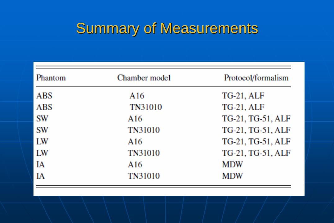
Summary of Measurements

Results Averaged over all Institutions Sorted by Dosimetry
Protocol/Formalism

*Johansson and Gorka, Elekta Physics Report: Reference No. SSM 2010/2201,
Project nr 4017003-006)
Take a closer look…


From previous slide: Average values
for ionization chambers with collecting volume 0.05 cm3
Liquid Water: 1.000 0.0015
Solid Water: 1.004 0.0014
ABS Plastic: 1.012 0.0034

Drzymala et al, Med Phys 42 (11) November, 2015


Conclusions
Four different procedures were carried out on four different phantoms, utilizing two small volume ion chambers and four protocols
All results were quite consistent
TG-51 modified by factors (announced but not yet published) following the formalism of Alfonso et al, yielded the overall best results
This procedure uses the Elekta ABS phantom supplied w every Elekta Gamma Knife in the world

References
Elekta white papers (available on request):
1. Accuracy of co-registration of planning images with Cone Beam CT images
2. Automatic positional delivery correction using a stereotactic CBCT in Leksell Gamma Knife Icon™
3. Design and performance characteristics of a Cone Beam CT system for Leksell Gamma Knife Icon ™
4. Geometric quality assurance for Leksell Gamma Knife Icon™
5. Automatic positional delivery correction using a stereotactic CBCT in Leksell Gamma Knife Icon™
6. Li et al “Impact of Immobilization on Intrafraction Motion for Dedicated Cobalt Radiosurgery Unit Using Cone Beam Computed Tomography” Int J Rad Onc Biol Phys Sep 2014 (abstract)