abdominal compartment syndrome and septic abdomen
TRANSCRIPT

Abdominal compartment syndrome and septic abdomen
asist.dr Aleksandar GluhovićKC Vojvodine
Novi Sad
Beograd, 28.04.2015.


DefinitionsWCACS, Antwerp Belgium 2007
• Intra-abdominal Pressure (IAP): Intrinsic pressure within the abdominal cavity
• Intra-abdominal Hypertension (IAH): An IAP > 12 mm Hg (often causing occult ischemia) without obvious organ failure
• Abdominal Compartment Syndrome (ACS): IAH with at least one overt organ failing

Types of IAH /ACS WCACS, Antwerp Belgium 2007
• Primary – Injury/disease of abdomino-pelvic region, “surgical”
• Secondary – Sepsis, capillary leak, burns, “medical”
• Recurrent – ACS develops despite surgical intervention

Physiologic Insult/Critical Illness
Ischemia Inflammatory response
Capillary leak
Tissue Edema (Including bowel wall and mesentery)
Intra-abdominal hypertension
Fluid resuscitation

IAP Interpretation
Pressure (mm Hg) Interpretation
• 0-5 Normal
• 5-10 Common in most ICU patients
• > 12 (Grade I) Intra-abdominal hypertension
• 16-20 (Grade II) Dangerous IAH - begin non-invasive interventions
• >21-25 (Grade III) Impending abdominal compartment syndrome - strongly consider decompressive laparotomy

Grades of IAH
Grade *IAP Organ failure
I 12-15 Absent
II 16-20 Absent
III 21-25 Absent
IV >25 Absent
**ACS >20 Present
*IAP = Intra-abdominal pressure.**ACS = Abdominal Compartment Syndrome.
Godat et al. World Journal of Emergency Surgery 2013 8:53 doi 10.1186/1749-7922-8-53


Physiologic Sequelae
Cardiac: • Increased intra-abdominal pressures cause:
– Compression of vena cava with reduced venous return– Elevated intra-thoracic pressure with multiple negative cardiac
effects
• The result:– Decreased cardiac output, increased SVR– Increased cardiac workload– Decreased tissue perfusion– Misleading elevations of CVP and PAWP– Cardiac insufficiency; cardiac arrest

Pulmonary: • Increased intra-abdominal pressures causes:
– Elevated diaphragm, reduced lung volumes & alveolar inflation, stiff thoracic cage,, increased interstitial fluid
The result:
– Elevated intrathoracic pressure (which further reduces venous return to heart, exacerbating cardiac problems)
– Increased peak pressures, reduced tidal volumes– Barotrauma - atelectasis, hypoxia, hypercarbia– ARDS (indirect - extrapulmonary)

Gastrointestinal: • Increased intra-abdominal pressures causes:
– Compression / Congestion of mesenteric veins and capillaries– Reduced cardiac output to the gut
The result:
– Decreased gut perfusion, increased gut edema and leak– Ischemia, necrosis– Bacterial translocation – Development and perpetuation of SIRS– Further increases in intra-abdominal pressure

Renal: • Elevated intra-abdominal pressure causes:
– Compression of renal veins, parenchyma– Reduced cardiac output to kidneys
The Result:– Reduced blood flow to kidney– Renal congestion and edema– Decreased glomerular filtration rate (GFR) – Renal failure, oliguria/anuria
• Mortality of renal failure in ICU is over 50% - • DO NOT WAIT for this to occur!
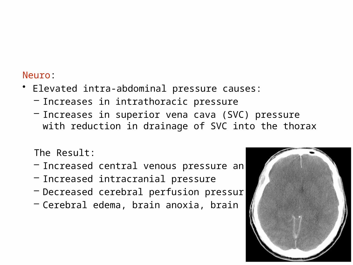
Neuro: • Elevated intra-abdominal pressure causes:
– Increases in intrathoracic pressure– Increases in superior vena cava (SVC) pressure with reduction in
drainage of SVC into the thorax
The Result:– Increased central venous pressure and IJ pressure– Increased intracranial pressure – Decreased cerebral perfusion pressure– Cerebral edema, brain anoxia, brain injury

Circling the Drain
Intra-abdominal Pressure
MucosalBreakdown
(Multi-System Organ Failure)
Bacterial translocation
Acidosis
Decreased O2 delivery
Anaerobic metabolism
Capillary leak
Free radical formation
MSOF

Risk factors for intra-abdominal hypertension and abdominal compartment syndrome
• Diminished abdominal wall compliance• Abdominal surgery• Major trauma• Major burns• Prone positioning
• Increased intra-luminal contents• Gastroparesis/gastric distention/ileus• Ileus• Colonic pseudo-obstruction• Volvulus
• Increased intra-abdominal contents• Acute pancreatitis• Distended abdomen• Hemoperitoneum/pneumoperitoneum or intra-peritoneal fluid collections• Intra-abdominal infection/abscess• Intra-abdominal or retroperitoneal tumors• Laparoscopy with excessive insufflation pressures• Liver dysfunction/cirrhosis with ascites• Peritoneal dialysis
Intensive Care Med. 2013 Jul; 39(7): 1190–1206.

• Capillary leak/fluid resuscitation• Acidosis• Damage control laparotomy• Hypothermia• Increased APACHE-II or SOFA score• Massive fluid resuscitation or positive fluid balance• Polytransfusion
• Others/miscellaneous• Age• Bacteremia• Coagulopathy• Increased head of bed angle• Massive incisional hernia repair• Mechanical ventilation• Obesity or increased body mass index• PEEP > 10• Peritonitis• Pneumonia• Sepsis• Shock or hypotension

Retained definitions from the original 2006 consensus statements
1. IAP is the steady-state pressure concealed within the abdominal cavity
2. The reference standard for intermittent IAP measurements is via the bladder with a maximal instillation volume of 25 mL of sterile saline
3. IAP should be expressed in mmHg and measured at end-expiration in the supine position after ensuring that abdominal muscle contractions are absent and with the transducer zeroed at the level of the midaxillary line
4. IAP is approximately 5–7 mmHg in critically ill adults
5. IAH is defined by a sustained or repeated pathological elevation in IAP ≥ 12 mmHg
6. ACS is defined as a sustained IAP > 20 mmHg (with or without an APP < 60 mmHg) that is associated with new organ dysfunction/failure

7. IAH is graded as follows
Grade I, IAP 12–15 mmHg Grade II, IAP 16–20 mmHg Grade III, IAP 21–25 mmHg Grade IV, IAP > 25 mmHg
8. Primary IAH or ACS is a condition associated with injury or disease in the abdominopelvic region that frequently requires early surgical or interventional radiological intervention
9. Secondary IAH or ACS refers to conditions that do not originate from the abdominopelvic region
10. Recurrent IAH or ACS refers to the condition in which IAH or ACS redevelops following previous surgical or medical treatment of primary or secondary IAH or ACS
11. APP = MAP − IAP

New definitions accepted by the 2013 consensus panel
12. A polycompartment syndrome is a condition where two or more anatomical compartments have elevated compartmental pressures
13. Abdominal compliance is a measure of the ease of abdominal expansion, which is determined by the elasticity of the abdominal wall and diaphragm. It should be expressed as the change in intra-abdominal volume per change in IAP
14. The open abdomen is one that requires a temporary abdominal closure due to the skin and fascia not being closed after laparotomy
15. Lateralization of the abdominal wall is the phenomenon where the musculature and fascia of the abdominal wall, most exemplified by the rectus abdominus muscles and their enveloping fascia, move laterally away from the midline with time

Final 2013 WSACS consensus management statementsRecommendations
1. Measuring IAP when any known risk factor for IAH/ACS is present in a critically ill or injured patient [GRADE 1C]
2. Studies should adopt the trans-bladder technique as the standard IAP measurement technique [not GRADED]
3. Use of protocolized monitoring and management of IAP versus not [GRADE 1C]
4. Efforts and/or protocols to avoid sustained IAH as compared to inattention to IAP among critically ill or injured patients [GRADE 1C]
5. Decompressive laparotomy in cases of overt ACS compared to strategies that do not use decompressive laparotomy in critically ill adults with ACS [GRADE 1D]
6. That among ICU patients with open abdominal wounds, conscious and/or protocolized efforts be made to obtain an early or at least same-hospital-stay abdominal fascial closure [GRADE 1D]
7. That among critically ill/injured patients with open abdominal wounds, strategies utilizing negative pressure wound therapy should be used versus not [GRADE 1C]

“Home Made” Pressure Transducer Technique
Home-made assembly:– Transducer– 2 stopcocks– 1 60 ml syringe, – 1 tubing with saline bag spike /
luer connector– 1 tubing with luer both ends– 1 needle / angiocath– Clamp for FoleyAssembled sterilely in proper
fashion

Bladder Pressure Monitoring: How to do it
Commercially available devices :• Foley Manometer – (Bladder
manometer)• CiMon (Gastric)• Spiegelberg (Gastric)• AbViser – (Bladder
transduction)
Advantages – Simple, standardized, reproducible, time-efficient, sterile

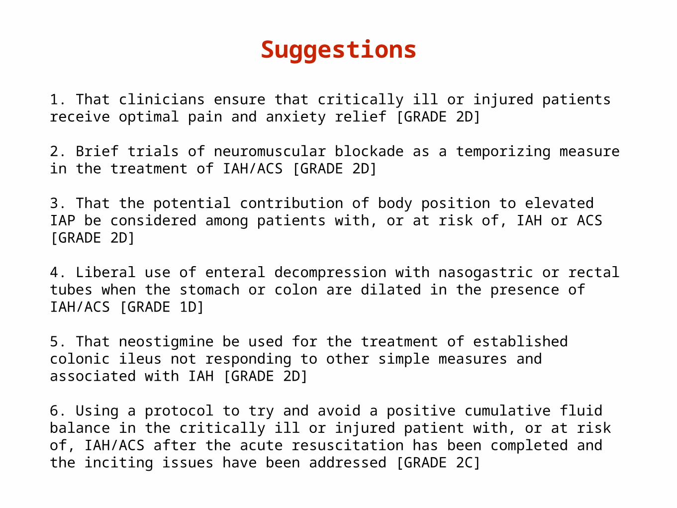
Suggestions
1. That clinicians ensure that critically ill or injured patients receive optimal pain and anxiety relief [GRADE 2D]
2. Brief trials of neuromuscular blockade as a temporizing measure in the treatment of IAH/ACS [GRADE 2D]
3. That the potential contribution of body position to elevated IAP be considered among patients with, or at risk of, IAH or ACS [GRADE 2D]
4. Liberal use of enteral decompression with nasogastric or rectal tubes when the stomach or colon are dilated in the presence of IAH/ACS [GRADE 1D]
5. That neostigmine be used for the treatment of established colonic ileus not responding to other simple measures and associated with IAH [GRADE 2D]
6. Using a protocol to try and avoid a positive cumulative fluid balance in the critically ill or injured patient with, or at risk of, IAH/ACS after the acute resuscitation has been completed and the inciting issues have been addressed [GRADE 2C]

7. Use of an enhanced ratio of plasma/packed red blood cells for resuscitation of massive hemorrhage versus low or no attention to plasma/packed red blood cell ratios [GRADE 2D]
8. Use of PCD to remove fluid (in the setting of obvious intraperitoneal fluid) in those with IAH/ACS when this is technically possible compared to doing nothing [GRADE 2C]. We also suggest using PCD to remove fluid (in the setting of obvious intraperitoneal fluid) in those with IAH/ACS when this is technically possible compared to immediate decompressive laparotomy as this may alleviate the need for decompressive laparotomy [GRADE 2D]
9. That patients undergoing laparotomy for trauma suffering from physiologic exhaustion be treated with the prophylactic use of the open abdomen versus intraoperative abdominal fascial closure and expectant IAP management [GRADE 2D]
10. Not to routinely utilize the open abdomen for patients with severe intraperitoneal contamination undergoing emergency laparotomy for intra-abdominal sepsis unless IAH is a specific concern [GRADE 2B]
11. That bioprosthetic meshes should not be routinely used in the early closure of the open abdomen compared to alternative strategies [GRADE 2D]

No recommendation
1. Use of abdominal perfusion pressure in the resuscitation or management of the critically ill or injured
2. Use of diuretics to mobilize fluids in hemodynamically stable patients with IAH after the acute resuscitation has been completed and the inciting issues have been addressed
3. Use of renal replacement therapies to mobilize fluid in hemodynamically stable patients with IAH after the acute resuscitation has been completed and the inciting issues have been addressed
4. Administration of albumin versus not, to mobilize fluid in hemodynamically stable patients with IAH after acute resuscitation has been completed and the inciting issues have been addressed
5. Prophylactic use of the open abdomen in non-trauma acute care surgery patients with physiologic exhaustion versus intraoperative abdominal fascial closure and expectant IAP management
6. Use of an acute component separation technique versus not to facilitate earlier abdominal fascial closure


Conclusion
Although IAH and ACS are common and frequently associated with poor outcomes, the overall quality of evidence available to guide
development of RECOMMENDATIONS was generally low.
Appropriately designed intervention trials are urgently needed for patients with IAH and ACS.