ablation of mir-10b suppresses oncogene- induced...
TRANSCRIPT

Tumor and Stem Cell Biology
Ablation of miR-10b Suppresses Oncogene-Induced Mammary Tumorigenesis and Metastasisand Reactivates Tumor-Suppressive PathwaysJongchan Kim1, Ashley N. Siverly1, Dahu Chen1, Min Wang1, Yuan Yuan2, Yumeng Wang2,Hyemin Lee1, Jinsong Zhang1,William J. Muller3, Han Liang2, Boyi Gan1, Xianbin Yang4,Yutong Sun5, M. James You6, and Li Ma1
Abstract
The invasive andmetastatic properties of many human tumorshave been associated with upregulation of the miRNA miR-10b,but its functional contributions in this setting have not been fullyunraveled. Here, we report the generation of miR-10b–deficientmice, in which miR-10b is shown to be largely dispensable fornormal development but critical to tumorigenesis. Loss of miR-10b delays oncogene-induced mammary tumorigenesis and sup-presses epithelial–mesenchymal transition, intravasation, andmetastasis in a mouse model of metastatic breast cancer. Amongthe target genes ofmiR-10b, the tumor suppressor genes Tbx5 andPten and the metastasis suppressor gene Hoxd10 are significantly
upregulated by miR-10b deletion. Mechanistically, miR-10b pro-motes breast cancer cell proliferation, migration, and invasionthrough inhibition of the expression of the transcription factorTBX5, leading to repression of the tumor suppressor genesDYRK1A and PTEN. In clinical specimens of breast cancer, theexpression of TBX5, HOXD10, and DYRK1A correlates withrelapse-free survival and overall survival outcomes in patients.Our results establish miR-10b as an oncomiR that drives metas-tasis, termed a metastamiR, and define the set of critical tumorsuppressor mechanisms it overcomes to drive breast cancer pro-gression. Cancer Res; 76(21); 6424–35. �2016 AACR.
IntroductionmiRNAs are approximately 22-nucleotide noncoding RNAs
that base-pair with mRNAs, leading to degradation of targetmRNAs and/or inhibition of their translation (1). Althoughgrowing numbers of miRNAs have been found to be involved inbreast cancer progression based on cell culture and xenograftmodels (2–4), their functions in themetastasis of autochthonousmammary tumors have not been demonstrated bymiRNA knock-out approaches. Elucidating the role of these miRNAs in normaldevelopment and in the initiation and metastasis of autochtho-nous tumorswill provide important insights into themechanismsof malignant progression.
It has been shown that certain miRNAs, such as miR-10b andmiR-9, are upregulated in metastatic cancer cells and promote
metastasis (5, 6). Thefirstmetastasis-regulatingmiRNA,miR-10b,was initially found to be expressed at elevated levels in metastaticcell lines and primary tumors frompatients withmetastatic breastcancer (5, 7). A growing body of evidence has demonstrated thatmiR-10b expression is also positively associated with high-grademalignancy ormetastasis in other cancer types, such as pancreaticcancer (8–10), glioblastoma (11–14), bladder cancer (15), andliver cancer (16). In addition, lymph node metastases expresshigher levels of miR-10b than paired primary tumors in multiplecancer types (17).
When overexpressed, miR-10b can induce motility and inva-siveness of various cancer cell lines (5, 15, 16, 18–21) andpromote metastasis in xenograft models (5, 15, 20) throughdirect targeting of HOXD10, NF1, KLF4, PTEN, and likely othergenes. Conversely, silencing miR-10b expression suppresses can-cer cell proliferation, migration, and invasion in vitro as well astumor growth ormetastasis in vivo (14, 18, 19, 22), suggesting thatmiR-10b is a potential target for antitumor or antimetastatictherapy. Notably, combining nanoparticle-encapsulated miR-10b inhibitors with low-dose doxorubicin achieved completedurable regression of metastases in a xenograft model of breastcancer (23). Intriguingly, miR-10b is secreted bymetastatic breastcancer cells via exosomes and, upon uptake, induces invasivenessof nonmalignant mammary epithelial cells (24).
In this study, we generated miR-10b–deficient mice to deter-mine the role of miR-10b in normal development and tumorprogression. Although genetic deletion ofmiR-10bdoes not causesubstantial developmental defects, it impedes mammary tumorinitiation, progression, and metastasis in a polyomavirus middleT (PyMT) model of breast cancer. We further identified TBX5 as afunctional target ofmiR-10b and identified two tumor suppressor
1Department of Experimental Radiation Oncology, The University ofTexas MD Anderson Cancer Center, Houston, Texas. 2Department ofBioinformatics and Computational Biology, The University of TexasMDAnderson Cancer Center, Houston,Texas. 3Goodman Cancer Cen-ter, McGill University, Montreal, Quebec, Canada. 4AM Biotechnolo-gies, Houston,Texas. 5DepartmentofMolecular andCellularOncology,The University of Texas MDAnderson Cancer Center, Houston, Texas.6Department of Hematopathology,TheUniversity of TexasMDAnder-son Cancer Center, Houston, Texas.
Note: Supplementary data for this article are available at Cancer ResearchOnline (http://cancerres.aacrjournals.org/).
Corresponding Author: Li Ma, The University of Texas MD Anderson CancerCenter, 6565MDAndersonBlvd., Houston, TX 77030. Phone: 713-792-6590; Fax:713-745-6141; E-mail: [email protected]
doi: 10.1158/0008-5472.CAN-16-1571
�2016 American Association for Cancer Research.
CancerResearch
Cancer Res; 76(21) November 1, 20166424
on July 8, 2018. © 2016 American Association for Cancer Research. cancerres.aacrjournals.org Downloaded from
Published OnlineFirst August 28, 2016; DOI: 10.1158/0008-5472.CAN-16-1571
genes, DYRK1A and PTEN, as target genes of the transcriptionfactor TBX5.
Materials and MethodsFor details, see Supplementary Materials and Methods.
Generation of miR-10b knockout micemiR-10b–mutantmicewere produced at Taconic Artemis.Mice
with the conditional knockout allele were generated after breed-ing to Flp-deleter mice for the removal of the selection marker(PuroR). Mice with the constitutive knockout allele were gener-ated after breeding to Cre-deleter mice. Both conditional andconstitutivemiR-10b–mutantmice weremaintained on aC57BL/6 background.
Tumor and metastasis studiesAll animal studies were performed in accordance with a
protocol approved by the Institutional Animal Care and UseCommittee of MD Anderson Cancer Center (Houston, TX).Mammary tumor-free survival was determined by daily palpa-tion. MMTV-PyMT;miR-10bþ/þ and MMTV-PyMT;miR-10b�/�
female mice were euthanized at 13, 16, and 19 weeks of age andthe endpoint (18–26 weeks, when mice met the institutionaleuthanasia criteria for tumor size and overall health condition);whole mammary glands or tumors and lung tissues werecollected, weighed, and prepared for histopathologic analysis.The presence of metastasis was determined by histopathologicreview of lung sections.
Histopathology and immunostainingMouse tissue samples were fixed in 10% buffered formalin
overnight, washed with PBS, transferred to 70% ethanol,embedded in paraffin, sectioned (5 mm thick), and stainedwith hematoxylin and eosin (H&E). Histopathologic examina-tion was performed on all tissue sections by a pathologist (M.J.You). Specific tissue areas were quantitated by counting pixelswith digital imaging software (Photoshop). For immunohisto-chemical staining, paraffin-embedded sections were deparaffi-nized in xylene and rehydrated in ethanol. Antigen wasretrieved using citric acid (pH 6.0, Dako). Endogenous perox-idase activity was blocked using 3% hydrogen peroxide in PBS.Sections were blocked with 5% BSA in PBS and incubated withthe primary antibody. The sections were then washed with PBSand incubated with a horseradish peroxidase (HRP)–conjugat-ed secondary antibody, followed by incubation with diamino-benzidine (Sigma).
Whole-mount staining of mammary glandsAfter euthanasia, the entire right #4 inguinal mammary gland
was collected from wild-type and miR-10b�/� female mice,placed onto a glass slide, stretched out, and air dried for a fewminutes. The tissues were fixed overnight in Carnoy solution,rinsed in PBS, and stained in Carmine Alum solution overnight.The tissues were then dehydrated in ethanol and xylene andmounted.
Blood collection and blood countPeripheral blood was drawn by retro-orbital sampling from
age- and sex-matchedwild-type andmiR-10b�/�mice. Bloodwascollected in EDTA-coated tubes to prevent clotting and subjected
to complete blood counting performed by the Department ofVeterinary Medicine & Surgery at MD Anderson Cancer Center.
Isolation and staining of circulating tumor cells from micePeripheral blood was collected via facial vein or retro-orbital
bleeding, and red blood cells were lysed with RBC lysing buffer(Gibco, A10492-01). Nucleated cells were spun onto the slidesusing cytospin and fixed in 10% formalin. For immunofluores-cent staining of the PyMT protein, cells were permeabilized with0.25% Triton X-100. The PyMT-specific primary antibody(Abcam, ab15085, 1:50) and anti-rat secondary antibody conju-gatedwith Fluorescein (Vector Laboratories, FI-4001, 1:100) wereused. After staining, cells weremounted usingmountingmediumwith DAPI (Vector Laboratories, H-1200). For circulating tumorcell (CTC) quantification, the ratio of PyMTþ;DAPIþ cells to totalDAPIþ cells was calculated in �200 fields.
qPCRTotal RNA from mouse tissues, mouse embryonic fibroblasts
(MEF), and human cells was isolated using the TRIzol reagent(Invitrogen) according to the manufacturer's protocol. Real-timePCR and data collection were performed on a CFX96 instrument(Bio-Rad).
Plasmids and tissue cultureHuman TBX5 cDNA was cloned into the pCMV-Tag2B vector
using restriction enzymes BamHI and EcoRI. 293FT cells werefrom Thermo Fisher Scientific in 2012, and the LM2 subline ofMDA-MB-231 cells was from Xiang Zhang (Baylor College ofMedicine, Houston, TX) in 2014. Both cell lines were cultured inDMEM medium supplemented with 10% FBS. Cell lines wereauthenticated by short tandem repeat analysis at MD Anderson'sCharacterized Cell Line Core Facility.
TransfectionFor overexpression, the MDH control, MDH-miR-10b, pCMV-
Tag2B control, and pCMV-Tag2B-TBX5 plasmids were transfectedinto cells using Lipofectamine 2000 (Life Technologies). Forknockdown, the negative control inhibitor (AMBiotechnologies),anti-miR-10b inhibitor (AM Biotechnologies), scrambled siRNA(Sigma), and TBX5 siRNA (Sigma, SASI_Hs01_00193647 andSASI_Hs01_00193648) oligonucleotides were transfected intocells using Oligofectamine (Life Technologies). The sequence andchemicalmodifications of themiR-10b antisense inhibitor are: 50-Cfs2AmCmAmAmAmUmUmCmGmGmUmUmCmUmAmCmAmGmGm
Gfs2Ufs2Am-30 (m¼ 20-OMe, fs¼ 20-F-30-monothioate, fs2¼ 20-F-30-dithioate).
Luciferase reporter assayFirefly luciferase reporter constructs containing the DYRK1A
promoter (D1A-Luc) and PTEN promoter (PTEN-Luc) were fromWalter Becker (RWTH Aachen University, Aachen, Germany) andJonathan Kurie (MD Anderson Cancer Center), respectively, andare describedpreviously (25–27). In 24-well plates, pCMV-Tag2B-TBX5 (þ, 50 ng; þþ, 400 ng) was transfected into 293FT cellsalong with the pRL-SV40 Renilla luciferase construct (0.5 ng) andthe firefly luciferase reporter construct (D1A-Luc or PTEN-Luc, 25ng). Two days after transfection, luciferase activity was measuredusing the Dual-Luciferase Reporter Assay System (Promega).Firefly luciferase activity was normalized with Renilla luciferaseactivity.
Genetic Dissection of miR-10b and Breast Cancer
www.aacrjournals.org Cancer Res; 76(21) November 1, 2016 6425
on July 8, 2018. © 2016 American Association for Cancer Research. cancerres.aacrjournals.org Downloaded from
Published OnlineFirst August 28, 2016; DOI: 10.1158/0008-5472.CAN-16-1571

Cell proliferation assayLM2 cells were transfected with the negative control inhibitor,
miR-10b inhibitor, scrambled siRNA, and/or TBX5 siRNA, and inseparate experiments, transfected with the pCMV-Tag2B orpCMV-Tag2B-TBX5 construct. Next day, 700 cells were plated in96-well plates. Onday 1, 2, 3, 4, and 5, cells werewashedwith PBSand stained with 0.05% crystal violet solution. Excess stainingsolution was washed with water, and the stained cells were airdried. For quantitation, crystal violet was extracted with 10%acetic acid, and optical density was measured using a multi-wellplate reader at 595 nm.
Migration and invasion assaysCell migration and invasion were measured using Boyden
Chambers (Corning, 3422 and 354480, respectively). LM2 cellswere transfected with the negative control inhibitor, miR-10binhibitor, scrambled siRNA, and/or TBX5 siRNA, and in separateexperiments, transfected with the pCMV-Tag2B or pCMV-Tag2B-TBX5 construct. Next day, cells were trypsinized, counted, andwashed with PBS to remove serum and growth factors. A total of1.5 � 104 and 7 � 104 cells were resuspended in serum-freemedium and added to the upper chamber for migration andinvasion assays, respectively. Serum-containing growth mediumwas added to the bottom chamber. Twenty hours later, cells on theupper surface of themembranewere removedwith a cotton swab,and cells on the lower surface of the membrane were stained andcounted.
Western blot analysisImmunoblotting was performed with precast gradient gels
(Bio-Rad) using standard methods. Briefly, mouse tissues weremechanically homogenized using an electric tissue homogenizer.Homogenized mouse tissues and human cells were lysed in theRIPA buffer (Cell Signaling Technology) containing proteaseinhibitors (Roche) and phosphatase inhibitors (Thermo FisherScientific). Proteins were separated by SDS-PAGE and blottedonto a PVDFmembrane (Bio-Rad).Membraneswere probedwiththe specific primary antibodies, followed by HRP-conjugatedsecondary antibodies. The bands were visualized by chemilumi-nescence (Denville Scientific).
TCGA and computational analysisTo assess genomic alterations of TBX5, HOXD10, andDYRK1A
genes, we used The Cancer Genome Atlas (TCGA) breast cancersomatic mutation data and copy number alteration data todetermine the alteration rates. To compare the mRNA expressionlevels of TBX5, HOXD10, orDYRK1A between normal and tumortissues, we used TCGA breast cancer RNA sequencing (RNA-Seq)data and performed t tests on the log2-transformed expressionvalues (i.e., RNA-Seq by expectation maximization, RSEM). Toassess the correlation of TBX5, HOXD10, or DYRK1A expressionwith clinical outcomes, we used the Kaplan–Meier plotter (28)andperformed a log-rank test to compare high and lowexpressiongroups.
Statistical analysisWe used a log-rank test for survival analysis andWilcoxon sum
rank test for tumor grade analysis. For other analyses, Student t test(two-tailed) was used to compare two groups for independentsamples. Data are presented as mean � SEM. P < 0.05 wasconsidered statistically significant.
ResultsGeneration and characterization of miR-10b knockout mice
To investigate the role of miR-10b in mammalian develop-ment, we generated mice with conditional and constitutive dele-tion of miR-10b on a C57BL/6 background by homologousrecombination and subsequent breeding to Flp- and Cre-deletermice, which bear a ubiquitously expressed Flp orCre recombinasetransgene (Fig. 1A and B). Using a probe specifically detectingmature miR-10b as opposed to mature miR-10a in Northern blotanalysis (Supplementary Fig. S1A and S1B), we confirmed thatmiR-10b expression was deficient in miR-10b–null embryos andin multiple organs of adult miR-10b�/� mice (Fig. 1C). MousemiR-10b is embedded in intron 4 ofHoxd4 and intron 1 ofHoxd3(Fig. 1A). Deletion of miR-10b did not alter the expression ofHoxd4 and Hoxd3 (Supplementary Fig. S1C and S1D).
From the intercrosses between miR-10b heterozygotes (miR-10bþ/�; Fig. 1B), we found that mice homozygous for miR-10bdeletion (miR-10b�/�) were born at the expectedMendelian ratio(Supplementary Fig. S2A). Compared with age- and sex-matchedwild-typemice, miR-10b–null animals did not display significantabnormalities in total body weight (Supplementary Fig. S2B), theweight of most organs (Supplementary Fig. S2C), overall survival(Supplementary Fig. S3A), fertility (Supplementary Fig. S3B), andcomplete blood cell count (Supplementary Fig. S4). A compre-hensive histopathologic examination did not detect significantaberrations in the majority of organs in miR-10b�/� mice (Sup-plementary Fig. S5A). In addition, whole-mount staining of themammary gland revealed no differences in ductal growth, ductalbranching, and terminal bud formation (Supplementary Fig.S5B). However, a moderate but significant change in the weightof the spleen (P¼ 0.0216), ovary (P¼ 0.02645), and uterus (P¼0.0334) was observed in miR-10b�/� mice (Fig. 1D and Supple-mentary Fig. S2C). Consistent with the increase in spleen size,spleens of miR-10b–deficient animals displayed expansion ofgerminal centers compared with those of age- and sex-matchedwild-typemice (Fig. 1E and F), althoughno lymphomawas foundin a 118-week follow-up.
miR-10b deficiency impedes mammary tumor formation andprogression in a PyMT mouse model of breast cancer
The mouse mammary tumor virus (MMTV)-PyMT transgenicmice on an FVB/N background develop palpable mammarytumors as early as 5 weeks of age and eventually show 100%tumor penetrance, and pulmonary metastases are observed in94%of tumor-bearing femalemice (29). InMMTV-PyMTmice ona C57BL/6 strain, tumor onset is delayed, although eventually alltransgenic females develop mammary tumors (30). BecauseMMTV-PyMT mice recapitulate the tumor stages, pathology,metastasis, and biomarkers of patients with metastatic breastcancer (31), this model is widely used to dissect the molecularevents at early and late stages of breast cancer progression.
As the miR-10b knockout mice were generated on a C57BL/6background, we bred these animals to MMTV-PyMT mice on aC57BL/6 strain. Both mature miR-10b and its precursor weredepleted in the mammary tumors of MMTV-PyMT;miR-10b�/�
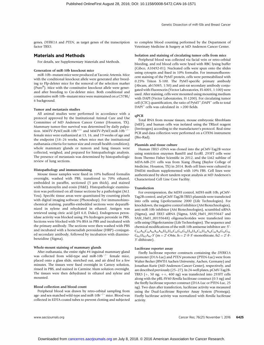
animals (Fig. 2A)without compensatory increase ofmiR-10a (Fig.2B). In MMTV-PyMT;miR-10bþ/þ mice, palpable mammarytumors appeared as early as 40 days of age, with a median onsetat 52 days; by 120 days of age, all females had palpable tumors(n ¼ 17; Fig. 2C). In contrast, MMTV-PyMT;miR-10b�/� mice
Kim et al.
Cancer Res; 76(21) November 1, 2016 Cancer Research6426
on July 8, 2018. © 2016 American Association for Cancer Research. cancerres.aacrjournals.org Downloaded from
Published OnlineFirst August 28, 2016; DOI: 10.1158/0008-5472.CAN-16-1571

developed palpable tumors starting at 73 days of age, with 50%of mice showing tumors by 103 days and 100% of mice showingtumors by 127 days of age (n ¼ 15; Fig. 2C). Thus, deletionof miR-10b significantly delayed the onset of PyMT-inducedmammary tumors (P ¼ 0.0001).
In addition to the effect on primary tumor onset, the weight ofmammary tumors inMMTV-PyMT;miR-10b�/�micewas reducedby32%(0.8052g vs. 0.5478 g,P¼0.0079) and40.1%(1.273g vs.0.7622 g, P ¼ 0.0167) at 13 and 16 weeks of age, respectively,compared with MMTV-PyMT;miR-10bþ/þ mice (Fig. 2D). How-ever, at the late stage, there was no significant difference in tumorweight (2.109 g�0.3049g inMMTV-PyMT;miR-10b�/�mice and2.032 g� 0.3341 gMMTV-PyMT;miR-10bþ/þmice at 19weeks ofage; P¼ 0.869). We performed histopathologic analysis of mam-mary tissues. At 13 weeks of age, 10 of 16 (62.5%) MMTV-PyMT;miR-10bþ/þ mice had full-blown adenocarcinoma of the mam-mary gland, whereas MMTV-PyMT;miR-10b�/� mice displayed
lesions ranging from focal hyperplasia to focal adenocarcinoma,with only 6 of 20 (30%, P¼ 0.0209) animals showing full-blownadenocarcinoma (Fig. 2E–G). At 19 weeks of age, the percentagesof full-blown adenocarcinoma were 100% (10/10) for MMTV-PyMT;miR-10bþ/þ mice and 64% (7/11) for MMTV-PyMT;miR-10b�/�mice, respectively (P¼ 0.04373; Fig. 2G). Consistent withthe inhibition of tumor growth, miR-10b–deficient mammarytumors exhibited a 71.3% decrease (P ¼ 0.0205) in cell prolif-eration without significant changes in apoptotic cell death (P ¼0.701), as gauged by phosphorylated histoneH3 (SupplementaryFig. S6A and S6B) and cleaved caspase-3 (Supplementary Fig. S6Cand S6D) staining, respectively.
miR-10b deficiency reduces metastasis of PyMT-inducedmammary tumors
Inorthotopic implantationmodels, overexpressionofmiR-10bin otherwise nonmetastatic breast cancer cells induced lung
miR-10b
1 1 2 3 4 5 6 2 3
miR-10b
1 2 3 4 5 6
PuroRTK
1 1 2 3 4 5 6 2 3
miR-10b PuroR
1 1 2 3 4 5 6 2 3
LHA = ~6 kb SHA = ~3 kb
Mouse genomic locus
Targeting vector
Targeted allele
Constitutive knockout allele after Cre recombination (deleted)
Hoxd3 Exon Hoxd4 Exon
0.4 kb
LHA: Long homology arm SHA: Short homology arm
F3 Site LoxP Site TK: Thymidine kinase Genotyping primer pair
1 1 2 3 4 5 6 2 3
miR-10b
Conditional knockout allele after Flp recombination (floxed)
WT KO WT KO WT KO WT KO
Kidney Testis Embryo Ovary
� miR-10b
� RNU6B
A
B C
% B
ody
wei
ght
*
D FE**
P = 0.0037
Ger
min
al c
ente
r ar
ea
(×10
3 pix
els)
0
100
200
300
400
500
600
WT KO
miR-10b WT miR-10b KO
+/+ +/f +/− f/f −/−700 bp600 bp500 bp400 bp300 bp200 bp
Spleen0.5
0.4
0.3
0.2
0.1
0.0WT KO
P = 0.0216
Wild-type (514 bp)Floxed (680 bp)
Deleted (203 bp)
��
�
Figure 1.
Targeted deletion of miR-10b inmice. A, targeting strategy used togeneratemiR-10b knockout alleles. B,PCR-based genotyping of wild-type(þ), floxed (f), and deleted (�) allelesof miR-10b. C, Northern blot analysisdemonstrates miR-10b deletion in thekidney, testis, ovary, and embryo ofage- and sex-matched wild-type(WT) and miR-10b�/� [knockout(KO)]mice.D, spleenweight (relativeto body weight) of age- and sex-matched wild-type (WT) and miR-10b�/� (KO) mice; n ¼ 8 mice pergroup. E,gross examination (top) andH&E staining (bottom) of spleens ofwild-type (WT) andmiR-10b�/� (KO)mice. Scale bars, 200 mm. F, germinalcenter areas were quantitated bypixel counts; n � 4 mice per group.P values in D and F are from a two-tailed, unpaired t test.
Genetic Dissection of miR-10b and Breast Cancer
www.aacrjournals.org Cancer Res; 76(21) November 1, 2016 6427
on July 8, 2018. © 2016 American Association for Cancer Research. cancerres.aacrjournals.org Downloaded from
Published OnlineFirst August 28, 2016; DOI: 10.1158/0008-5472.CAN-16-1571
metastasis (5), and treatment with miR-10b antagomirs reducedmetastasis formation by otherwise highly malignant mammarytumor cells (22). In the PyMT-driven mammary tumor model,
miR-10b deficiency did not completely abolish metastasis;instead, it moderately but significantly prolonged metastasis-freesurvival (median metastasis-free survival: 160 days vs. 175 days,
Rel
ativ
e le
vels
to R
NU
6B
P = 0.719
G
0
1
2
3
4
5
PyMT;WT PyMT;KO
Tum
or a
rea
(× 10
6pi
xels
)
**
P = 0.0029
A B
C
Prob
abilit
y (%
) *** P = 0.0001
Rel
ativ
e le
vels
to R
NU
6B
Rel
ativ
e le
vels
to R
NU
6B
***
P = 0.002
P = 0.0448
D
P = 0.0079 P = 0.0167
***
FE PyMT;WT PyMT;KO
PyMT;WT PyMT;KO
13 weeks 19 weeks
PyMT;WT PyMT;KO
Breast adenocarcinoma
Small foci of breast adenocarcinoma
Very small foci of breast adenocarcinoma
Hyperplasia
Focal hyperplasia
*P = 0.0209 *P = 0.04373
Per
cent
(%
)
100
80
60
40
20
0
Figure 2.
Deletion of miR-10b impedes mammary tumor initiation, growth, and progression. A and B, qPCR of mature miR-10b (A, left), the miR-10b precursor (A, right), andmature miR-10a (B) in mammary tumors of age-matched MMTV-PyMT;miR-10b WT and MMTV-PyMT;miR-10b KO mice; n ¼ 3 mice per group. C, mammarytumor-free survival of MMTV-PyMT;miR-10b WT (n ¼ 17) and MMTV-PyMT;miR-10b KO (n ¼ 15) mice. The P value was from a log-rank test. D, mammary tumorweight of MMTV-PyMT;miR-10b WT and MMTV-PyMT;miR-10b KO mice at 13 and 16 weeks of age; n � 8 mice per group. E and F, H&E staining (E) and tumorarea quantification (F) of mammary tissue sections from 13-week-old MMTV-PyMT;miR-10b WT and MMTV-PyMT;miR-10b KO mice. Scale bars (E), 100 mm; n ¼ 3mice per group in F. G, histopathologic examination of mammary glands (n ¼ 16, 20, 10, and 11 mice per group, from left to right). P values were fromWilcoxon rank-sum test. P values in A, B, D, and F are from a two-tailed, unpaired t test.
Kim et al.
Cancer Res; 76(21) November 1, 2016 Cancer Research6428
on July 8, 2018. © 2016 American Association for Cancer Research. cancerres.aacrjournals.org Downloaded from
Published OnlineFirst August 28, 2016; DOI: 10.1158/0008-5472.CAN-16-1571

P ¼ 0.0044; Fig. 3A). To quantitate the effect on metastasis, weassessed metastatic lesions in H&E-stained sections of lungs frommice at 24 to 26 weeks of age. Compared withMMTV-PyMT;miR-10bþ/þ animals, MMTV-PyMT;miR-10b�/� mice showed 68.3%and 90.3% reduction in the number (10.4 foci vs. 3.3 foci, P ¼0.0283) and area (158.0 � 103 pixels vs. 15.27 � 103 pixels, P ¼0.0384) of pulmonary metastatic foci, respectively (Fig. 3B andC). Similar to primary tumors, metastatic tumor cells in the lungsof MMTV-PyMT;miR-10b�/� mice were also less proliferativethan those inMMTV-PyMT;miR-10bþ/þmice without an increasein apoptosis (Supplementary Fig. S6E–S6G).
miR-10b deficiency suppresses the mesenchymal phenotypeand intravasation of mammary tumor cells
Previous studies demonstrated the activation of epithelial–mesenchymal transition (EMT) in the MMTV-PyMT model (32,
33). Therefore, we examined the mesenchymal markers vimentinand N-cadherin and the epithelial marker E-cadherin in mousemammary tumors. In MMTV-PyMT;miR-10b�/� tumors, vimen-tin andN-cadherin levels were reduced and E-cadherin expressionwas increased (Fig. 3D and E). To determine the impact of miR-10b depletion on tumor cell intravasation, we compared thenumber of CTCs in the peripheral blood collected fromMMTV-PyMT;miR-10bþ/þ and MTV-PyMT;miR-10b�/� mice, byusing an antibody that could specifically detect PyMT-positivemammary tumor cells that metastasized to the lung (Supplemen-tary Fig. S7). In peripheral blood samples, the percentages of CTCsinMMTV-PyMT;miR-10b�/�micewere 0.59%, 1.6%, and3.7%at13, 16, and 19 weeks of age, respectively, which was significantlyless than those in MMTV-PyMT;miR-10bþ/þ mice (2.4%, 4.1%,and 6.3% at 13, 16, and 19 weeks of age, respectively; Fig. 3F andG). These results suggest that the loss of miR-10b inhibits the
*
P = 0.0283
P = 0.0384
*
PyMT; WT PyMT;KO0
5
Metastasis-free survival (days)
# of
Met
asta
tic fo
ciM
etas
tatic
are
a (×
103
pixe
ls)
00
50
100
PyMT;WT PyMT;KO
50 100 150 200 250
10
15
20
25
PyMT;WT PyMT;KO0
100
200
300
400
500
C
B
A
PyMT;WT PyMT;KO
D
PyMT;WT PyMT;KO
Vimentin/E-cadherin/DAPI
E
PyMT;WT PyMT;KO
PyMT/DAPI
F
Prob
abilit
y (%
) **P = 0.0044
G 8
7
6
5
4
3
2
1
013 16 19 (weeks)
% o
f CT
Cs
P = 0.019*
P = 0.00059***
P = 0.035*PyMT;WTPyMT;KO
PyMT;WT PyMT;KO
140
50
100
140
E-Cadherin
Vimentin
Hsp90
N-Cadherin
1.1 1.0 0.8 1.1 1.4 1.2 1.5 1.1
Figure 3.
Deletion of miR-10b inhibits EMT,intravasation, and metastasis. A,metastasis-free survival of MMTV-PyMT;miR-10b WT (n ¼ 67) andMMTV-PyMT;miR-10b KO (n ¼ 53)mice. The P value was from a log-rank test. B, H&E staining of lungs ofMMTV-PyMT;miR-10b WT andMMTV-PyMT;miR-10b KO mice at 24to 26 weeks of age. The bottompanels are high-magnificationimages of the boxed areas in the toppanels. Arrows, metastatic foci;scale bars, 1 mm (top) and 200 mm(bottom). C, quantitation of thenumber (top) and area (bottom) ofmetastatic foci in the lungs ofMMTV-PyMT;miR-10b WT and MMTV-PyMT;miR-10b KO mice at 24 to 26weeks of age; n ¼ 7 mice per group.D and E, immunoblotting (D) andimmunofluorescent staining (E) ofEMT markers in mammary tumors ofMMTV-PyMT;miR-10b WT andMMTV-PyMT;miR-10b KO mice.Scale bars (E), 200 mm. F and G,CTCs were immunostained with aPyMT-specific antibody (green), andnuclei were stained with DAPI (blue;F). G, the percentages of CTCs werequantitated. Scale bars (F), 20 mm; n�4mice per group inG. P values inCand G are from a two-tailed,unpaired t test.
Genetic Dissection of miR-10b and Breast Cancer
www.aacrjournals.org Cancer Res; 76(21) November 1, 2016 6429
on July 8, 2018. © 2016 American Association for Cancer Research. cancerres.aacrjournals.org Downloaded from
Published OnlineFirst August 28, 2016; DOI: 10.1158/0008-5472.CAN-16-1571

ability of mammary tumor cells to disseminate, which is notsimply due to the effect on the primary tumor considering similarprimary tumor sizes at 19 weeks.
miR-10b regulates the expression of Tbx5, Hoxd10, and Pten invivo
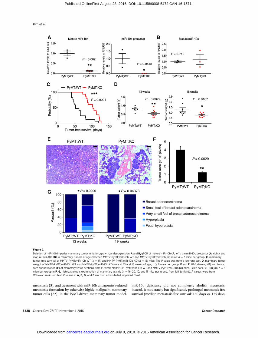
To identify miR-10b target genes whose expression is upregu-lated upon genetic deletion of miR-10b, we performed qPCRanalysis of approximately 100 genes, which were predicted com-putationally to be miR-10b targets (TargetScan, mouse; version6.0). In this initial screen, 10 genes showed a more than 1.5-foldincrease in their expression levels in miR-10b–null MEFs, com-pared with wild-type MEFs (Supplementary Fig. S8). These 10genes were tested again to confirm their upregulation bymiR-10bdepletion. Among the eight confirmed targets (Tbx5, Myt1l,Ppargc1b, Tnrc6b, Mycbp, Arnt, Prtg, and Gpcpd1), Tbx5 (T-box5) stood out as the most significantly upregulated gene (Fig. 4A).In addition, Hoxd10 (homeobox D10), a metastasis suppressorgene previously reported by many groups to be targeted by miR-10b (5, 15, 16, 22–24, 34–37), was upregulated in miR-10b–deficient MEFs by 1.4-fold (Supplementary Fig. S8) and in mam-mary tumors fromMMTV-PyMT;miR-10b�/�mice by 2-fold (Fig.4B and C).
miR-10a andmiR-10b have been reported to directly target the30-untranslated region of TBX5 (38). Consistent with the MEFanalysis, Tbx5mRNA levels were upregulated by 4.8-fold in miR-
10b–deficient PyMT tumors relative to tumors from MMTV-PyMT;miR-10bþ/þ mice (Fig. 4D). This upregulation was alsoobserved at the protein level (Fig. 4B and E). In addition, tworecent studies reported that miR-10b directly targets PTEN (21,39). Indeed, compared with MMTV-PyMT;miR-10bþ/þ mice,MMTV-PyMT;miR-10b�/� mice displayed upregulation of Ptenand downregulation of phospho-Akt in both primary mammarytumors (Fig. 4B and Supplementary Fig. S9A) and lungmetastases(Supplementary Fig. S9B).
DYRK1A and PTEN are TBX5 target genes and are upregulatedby miR-10b deletion
PTEN is a well-established dose-dependent tumor suppressorin breast cancer (40), and loss of PTEN expression has been foundin human mammary tumors (41, 42). TBX5 expression is down-regulated in colon cancer, and restored expression of TBX5 incolon tumor cells inhibited proliferation and migration (43),suggesting that TBX5 is a potential tumor suppressor. HOXD10expression is progressively lost in mammary tumors with increas-ing degrees of malignancy (44, 45), and reexpression of HOXD10in breast cancer cells suppressedmigration and invasion in vitro aswell as tumor progression in vivo (45), presumably by repressinggenes involved in cell movement and extracellular matrix remo-deling (46). However, the role of the transcription factor TBX5 inbreast cancer is unknown.
A
0 0.5 1 1.5 2 2.5
Fold change of mRNA levelsin miR-10b KO MEFs
Tbx5Myt1l
Ppargc1bTnrc6bMycbp
ArntPrtg
Gpcpd1D
B
C
PyMT;WT PyMT;KO
Tbx5
Pten
p-Akt(T308)
Akt
Hsp90
70
50
50
50
100
Hoxd1050
E
P = 0.01143
2
1
0
10
8
6
4
2
3
2
1
0
0
PyMT;WT PyMT;KO
PyMT;WT PyMT;KO
PyMT;WT PyMT;KO
*
Rel
ativ
e H
oxd1
0 pr
otei
n le
vels
*
P = 0.0108
Rel
ativ
eTbx5
mR
NA
leve
ls
*
P = 0.0202
Rel
ativ
e Tb
x5 p
rote
in le
vels
Figure 4.
Tbx5, Hoxd10, and Pten areupregulated by miR-10b deletion inmice. A, qPCR of miR-10b targetsin miR-10b–null MEFs. B,immunoblotting of Tbx5, Hoxd10,Pten, phospho-Akt, Akt, and Hsp90 inmammary tumors of MMTV-PyMT;miR-10b WT and MMTV-PyMT;miR-10b KO mice at 24 to 26 weeks of age;n¼ 4mice per group.C, quantificationof Hoxd10 protein levels in B. D, qPCRof Tbx5 in mammary tumors of MMTV-PyMT;miR-10b WT and MMTV-PyMT;miR-10b KO mice at 24 to 26 weeks ofage; n � 7 mice per group. E,quantification of Tbx5 protein levels inB. P values in C–E are from a two-tailed, unpaired t test.
Kim et al.
Cancer Res; 76(21) November 1, 2016 Cancer Research6430
on July 8, 2018. © 2016 American Association for Cancer Research. cancerres.aacrjournals.org Downloaded from
Published OnlineFirst August 28, 2016; DOI: 10.1158/0008-5472.CAN-16-1571
To identify TBX5-regulated genes in breast cancer, we analyzedeight publicly available transcriptome profiling datasets fromhumanbreast cancer specimens and identifiedfive genes (ADCY1,FAM134B, DYRK1A, FGFR2, and PTEN) whose expression pos-itively correlated with TBX5. Among them, the genomic loci ofDYRK1A, FGFR2, and PTEN were found to be occupied by TBX5based on a previous chromatin immunoprecipitation sequencingstudy (Supplementary Fig. S9C; ref. 47). To validate whether theexpression of these three genes is indeed activated by TBX5, weoverexpressed TBX5 in 293FT cells and observed upregulation ofDYRK1A and PTEN mRNA levels (Supplementary Fig. S9D).
Theprotein kinaseDYRK1A (dual-specificity tyrosine-(Y) phos-phorylation–regulated kinase 1A) has recently been shown topromote quiescence and senescence (48), inhibit tumor growthand stemness (49), and increase chemosensitivity (50). To deter-mine whether TBX5 activates the transcription of DYRK1A and
PTEN, we performed luciferase reporter assays and found that theactivity of theDYRK1A (Fig. 5A and Supplementary Fig. S9E) andPTEN (Fig. 5B and Supplementary Fig. S9E) promoters wasincreased by TBX5 in a dose-dependent manner. We then exam-inedDyrk1a and PtenmRNA levels inmouse tumors and observedupregulation of both genes in miR-10b–deficient PyMT mam-mary tumors (Fig. 5C and D). Similar to the increase in Ptenprotein levels (Fig. 4B and Supplementary Fig. S9A and S9B),MMTV-PyMT;miR-10b�/� mice exhibited upregulation of thepercentage of Dyrk1a-positive cells and Dyrk1a protein levels inboth primary mammary tumors (Fig. 5E–G) and lung metastases(Fig. 5H and I).
TBX5 is a functional target of miR-10bTo determine whether miR-10b downregulates DYRK1A and
PTEN expression through TBX5, we transfected 293FT cells with
9080706050403020100
TBX5 − − + + ++D1A-Luc − + − + +
Nor
mal
ized
act
ivity
P = 0.00026
***
P = 0.00075
***
P < 0.0001
****
Rel
ativ
eP
ten
mR
NA
leve
ls 8
6
4
2
0
*
P = 0.027
PyMT;WT PyMT;KO
**
P = 0.004
PyMT;WT PyMT;KO
Rel
ativ
eD
yrk1
am
RN
A le
vels 4
3
2
1
0
9080706050403020100
PTEN-Luc− + − + +TBX5 − − + + ++
P = 0.015***
P = 0.0013**
P = 0.64
P < 0.0001****
P = 0.0012**P < 0.0001****
Nor
mal
ized
act
ivity
PyMT;WT PyMT;KODyrk1a/DAPI
PyMT;WT PyMT;KO
% o
f Dyr
k1a+
cel
ls
100
80
60
40
20
0
P < 0.0001
****
H
PyM
T;W
TP
yMT
;KO
Dyrk1a/DAPI H&E I
PyMT;WT PyMT;KO
90
75
60
45
30
15
0
P = 0.0031
% o
f D
yrk1
a+ c
ells
**
G PyMT;WT PyMT;KO
100
100
Dyrk1a
Hsp90
A B C
D
E
F
Figure 5.
DYRK1A and PTEN are TBX5 targetgenes and are upregulated inmammary tumors and lungmetastasesof MMTV-PyMT;miR-10b�/� mice. Aand B, luciferase reporter assays showthat the promoters of DYRK1A (A) andPTEN (B) are activated by TBX5 in adose-dependent manner. D1A-Luc andPTEN-Luc are reporter constructscontaining the human DYRK1A (1,497bp) and PTEN (black bars, 1,064 bp;white bars, 1,978 bp) promoter regions,respectively. C and D, qPCR of Dyrk1a(C) and Pten (D) in mammary tumorsof MMTV-PyMT;miR-10b WT andMMTV-PyMT;miR-10b KO mice. E andF, immunofluorescent staining ofDyrk1a (E) and the percentages ofDyrk1a-positive cells (F) in mammarytumors of age-matched MMTV-PyMT;miR-10bWT andMMTV-PyMT;miR-10bKO mice. Scale bars (E), 50 mm; n ¼ 4mice per group in F.G, immunoblottingof Dyrk1a and Hsp90 in mammarytumors of MMTV-PyMT;miR-10b WTand MMTV-PyMT;miR-10b KO mice 24to 26 weeks of age; n ¼ 4 mice pergroup. H and I, immunofluorescentstaining of Dyrk1a (H) and thepercentages of Dyrk1a-positive cells (I)in lung metastases of age-matchedMMTV-PyMT;miR-10b WT and MMTV-PyMT;miR-10b KOmice. Scale bars (H),50 mm; n ¼ 4 mice per group in I.P values inA–D, F, and I are from a two-tailed, unpaired t test.
Genetic Dissection of miR-10b and Breast Cancer
www.aacrjournals.org Cancer Res; 76(21) November 1, 2016 6431
on July 8, 2018. © 2016 American Association for Cancer Research. cancerres.aacrjournals.org Downloaded from
Published OnlineFirst August 28, 2016; DOI: 10.1158/0008-5472.CAN-16-1571

miR-10b in the absence or presence of ectopic TBX5 expression.Overexpression of miR-10b decreased mRNA levels of TBX5,DYRK1A, and PTEN, and ectopic expression TBX5 in miR-10b–overexpressing cells restored DYRK1A and PTEN expression(Fig. 6A). Conversely, we transfected 293FT cells with miR-10bantisense inhibitors in the absence or presence of TBX5 siRNA.Consistent with our observations in miR-10b knockout mice,antisense inhibitors of miR-10b upregulated TBX5, DYRK1A, andPTEN expression, which could be reversed by knockdownof TBX5(Fig. 6B). Similarly, we observed the same effects in the LM2 lungmetastatic subline of theMDA-MB-231 human breast cancer cells(Fig. 6C and D; ref. 51). Therefore, gain- and loss-of-functionanalyses demonstrated thatmiR-10b suppresses TBX5 expression,leading to downregulation of DYRK1A and PTEN.
We asked whether TBX5 mediates the functions of miR-10b inbreast cancer cells. Consistent with the phenotypes of MMTV-PyMT;miR-10b�/� mice, transfecting the LM2 metastatic breast
cancer cells with miR-10b antisense inhibitors suppressed cellproliferation, migration, and invasion, which could be reversedby TBX5 siRNA (Fig. 6E and F).Moreover, overexpression of TBX5markedly inhibited the proliferation, migration, and invasion ofLM2 cells (Supplementary Fig. S10A–S10C). Collectively, theseresults suggest that TBX5 is a functional target of miR-10b.
TBX5,HOX10, andDYRK1A aredownregulated inhumanbreastcancer and correlate with clinical outcomes
Using miR-10b knockout mice and human cell lines, weidentified Tbx5, Hoxd10, and Pten as the targets of miR-10b, andDYRK1A and PTEN as the target genes of TBX5. Compared withPTEN, the relevance of TBX5, HOXD10, and DYRK1A in humanbreast cancer is less defined. We analyzed the breast cancer datafrom TCGA (52). Although genomic alterations of TBX5,HOXD10, and DYRK1A genes in human breast cancer were rare(TBX5, 0.83%; HOXD10, 0.93%; and DYRK1A, 2.0%), their
B
C
F
Anti-10b: − − + +si-TBX5: − + − +
3,000
2,500
2,000
1,500
1,000
500
0
P = 0.012*
P = 0.025*
P = 0.045*
P = 0.012*P = 0.021*
P = 0.0022**
P = 0.00023***
P = 0.035*
# of
Mig
rate
d/in
vade
d ce
lls
MigrationInvasion
E
A 5.04.54.03.53.02.52.0
1.5
1.0
0.5
0
TBX5 DYRK1APTEN
(×10
5 )
10b: − − + + − − + +TBX5: − + − + − + − +
50
40
35
20
10
0
miR-10b
Rel
ativ
e le
vels
6
5
4
3
2
1
0
TBX5 DYRK1APTEN
1.2
1.0
0.8
0.6
0.4
0.2
0Anti-10b: − − + + − − + +si-TBX5: − + − + − + − +
miR-10b
Rel
ativ
e le
vels
D
1.4
1.2
1.0
0.8
0.6
0.4
0.2
0Anti-10b: − − + +si-TBX5: − + − +
miR-10b
Rel
ativ
e le
vels
1.2
1.0
0.8
0.6
0.4
0.2
0
Rel
ativ
e O
D a
t 595
nm
1 2 3 4 5 (days)
***
**
***
*
Anti-10b /si-TBX5
−/−−/++/−+/+
P=
0.0
0016
P =
0.0
0024
P=
0.0
29P
= 0
.002
4
50
100
50
50
50
100
− − + + : anti-10b− + − + : si-TBX5
TBX5
DYRK1A
PTEN
p-AKT
AKT
HSP90
Figure 6.
TBX5 is a functional target of miR-10b. A,qPCRofmiR-10b, TBX5, DYRK1A, andPTENin 293FT cells transfected with miR-10band TBX5, alone or in combination. B,qPCRofmiR-10b, TBX5, DYRK1A, andPTENin 293FT cells transfected with miR-10bantisense inhibitors and TBX5 siRNA, aloneor in combination. C, qPCR of miR-10b inLM2 cells transfected with miR-10bantisense inhibitors and TBX5 siRNA, aloneor in combination. D, immunoblotting ofTBX5, DYRK1A, PTEN, phospho-AKT, AKT,and HSP90 in LM2 cells transfected withmiR-10b antisense inhibitors and TBX5siRNA, alone or in combination. E and F,growth curves (E) and migration andinvasion assays (F) of LM2 cells transfectedwith miR-10b antisense inhibitors andTBX5 siRNA, alone or in combination; n¼ 4and three wells per group in E and F,respectively. OD, optical density. P valuesare from a two-tailed, unpaired t test.
Kim et al.
Cancer Res; 76(21) November 1, 2016 Cancer Research6432
on July 8, 2018. © 2016 American Association for Cancer Research. cancerres.aacrjournals.org Downloaded from
Published OnlineFirst August 28, 2016; DOI: 10.1158/0008-5472.CAN-16-1571
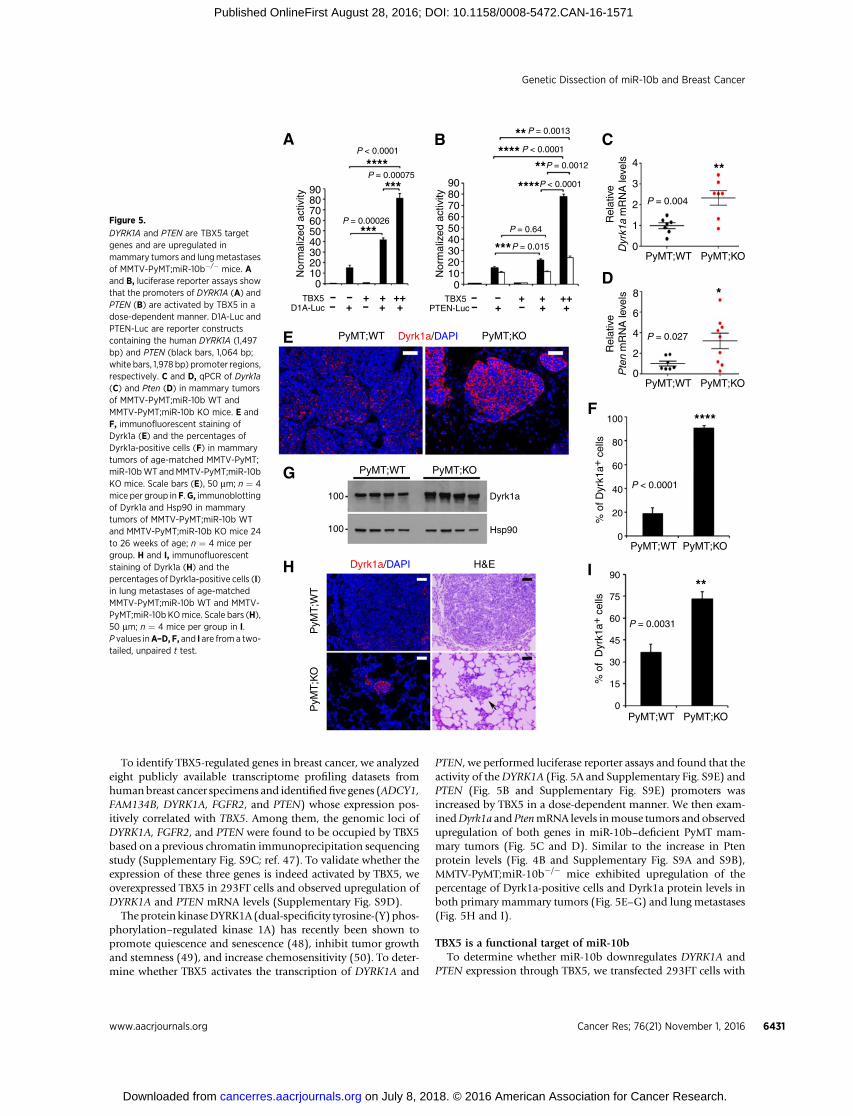
mRNA levels were significantly downregulated in breast tumorsrelative to normal mammary tissues (Fig. 7A–C). We then per-formed Kaplan–Meier plotter (28) analyses. Compared withpatients with high TBX5 expression in their breast tumors,patients with low TBX5 expression hadworse relapse-free survival(Fig. 7D) and overall survival (Supplementary Fig. S11A). Sim-ilarly, the expression of HOXD10 and DYRK1A also correlatedwith relapse-free survival (Fig. 7E and F) and overall survival(Supplementary Fig. S11B and S11C) of breast cancer patients.These data suggest that miR-10b–regulated genes are humanbreast cancer–relevant tumor suppressors or metastasis suppres-sors (Supplementary Fig. S11D).
DiscussionIn mammals, the miR-10 family consists of miR-10a and miR-
10b. Their coding genes are locatedwithin twodifferentHOX geneclusters whose protein products play important roles in develop-ment. Moreover, miR-10a and miR-10b target specific HOX
mRNAs, leading to the speculation that these two miRNAs mightbe developmental regulators (53, 54). Surprisingly, however,extensive analyses did not reveal substantial phenotypic differ-ences in miR-10b–deficient mice except germinal center expan-sion in the spleen. Similarly, miR-10a–null mice were indistin-guishable from littermate controls in terms of development,growth, overall survival, and incidence of spontaneous tumori-genesis (55).
Whereas the functional redundancy of miR-10a and miR-10bremains to be determined, a number of previous knockout studiesdemonstrated that many conserved miRNAs are dispensable foranimal development and viability; however, some of these miR-NAs play critical roles in stress responses or pathologic processes,including cardiac stress, vascular injuries, intestinal injuries, andoncogenic stress, and thus mice deficient in these miRNAs exhibitnotable phenotypes in response to external or internal perturba-tions (56). In the current study, genetic deletion of miR-10bin a PyMT-driven mouse model of breast cancer significantlyimpeded mammary tumor initiation, growth, progression, and
A
C
P = 4.4 ×10⁻¹²
n = 1,062 n = 113
Tumor Normal
98
76
54
3
76
54
32
11.
00.
811.5
11.0
10.5
10.0
0.6
0.4
0.2
0.0
0 50 100 150 200 250
FE
P = 4.4 ×10⁻¹⁸
n = 1,062n = 113
Tumor Normal
HO
XD
10 E
xpre
ssio
n (lo
g 2 R
SE
M)
TB
X5
Exp
ress
ion
(log 2
RS
EM
)D
YR
K1A
Exp
ress
ion
(log 2
RS
EM
)
B
D
P = 8.2 ×10⁻⁵
n = 1,062 n = 113
Tumor Normal
0.0
0.2
0.4
0.6
0.8
1.0
Low DYRK1A
High DYRK1A
0 50 100 150 200 250Relapse-free survival (months)
Pro
babi
lity
P = 2 ×10⁻⁴
Relapse-free survival (months)
Pro
babi
lity
P = 8.1 ×10⁻¹⁴
Low HOXD10
High HOXD10
0.0
0
.2
0.4
0
.6
0.8
1
.0
0 50 100 150 200 250
Relapse-free survival (months)
Pro
babi
lity
P = 0.0037
Low TBX5
High TBX5
Figure 7.
TBX5, HOXD10, and DYRK1A aredownregulated in human breastcancer and are associated withrelapse-free survival. A–C, box plotscomparing TBX5 (A), HOXD10 (B), orDYRK1A (C) expression in normalbreast tissues and breast tumors,based on the RNA-Seq data fromTCGA. Boxes,median and interquartilerange; whiskers, minimum andmaximum. P values were from a two-tailed, unpaired t test. D–F, Kaplan–Meier curves of relapse-free survival ofbreast cancer patients stratified byTBX5 (D), HOXD10 (E), or DYRK1A (F)expression levels; n¼ 1,660 patients. Pvalues are from a log-rank test.
Genetic Dissection of miR-10b and Breast Cancer
www.aacrjournals.org Cancer Res; 76(21) November 1, 2016 6433
on July 8, 2018. © 2016 American Association for Cancer Research. cancerres.aacrjournals.org Downloaded from
Published OnlineFirst August 28, 2016; DOI: 10.1158/0008-5472.CAN-16-1571

lung metastasis. The observed delay in oncogene-induced mam-mary tumor formation has likely been masked in previous xeno-graft models in which highly aggressive and late-stage tumor cellswere used (5); this underscores the importance of using autoch-thonous tumor models to track the early changes during tumorinitiation. Moreover, despite a dramatic reduction in the numberand area of metastatic foci in the lung, metastasis was notcompletely abrogated by miR-10b deletion, suggesting that othermetastasis genes and pathways are operational in this model. Inaddition, despite no compensatory increase of miR-10a expres-sion,miR-10amight partially compensate for the loss ofmiR-10bat the functional level.
Among the target genes of miR-10b, the tumor suppressorgenes Tbx5 and Pten and the metastasis suppressor gene Hoxd10are significantly upregulated by miR-10b deletion in mice, sug-gesting that these genes are physiologically relevant bona fidemiR-10b targets. In addition, we found that PTEN and anothertumor suppressor, DYRK1A, are target genes of the transcriptionfactor TBX5 and that both genes are downregulated bymiR-10b ina TBX5-dependent manner. Furthermore, TBX5, HOXD10, andDYRK1A are underexpressed in human breast tumors and areassociated with overall survival and relapse-free survival out-comes. Taken together with the well-established tumor-suppres-sing role of PTEN in breast cancer, these data suggest thatmiR-10bmay downregulate multiple tumor suppressors or metastasissuppressors, ultimately contributing to malignant progression.Inhibition of miR-10b may provide therapeutic opportunities byreactivating downstream tumor-suppressing and metastasis-sup-pressing pathways.
Disclosure of Potential Conflicts of InterestNo potential conflicts of interest were disclosed.
Authors' ContributionsConception and design: J. Kim, Y. Sun, L. MaDevelopment of methodology: J. Kim, J. ZhangAcquisition of data (provided animals, acquired and managed patients,provided facilities, etc.): D. Chen, M. Wang, W.J. Muller, B. GanAnalysis and interpretation of data (e.g., statistical analysis, biostatistics,computational analysis): J. Kim, Y. Yuan, Y. Wang, H. Liang, M.J. You, L. MaWriting, review, and/or revision of themanuscript: J. Kim, A.N. Siverly, Y. Sun,L. MaAdministrative, technical, or material support (i.e., reporting or organizingdata, constructing databases): A.N. Siverly, H. Lee, X. YangStudy supervision: Y. Sun, L. MaOther (provided technical assistance and materials): M. Wang, X. Yang
AcknowledgmentsWe thank Department of Veterinary Medicine & Surgery and the Histology
Core Laboratory atMDAndersonCancerCenter for technical assistance andDrs.Walter Becker, Jonathan Kurie, and Xiang Zhang for providing reagents.
Grant SupportL. Ma is supported by NIH grants R01CA166051 and R01CA181029, a
CPRIT grant RP150319, a Clark Fellows Award, and a Stand Up To CancerInnovative Research Grant, Grant Number SU2C-AACR-IRG-07-16. Stand UpTo Cancer is a program of the Entertainment Industry Foundation. Researchgrants are administered by the American Association for Cancer Research, thescientific partner of SU2C. M.J. You is supported in part by NIHR01CA164346,CPRITRP140402, and Center for Genetics and Genomics, Center for Inflam-mation andCancer, Institutional ResearchGrant, and Sister InstitutionNetworkfund of MD Anderson Cancer Center.
The costs of publication of this articlewere defrayed inpart by the payment ofpage charges. This article must therefore be hereby marked advertisement inaccordance with 18 U.S.C. Section 1734 solely to indicate this fact.
Received June 6, 2016; revised August 17, 2016; accepted August 18, 2016;published OnlineFirst August 28, 2016.
References1. Bartel DP. MicroRNAs: genomics, biogenesis, mechanism, and function.
Cell 2004;116:281–97.2. Ma L, Weinberg RA. Micromanagers of malignancy: role of microRNAs in
regulating metastasis. Trends Genet 2008;24:448–56.3. Piao HL, Ma L. Non-coding RNAs as regulators of mammary develop-
ment and breast cancer. J Mammary Gland Biol Neoplasia 2012;17:33–42.
4. Zhang J,MaL.MicroRNA control of epithelial-mesenchymal transition andmetastasis. Cancer Metastasis Rev 2012;31:653–62.
5. Ma L, Teruya-Feldstein J, Weinberg RA. Tumour invasion and metastasisinitiated by microRNA-10b in breast cancer. Nature 2007;449:682–8.
6. Ma L, Young J, PrabhalaH, PanE,MestdaghP,MuthD, et al.miR-9, aMYC/MYCN-activated microRNA, regulates E-cadherin and cancer metastasis.Nat Cell Biol 2010;12:247–56.
7. EdmondsMD, Hurst DR, Vaidya KS, Stafford LJ, Chen D,Welch DR. Breastcancermetastasis suppressor 1 coordinately regulatesmetastasis-associatedmicroRNA expression. Int J Cancer 2009;125:1778–85.
8. Bloomston M, Frankel WL, Petrocca F, Volinia S, Alder H, Hagan JP,et al. MicroRNA expression patterns to differentiate pancreatic adeno-carcinoma from normal pancreas and chronic pancreatitis. JAMA 2007;297:1901–8.
9. Preis M, Gardner TB, Gordon SR, Pipas JM, Mackenzie TA, Klein EE, et al.MicroRNA-10b expression correlateswith response to neoadjuvant therapyand survival in pancreatic ductal adenocarcinoma. Clin Cancer Res2011;17:5812–21.
10. Nakata K, Ohuchida K, Mizumoto K, Kayashima T, Ikenaga N, Sakai H,et al. MicroRNA-10b is overexpressed in pancreatic cancer, promotes itsinvasiveness, and correlates with a poor prognosis. Surgery 2011;150:916–22.
11. Ciafre SA, Galardi S, Mangiola A, Ferracin M, Liu CG, Sabatino G, et al.Extensive modulation of a set of microRNAs in primary glioblastoma.Biochem Biophys Res Commun 2005;334:1351–8.
12. Huse JT, Brennan C, Hambardzumyan D, Wee B, Pena J, RouhanifardSH, et al. The PTEN-regulating microRNA miR-26a is amplified in high-grade glioma and facilitates gliomagenesis in vivo. Genes Dev 2009;23:1327–37.
13. Sasayama T, Nishihara M, Kondoh T, Hosoda K, Kohmura E. MicroRNA-10b is overexpressed in malignant glioma and associated with tumorinvasive factors, uPAR and RhoC. Int J Cancer 2009;125:1407–13.
14. Gabriely G, Yi M, Narayan RS, Niers JM, Wurdinger T, Imitola J, et al.Human glioma growth is controlled by microRNA-10b. Cancer Res 2011;71:3563–72.
15. Xiao H, Li H, Yu G, Xiao W, Hu J, Tang K, et al. MicroRNA-10b promotesmigration and invasion through KLF4 and HOXD10 in human bladdercancer. Oncol Rep 2014;31:1832–8.
16. Liao CG, Kong LM, Zhou P, Yang XL,Huang JG, ZhangHL, et al.miR-10b isoverexpressed in hepatocellular carcinoma and promotes cell prolifera-tion,migration and invasion through RhoC, uPAR andMMPs. J TranslMed2014;12:234.
17. Baffa R, FassanM, Volinia S, O'Hara B, Liu CG, Palazzo JP, et al. MicroRNAexpression profiling of human metastatic cancers identifies cancer genetargets. J Pathol 2009;219:214–21.
18. Chai G, Liu N, Ma J, Li H, Oblinger JL, Prahalad AK, et al. MicroRNA-10bregulates tumorigenesis in neurofibromatosis type 1. Cancer Sci 2010;101:1997–2004.
19. Tian Y, Luo A, Cai Y, Su Q, Ding F, Chen H, et al. MicroRNA-10b promotesmigration and invasion through KLF4 in human esophageal cancer celllines. J Biol Chem 2010;285:7986–94.
Kim et al.
Cancer Res; 76(21) November 1, 2016 Cancer Research6434
on July 8, 2018. © 2016 American Association for Cancer Research. cancerres.aacrjournals.org Downloaded from
Published OnlineFirst August 28, 2016; DOI: 10.1158/0008-5472.CAN-16-1571

20. Li G, Wu Z, Peng Y, Liu X, Lu J, Wang L, et al. MicroRNA-10b induced byEpstein-Barr virus-encoded latent membrane protein-1 promotes themetastasis of human nasopharyngeal carcinoma cells. Cancer Lett 2010;299:29–36.
21. Liu S, Sun J, Lan Q. TGF-beta-induced miR10a/b expression promoteshuman glioma cell migration by targeting PTEN. Mol Med Rep 2013;8:1741–6.
22. Ma L, Reinhardt F, Pan E, Soutschek J, Bhat B, Marcusson EG, et al.Therapeutic silencing ofmiR-10b inhibitsmetastasis in amousemammarytumor model. Nat Biotechnol 2010;28:341–7.
23. Yoo B, Kavishwar A, Ross A, Wang P, Tabassum DP, Polyak K, et al.Combining miR-10b-targeted nanotherapy with low-dose doxorubicinelicits durable regressions of metastatic breast cancer. Cancer Res 2015;75:4407–15.
24. Singh R, Pochampally R, Watabe K, Lu Z, Mo YY. Exosome-mediatedtransfer of miR-10b promotes cell invasion in breast cancer. Mol Cancer2014;13:256.
25. Virolle T, Adamson ED, Baron V, Birle D, Mercola D, Mustelin T, et al. TheEgr-1 transcription factor directly activates PTEN during irradiation-induced signalling. Nat Cell Biol 2001;3:1124–8.
26. Sheng X, Koul D, Liu JL, Liu TJ, Yung WK. Promoter analysis of tumorsuppressor gene PTEN: identification of minimum promoter region. Bio-chem Biophys Res Commun 2002;292:422–6.
27. Maenz B, Hekerman P, Vela EM, Galceran J, Becker W. Characterization ofthe humanDYRK1A promoter and its regulation by the transcription factorE2F1. BMC Mol Biol 2008;9:30.
28. Gyorffy B, Lanczky A, Eklund AC, Denkert C, Budczies J, Li Q, et al. Anonline survival analysis tool to rapidly assess the effect of 22,277 genes onbreast cancer prognosis using microarray data of 1,809 patients. BreastCancer Res Treat 2010;123:725–31.
29. Guy CT, Cardiff RD, Muller WJ. Induction of mammary tumors byexpression of polyomavirusmiddle T oncogene: a transgenicmousemodelfor metastatic disease. Mol Cell Biol 1992;12:954–61.
30. Davie SA, Maglione JE, Manner CK, Young D, Cardiff RD, MacLeod CL,et al. Effects of FVB/NJ and C57Bl/6J strain backgrounds on mammarytumor phenotype in inducible nitric oxide synthase deficient mice. Trans-genic Res 2007;16:193–201.
31. Lin EY, Jones JG, Li P, Zhu L, Whitney KD, Muller WJ, et al. Progression tomalignancy in the polyoma middle T oncoprotein mouse breast cancermodel provides a reliable model for human diseases. Am J Pathol2003;163:2113–26.
32. Trimboli AJ, Fukino K, de Bruin A, Wei G, Shen L, Tanner SM, et al. Directevidence for epithelial-mesenchymal transitions in breast cancer. CancerRes 2008;68:937–45.
33. Ye X, TamWL, Shibue T, Kaygusuz Y, Reinhardt F, Ng Eaton E, et al. DistinctEMT programs control normal mammary stem cells and tumour-initiatingcells. Nature 2015;525:256–60.
34. Liu Z, Zhu J, Cao H, Ren H, Fang X. miR-10b promotes cell invasionthrough RhoC-AKT signaling pathway by targeting HOXD10 in gastriccancer. Int J Oncol 2012;40:1553–60.
35. Nakayama I, ShibazakiM, Yashima-Abo A,Miura F, Sugiyama T,Masuda T,et al. Loss of HOXD10 expression induced by upregulation of miR-10baccelerates themigration and invasion activities of ovarian cancer cells. Int JOncol 2013;43:63–71.
36. Sun L, YanW,Wang Y, SunG, LuoH, Zhang J, et al. MicroRNA-10b inducesglioma cell invasion by modulating MMP-14 and uPAR expression viaHOXD10. Brain Res 2011;1389:9–18.
37. Yu X, Li Z, Shen J, Wu WK, Liang J, Weng X, et al. MicroRNA-10bpromotes nucleus pulposus cell proliferation through RhoC-Akt path-way by targeting HOXD10 in intervetebral disc degeneration. PLoS One2013;8:e83080.
38. Wang F, Yang XY, Zhao JY, Yu LW, Zhang P, Duan WY, et al. miR-10a andmiR-10b target the 30-untranslated region of TBX5 to repress its expression.Pediatr Cardiol 2014;35:1072–9.
39. Mussnich P, D'Angelo D, Leone V, Croce CM, Fusco A. The high mobilitygroup A proteins contribute to thyroid cell transformation by regulatingmiR-603 and miR-10b expression. Mol Oncol 2013;7:531–42.
40. Alimonti A,CarracedoA,Clohessy JG, Trotman LC,NardellaC, EgiaA, et al.Subtle variations in Pten dose determine cancer susceptibility. Nat Genet2010;42:454–8.
41. Perez-Tenorio G, Alkhori L, Olsson B, Waltersson MA, Nordenskjold B,Rutqvist LE, et al. PIK3CA mutations and PTEN loss correlate with similarprognostic factors and are not mutually exclusive in breast cancer. ClinCancer Res 2007;13:3577–84.
42. Zhang J, Zhang P,Wei Y, PiaoHL,WangW,Maddika S, et al. Deubiquityla-tion and stabilization of PTEN by USP13. Nat Cell Biol 2013;15:1486–94.
43. Yu J, Ma X, Cheung KF, Li X, Tian L,Wang S, et al. Epigenetic inactivation ofT-box transcription factor 5, a novel tumor suppressor gene, is associatedwith colon cancer. Oncogene 2010;29:6464–74.
44. MakiyamaK,Hamada J, TakadaM,MurakawaK, Takahashi Y, TadaM, et al.Aberrant expression of HOX genes in human invasive breast carcinoma.Oncol Rep 2005;13:673–9.
45. Carrio M, Arderiu G, Myers C, Boudreau NJ. Homeobox D10 inducesphenotypic reversion of breast tumor cells in a three-dimensional culturemodel. Cancer Res 2005;65:7177–85.
46. Myers C, Charboneau A, Cheung I, Hanks D, Boudreau N. Sustainedexpression of homeobox D10 inhibits angiogenesis. Am J Pathol 2002;161:2099–109.
47. He A, Kong SW, Ma Q, Pu WT. Co-occupancy by multiple cardiac tran-scription factors identifies transcriptional enhancers active in heart. ProcNatl Acad Sci U S A 2011;108:5632–7.
48. Litovchick L, Florens LA, Swanson SK, Washburn MP, DeCaprio JA.DYRK1A protein kinase promotes quiescence and senescence throughDREAM complex assembly. Genes Dev 2011;25:801–13.
49. Lee SB, Frattini V, Bansal M, Castano AM, Sherman D, Hutchinson K, et al.An ID2-dependent mechanism for VHL inactivation in cancer. Nature2016;529:172–7.
50. Liu Q, Liu N, Zang S, Liu H, Wang P, Ji C, et al. Tumor suppressor DYRK1Aeffects on proliferation and chemoresistance of AML cells by downregulat-ing c-Myc. PLoS One 2014;9:e98853.
51. Minn AJ, Gupta GP, Siegel PM, Bos PD, Shu W, Giri DD, et al. Genes thatmediate breast cancer metastasis to lung. Nature 2005;436:518–24.
52. The Cancer Genome Atlas Research Network. Comprehensive molecularportraits of human breast tumours. Nature 2012;490:61–70.
53. Tehler D, Hoyland-Kroghsbo NM, Lund AH. The miR-10 microRNAprecursor family. RNA Biol 2011;8:728–34.
54. Lund AH. miR-10 in development and cancer. Cell Death Differ 2010;17:209–14.
55. Stadthagen G, Tehler D, Hoyland-Kroghsbo NM, Wen J, Krogh A, JensenKT, et al. Loss of miR-10a activates lpo and collaborates with activatedWntsignaling in inducing intestinal neoplasia in female mice. PLoS Genet2013;9:e1003913.
56. Vidigal JA, Ventura A. The biological functions of miRNAs: lessons fromin vivo studies. Trends Cell Biol 2015;25:137–47.
www.aacrjournals.org Cancer Res; 76(21) November 1, 2016 6435
Genetic Dissection of miR-10b and Breast Cancer
on July 8, 2018. © 2016 American Association for Cancer Research. cancerres.aacrjournals.org Downloaded from
Published OnlineFirst August 28, 2016; DOI: 10.1158/0008-5472.CAN-16-1571
2016;76:6424-6435. Published OnlineFirst August 28, 2016.Cancer Res Jongchan Kim, Ashley N. Siverly, Dahu Chen, et al. Pathways
Tumor-SuppressiveTumorigenesis and Metastasis and Reactivates Ablation of miR-10b Suppresses Oncogene-Induced Mammary
Updated version
10.1158/0008-5472.CAN-16-1571doi:
Access the most recent version of this article at:
Material
Supplementary
http://cancerres.aacrjournals.org/content/suppl/2016/08/26/0008-5472.CAN-16-1571.DC1
Access the most recent supplemental material at:
Cited articles
http://cancerres.aacrjournals.org/content/76/21/6424.full#ref-list-1
This article cites 56 articles, 11 of which you can access for free at:
Citing articles
http://cancerres.aacrjournals.org/content/76/21/6424.full#related-urls
This article has been cited by 1 HighWire-hosted articles. Access the articles at:
E-mail alerts related to this article or journal.Sign up to receive free email-alerts
Subscriptions
Reprints and
To order reprints of this article or to subscribe to the journal, contact the AACR Publications Department at
Permissions
Rightslink site. Click on "Request Permissions" which will take you to the Copyright Clearance Center's (CCC)
.http://cancerres.aacrjournals.org/content/76/21/6424To request permission to re-use all or part of this article, use this link
on July 8, 2018. © 2016 American Association for Cancer Research. cancerres.aacrjournals.org Downloaded from
Published OnlineFirst August 28, 2016; DOI: 10.1158/0008-5472.CAN-16-1571