about omics group - d2cax41o7ahm5l.cloudfront.net omics group omics group ... hyderabad, bengaluru...
TRANSCRIPT

About OMICS GroupAbout OMICS GroupAbout OMICS GroupAbout OMICS GroupAbout OMICS GroupAbout OMICS GroupAbout OMICS GroupAbout OMICS Group
OMICS Group International is an amalgamation of Open Accesspublications and worldwide international science conferences andevents. Established in the year 2007 with the sole aim of making theinformation on Sciences and technology ‘Open Access’, OMICSGroup publishes 400 online open access scholarly journals in allaspects of Science, Engineering, Management and Technologyjournals. OMICS Group has been instrumental in taking thejournals. OMICS Group has been instrumental in taking theknowledge on Science & technology to the doorsteps of ordinarymen and women. Research Scholars, Students, Libraries,Educational Institutions, Research centers and the industry are mainstakeholders that benefitted greatly from this knowledgedissemination. OMICS Group also organizes 300 Internationalconferences annually across the globe, where knowledge transfertakes place through debates, round table discussions, posterpresentations, workshops, symposia and exhibitions.

About OMICS Group ConferencesAbout OMICS Group ConferencesAbout OMICS Group ConferencesAbout OMICS Group ConferencesAbout OMICS Group ConferencesAbout OMICS Group ConferencesAbout OMICS Group ConferencesAbout OMICS Group Conferences
OMICS Group International is a pioneer and leading science event
organizer, which publishes around 400 open access journals andconducts over 300 Medical, Clinical, Engineering, Life Sciences,Pharma scientific conferences all over the globe annually with thesupport of more than 1000 scientific associations and 30,000editorial board members and 3.5 million followers to its credit.editorial board members and 3.5 million followers to its credit.
OMICS Group has organized 500 conferences, workshops andnational symposiums across the major cities including SanFrancisco, Las Vegas, San Antonio, Omaha, Orlando, Raleigh,Santa Clara, Chicago, Philadelphia, Baltimore, United Kingdom,Valencia, Dubai, Beijing, Hyderabad, Bengaluru and Mumbai.

ADVANCES IN GYNAECOLOGIC CYTOLOGY
Role of immunomarkers in increasing diagnostic accuracy of lesions of cervix
Dr Nandini N,Manoli (Prof,Patho),
Dr Nandish S Manoli (Prof,OBG),
Dr Shweta Kulkarni (PG,Patho),
Dr A.P Chandrashekar (Prof & HOD OBG),
Dr Anjali Siddesh (Prof, OBG)
JSS Medical College, a constituent of JSS University, Mysore, Karnataka, India

INTRODUCTION:
• Cancer remains one of the
world’s leading causes of
death and a major health and
economic burden.
Worldwide, cervical cancer is • Worldwide, cervical cancer is
the third most commonly
diagnosed cancer in women
(approx 530000 new cases)
resulting in 275000 deaths
annually. World wide cancer incidence globe scan 2008

INTRODUCTION:
• Since the pap test was introduced in the 1940s, there has been an approximately
• 70% reduction in the incidence of squamous cell cervical cancers in many developed countries by the many developed countries by the application of organized and opportunistic screening programmes.
• The efficacy of the pap test, however, is hampered by high interobserver variability and high false negative and false positive rates
RHO Cervical cancer

INTRODUCTION:
• Investigators have attempted by various
means to enhance the sensitivity of the
pap test.
• First, by the introduction of liquid based
Manual liquid based cytology-normal smear
• First, by the introduction of liquid based
methods to address issues of specimen
collection and preparation and later,
• use of computer assisted screening
systems to address the screening errors
and to improve the screening efficiency
and disease detection. Image analysis in pap smear screening

INTRODUCTION:
• High risk HPV DNA testing has a very high sensitivity for the detection of high grade cervical disease,
• It has a very low specificity and positive predictive value.
• The use of biomarkers has demonstrated the ability to overcome the issues with both false positive and false negative results
• leading to improved positive predictive value of cervical screening results.
Signal amplification methods -HC2 test

INTRODUCTION:
• Numerous protein bio-markers for the detection of cervical disease have been identified.
• Many of these proteins are involved in cell cycle regulation, signal transduction, DNA replication and cellular proliferation.
Nature Reviews Cancer 3, 217-226 (March 2003)Peter Baldwin, Ronald Laskey & Nicholas Coleman
A simplified diagram illustrating cell-cycle functions of candidate biomarkers of cervical neoplasia

INTRODUCTION:
• Biomarkers currently under investigation for use in cervical cancer screening
• That appear to improve the detection of women at greatest risk for developing cervical cancer, include Ki-67, P16INK4A, BD Pro Ex c and HPVL1
• These biomarkers are reported
• to have a role in the triage of indeterminate cytology cases,
• discrimination of true high grade cervical dysplasia from mimics in histology
• serve as predictive markers to identify lesions most likely to progress to high grade cervical disease and cancer.

P-16 INK4A
• The protein P-16 INK4A derived from the host P-16 INK4A /CDKN2A tumor suppressor gene, the protein has been identified as a biomarker for transforming HPV infection and therefore can be used as a surrogate
Normal Ectocervix-cell block(H&E,400x)
therefore can be used as a surrogate marker of HR-HPV infection.
• The protein accumulates in the nucleus and cytoplasm of affected cells and can be detected by immunocytochemistry.
p16 ink4a Negative in Normal Cervi x-Cell Block,
p16 ink4a positive in HSIL of cervi x-Cell Block

P-16 INK4A
• Several studies have tested it in either LBC or cell block preparations and the majority have demonstrated the effectiveness of P-16 INK4A for
a) Pap smear interpreted b) H and E cell block effectiveness of P-16 for improving the cytological detection of HSIL.
• These studies showed th
P-16 INK4A has good specificity (SP) and positive predictive value (PPV).
,
REF p16INK4a immunocytochemistry on cell blocks as an adjunct to
cervical cytology: Potential reflex testing on specially prepared cell blocks
from residual liquid-based cytology specimens
Shidham VB, Mehrotra R, Varsegi G, D'Amore KL, Hunt B, Narayan R -
Cytojournal (2011)
a) Pap smear interpreted
HSIL,
b) H and E cell block
section containing “microbiopsies
c) p16-stained cell block section showing true nuclear positivity
d) biopsy showing invasive squamous cell carcinoma

MIB-1 (Ki-67)
• Ki-67 is an antigen that identifies proliferating cells and is expressed in all phases of the cell cycle. MIB-1 is a monoclonal antibody that detects this antigen in the nuclei of fixed cells or tissues embedded in paraffin.embedded in paraffin.
• When HPV infection leads to increased epithelial cell proliferation in infected tissues, increased Ki-67 staining can be
an indicator of HPV infection.
SCC was the diagnosis on cell block finding. Strong and diffuse staining was observed for Ki-67 (B). Original magnification ×400 (A, B). HE staining (A); DAB staining (B).
ref p16INK4A and Ki-67 immunostaining on cell blocks from residual ThinPrep material is helpful in identifying significant preneoplastic cervical lesions Huiqiong Baoa,Yilin Wub

MIB-1 (Ki-67)
• In dysplasia and carcinoma,Ki-67 expression extends above the basal one third of the epithelium and the thickness of the epithelium and the number of positive cells increases.
Normal Cervix-Cell Block,(H&E,400)number of positive cells increases.
• There is a significant positive correlation between ascending grade of squamous intraepithelial lesion and labelling index.
Block,(H&E,400)
ki-67positive in HSIL-Cell Block

BD-Pro Ex C
• BD-pro ex C is a protein based biomarker reagent containing antibodies to the nuclear proteins minichromosome maintenance protein 2 (MCM2) and topoisomerase II alpha (TOP2A), proteins that have been shown to accumulate in HPV transformed cells.
• They are both over expressed when the S phase cell cycle induction is aberrant.
• Advantages of these are exclusively nuclear biomarkers which are easier to detect than those producing cytoplasmic staining.

BD-Pro Ex C
Biomarker expression in low-grade squamous intraepithelial lesions and high-grade squamous intraepitheliallexpression detected in liquid-based cytology samples and Cytoactiv HPV L1 staining performed on conventionalPap smears lesions in cervical cytology specimens. Ki-67, p16INK4a, BD ProEx C, and BD SurePath Plus
Ref Charlotte A. Brown, et all Role of Protein Biomarkers in the Detection of High-Grade Disease in Cervical Cancer Screening Programs Journal of Oncology Volume 2012 (2012),

L1 capsid protein• L1 is primarily the name of the major capsid protein of HPVs.
• L1 is also the name of an • L1 is also the name of an antibody against a protein of the HPV16 capsid that is expressed in the early productive phase of the viral life cycle and is progressively lost during cervical carcinogenesis.
REF Ralf Hilfrich1
HPV L1 Detection as a Prognostic Marker for Management of HPV High Risk Positive Abnormal Pap Smears chap 4"Human Papillomavirus and Related Diseases From Bench to Bedside A Diagnostic and Preventive Perspective", book edited by Davy Vanden Broeck, ISBN 978-953-51-1072-9, Published: April 30, 2013 under CC BY 3.0 license
HPV life cycle with L 1 protein

L1 capsid protein
• The combination of L1 and
P-16 INK4A antibodies in LBC samples and cell blocks has been proposed for prognostic prediction of LSIL.prediction of LSIL.
L1 PROTEIN positive cells
a) normal cervix,( H&E)
b)L1ProteinPositivenormalcervical biopsy
c) HSIL of cervix (H&E)
d)L1 protein negative in HSIL cervix

E-Cadherin and β-catenin
• The disruption of intercellular adhesions is an important component of the acquisition of invasive properties in epithelial malignancies.
• Alterations in the cell-cell adhesion complex E-cadherin/β catenin, have been implicated in the oncogenesis of carcinomas arising from various anatomic sites and have been correlated with adverse clinico pathological parameters.
E-cadherin in Normal Cervix

E-Cadherin and β-catenin
• Impairment of E-cadherin and β-catenin expression is very frequent in early stage cervical cancers.
• Reduced expression of E-cadherin is significantly associated with overall significantly associated with overall survival and disease free survival in the patients with cervical carcinoma
• It serves as an indicator of aggressive clinical behaviour and could suggest the use of adjuvant therapy in early stages of the disease.
E-cadherin in CIN III of Cervix: D. Mocuta, D. Craiut, T. Pop, Elena Lazar
The possible role of p16, e-cadherin and bcl-2 expression in prognosis of cervical precancerous lesions. Journal of medical and experimental research

Recent Advances:
• Chromosome studies shows genetic changes in chromosome 3.,which include changes in 3p loss and 3q gain.
• 3q gainA gain at the segment between chromosome band 3q24/25 and band 3q28 are associated with HSIL.3q24/25 and band 3q28 are associated with HSIL.
• These observations have led to the hypothesis that this genetic aberration might play a pivotal role in the transition from pre-invasive lesions to invasive cervical cancer
• 3q abnormalitiesHuman telomerase RNA gene (hTERC) and PIK3CA gene are located in Chromosome segment 3q26.

Figure 1. Cells positive and negative for 3q26 gain.
Heitmann ER, Lankachandra KM, Wall J, Harris GD, et al. (2012) 3q26 Amplification Is an Effective Negative Triage Test for LSIL: A Historical Prospective Study. PLoS ONE 7(7): e39101. doi:10.1371/journal.pone.0039101http://www.plosone.org/article/info:doi/10.1371/journal.pone.0039101

Categories of lesions
Number of lesions(60)
P-16 Ki-67 Ecadherin
Squamous cell carcinoma
2 ++ ++ _ _
Table showing the different categories of cervical lesions and markers
Dysplasia 21 + + _
Inflammatory conditions
20 _ _ +
Normalsmears(NILM)
17 _ _ ++
Recent advances:
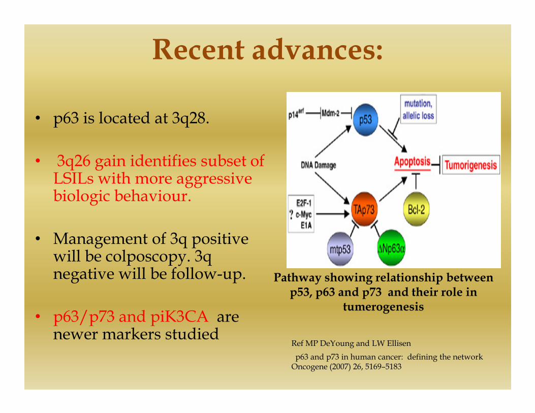
• p63 is located at 3q28.
• 3q26 gain identifies subset of LSILs with more aggressive biologic behaviour.
• Management of 3q positive will be colposcopy. 3q negative will be follow-up.
• p63/p73 and piK3CA are newer markers studied
Pathway showing relationship between p53, p63 and p73 and their role in
tumerogenesis
p63 and p73 in human cancer: defining the network Oncogene (2007) 26, 5169–5183
Ref MP DeYoung and LW Ellisen

Markers tested in combinations
• Several studies have tested more
than one biomarker on the same
sample.
• The complementary role of P16 • The complementary role of P16
and MIB-1 on LBC and Cell Block
preparations which in
combination is compared to LBC,
improves diagnostic accuracy for
HSIL and squamous cell
carcinoma.
A, p16 immunostaining in cervical intraepithelial neoplasia (CIN) 1 with a focal, patchy staining pattern (×200). B, ProExCimmunostaining in CIN 1 with a basal layer staining pattern (×200). C, p16 immunostaining in CIN 3 with a diffuse staining pattern (×200). D, ProExC immunostaining in CIN 3 with a diffuse staining pattern (×200). E, p16 immunostaining in carcinoma (×200). F, ProExC immunostaining in carcinoma (×200)
Ref Ming Guo, MD, Amy C. Baruch, MD, Elvio G. Silva, MD, Yee Jee Jan, MD, E. Lin, MD, Nour Sneige, MD, Michael T. Deavers, MDEfficacy of p16 and ProExC Immunostaining in the Detection of High-grade Cervical Intraepithelial Neoplasia and CervicalCarcinoma. Am J Clin Pathol. 2011;135:212-220.

Conclusion:
• There are different methods for early detection of cervical cancer.
They are
• A) Biomarkers on tissue,• A) Biomarkers on tissue,
• B) Serum levels of various human markers,
• C) HPV testing
• D) gene profiling.

Conclusion:
• The biomarkers or immunomarkers can be studied on various cytological specimens like
• a) LBC,
• b) Cell block • b) Cell block
• C) Histopathological biopsies
• To increase the sensitivity, specificity and diagnostic accuracy of cervical cancer

Conclusion:
• These marker studies are cost effective as compared to HPV testing which is more accurate
• Thus improvement in diagnostic accuracy and cost effectiveness makes it useful to include biomarkers in effectiveness makes it useful to include biomarkers in single or in combinations in cervical cancer screening programme.
• These along with clinical colposcopy, VIA, conventional pap smear screening, LBC, cell block and histopathological biopsies will help to decrease the deaths by cervical cancer in developing countries.

References
• Arbyn M, Castellsague X, de Sanjose S, Bruni L, Saraiya M Bray F, Ferlay J. Worldwide burden of cervical cancer in Ann oncol ;22:2675-2686,2008.
• D P Malinowski. Multiple biomarkers in molecular oncology. I.molecular diagnostics application in cervical cancer detection’. Expert review of molecular diagnostics, 7117-131,2007.review of molecular diagnostics, 7117-131,2007.
• Pinto AP, Degen M, Villa LL, Cites ES. Immunomarkers in gynaecologic cytology. The search for the ideal ‘Biomolecular papanicoloau test’, Acta cytol, 56;109-121:2012.
• Nieh S F, Chen TY Chu, HC Lai and E Fu. Expression of P-16 INK4A in papanicoloau smears containing atypical squamous cells of undetermined significance from the uterine cervix. Gynaecologic oncology 91, p-201-208,2003

References
• Peter Baldwin, Ronald Laskey & Nicholas ColemanNature Reviews Cancer 3, 217-226 (March 2003)
• Charlotte A. Brown, et all Role of Protein Biomarkers in the Detection of High-Grade Disease in Cervical Cancer Screening Programs Journal of Oncology Volume 2012 (2012)
• Ralf HilfrichHPV L1 Detection as a Prognostic Marker for Management of HPV High Risk Positive Abnormal Pap Smears
• Ming Guo, MD, Amy C. Baruch, MD, Elvio G. Silva, MD, Yee Jee Jan, MD, E. Lin, MD, Nour Sneige, MD, Michael T. Deavers, MD Efficacy of p16 and ProExC Immunostaining in the Detection of High-grade Cervical Intraepithelial Neoplasia and Cervical Carcinoma. Am J ClinPathol. 2011;135:212-220.
• MP DeYoung and LW Ellisen p63 and p73 in human cancer: defining the network Oncogene (2007) 26, 5169–5183

Acknowledgment
• Department of Pathology H.O.D, Faculty, Postgraduates and Technicians. JSS Medical College, Mysore.
• Department of Pathology , Kidwai Institute of Oncology, Bangalore.
• Department of OBG, JSS Medical College, Mysore.

JSS MEDICAL COLLEGE

Let Us Meet AgainLet Us Meet AgainLet Us Meet AgainLet Us Meet Again
We welcome you all to our future We welcome you all to our future conferences of OMICS Group conferences of OMICS Group
International International
Please Visit:Please Visit:
www.omicsgroup.comwww.omicsgroup.com
www.conferenceseries.comwww.conferenceseries.com
http://pathology.conferenceseries.com/http://pathology.conferenceseries.com/