about the pagination of this ebook - aswarphysics.weebly.com · about the pagination of this ebook...
TRANSCRIPT


About the pagination of this eBook
Due to the unique page numbering scheme of this book, the
electronic pagination of the eBook does not match the pagination
of the printed version. To navigate the text, please use the
electronic Table of Contents that appears alongside the eBook or
the Search function.
For citation purposes, use the page numbers that appear in the text.

Encyclopædia Britannica, Inc.
Chicago ■ London ■ New Delhi ■ Paris ■ Seoul ■ Sydney ■ Taipei ■ Tokyo
Britannica Illustrated Science LibraryBritannica Illustrated Science Library
HUMAN BODY IHUMAN BODY I

© 2008 Editorial Sol 90All rights reserved.
Idea and Concept of This Work: Editorial Sol 90
Project Management: Fabián Cassan
Photo Credits: Corbis, ESA, Getty Images, Graphic News,NASA, National Geographic, Science Photo Library
Illustrators: Guido Arroyo, Pablo Aschei, Carlos FranciscoBulzomi, Gustavo J. Caironi, Hernán Cañellas, Leonardo César,José Luis Corsetti, Vanina Farías, Manrique Fernández Buente,Joana Garrido, Celina Hilbert, Inkspot, Jorge Ivanovich, IvánLonguini, Isidro López, Diego Martín, Jorge Martínez, MarcoMenco, Marcelo Morán, Ala de Mosca, Diego Mourelos, LauraMourelos, Pablo Palastro, Eduardo Pérez, Javier Pérez, ArielPiroyansky, Fernando Ramallo, Ariel Roldán, Marcel Socías,Néstor Taylor, Trebol Animation, Juan Venegas, ConstanzaVicco, Coralia Vignau, Gustavo Yamin, 3DN, 3DOM studio
Composition and Pre-press Services: Editorial Sol 90Translation Services and Index: Publication Services, Inc.
Portions © 2008 Encyclopædia Britannica, Inc.Encyclopædia Britannica, Britannica, and the thistle logo areregistered trademarks of Encyclopædia Britannica, Inc.
Britannica Illustrated Science Library Staff
EditorialMichael Levy, Executive Editor, Core EditorialJohn Rafferty, Associate Editor, Earth SciencesWilliam L. Hosch, Associate Editor, Mathematics and
ComputersKara Rogers, Associate Editor, Life SciencesRob Curley, Senior Editor, Science and TechnologyDavid Hayes, Special Projects Editor
Art and CompositionSteven N. Kapusta, DirectorCarol A. Gaines, Composition SupervisorChristine McCabe, Senior Illustrator
Media AcquisitionKathy Nakamura, Manager
Copy DepartmentSylvia Wallace, DirectorJulian Ronning, Supervisor
Information Management and RetrievalSheila Vasich, Information Architect
Production ControlMarilyn L. Barton
ManufacturingKim Gerber, Director
Britannica IllustratedScience LibraryBritannica IllustratedScience Library
Encyclopædia Britannica, Inc.
Jacob E. Safra, Chairman of the Board
Jorge Aguilar-Cauz, President
Michael Ross, Senior Vice President, Corporate Development
Dale H. Hoiberg, Senior Vice President and Editor
Marsha Mackenzie, Director of Production
International Standard Book Number (set): 978-1-59339-797-5
International Standard Book Number (volume): 978-1-59339-809-5
Britannica Illustrated Science Library: Human Body I 2008
Printed in China
www.britannica.com

Human Body I

Contents
What Are WeMade Of?Page 6
Bones andMusclesPage 18
Internal Systemsand OrgansPage 34
The Sensesand SpeechPage 68
ControlCentersPage 80

What are cells like, and how do they formtissue? What is blood, and why are proteinsso important? The heart, usually thought of asthe wellspring of love and the emotions, isactually the engine of the circulatory system.It is because of the heart that all the cells ofthe body receive a constant supply ofnutrients, oxygen, and other essentialsubstances. The heart is so powerful that itpumps about 10 pints (4.7 l) of blood perminute. The nervous system is the mostintricate of all the body's systems. It works
A LIVING STRUCTUREThe skeleton consists of206 separate bones, which differ in form, size,and name. It supports and shapes the body,protects the internalorgans, and—in the bone marrow of certainbones—manufacturesvarious types of blood cells.
every second of every day, gatheringinformation about the organism and itssurroundings and issuing instructions so thatthe organism can react. It is this computerthat permits us to think and remember andthat makes us who we are.
The nervous system is a complex networkof sensory cells, originating in the brainand spinal cord, that transmits signals
throughout the body, employing a caravan ofchemical messengers to make sense of thismarvelous complex that we catalogue astouch, taste, smell, hearing, and vision. In fact,at this precise moment, because of anextraordinary relationship between our eyesand our brain, we are able to see andunderstand what we are reading. Moderncameras are designed on the same basicprinciples as our eye, but they have never beenable to equal the visual power of the eye. Thefocus and the automatic aperture of the humaneye are perfect. Our ears share a similarcomplexity and allow us to have excellenthearing. The external ear operates by receivingsound waves in the air. Sound waves travelthrough the auditory canal and are transmittedby the bones of the intermediate ear towardthe cochlea, which contains liquid and isspiraled like the shell of a small sea snail. Thecochlea converts waves of air into vibrations ofliquid, which are detected by special filamentsin the ear that are of many lengths and thatdetect sound waves of different lengths. Thesefilaments then transmit nerve impulses to thebrain and provide us with our ability tointerpret what we hear. This book will alsotell you about the function of our skin, thelargest organ of the body, which serves as anelastic barrier covering and protectingeverything inside our bodies. Captivatingimages will show you how each of ourextraordinary body systems function, andincredible facts will help you understand whythe human body is so amazing.
How can we understand what we are?What are we made of? Are we awarethat all that we do—including reading
this book—is the work of a marvelousmachine? We know very little about how weare able to be conscious of our own actions;nevertheless, even though we are usually notvery aware of it, this community of organsthat is the body—an integrated system thatincludes the brain, heart, lungs, liver, kidneys,muscles, bones, skin, and endocrine glands—acts together in exquisitely regulatedharmony. It is interesting that variousmechanisms work together to keep thetemperature of the body at 98.6° F (37° C);
thanks to the dynamic structure of bonesand cartilage, the body is maintained inperfect balance. The body also has afantastic ability to transform the food it
ingests into living tissues, bones, andteeth, all of which contribute to its growth.By this same process, we obtain the energyfor working and playing. It is hard toimagine that not long ago the cells of thebody of the person reading this book were
autonomous and were duplicatingthemselves freely within the walls of amother's uterus. Certainly no onereading this book could recognize herself
or himself in those cells. Nevertheless,each cell carried within it the informationnecessary for the development of thatperson. Everything that happens inside us istruly fascinating. Therefore, we invite you toenjoy this book. It is full of incredible factsand illustrations that will show you thecomplex ways each part of the body works.
A PerfectMachine

What Are We Made Of?
To understand the truest andmost elementary characteristicsof life, we must begin with thecell-the tiny organizingstructure of life in all its forms.
Most cells are too small to beobserved with the naked eye, but theycan be distinguished easily through anordinary microscope. Human bodytissues are groups of cells whose size
and shape depend on the specifictissue to which they belong. Did youknow that an embryo is a mass ofrapidly dividing cells that continue todevelop during infancy? We invite you
to turn the page and discover manysurprising things in this fascinatingand complex world.
UNDIVIDED ATTENTION 8-9
WATER AND LIQUIDS 10-11
THE CELL 12-13
MITOSIS 14-15
SYSTEMS OF THE BODY 16-17
MITOSISAn enlarged view that showsthe process of mitosis, themost common form ofcellular division

HUMAN BODY I 98 WHAT ARE WE MADE OF?
UndividedAttentionFrom birth the infant's braincells develop rapidly,making connections that canshape all of life'sexperiences. The first threeyears are crucial. Whenneurons receive visual,auditory, or gustatory stimuli,they send messages thatgenerate new physicalconnections withneighboring cells. The signalsare sent through a gap calleda synapse by means of acomplex electrochemicalprocess. What determinesthe formation of a person'ssynapses and neuralnetworks? One key factor isbelieved to be the undividedattention and mental effortexerted by the person.
THE SENSE OF TOUCHIt is predominant in the fingers and hands. Theinformation is transmitted through neurotransmitters,nerves that carry these impulses to the brain andthat serve to detect sensations such as cold, heat,pressure, and pain.
SKINThe skin is one of themost important organs of thebody. It contains approximatelyfive million tiny nerve endingsthat transmit sensations.
LearningEach child has his or her own intellectual filter; thequality of the filter depends on undivided attention andon how the child responds to a broad variety of stimuli.
BrainAt birth the infant brain contains 100billion neurons. That is about as manynerve cells as there are stars in theentire Milky Way Galaxy! Then as theinfant receives messages from thesenses, the cerebral cortex begins itsdynamic development.
RespirationRespiration is usually an involuntary,automatic action that allows us to take inthe oxygen we need from the air and exhalecarbon dioxide. These gases are exchangedin the pulmonary alveoli.
NeuronsEach neuron in the brain can beconnected with several thousand otherneurons and is capable of receiving100,000 signals per second. The signalstravel through the nervous
system at a speed of 225 miles per hour(360 km/h). Thanks to this complexcommunication network, the brain iscapable of remembering, calculating,deciding, and thinking.
A WORLD OF SENSATIONSThe tongue recognizes four tastes (sweet,salty, sour, and bitter), and the nasal fossascontain cells that have more than 200 millionfilaments, called cilia, which are capable ofdetecting thousands of odors.
DENDRITESThey are the branchesthrough which a neuronreceives and sends messages.With this system each neuroncan be stimulated bythousands of other neurons,which in turn can stimulateother neurons, and so forth. 3 pounds
(1.4 kg) IS THE WEIGHT OF A HUMAN BRAIN.
225(360 km/h)
THE VELOCITY OF THE NERVOUSSYSTEM'S SIGNALS
milesper hour

HUMAN BODY I 1110 WHAT ARE WE MADE OF?
Water and FluidsW
ater is of such great importance that it makes up almosttwo thirds of the human body by weight. Water is present in allthe tissues of the body. It plays a fundamental role in digestion and
absorption and in the elimination of indigestible metabolic waste. Water alsoserves as the basis of the circulatory system, which uses blood to distributenutrients to the entire body. Moreover, water helps maintain body temperatureby expelling excess heat through the skin via perspiration and evaporation.Perspiration and evaporation of water account for most of the weight a personloses while exercising.
N 3% NITROGENPresent in proteinsand nucleic acids
Water Balance and FoodIn its continuous process of taking in andeliminating water, one of the most
important functions of the body is to maintain acontinuous equilibrium between the water thatenters and the water that leaves the body.Because the body does not have an organ orother place for storing water, quantities that arelost must be continuously replenished. Thehuman body can survive for several weekswithout taking in food, but going without waterfor the same length of time would have tragicconsequences. The human being takes in about2.5 to 3 quarts (2.5-3 l) of water per day. Abouthalf is taken in by drinking, and the rest comesfrom eating solid food. Some foods, such as fruitsand vegetables, consist of 95 percent water.Eggs are 90 percent water, and red meat andfish are 60 to 70 percent water.
HOW THIRST IS CONTROLLEDThirst is the sensation throughwhich the nervous system informsits major organ, the brain, that thebody needs water. The controlcenter is the hypothalamus. If theconcentration of plasma in the bloodincreases, it means the body is losingwater. Dry mouth and a lack ofsaliva are also indications that thebody needs water.
HOW WATER IS ABSORBEDWater for the body is obtainedprimarily by drinking and ingestingfood and through internal chemicalreactions.
HOW WATER ISELIMINATED
Water is expelled not only withurine but also with sweat, throughthe elimination of feces, and throughevaporation from the lungs and skin.
50%of the watercomes from
ingesting fluids.
35%of the water
is obtained from food.
15%comes from
metabolicactivities.
60%is eliminated with urine.
18%is eliminated bysweating and throughevaporation from the skin.
14%is eliminated duringexhalation by thelungs.
8%is eliminatedin excrement.
C 18% CARBONPresent in allorganic molecules
O 65% OXYGENPresent in water and inalmost all organic molecules
H 10% HYDROGENPresent in water,nutrients, andorganic molecules
Chemical Elements The body contains many chemical elements. The most common are oxygen, hydrogen,carbon, and nitrogen, which are found mainly in proteins. Nine chemical elements arepresent in moderate amounts, and the rest (such as zinc) are present only in very smallamounts, so they are called trace elements.
0.004% IRONFluids and tissues, bones,proteins. An iron deficiencycauses anemia, whosesymptoms include fatigueand paleness. Iron isessential for the formationof hemoglobin in the blood.
THE PERCENTAGE OF A PERSON'SWEIGHT THAT IS DUE TO WATER. INGENERAL, A 10 PERCENT LOSS OF WATERLEADS TO SERIOUS DISORDERS, AND ALOSS OF 20 PERCENT RESULTS IN DEATH.
60%
SULFUR 0.3%Contained in numerousproteins, especially in thecontractile proteins
S
POTASSIUM 0.3%Nerves and muscles;inside the cell
K
SODIUM 0.15%Fluids and tissues, inthe form of salt
Na
MAGNESIUM 0.05%Lungs, kidneys, liver,thyroid, brain, muscles,heart
Mg
PHOSPHORUS 1%Urine, bonesP
CHLORINE 0.2%maintains theequilibrium of waterin the blood.
Cl
CALCIUM 1.5% Bones, lungs, kidneys,liver, thyroid, brain,muscles, heart
Ca
0.0004% IODINEUrine, bones. Whenconsumed, iodine passesinto the blood and fromthere into the thyroid gland.Among its other functions,iodine is used by the thyroidto produce growthhormones for most of theorgans and for braindevelopment.
Fe
I
ProteinsProteins are formed throughthe combination of the fourmost common chemicalelements found in the body.Proteins include insulin, whichis secreted by the pancreas toregulate the amount ofsugar in the blood.

12 WHAT ARE WE MADE OF? HUMAN BODY I 13
The CellI
t is the smallest unit of the human body—and of allliving organisms—able to function autonomously. It isso small that it can be seen only with a microscope.
Its essential parts are the nucleus and cytoplasm,which are surrounded by a membrane. Each cellreproduces independently through a process calledmitosis. The animal kingdom does have single-celled organisms, but in a body such as that ofa human being millions of cells are organizedinto tissues and organs. The word “cell”comes from Latin; it is the diminutive ofcella, which means “hollow.” The scienceof studying cells is called cytology.
MATHIAS SCHLEIDEN
NUCLEUS
ROUGH ENDOPLASMIC
RETICULUM
MITOCHONDRIA
THEODOR SCHWANN
Cell TheoryBefore the invention of themicroscope, it was impossible to
see cells. Some biological theories weretherefore based on logical speculationsrather than on observation. People believedin “spontaneous generation” because it wasinconceivable that cells would regenerate.The development of the microscope,including that of an electronic version inthe 20th century, made detailedobservation of the internal structure of thecell possible. Robert Hooke was the first tosee dead cells in 1665. In 1838 MathiasSchleiden observed living cells, and in 1839,in collaboration with Theodor Schwann, hedeveloped the first theory of cells: that allliving organisms consist of cells.
MitochondriaThe mitochondria provide large amounts ofenergy to the cell. They contain a variety of
enzymes that, together with oxygen, degradeproducts derived from glycolysis and carry outcellular respiration. The amount of energyobtained in this process is almost 20times as great as that released byglycolysis in the cytoplasm.Mitochondria are very differentfrom other organelles becausethey have a unique structure: anexternal membrane enclosing aninternal membrane with a greatnumber of folds that delimitthe internal area, ormitochondrial matrix. Inaddition, the mitochondriahave a circular chromosomesimilar to that of bacteriathat allows the mitochondriato replicate. Cells that need arelatively large amount ofenergy have manymitochondria because thecells reproduce frequently.
TRANSPORT MECHANISMSThe cell membrane is a semipermeable barrier. The cellexchanges nutrients and waste between its cytoplasmand the extracellular medium via passive and activetransport mechanisms.
DIFFUSION It is a passivetransport mechanism in whichthe cell does not use energy. Theparticles that cross the cellmembrane do so because of aconcentration gradient. For example,water, oxygen, and carbon dioxidecirculate by diffusion.
FACILITATED DIFFUSIONPassive transport in whichsubstances, typically ions (electricallycharged particles), that becauseof their size could not otherwisepenetrate the cell's bilayer can do sothrough a pore consisting of proteins.Glucose enters the cell in this way.
ACTIVE TRANSPORT It occursby means of proteins and requiresenergy consumption by the cellbecause the direction of ion transportis against the concentration gradient.In some cells, such as neurons, theNa+/K+ pump uses active transportto move ions into or out of the cell.
UNDER THE MICROSCOPEThis cell has been magnified4,000 times with an electronmicroscope. The nucleus isclearly visible, along with sometypical organelles in the green-colored cytoplasm.
CENTRIOLESThey are cylindrical,hollow structures thatare part of thecytoskeleton.
NUCLEUSThe nucleus consistsof chromatin andregulates cellmetabolism, growth,and reproduction.
POREA discontinuity inthe nuclearmembraneformed byproteins
SMOOTH ENDOPLASMICRETICULUMVarious membranes, whosefunctions include transportand synthesis. They aretube-shaped and do nothave ribosomes.
VESICLEA closedcompartment.It transportsor digests cellproducts andresidues.
NUCLEOLEThe nucleole canbe single ormultiple. Thenucleole consistsof ribonucleicacid and proteins.
CELLULAR MEMBRANEThe covering of the cellsurrounding the cytoplasm.It is also known as theplasma membrane.
VACUOLETransports andstores ingestedmaterials, waste,and water
DNAIt is organizedinto chromosomeswithin the nucleus.DNA is genetic materialthat contains informationfor the synthesis andreplication of proteins.
GOLGI APPARATUSThis structure processes
proteins produced by therough endoplasmatic
reticulum and places themin sacs called vesicles.
CYTOPLASMThe region locatedbetween the plasmamembrane and thenucleus. It containsorganelles.
MITOCHONDRIAAn organelle of theeukaryotic cell responsiblefor cellular respiration
LYSOSOMEThis is the “stomach”of the cell because it
breaks down wastemolecules with its
enzymes.
RIBOSOMEThis organelle iswhere the laststages of proteinsynthesis takeplace.
CYTOSKELETONComposed of fibers,the cytoskeleton isresponsible for cell
motion, orcytokinesis.
ROUGHENDOPLASMATIC
RETICULUMA labyrinthine assembly of
canals and membranousspaces that transport
proteins and are involvedin the synthesis of
substances.
PEROXISOMEOrganelles presentin eukaryotes thatfunction to metabolizeand eliminate toxicsubstances from cells
100 billionTHE AVERAGE NUMBER OF CELLS IN THEBODY OF AN ADULT. ONE CELL ALONE CANDIVIDE UP TO 50 TIMES BEFORE DYING.

14 WHAT ARE WE MADE OF? HUMAN BODY I 15
MitosisI
t is the cell-division process that results in the formation ofcells that are genetically identical to the original (or mother)cell and to each other. The copies arise through replication
and division of the chromosomes, or genetic material, in such away that each of the daughter cells receives a similar inheritanceof chromosomes. Mitosis is characteristic of eukaryotic cells. Itensures that the genetic information of the species and theindividual is conserved. It also permits the multiplication of cells,which is necessary for the development, growth, and regeneration of the organism. The word “mitosis” comes from the Greek mitos,which means “thread,” or “weave.”
THE ESTIMATED NUMBER OF CELLSREPLACED EVERY SECOND IN THE HUMANBODY THROUGH CELLULAR DIVISION
50,000
50 MITOSES MARK THELIFETIME OF A CELL ANDARE KNOWN AS THE“HAYFLICK LIMIT.” THISIDEA IS NAMED AFTERLEONARD HAYFLICK, WHO IN1961 DISCOVERED THAT THESECTION OF DNA CALLEDTHE TELOMERE INFLUENCESCELL LIFE SPAN.
Limit
The Ever-Changing SkinMitosis, or cellular division, occurs intensely within theskin, a fundamental organ of the sense of touch. The dead
cells on the surface are continuously being replaced by new cells,which are produced by mitosis in the lowest, or basal, layer. Fromthere the cells move upward until they reach the epidermis, theouter layer of the skin. A person typically sheds 30,000 deadskin cells every minute.
AntioxidantsAntioxidants are various types of substances (vitamins,enzymes, minerals, etc.) that combat the pernicious effects of
free radicals—molecules that are highly reactive and form as a resultof oxidation (when an atom loses an electron), which is often causedby coming into contact with oxygen. A consequence of this oxidativeaction is the aging of the body. One action of antioxidants is theregulation of mitosis. Preventive geriatrics has focused on usingantioxidants to prevent disease and to slow aging, in part becauseproperly regulated mitosis is fundamental to these processes.
CENTROMERE
SUPERFICIALCELLS
GRANULARCELLS
SPINOUS CELLS
BASAL CELLS
SPINDLEFILAMENT
CELLULARMEMBRANE
CENTRIOLE
CYTOPLASM
CHROMATIN
NUCLEUS
NUCLEUS
ORGANELLES
SISTERCHROMOSOMES
CHROMATIDCHROMOSOME
SHEDDING SUPERFICIAL CELLS LAYERS OF THE SKIN
1. INTERPHASEAn independent stagethat precedes mitosis.The chromatinconsists of DNA.
2. PROPHASEIn prophase the chromatincondenses to form chromosomes.The karyotheca (nuclearenvelope) begins to disappear.Chromosomes are formed by twochromatids that are joinedtogether by a centromere.
3. METAPHASEIt is characterized by theappearance of the spindle. Thecentromere—the “center” ofeach chromosome—and thechromatids are joined togetherand align at the center of thespindle complex. The nuclearmembrane disappears.
4. ANAPHASEIn this crucial stage the copies ofgenetic information separate: thechromatids move apart and formsister chromosomes that migrate toopposite poles of the cell.
5. TELOPHASEThe spindle disappears, and anew nuclear membrane beginsto form around each new set ofchromosomes. The membranedivides, resulting in two newcells that are identicaldaughters of the original cell.
NUCLEUS

16 WHAT ARE WE MADE OF? HUMAN BODY I 17
Systems of the BodyT
he body has various systems with different functions. Thesefunctions range from reproducing a cell to developing a newhuman being, from circulating the blood to capturing
oxygen from the air, and from processing food throughgrinding and chemical transformations to absorbing nutrientsand discarding waste. These functions act in harmony, andtheir interaction is surprisingly efficient.
MuscularSystem
Its function is to define the shape of the organism andprotect it. The muscular system is essential forproducing movement. It consists of muscles, organsmade of fleshy tissue, and contractile cells. There aretwo types of muscles: striated and smooth. Striatedmuscles are attached to the bones and governvoluntary movement. Smooth muscles also obey thebrain, but their movement is not under voluntarycontrol. The myocardium, the muscle tissue of theheart, is unique and is in a class by itself. See page 30.
RespiratorySystemAir from the external world enters the bodythrough the upper airways. The centralorgans, the lungs, absorb oxygen and expelcarbon dioxide. The lungs send oxygenatedblood to all the cells via the circulatorysystem and in turn receive blood that requirespurification. See page 46.
EndocrineSystemThe endocrine system is formed by glands thatare distributed throughout the body. Itsprimary function is to produce approximately50 hormones, the body's chemical messengers.The endocrine system secretes the hormonesinto the bloodstream so that they can reachthe organs they are designed to influence,excite, or stimulate for such activities asgrowth and metabolism. See page 62.
MALE
The various male organs contributeone of the two cells needed tocreate a new human being. Twotesticles (or gonads) and a penis arethe principal organs of the system.The system is continuously active,producing millions of tiny cells calledspermatozoa. See page 64.
ReproductiveSystemFEMALE
A woman's internal organs are the vagina, theuterus, the ovaries, and the fallopian tubes.The basic functions of these organs are theproduction of ova and the facilitation offertilization of an ovum by a spermatozoon (amature male sperm cell). When fertilizationoccurs, it sets a group of processes in motionthat result in pregnancy. See page 66.
Skeletal SystemThe skeleton, or skeletal system, is a solid structureconsisting of bones that are supported by ligamentsand cartilage. The main functions of the systemare to give the body form and to support it, tocover and protect the internal organs, and toallow motion to occur. The skeleton alsogenerates red blood cells (callederythrocytes). See page 20.
Circulatory SystemThis system carries blood to and from the heart and reaches theorgans and cells in every part of the body. The supreme pump—theheart—drives the vital fluid—blood—through the arteries andcollects it by means of the veins, with a continuous driving impulsethat makes the heart the central engine of the body. See page 36.
NervousSystemThe central nervous system consists of thebrain, which is the principal organ of thebody, along with the spinal cord. Theperipheral nervous system consists of thecranial and spinal nerves. Together theysend external and internal sensations to thebrain, where the sensations are processedand responded to whether the person isasleep or awake. See page 82.
Lymphatic SystemIts basic functions are twofold. One is to defend the bodyagainst foreign organisms, such as bacteria or viruses. Theother is to transport interstitial fluid and substances from thedigestive system into the bloodstream via the lymphaticdrainage system. See page 42.
Digestive SystemThis system is a large tract that changes form andfunction as it goes from the mouth to the rectum andanus, passing through the pharynx, the esophagus, thestomach, and the small and large intestines. The liver andpancreas help process ingested food to extract itschemical components. Some of these components arewelcome nutrients that are absorbed by the system, butothers are useless substances that are discarded andeliminated. See page 50.
Urinary SystemThis system is a key system for homeostasis—that is,the equilibrium of the body's internal conditions. Itsspecific function is to regulate the amount of waterand other substances in the body, discarding any thatare toxic or that form an unnecessary surplus. Thekidneys and the bladder are the urinary system'sprincipal organs. The ureters transport the urine fromthe kidneys to the bladder, and the urethra carriesthe urine out of the body. See page 58.

JOINTS 28-29
MUSCULAR SYSTEM 30-31
MUSCLE FIBER 32-33
Bones and Muscles
The musculoskeletal systemconsists of the skeletal systemof bones, attached to each otherby ligaments to form joints, andthe skeletal muscles, which use
tendons to attach muscles to bone. Theskeleton gives resistance and stabilityto the body and serves as a supportstructure for the muscles to work andproduce movement. The bones also
serve as a shield to protect the internalorgans. In this chapter you will see indetail—even down to the inside of amuscle fiber—how each part works.Did you know that bones are constantly
being regenerated and that, besidessupporting the body, they are chargedwith producing red blood cells? In thischapter you will find incredible images,curiosities, and other information.
SKELETON 20-21
BONE TISSUE 22-23
CRANIUM AND FACE 24-25
THE GREAT AXIS OF THE BODY 26-27
MUSCLES OF THE THORAXThey play an important role inbreathing by facilitating thecontraction and expansion ofthe thoracic cavity.

Skeleton
20 BONES AND MUSCLES HUMAN BODY I 21
The skeleton, or the skeletal system, is a strong,resistant structure made up of bones and theirsupporting ligaments and cartilage. The skeleton
gives the body form and structure, covers andprotects the internal organs, and makes movementpossible. The bones store minerals and produce bloodcells in the bone marrow.
CRANIUM Holds and protectsthe brain
INFERIOR MAXILLARYThe only movable boneof the head, it forms themandible (or jaw).
SHOULDERBLADEJoins to thehumerus
Sexual DifferencesBone structure is basically the same forboth sexes. In women, though, the center
opening of the pelvis is larger in order for aninfant's head to pass through it during childbirth.The pelvic girdle is formed by two coxal, or hip,bones, which are joined in the rear with thesacral bone and are fused together in the frontin the pubis. The pelvic girdle is involved in thejoining of the hips, where it connects to thefemur (thigh bone), serving the function oftransmitting weight downward from the upperpart of the body. The pelvic girdle and sacrumform the pelvis, which contains the organs ofthe digestive, reproductive, and urinary systems.
Types of BonesDepending on their characteristics, such assize or shape, the bones of the human body
are generally classified as follows:SHORT BONES: have a spherical or conical shape.The heel bone is a short bone.LONG BONES: have a central section that lies betweentwo end points, or epiphyses. The femur is a long bone.FLAT BONES: form thin bony plates. Most bones of
the cranium are flat bones.IRREGULAR BONES: take various shapes. Thesphenoids (“wedgelike” bones) in the skull areirregular bones.SESAMOID BONES: are small and round. Thepatella and the bones between tendons andin the joints of the hands and feet aresesamoid bones.
Well-Defined FormThe structure of the skeleton can be described as avertical column of chained vertebrae with a pair of
limbs at each end and topped off by the cranium. The upperlimbs, or arms, are connected to the shoulder blades andclavicles in what is called the scapular belt, and the lowerlimbs, or legs, are connected at the hips, or pelvic belt. Thejoints reach such a level of perfection that modern engineeringoften uses them as a model in the study of levers whendesigning such objects as cranes or desk lamps. Although thebones that make up the skeleton are solid, they have a flexiblestructure and to a large degree consist of spongy tissue.Nevertheless, a small bone is capable of supporting up to9 tons without breaking. A comparable weight wouldcrush a block of concrete. For a long timeanatomists thought that bones themselves werenot alive and that their strength merelyprovided support for the other organs.Modern medicine recognizes that bonesare actively living, furnished withnerves and supplied with blood.
The body has 80 of these bones, which belong tothe part of the skeleton formed by the spinal
column, the ribs, and the cranium.
Axial BonesTHESE COMPRISE THE OTHER 126 BONES: THOSE OF THE ARMS, SHOULDERS,
HIPS, AND LEGS. THESE BONES PERMIT A GREAT RANGE OF MOTION.
Appendicular Bones
THE LENGTH OF THE SHORTESTBONE OF THE BODY. IT IS THESTIRRUP, A BONE IN THE EAR.
0.12 inches(3 mm)
THE SIZE OF THE LARGESTBONE OF THE BODY, THE FEMUR
17inches(43 cm)
In the Renaissance, the cradle ofmodernity, Leonardo da Vinci was oneof the first to make precise drawings ofhuman bones. Such drawings wereneeded for studying anatomy sincethere were no photographs or X-rays.
Leonardo
The total number of bones in the body isbetween 206 and 208, depending on theindividual. The variation occurs with the
supernumerary bones (bones of the skull) andthe sesamoids (bones found in the joints of thehands and feet or embedded within tendons).
208 bones
OCCIPITAL BONEForms part of theback of thecranium
CARPALSThe bones of the wrist
METACARPALSThe bones ofthe palm of thehand
COCCYX (TAILBONE)
CLAVICLEConnects the shoulderblade with the sternum
SPINAL COLUMNThe core of the body'sstructure
STERNUMConnectedto the ribsby bands ofcartilage
PELVISContains andsupports theabdominalorgans
HUMERUSThe bone of theupper part of thearm, extendingfrom the shoulderto the elbow
SACRUM
ILIUMForms theposterior, or back,part of the pelvis
RIBSSurround andprotect theheart and thelungs
CUBITUMThe inside boneof the forearm
RADIUSThe shorter boneof the forearm
CALCANUMHeel bone, thelargest bone ofthe foot
PHALANGESThe bones ofthe fingers
KNEECAPThe knee bone, orpatella, which isenveloped by tendons
FIBULAThe thin outsidebone of the lowerpart of the leg
FEMURThe thigh bone, thelargest bone in thebody. It extends fromthe hip to the knee.
TIBIAThe bone thatsupports most of theweight of the lowerpart of the leg
TARSALSAnkle bones
METATARSALSFive small bonesbetween the ankleand the toes
PHALANGESBones of the toes
SACRUM
COXALS
SACROILIAC The joint that transmitsthe weight of the bodyfrom the spinal columnto the pelvis
TWO TYPES OF BONE CELLS
22 BONES AND MUSCLES HUMAN BODY I 23
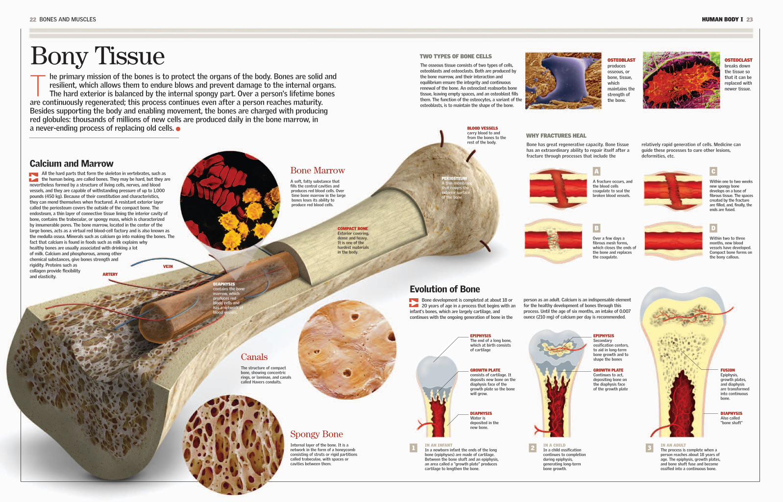
Bony TissueT
he primary mission of the bones is to protect the organs of the body. Bones are solid andresilient, which allows them to endure blows and prevent damage to the internal organs.The hard exterior is balanced by the internal spongy part. Over a person's lifetime bones
are continuously regenerated; this process continues even after a person reaches maturity.Besides supporting the body and enabling movement, the bones are charged with producingred globules: thousands of millions of new cells are produced daily in the bone marrow, ina never-ending process of replacing old cells. BLOOD VESSELS
carry blood to andfrom the bones to therest of the body.
PERIOSTEUM A thin membranethat covers theexterior surfaceof the bone
OSTEOBLASTproducesosseous, orbone, tissue,whichmaintains thestrength ofthe bone.
Bone MarrowA soft, fatty substance thatfills the central cavities andproduces red blood cells. Overtime bone marrow in the largebones loses its ability toproduce red blood cells.
OSTEOCLASTbreaks downthe tissue sothat it can bereplaced withnewer tissue.
IN AN INFANTIn a newborn infant the ends of the longbone (epiphyses) are made of cartilage.Between the bone shaft and an epiphysis,an area called a “growth plate” producescartilage to lengthen the bone.
EPIPHYSISSecondaryossification centers,to aid in long-termbone growth and toshape the bones
GROWTH PLATEContinues to act,depositing bone onthe diaphysis faceof the growth plate
GROWTH PLATEconsists of cartilage. Itdeposits new bone on thediaphysis face of thegrowth plate so the bonewill grow.
EPIPHYSISThe end of a long bone,which at birth consistsof cartilage
CanalsThe structure of compactbone, showing concentricrings, or laminae, and canalscalled Havers conduits.
COMPACT BONEExterior covering,dense and heavy.It is one of thehardest materialsin the body.
FUSIONEpiphysis,growth plates,and diaphysisare transformedinto continuousbone.
DIAPHYSISWater isdeposited in thenew bone.
DIAPHYSISAlso called“bone shaft”
Calcium and MarrowAll the hard parts that form the skeleton in vertebrates, such asthe human being, are called bones. They may be hard, but they are
nevertheless formed by a structure of living cells, nerves, and bloodvessels, and they are capable of withstanding pressure of up to 1,000pounds (450 kg). Because of their constitution and characteristics,they can mend themselves when fractured. A resistant exterior layercalled the periosteum covers the outside of the compact bone. Theendosteum, a thin layer of connective tissue lining the interior cavity ofbone, contains the trabecular, or spongy mass, which is characterizedby innumerable pores. The bone marrow, located in the center of thelarge bones, acts as a virtual red blood-cell factory and is also known asthe medulla ossea. Minerals such as calcium go into making the bones. Thefact that calcium is found in foods such as milk explains whyhealthy bones are usually associated with drinking a lotof milk. Calcium and phosphorous, among otherchemical substances, give bones strength andrigidity. Proteins such ascollagen provide flexibilityand elasticity.
Evolution of BoneBone development is completed at about 18 or20 years of age in a process that begins with an
infant's bones, which are largely cartilage, andcontinues with the ongoing generation of bone in the
person as an adult. Calcium is an indispensable elementfor the healthy development of bones through thisprocess. Until the age of six months, an intake of 0.007ounce (210 mg) of calcium per day is recommended.
Spongy BoneInternal layer of the bone. It is anetwork in the form of a honeycombconsisting of struts or rigid partitionscalled trabeculae, with spaces orcavities between them.
The osseous tissue consists of two types of cells,osteoblasts and osteoclasts. Both are produced bythe bone marrow, and their interaction andequilibrium ensure the integrity and continuousrenewal of the bone. An osteoclast reabsorbs bonetissue, leaving empty spaces, and an osteoblast fillsthem. The function of the osteocytes, a variant of theosteoblasts, is to maintain the shape of the bone.
WHY FRACTURES HEAL
Bone has great regenerative capacity. Bone tissuehas an extraordinary ability to repair itself after afracture through processes that include the
relatively rapid generation of cells. Medicine canguide these processes to cure other lesions,deformities, etc.
A
A fracture occurs, andthe blood cellscoagulate to seal thebroken blood vessels.
1 IN A CHILDIn a child ossificationcontinues to completionduring epiphysis,generating long-termbone growth.
2 IN AN ADULTThe process is complete when aperson reaches about 18 years ofage. The epiphysis, growth plates,and bone shaft fuse and becomeossified into a continuous bone.
3
B
Over a few days afibrous mesh forms,which closes the ends ofthe bone and replacesthe coagulate.
C
Within one to two weeksnew spongy bonedevelops on a base offibrous tissue. The spacescreated by the fractureare filled, and, finally, theends are fused.
D
Within two to threemonths, new bloodvessels have developed.Compact bone forms onthe bony callous.
VEIN
ARTERY
DIAPHYSIScontains the bonemarrow, whichproduces redblood cells andhas a network ofblood vessels.

24 BONES AND MUSCLES HUMAN BODY I 25
Cranium and FaceT
he cranium surrounds and protects the brain, cerebellum, and cerebral trunk (sometimescalled the encephalus). In an adult the cranium consists of eight bones that form the skull andthe base of the cranium. The face is the anterior part of the skull. It consists of 14 bones, all
of which are fixed except the lower maxillary, which makes up the mandible. The total number ofbones in the head as a whole exceeds the total of the face and cranium (22) because it includes thelittle bones of the middle ear.
Cranial SinusesThe sinuses are air-filled cavities whose principal knownfunction is to humidify and heat the air that enters therespiratory tract via the nose. The sinuses reduce theweight of the head, and they also act as resonancecavities, giving the voice its timbre. The sinuses arecovered by a moist membrane and are connected viasmall openings with the interior of the nasal cavity.When the sinuses become inflamed or filled with mucus,there is a risk of infection.
VibrationWhen a person speaks, the bonesof the cranium vibrate. In Japana technology was developedbased on this vibration. In 2006the firefighters of the Madridmunicipality in Spain adoptedthis technology. A helmet,furnished with a cranial contactmicrophone, amplifies thevibrations produced in the bonesof the cranium during speechand sends them to radioequipment.
FRONTAL SINUS
ETHMOID SINUS
SPHENOID SINUS
MAXILLARY SINUS
Foramen MagnumIn Latin this term means “big hole.” It is a circularopening, also called the occipital orifice, which islocated at the base of the cranium. The foramenmagnum allows for the passage of the spinalcolumn, the medulla oblongata, the vertebralarteries, and the spinal nerve. The placement of theforamen magnum toward the bottom of the skull isassociated with more highly evolved species.
The cranium can be comparedto a sphere, which consists of
separate bones at birth and closescompletely at maturity. Thenarrow separations between thebones, which appear as lines inthe fetus for the first months ofits life, are called sutures.Spaces called fontanels formwhere the sutures meet. Theirseparation has the functionalpurpose of allowing the brainto grow. Therefore, when braingrowth is complete, the spherecloses tightly, because itsfunction is to protect the brain.
Cranial Bones (8)
PARIETAL (2)The superior and lateral parts of the cranium
OCCIPITAL (1)Together with the temporals,
it forms the base of the cranium.
FRONTAL (1)It makes up the forehead.
TEMPORAL (2)The lateral part of the cranium
SPHENOID (1)The front part of the base of the cranium and part of the orbital bone (eye socket)
ETHMOID (1)Upper part of the nasal cavity
Facial Bones (14)
ZYGOMATIC (2)The cheekbones
PALATINES (2)Internal bones that form
the roof of the mouth
LACHRYMAL BONES (2)form the eye socket.
SUPERIOR MAXILLARIES (2)The upper mandible
NASAL CONCHAS (2)Independent of
the ethmoid conchas
VOMER (1)divides the nasal cavity
into two halves.
NASAL BONE (2)forms the bridge of the nose
(the rest of the nose is cartilage).
INFERIOR MAXILLARY (1)constitutes the mandible
and is the only facial bone that can move freely.
FORAMENMAGNUM
22THE TOTAL NUMBER OF BONESIN THE CRANIUM
83(1,360 cu cm)THE TYPICAL VOLUMEOF THE CRANIUM
Suturesand Fontanels
cubicinches
9THE WEIGHT OF ANADULT HUMAN HEAD
pounds(4 kg)

26 BONES AND MUSCLES HUMAN BODY I 27
The vertebral, or spinal, column is the flexible axis that lends supportto the body. It consists of a series of bones jointed together in aline, or chain, called the vertebrae. The spinal column forms a
protective inner channel through which the spinal cord runs. The ribsperform a similar function, wrapping and shielding the vital internalorgans, which include the heart and lungs.
DownwardsAll the vertebrae except the cervicalaxis and atlas have a cylindrical body,which gives them a particularcharacteristic: as they approach thepelvis they tend to be longer andstronger.
CARPALS (8)1. LUNATE2. PISIFORM3. TRIQUETRUM4. TRAPEZIUM5. TRAPEZOID6. CAPITATE7. SCAPHOID8. HAMATE
TARSUS (7)1. MEDIAL CUNEIFORM2. INTERMEDIATECUNEIFORM3. LATERAL CUNEIFORM4. TALUS5. TARSAL SCAPHOIDS6. CALCANEUS7. CUBOIDS
METACARPALS (5)
CARPALS (8)
METATARSALS (5)
PHALANGES (14)
PHALANGES (14)
Bones of the Hands and FeetEach hand (see the drawing below) has 27 bones, and eachfoot (see above) has 26. The hand has great mobility, and eachof its fingers (five in all) has three phalanges (distal, medial,and proximal), except for the thumb, which has two. Thecomplex of carpal bones makes up the wrist and is connectedto the forearm. The metacarpal bone sustains the medial part.The feet function in a similar manner; the toes have first,second, and third phalanges, except for the big toe.
Stability and MotionThe vertebrae have a centrum that allowsthem to support the body's weight, each
vertebra upon the next, as well as the weight of therest of the body. The vertebrae also have extensionsthat allow them to articulate with other vertebrae oract as supports for the ligaments and the muscles.This system gives the axis of the body both strengthand flexibility. In addition, most of the nerves of theperipheral system (that is, those responsible for
voluntary movement, for pain, and for the sense oftouch) are connected to the spinal cord inside thespinal column. In the centrum the vertebrae areseparated from each other by intervertebraldisks that are made of cartilage and have agelatinous interior. When an intervertebraldisk is damaged, some of this material canescape and pinch a nerve. This condition,called a herniated disk, can be very painful.
1
1
2
3
47
8
5
6
2
3
76
45
SACRUMThis bone isformed by fivefused vertebrae.
COCCYX This bone iscomposed of fourfused vertebrae.
The Ribs and the Rib CageThe 12 pairs of ribs, which also extend from thespinal column, protect the heart, lungs, majorarteries, and liver. These bones are flat andcurved. The seven upper pairs are called “trueribs,” and they are connected to the sternum (aflat bone consisting of fused segments) by
cartilage. The next two or three pairs (called“false ribs”) are connected indirectly. Theremaining pairs (“floating ribs”) are notattached to the sternum. The rib cage,formed by the ribs and its muscles, is flexible:it expands and contracts during breathing.
SACRALCANALNerves passthrough thesacral canal.
BLADE
LUMBAR VERTEBRAEThere are five of them, andthey bear the weight of theupper part of the body.
The Three CurvesThe three types of naturalcurvature in the spinalcolumn include cervicallordosis (forward, or inward,bending in the cervicalregion of the spine),kyphosis (outward bendingof the thoracic region of thespine), and lumbar lordosis(forward bending of thelower back). Shown hereis the right side of thespinal column.
AXISThe second cervicalvertebra. Together withthe atlas, it permits themovement of the head.
CERVICALThese sevenvertebrae (includingthe atlas and theaxis) support thehead and the neck.
THORACIC, OR DORSAL,VERTEBRAEThere are 12, and they arejoined to the ribs.
ATLASThis bone is the first ofthe seven cervical bones;it unites the spinalcolumn with the head.
PARTS OF THE VERTEBRAE
1. SPINAL APOPHYSIS2. TRANSVERSE
APOPHYSIS (2)3. ARTICULAR
APOPHYSIS (4) (2 SUPERIOR AND 2 INFERIOR)
4. LAMINAE (2)5. PEDICULAE (2)6. FORAMEN MAGNUM7. BODY
1
2
3
76
4
5
STERNUM
LUNG
LIVER
DIAPHRAGM
HEART
SPLEEN
STOMACH
RIBCARTILAGE
33 bonesOR VERTEBRAE, MAKE UP THESPINAL COLUMN. DEPENDING ON THEINDIVIDUAL, SOMETIMESTHERE ARE 34. THEY ARECONNECTED BY DISKS OFCARTILAGE THAT ACT ASSHOCK ABSORBERS. THESACRUM AND THE COCCYX AREA RUDIMENTARY TAIL LOSTDURING EVOLUTION.
The Great Axis of the Body

HUMAN BODY I 2928 BONES AND MUSCLES
The KneeThe knee is the biggestjoint of the body. It
maintains its stability because it isconstrained by four ligaments: theanterior and posterior cruciate andthe internal and external lateral. Theligaments link the femur (the thighbone) with the tibia (a bone of theleg). The knee is protected by thekneecap, a bony disk covered withcartilage that encases the anteriorand superior part of the kneejoint. Like the majority of thejoints, it is synovial.
FEMURThe thigh bone,which is theupper region ofthe lower limb
MUSCLE
MUSCLE
TIBIAThe larger ofthe two bonesof the lower leg
KNEECAPProtectivebony diskcovered withcartilage
SYNOVIALMEMBRANEproduces thesynovial liquid.
PATELLARLIGAMENTThis ligamentcrosses overthe kneecapand encases it.
MENISCUSFibrouscartilage thathelps theweight-supportingbones toabsorb a blow
EXTERNALLIGAMENTSStabilize the jointduring movement.The knee also hasinternal ligaments.
ARTERYThe femoral artery(artery of the femur)changes into thepopliteal artery at theposterior face of theknee. Like all arteriesit carries oxygenatedblood from the heart.
JointsT
hey are the structures where two or more bones come together, either directly or bymeans of strong fibrous cords called ligaments. The skeleton has movement thanks toits joints. Most joints, like the knee, are synovial joints. They are characterized by
mobility, versatility, and lubrication. The muscles that surround them contract to causemovement. When they work as a whole, the bones, muscles, and joints—together with thetendons, ligaments, and cartilage—constitute a grand system that governs the motoractivity of the body and allows us to carry out our daily physical activities.
Flexion
Extension
Circumduction
MOVEMENTSThe complex of joints,together with the musclesand bones, allows thebody to performnumerous actions, withmovements that includeturns and twists.
HypermobilityThe versatility of the joints refersto their characteristic range of
motion. Just as there are mobile,semimobile, and fixed joints, there is alsoa group of joints that are hypermobile.Such joints are less common but areeasily recognizable, especially in childrenand adults who have not lost theflexibility of their joints. The elbows,wrists, fingers, and knees can at an earlyage and in certain individuals have agreater-than-normal range of motion.For people with hypermobile joints thisextra range of motion can beaccomplished without difficulty or riskof dislocation.
Rotation
Abduction
DorsiflexionPlantarFlexion
Adduction
FIBULAThe smallest boneof the lower leg
A CHARACTERISTIC OF THE JOINTSIS THAT THEY CAN MAKE A SOUND,SUCH AS THAT MADE WHENSOMEONE CRACKS HER OR HISKNUCKLES. THIS IS BECAUSE THEREIS AN EXPLOSIVE RELEASE OF GASTHAT PERMITS A SHOCK-ABSORBINGFLUID TO FLOW IN THE JOINT.
Noise
IN THIS YEAR PROFESSORKENJI TAKAGI OF JAPANUSED A CYSTOSCOPE FOR THE FIRST INTERNAL OBSERVATION OF THE KNEE. Technological advances nowpermit arthroscopy to makeprecise observations for diagnosis.
1918
IN THE FORM OF A PIVOTThe joint of the upper bones of the neck.One bone is nested within the other andturns within it. This is the case of the atlasand the axis, in the upper part of the neck,which allow the head to turn from side toside. This is a limited movement.
SPHEROIDArticulation of the shoulder.A bone that has a spherical endthat can be inserted into anotherbone. The motion is extremelyvaried, such as that of theshoulders.
HINGEArticulation of the knee. One bonewith a cylindrical end is inserted intothe patellar groove of the other. Thereis flexion and extension, as in the knee.
PLANEArticulation of the foot. Twosurfaces that slide, one on top of theother, forward, backward, and sideways,as in some joints of the foot and wrist.
ELLIPSOIDThe joint betweenthe humerus and theradius. A bone with anoval end is inserted intothe cavity of anotherbone. The motion isvaried, but there isminimal rotation, as isthe case for the wrists.
BASAL JOINTThe joint at the baseof the thumb. The endsof the two bones cometogether at a rightangle. This allows themto turn, and they movebackward and forward,as occurs with thethumbs.
MobileThese are also called diarthroses; theyare the joints with the greatest range ofmotion. The ends of the bones linkedtogether are structured in various waysthat facilitate their movement relative toeach other, while ensuring the stability ofthe joint. Most joints in the body are ofthis type.
SemimobileAlso known as amphiarthroses. Thesurfaces of the bone that make contacthave cartilaginous tissue. One exampleis the vertebral joints: they have littleindividual movement, but as a wholethey have ample flexion, extension, androtation.
FixedAlso known as synarthroses. Most fixedjoints are found in the cranium and haveno need for motion because their primaryfunction is to protect internal organs.They are connected by bone growth orfibrous cartilage and are extremely rigidand very tough.
Where thepatellar tendon
connects to the bone

Muscular System
30 BONES AND MUSCLES HUMAN BODY I 31
The muscles are organs formed by fleshy tissue consisting of contractilecells. They are divided into striated, smooth, and, in a unique case,cardiac (the myocardium is the muscular tissue of the heart). Muscles
shape and protect the organism. The muscles of the skeleton are attachedto the bones to permit voluntary movement, which is consciously directedby the brain. The smooth muscles are also directed by the brain, but theirmotion is not voluntary, as in the case of digestion. These muscles getmost of their energy from alimentary carbohydrates, which can be storedin the liver and muscles in the form of glycogen and can later pass into theblood and be used as glucose. When a person makes a physical effort,there is an increased demand for both oxygen and glucose, as well as anincrease in blood circulation. A lack of glucose leads to fatigue.
FRONTAL MUSCLE wrinkles the forehead.
ORBICULAR MUSCLEallows blinking.
STERNOCLEIDOMASTOIDallows the head to turn and move forward.
PECTORALIS MAJORstretches the arm forward. It turns it and brings it close to the body.
BRACHIAL BICEPbends the arm at the elbow.
EXTERNAL OBLIQUEturns the trunk and bends it to both sides.
RECTUS ABDOMINISbends the trunk forward.
SPLENIUSkeeps the head erect.
TRAPEZIUMturns the head and theshoulders forward. Itstabilizes the shoulders.
OCCIPITALpulls the scalp backward.
ANTERIOR TIBIAlifts the foot and isconnected to the metatarsalbones of the foot.
EXTENSOR DIGITORUMLONGUSCalled the “pedis,” itconnects to the dorsal partof the foot.
FEMORAL QUADRICEPSA powerful muscular complexthat stretches the knee whena person runs and kicks. Thequadriceps include fourmuscles, with their upperextremes connected to thefemur and the pelvis and theirlower extremes anchored inthe tibia. When the musclescontract, the lower part ofthe leg is thrust forward.
STRIATEDThey are also called “skeletal” (because theycover the skeleton) and “voluntary.” Theyare composed of cells and fibers thatcontract rapidly.
CARDIACComposed of small interconnected fibers,which maintain the rhythmic andcontinuous pumping of the heart.
SMOOTHPerform unconscious actions such asdigestion. Their fibers contract slowly overan extended period of time.
ACHILLES TENDONconnects the gastrocnemius tothe calcaneus bone (talus bone).
GASTROCNEMIUSAlso called “twins.”There are two, and theyextend from the femurto the calcaneus. Theybend the leg.
DELTOIDA triangular muscle surroundingthe shoulder. It lifts the arm tothe side and causes it to swingwhen walking.
FEMORAL BICEPbends the leg atthe knee.
Clearly, a lot fewermuscles are needed tosmile than to frown.
FOREHEAD
WRINKLE THEEYEBROWS
UPPER LIPELEVATOR
ZYGOMATICMINOR
MUSCLES FORFROWNING
MUSCLES FOR SMILING
GLUTEUS MAXIMUSextends from the hipto the thigh.
BRACHIAL TRICEPstretches the arm at the elbow.
When the Skeleton MovesThe great number of muscles ofvoluntary action available to the human
body makes possible thousands of distinctmovements. Actions from the simple blink ofan eyelid to the twisting of a belt areaccomplished by muscular action. The eyemuscles involve the most activity becausethey carry out 100,000 movements per day.Some 30 muscles control all the movementsof the face and define an infinite possiblecombination of facial expressions. It iscalculated that to pronounce one word, theorgans for speech and respiration move some70 muscles. The stirrup muscle, whichcontrols the stirrup of the ear, is one of thesmallest in the body. It measuresapproximately 0.05 inch (1.2 mm). There areother muscles that are very large, includingthe latissimus dorsi of the shoulder. The foothas 40 muscles and more than 200ligaments. Because the muscles areconnected by a great number of nerves, alesion or blow causes the brain to react,
producing pain. Approximately 40 percent ofthe total weight of the body consists of themuscular system. When the organismreduces the quantity of calories it normallyingests (for example, when a person goes ona diet), the first thing the body loses iswater, which is reflected in a rapid weightloss. Then the metabolism adapts to the diet,and the body resorts to using up muscletissue before drawing on the fats stored forburning calories. For this reason, when thediet begins this second phase, theconsequences can be lack of vigor and loss ofmuscle tone, which is recovered when thediet returns to normal.
OR VOLUNTARY MUSCLES ARE IN THETYPICAL HUMAN BODY.
650 skeletalmuscles
RISORIUS
ZYGOMATICMAJOR
OCULAR ORBIT
NASAL
LOWER LIPDEPRESSOR
MENTALISMUSCLE
PLATYSMA
THE THREE TYPES OF MUSCLES

HUMAN BODY I 3332 BONES AND MUSCLES
Muscular FiberA
fiber is the long, thin cell that, when organizedby the hundreds into groups called fascicles,constitutes the muscles. It is shaped like an
elongated cylinder. The amount of fiber presentvaries according to the function accomplished byeach muscle. Fibers are classified as white, whichcontract readily for actions that require force andpower, and red, which perform slow contractions inmovements of force and sustained traction. Eachmuscle fiber contains in its structure numerousfilaments called myofibers. Myofibers, inturn, have two classes of proteinfilaments: myosin, also called thickfilaments, and actin, or thin filaments.Both kinds of fibers are arranged intiny matrices called sarcomeres.
MUSCLEComposed ofhundreds of fiberbundles
MUSCLEFIBER
MYOFIBRILA filament that usually has asticklike form and that is foundinside a muscle fiber
PERINEURIUMThe sheath of connectivetissue that surroundseach fascicle
AXONThe extension of thenerve cell, whose endmakes contact with themuscle and other cells
SARCOMEREEach smallinternal cylinderof the myofibril,consisting ofactin and myosin
CONNECTEDFILAMENTSActin and myosin arelinked through thesefilaments.
MYOSIN AND ACTIN FILAMENTSThe actin and myosinfilaments overlapeach other to causemuscular contraction.
Z BANDmarks theboundarybetweensarcomeres.
THE HEAD OF A MOLECULEThe head of a myosin moleculeextends. It makes contactwith the actin, and the myocinand actin overlap each other,producing a muscularcontraction.
THICK MYOFILAMENT (MYOSIN)The principal protein in thethick muscles, whichenables the reaction thatleads to contraction
THIN MYOFILAMENT(ACTIN)determines muscularcontraction whenlinked with myosin.
The order to contract given by thenervous system ceases, and themuscle fibers return to a positionof rest. This happens to all muscles,regardless of the duration ofcontraction.
Relaxation
The nervous system orders the musclefibers, no matter which type, toshorten. In order to create musclecontraction, calcium is released withinthe muscle cell, which allows the actinand the myosin to come together andoverlap each other.
Contraction
THE LENGTH AMUSCLE FIBER CAN
REACH
12 inches(30 cm)
THE POTENTIALCONTRACTION OF AMUSCLE FIBER IN TERMSOF THE FIBER'S LENGTH
70%
Marathon runners may haveas much as 90 percent red,or slow, fibers in their twinmuscles. Champions in the100-meter dash have only20 to 25 percent.
Running
SpecializationThe quantity of muscle fiber varies according to the sizeand function of the muscle. Also, the same muscle can
combine white fibers (rapid contracters) and red fibers (slowcontracters). Even though their percentages differ from oneperson to the next, the composition of the muscles of the upperlimbs tends to be the same as that of the lower in the sameperson. In other words, the relation between motor neurons andmuscle fibers is inscribed in a person's genes. Depending on thetype of neuron that stimulates them, the fibers are differentiatedinto slow fibers (when the neuron or motor neuron innervatesbetween five and 180 fibers) and rapid fibers (when the neuroninnervates between 200 and 800 fibers). The neurons and thefiber constitute what is called a motor unit.
A Bone LeverIn a lever system a force is applied to one end of abar that is placed on a fixed point of support (thefulcrum) to move a weight at the other end. In the
body the bones are the bars, and the joints actlike a fulcrum. The force is proportional to themuscular contraction.
FASCICLEEach of the hundreds of fiber bundles thatmake up one muscle
CAPILLARIESThese bring blood tothe muscle fibers.
FIRST CLASS LEVERThe joint is located between themuscular contraction and the bodypart that is moved. Examples arethe muscles that pull the craniumto move the head backward.
1 SECOND CLASS LEVERThe body part that is moved islocated between the joint and themuscular contraction. Examplesare the muscles of the calf that liftthe heel.
2 THIRD CLASS LEVERThe most common type in the body,where the muscular contraction isapplied between the joint and thebody part moved. Examples are themuscles that bend the elbow.
3OppositesThe muscles contract or relax according to the movementto be accomplished. To make the brain's directive takeeffect, the muscles involved carry out opposing actions.
EXTENDEDARM
FLEXED ARM
RelaxedBiceps
ContractedTriceps
ContractedBiceps
RelaxedTriceps
Force
Fulcrum
Weight
Force
Fulcrum
WeightForce
Fulcrum
Weight

KIDNEYS 60-61
ENDOCRINE SYSTEM 62-63
MALE REPRODUCTIVE SYSTEM 64-65
FEMALE REPRODUCTIVE SYSTEM 66-67
Internal Systems and Organs
It is difficult to explain that thesexual attraction between a man andwoman—something that appears tobe so natural and intimate—is achemical phenomenon. What is
certain is that when a couple feels theyare in love, it is because hormones havegone into action. Without them, amorousthoughts and sexual fantasies would bedrab and dull. We invite you to find out to
what extent hormones determine manyof our actions and also to investigate indetail, one by one, how the body'ssystems function. You will learn tounderstand how various organs of the
body work as a team. Although eachorgan accomplishes specific tasks onits own, they all communicate witheach other, and together they form acomplete human being.
LUNGS 48-49
DIGESTIVE SYSTEM 50-51
STOMACH 52-53
LIVER, PANCREAS, BILE 54-55
LARGE AND SMALL INTESTINE 56-57
URINARY SYSTEM 58-59
CIRCULATORY SYSTEM 36-37
ALL ABOUT THE HEART 38-39
COMPONENTS OF THE BLOOD 40-41
LYMPHATIC SYSTEM 42-43
GANGLIA 44-45
RESPIRATORY SYSTEM 46-47
THE CHEMISTRY OF LOVEEven a light kiss results inthe release of adrenaline,causing a sensation ofeuphoria and joy.

Circulatory System
36 INTERNAL SYSTEMS AND ORGANS HUMAN BODY I 37
Its function is to carry blood to and from all the organs of the body. Todrive the constant movement of the blood, the system uses the pumpingof the heart, the organ that acts as the system's engine. The arteries
bring oxygen-rich blood to all the cells, and the veins retrieve the blood sothat it can be oxygenated once again and so that wastes can be removed.
VeinsThe veins are the conduits that transportdeoxygenated blood back toward the heart afterit has traveled to different parts of the body. Theveins have thin walls with less muscular fiber andless elasticity than the arteries. The principalveins have valves to prevent the reflux of blood,forcing it to travel in only one direction.
CapillariesThese are branchings of the arterioles, smallvessels into which the arteries are subdivided.The capillaries are tiny, and they come togetherto form small veins, which combine to form largerveins. The capillaries are crucial in the exchangeof oxygen, nutrients, and waste, and they form anetwork to carry out this activity. Ten capillariestogether are as thick as a human hair.
THE EXTERNAL DIAMETER OF THE AORTA (THELARGEST ARTERY) ANDTHE VENA CAVA (THELARGEST VEIN)
1 inch(2.5 cm)
TEMPORAL ARTERYruns along the side ofthe head.
SUPERIOR VENA CAVAbrings the blood fromthe upper part of thebody for purification.
The superior vena cavaand the inferior vena
cava together form thelargest vein.
INFERIOR VENA CAVAtakes blood arrivingfrom the area below
the diaphragm andbrings it up to the
heart.
LEFT PRIMITIVEILIAC VEINThis is the primaryvein of the hip area.
JUGULAR VEINSThere are two oneach side of the neck:the internal and theexternal.
LEFT CAROTID ARTERYruns along the neck andsupplies blood to thehead.
AORTIC ARTERY (AORTA)The body's principal artery
HEARTThe greatengine
HUMERAL ARTERY(Axillary) The right one arisesfrom the brachiocephalictrunk and the left from theaortic arch.
TUNICAADVENTITIA
ELASTICMEMBRANET
TUNICAMEDIA
OUTSIDE OF TUNICAINTIMA
INSIDE OF TUNICAINTIMA
PULMONARY ARTERYcarries blood to the lungs.
PALMAR VENOUS ARCHchannels the hand'svenal blood flow.
FEMORAL ARTERYcarries oxygenatedblood along the thigh.
TIBIAL ARTERYirrigates the leg.
BLOODDISTRIBUTIONDURINGCIRCULATION
SUBCLAVIAN VEINconnects the axillary withthe superior vena cava.
A System That Goes Around The center of the system is the heart, which, together with anetwork of vessels, forms the cardiovascular machinery. This vital
engine beats more than 30 million times a year-approximately 2 billiontimes in a person's lifetime. With each beat it pumps about 5 cubicinches (82 ml) of blood. This means that an adult heart could fill a2,000-gallon (8,000-l) tank in just one day. Beginning at the heart, thecirculatory system completes two circuits: the main, or systemic,circulation via the aortic artery and the minor, or pulmonary,circulation. The main circulation brings oxygenated blood to thecapillary system, where the veins are formed; the minor circulationbrings oxygen-poor blood through the pulmonary artery to be enrichedwith oxygen and to have carbon dioxide removed from it, a processcalled hematosis. Other secondary circuits are the hepatic portalsystem and the hypophyseal portal system.
TRUNCUS OF THEPORTAL VEIN
It terminates in thesinusoids of the liver.
RENAL VEINBlood exits the kidneys
through this vein.
TEMPORAL VEINruns along the sideof the head.
RADIAL ARTERYruns along the radialside of the forearm.
FEMORAL VEINruns along the thigh,channeling thedeoxygenated bloodtoward the heart.
TIBIAL VEIN
LEFT PRIMITIVEILIAC ARTERYprovides blood to thepelvis and the legs.
THE TOTAL LENGTH OF THEBLOOD VESSELS. NINETY-EIGHT PERCENT OF THEMARE CAPILLARIES.
60,000 miles(100,000 km)
THE RANGE IN DIAMETER OFCAPILLARIES THE AVERAGELENGTH IS 0.04 INCH (1 MM).
0.00001to 0.1 inch(0.001 to 0.2 mm)
67% VEINS
17% ARTERIES
11% HEART
5% CAPILLARIES
EXTERNAL MEMBRANE
INTERNAL COVERING
VALVE
MUSCULAR MEMBRANE
CAPILLARY WALL
NUCLEUS
ArteriesMuscular elastic blood vessels. Theirfunction is to bring oxygenated bloodfrom the heart (from the primaryartery, the aorta) to all the cells of thebody. Arteries have thick walls,allowing them to withstand the highpressure of the blood.

38 INTERNAL SYSTEMS AND ORGANS HUMAN BODY I 39
The heart is the engine of the circulatory apparatus: it supplies 10 pints (4.7 l) ofblood per minute. Its rhythmic pumping ensures that blood arrives in every partof the body. The heart beats between 60 and 100 times per minute in a person
at rest and up to 200 times per minute during activity. The heart is a hollow organ,the size of a fist; it is enclosed in the thoracic cavity in the center of the chest abovethe diaphragm. The name of the stomach's entrance, or cardias, comes from the Greekword for heart, kardia. Histologically, one can distinguish three layers of tissue in theheart, starting from the inside out: the endocardium, the myocardium, and the pericardium.
DIASTOLICThe atria and the ventriclesare relaxed. The blood,supercharged with carbondioxide, flows from all thecorners of the body andenters the right atrium, whilethe blood that wasoxygenated through thework of the lungs returns tothe left part of the heart.
THE SEQUENCE OF THE HEARTBEAT
SUPERIORVENA CAVAbrings theblood to beoxygenatedfrom the lowerpart of thebody.
RIGHT ATRIUMIt sends theblood throughthe tricuspidvalve to theright ventricle.
LEFTVENTRICLEreceives theoxygenatedblood via themitral valve.
LEFT ATRIUMreceives theoxygenated bloodfrom the lungs
TRICUSPIDVALVEopens so thatblood can passfrom the atrium tothe ventricle andthen closes toprevent it fromgoing back.
PAPILLARYMUSCLES
MITRAL VALVEThis valve, also knownas the bicuspid valve,opens the path for theblood from the leftauricle toward theventricle and thenprevents it fromreturning.
SEPTUMThe interventricularwall that separatesthe two inferiorcavities
PULMONARYVALVEThrough this valveblood to beoxygenated passesfrom the rightventricle toward thepulmonary artery.
AORTAThe principalartery of thebody.Oxygenatedblood exitsthrough thisartery.
AORTIC VALVEregulates the passage ofthe oxygenated bloodtoward the aorta.
VALVESThe valves control the bloodflow between the atria and theventricles. In the graphic above(right) the pressure of theblood pumped by the heartforces the valve open. Thegraphic below shows that oncethe blood has entered, its ownweight leads to a pressurereversal that causes the valveto close.
IS THE AVERAGE WEIGHT OFAN ADULT HEART (RANGE: 7 TO14 OUNCES [200 TO 400 G]).
The Return Flow of BloodThese cells are phantom cells,because all they contain is a large
amount of hemoglobin, a protein thathas a great affinity for combining withoxygen. The red blood cells, whichcirculate in the blood, bring oxygen tothe cells that need it, and they alsoremove a small part of the carbondioxide that the cells are discardingas waste. Because they cannotreproduce themselves, they mustbe replaced by new red bloodcells that areproduced by thebone marrow.
AORTA
PULMONARYVEIN
PORTALVEIN
PULMONARYARTERY
Network ofvessels in theupper part of
the body
Network ofvessels in thelower part of
the body
Network of vesselsin the liver
Network ofvessels in theright lung
Network ofvessels in
the left lung
Network ofvessels in the
digestiveapparatus
SUPERIORVENA CAVA
INFERIORVENA CAVA
IS THE APPROXIMATE NUMBER OFTIMES THAT THE HEART BEATS PERMINUTE. IT PUMPS 2,000 GALLONS(8,000 L) OF BLOOD PER DAY.
70
A RED BLOOD CELLTRAVERSES THE BODY IN 20 SECONDS.THEREFORE, THEDISTANCE THAT ITTRAVELS AMOUNTSTO 12,000 MILES(19,000 KM).
1
ATRIAL SYSTOLEThe atria contract to pushthe blood down toward theventricles. The rightventricle receives the bloodthat will have to be sent tothe lungs to be oxygenated.The left ventricle receivesblood coming from thelungs, which is alreadyoxygenated and must bepumped toward the aorta.
2
VENTRICULAR SYSTOLEThe ventricles contractafter a brief pause. Thesystole, or contraction, ofthe right ventricle sendsimpure blood to the lungs.The contraction of the leftventricle pumps thealready oxygenated bloodtoward the aorta; it isready for distributionthroughout the body.
3RIGHTVENTRICLEreceives theblood from itsatrium andpumps it to thepulmonaryvalve.
TENDINOUS CORDSThese are the smallfibrous threadswhosefunction is to fastenthe ends of the tricuspidvalve to the heart wall.
LEFT
RIGHT
VALVE
TENDINOUSCORDS
All About the Heart
10ounces(300 g)
20 seconds

40 INTERNAL SYSTEMS AND ORGANS
Components of the BloodT
he blood is a liquid tissue composed of water, dissolved substances, and blood cells. Theblood circulates inside the blood vessels thanks to the impulse it receives from thecontraction of the heart. A principal function of the blood is to distribute nutrients to all the
cells of the body. For example, the red blood cells (erythrocytes) carry oxygen, which associateswith the hemoglobin, a substance in the cell responsible for the blood's red color. The blood alsocontains white blood cells and platelets that protect the body in various ways.
THE APPROXIMATE VOLUME OFBLOOD PRESENT IN A HUMAN ADULT
5 quarts (4.7 l)
Blood ComponentsThe blood is a tissue, and assuch it is characterized by the
same type of cells and intercellularsubstance as tissue. It isdistinguished from the rest of thetissues in the human body by anabundance of intercellular material,
which consists primarily of water.The intercellular material, calledplasma, is yellow, and it containsabundant nutrients and othersubstances, such as hormones andantibodies, that take part in variousphysiological processes.
White Blood Cells,or LeukocytesThis is what a leukocyte, or whiteblood cell, looks like swimming inblood plasma. They are called whitebecause that is their color whenviewed under a microscope.
Plateletsare cell fragments thathave separated from themegakaryocytes, cellslocated in the bonemarrow. They have a rolein blood coagulation. Nextto the red blood cells, theplatelets are the mostabundant component ofthe blood.
Red Blood CellsThese cells are phantom cells, because all they containis a large amount of hemoglobin, a protein that has agreat affinity for combining with oxygen. The redblood cells, which circulate in the blood, bring oxygento the cells that need it, and they also remove a smallpart of the carbon dioxide that the cells are discardingas waste. Because they cannot reproduce themselves,they must be replaced by new red blood cells that areproduced by the bone marrow.
PlasmaRed and white blood cells and platelets(which contribute to coagulation)make up 45 percent of the blood. Theremaining 55 percent is plasma, a fluidthat is 90 percent water and the restvarious nutrients.
Red Blood Cells 4 to 6 million
White Blood Cells 4,500 to 11,000
Platelets 150,000 to 400,000
Normal pH 7.40
COMPONENTS OF THE BLOOD PER 0.00006 cubicinch (1 cu ml)
DAILY PRODUCTION IN MILLIONS
200,000
10,000
400,000
Red Blood CellsWhite Blood Cells
Platelets
COMPOSITIONGRANULOCYTES Neutrophils
Eosinophils
Basophils
AGRANULOCYTES Lymphocytes
Monocytes
0.0003 INCH (0.008 MM)
90% Water8% Protein2% other(salts, nutrients,glucose, amino acidfats, and waste)
0.0003 INCH (0.008 MM)
0.0003 INCH (0.008 MM)
HUMAN BODY I 41
Each person belongs to a bloodgroup. Within the ABO system thegroups are A, B, AB, and O. Eachgroup is also identified with anantigen, or Rh factor, that ispresent in the red blood cells of 85percent of the population. It is of
vital importance to know whatblood group a person belongs to soas to give only the right type duringa blood transfusion. The immunesystem, via antibodies and antigens,will accept the body's own bloodtype but will reject the wrong type.
GROUP AAn individual with red blood cells with antigen A in its membranesbelongs to blood group A, and thatperson's plasma has antibodies against type B. These antibodiesrecognize red blood cells with antigenB in their membranes as foreign.
FLEXIBILITYRed blood cells areflexible and take on abell shape in order topass through thethinnest blood vessels.
COMPATIBILITYDonors of group O can give blood to any group,but group AB donors can give only to otherswith AB blood. The possibility of blood donationdepends on the antibodies of the recipient.
A B AB0
A B AB0
ANTI-B ANTIBODY
ANTIGEN A
ANTIGEN B
ANTI-AANTIBODY
BICONCAVE FORM BELL-SHAPED
GROUP BMembers of this group haveantigen B in the membrane oftheir red blood cells and anti-Aantibodies in their blood plasma.
GROUP ABMembers of this group haveantigen A and B in themembrane of their red bloodcells and no antibodies in theirblood plasma.
GROUP OMembers of this group have noantigens in the membranes of theirerythrocytes and anti-A and anti-Bantibodies in their blood plasma
1
2
3
4
The Blood Groups
ANTIGEN A
ANTIGEN B
ANTI-BANTIBODY
ANTI-AANTIBODY
THE BLOOD MAINTAINSTHE BODY AT THISAVERAGE TEMPERATURE.
98.6º F(37º C)
IS THE PORTIONOF BODY WEIGHTREPRESENTED BYTHE BLOOD.
7%

Lymphatic System
42 INTERNAL SYSTEMS AND ORGANS HUMAN BODY I 43
It accomplishes two basic functions: defense against foreign organisms (suchas bacteria) and aid with transport of liquid and matter via the circulation ofthe lymph from the interstices of the tissue and from the digestive apparatus
to the blood. About 3 to 4 quarts (2.8-3.7 l) of the liquid circulating in the systemdo not return. This liquid is known as lymph, and it is reabsorbed into the plasmaonly through the lymphatic vessels. The lymph contains cells called lymphocytesand macrophages, which are part of the immune system.
One part of the liquid that exits fromblood flow and distributes itself in thebody returns only through the action of
the lymphatic tissue, which reabsorbs itvia the lymphatic capillaries and returnsit to the blood via the lymphatic vessels.
SPLEENThe largest lymphatic organ, itperforms specific tasks, suchas filtering the blood,producing white blood cells,and eliminating old blood cells.It also stores blood. Thespleen can weigh between 3and 9 ounces (100 and 250g). It is about 5 inches (12 cm)long and 3 inches (7 cm) wide.
THYMUSA gland consisting of twolobes, located in the uppersection of the sternum. Itdevelops during puberty andthen begins to decline,transforming itself into amass of connective tissue.The thymus transforms bloodcells produced in the bonemarrow into specialized Tlymphocytes.
PEYER'S PATCHLymphatictissuelocated inthe lowerregion ofthe smallintestine
BONE MARROWThe bonemarrowgenerates white bloodcells, orlymphocytes,within thebones.
LEFT SUBCLAVIAN VEINHas the same function asthe right subclavian vein.The name derives from its
location beneath the clavicle.
TONSILSSimilar to the ganglia,their tissue detectsinvading organisms.
SPLEENThe mainlymphorgan forthe entirebody
LYMPHATIC VESSELS
receive the lymphfrom the lymphatic
capillaries.
POPLITEAL LYMPH NODES
are located behind theknees, and they filter
the lymph from thelower extremities.
THYMUStransforms the
white blood cellsin the bone
marrow into Tlymphocytes.
THORACIC DUCTsends the lymph
to the leftsubclavian vein.
INGUINALLYMPH NODESfilter the lymphfrom the lowerregions of the
body.
1 The lymphatic systemgenerates lymphocytes (alsofound in the blood and in othertissue) and macrophages.Together they constitute theimmune system. Here invadingbacteria are devoured by amacrophage, and the Blymphocytes take informationfrom the surface of thebacteria that they need to“recognize” other similarbacteria.
2 The B lymphocytes are activatedand upon recognizing a pathogendivide themselves into plasmaticcells and memory cells. Theplasmatic cells secrete thousandsof antibody molecules persecond, which are carried by theblood to the site of the infection.The memory cells retain theantigen information, and, whenfaced with a new invasion, willonce again divide rapidly in orderto deal with it.
3 The antibodies, also called“immunoglobin,” are proteinmolecules in the form of a “Y,”with arms unique to eachspecific type of antibody. It isthis feature that attaches themto a specific antigen. Theirfunction is to “mark” invaders,which can then be destroyed bythe macrophages.
Immune Response
Lymphatic Tissue
RIGHT SUBCLAVIAN VEIN
brings the lymph from theupper part of the body to
the lymphatic duct.
AXILLARYLYMPHATICGANGLIAThe lymph fromthe chest andthe arms isfiltered justabove thearmpits.
Lymphatic NetworkThis network contains vesselsthat extend throughout the body
and that filter the liquid that comesfrom the area surrounding the cells. Thelymph circulates in only one directionand returns to the blood through thewalls of small blood vessels. There arevalves that prevent the lymph from
flowing in the opposite direction. Thelymph nodes filter harmfulmicroorganisms from the lymph, whichreturns via blood vessels to maintainthe equilibrium of the body's fluids.Together with the white blood cells, thelymph nodes are in charge ofmaintaining the immune system.
ARTERIOLE
BLOOD CAPILLARY
LYMPHATIC CELL
LYMPHATIC CAPILLARY
VENULE
DIRECTION OF BLOOD FLOW
LYMPHATIC CELL
INTERSTITIALLIQUIDpenetratesthrough theultra-fine spacesin the tissues.
CAPILLARYCELLSlie along, butdo not impede,the passage of fluid.
VALVEopens whenthe liquid haspassed.
LATERALAORTIC NODES
Together with the thymusand the spleen, bonemarrow constitutes thelymphatic system tissues,whose function is tomature the lymphocytes.
BONEMARROW
THE AMOUNT OF LIQUID THATLEAVES THE BLOOD ANDPASSES THROUGH THE SYSTEMDAILY, MOVING THROUGH THETISSUES AND RETURNING TOTHE BLOODSTREAM
6 gallons (24 l)

44 INTERNAL SYSTEMS AND ORGANS HUMAN BODY I 45
Lymph NodeA
lso called a lymph gland, this node has a round shape and is about0.4 inch (1 cm) in diameter. Lymph nodes are distributed throughoutthe body—in the neck, armpits, groin, and popliteal bone (behind the
knees), as well as in the thorax and abdomen. The lymphatic vessels are theducts for the lymph and the pathways for communication among thelymph nodes. The battle of the immune system against invading germstakes place within the nodes, which then enlarge because of inflammation.
THE AREA OF THE SKINCOVERED BY SWEATGLANDS, A PART OF THENATURAL DEFENSES THATCOMPLEMENT THE WORKOF THE GANGLIA IN THEIMMUNE SYSTEM
100(600 sq cm)
THE COLOR OF INFLAMED SKINWHEN BACTERIAL ACTION IN A WOUND CAUSESVASODILATION. THIS OCCURSBECAUSE THE BLOOD VESSELSEXPAND TO INCREASE BLOODFLOW AS A MEANS OF DEFENSE.
Red
InvadersDisequilibrium can be causedin the homeostatic
mechanisms of the human body,causing disease that may or may notbe infectious. Noninfectious diseaseis usually produced by heredity,
external factors, or lifestyle.Infections are brought on byparasitic organisms, such asbacteria, viruses, fungi, and protozoa(single-celled organisms belonging tothe protist kingdom).
Natural DefensesBesides the immune system,composed in part by the lymphatic
system, the body has another group ofresources called natural defenses, whichpeople possess from birth. The body's firstdefensive barrier is the skin. If pathogenicagents succeed in passing through itsfilters, however, both the blood and thelymph possess specialized antimicrobialcells and chemical substances.
A Defensive FilterThe glands are covered with asheath of connective tissue, which
in turn forms an interior network thatconsists of clusters filled withlymphocytes. Their immunologicalfunctions are to filter the fluid that arrivesvia both the sanguine and lymphaticafferent veins, which then goes toward theheart to be returned to circulation via theefferent vessels and to produce immunecells for attacking and removing bacteriaand carcinogenic cells.
RESISTANTCAPSULEHas the functionof envelopingand protectingthe ganglia
B LYMPHOCYTESacquire their
immunologicalcapacity in the
bone marrow andin the liver of
the fetus.
RETICULARFIBERSThe networks thatsupport the lymphnodes
VEIN
ARTERY
T CELLSSpecializedlymphocytescreated in thethymus to helpdetect antigens
EFFERENTLYMPHATIC VESSELThe conduit for thelymph that exits theganglia and returns tothe bloodstream
VALVEregulates the passageof the lymph andprevents its reflux.
LYMPHOCYTESWhite blood cellsthat, together withthe macrophages,are the basis ofthe cellularcomponent of theimmune system.
GERMINAL CENTER The area that contains B lymphocytes. There aretwo types: B cells, whichproduce antibodies, andT cells.
MACROPHAGESTogether with thelymphocytes, they arethe basis of the immunesystem. They devour theinvading bodies that aredetected.
AFFERENT LYMPHATIC VESSELThe afferent vesselscarry the lymphaticliquid from the bloodto the ganglia, orlymphatic nodes.
A BACTERIAare found by the billions in anymedium. Not all of them are harmful.Bacteria known as germs arepathogenic and release poisonoussubstances called toxins.
B VIRUSESare not really living beings butchemical packages. They consist ofgenetic material. When they enterthe body, they invade a cell, wherethey reproduce and then spread.
C PROTOZOAare organisms that typically live inwater and in soil. There are about 30pathogenic species, which canproduce a range of diseases fromsleeping sickness and severe diarrheato malaria.
SWEAT GLANDsecretes sweat, which helps tocontrol body temperature, toeliminate toxins, and to protectthe skin immunologically.
MUCOUS SECRETIONSThese secretions, called mucus,form in the upper and lowerrespiratory tracts, where theycapture bacteria and carry themto the throat to be spit out.
SALIVARY GLANDproduces saliva, which containsbactericidal lysozymes.
INTESTINAL MUCOSAThe goblet cells in thismembrane produce adefensive mucus.
VAGINAL BACTERIAUnder normal conditions, theseare inoffensive, and they occupyareas that could be invaded bypathogenic bacteria.
SEBACEOUS GLAND Located on the surface of theskin, this gland secretes a fattysubstance called sebo.
LACHRYMAL GLANDSecretes tears that protect theeyes. Tears, like saliva andperspiration, kill bacteria.
squareinches
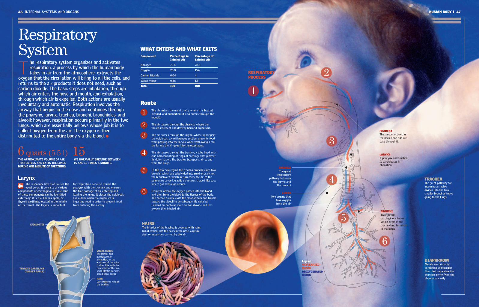
46 INTERNAL SYSTEMS AND ORGANS HUMAN BODY I 47
RespiratorySystemT
he respiratory system organizes and activatesrespiration, a process by which the human bodytakes in air from the atmosphere, extracts the
oxygen that the circulation will bring to all the cells, andreturns to the air products it does not need, such ascarbon dioxide. The basic steps are inhalation, throughwhich air enters the nose and mouth, and exhalation,through which air is expelled. Both actions are usuallyinvoluntary and automatic. Respiration involves theairway that begins in the nose and continues throughthe pharynx, larynx, trachea, bronchi, bronchioles, andalveoli; however, respiration occurs primarily in the twolungs, which are essentially bellows whose job it is tocollect oxygen from the air. The oxygen is thendistributed to the entire body via the blood.
Component Percentage in Percentage of Inhaled Air Exhaled Air
Nitrogen 78.6 78.6
Oxygen 20.8 15.6
Carbon Dioxide 0.04 4
Water Vapor 0.56 1.8
Total 100 100
The air enters the nasal cavity, where it is heated,cleaned, and humidified (it also enters through themouth).
The air passes through the pharynx, where thetonsils intercept and destroy harmful organisms.
The air passes through the larynx, whose upper part,the epiglottis, a cartilaginous section, prevents foodfrom passing into the larynx when swallowing. Fromthe larynx the air goes into the esophagus.
The air passes through the trachea, a tube lined withcilia and consisting of rings of cartilage that preventits deformation. The trachea transports air to andfrom the lungs.
In the thoracic region the trachea branches into twobronchi, which are subdivided into smaller branches,the bronchioles, which in turn carry the air to thepulmonary alveoli, elastic structures shaped like sacswhere gas exchange occurs.
From the alveoli the oxygen passes into the bloodand then from the blood to the tissues of the body.The carbon dioxide exits the bloodstream and travelstoward the alveoli to be subsequently exhaled.Exhaled air contains more carbon dioxide and lessoxygen than inhaled air.
1
2
3
4
5
6
HAIRS The interior of the trachea is covered with hairs(cilia), which, like the hairs in the nose, capturedust or impurities carried by the air.
TRACHEAThe great
respiratorypathway between
the larynx andthe bronchi
LARYNXA pharynx and trachea.It participates inphonation.
PHARYNXThe muscular tract inthe neck. Food and airpass through it.
LUNGSTwo organs that
take oxygenfrom the air
EPIGLOTTIS
THYROID CARTILAGE(ADAM'S APPLE)
VOCAL CORDSThe larynx alsoparticipates inphonation, or theemission of the voice.It does this with thetwo lower of the foursmall elastic muscles,called vocal cords.
RINGCartilaginous ring ofthe trachea
BRONCHITwo fibrouscartilaginous tubes,which begin in thetrachea and terminatein the lungs
DIAPHRAGMMembrane primarilyconsisting of muscularfiber that separates thethoracic cavity from theabdominal cavity
LegendOXYGENATEDBLOODDEOXYGENATEDBLOOD
THE APPROXIMATE VOLUME OF AIRTHAT ENTERS AND EXITS THE LUNGSDURING ONE MINUTE OF BREATHING
6 quarts (5.5 l)WE NORMALLY BREATHE BETWEEN15 AND 16 TIMES A MINUTE.
15
WHAT ENTERS AND WHAT EXITS
Route
LarynxThe resonance box that houses thevocal cords; it consists of various
components of cartilaginous tissue. Oneof these components can be identifiedexternally: it is the Adam's apple, orthyroid cartilage, located in the middleof the throat. The larynx is important
for respiration because it links thepharynx with the trachea and ensuresthe free passage of air entering andleaving the lungs. It closes the epiglottislike a door when the organism isingesting food in order to prevent foodfrom entering the airway.
1
2
3
4
5
6
TRACHEAThe great pathway forincoming air, whichdivides into the twosmaller bronchial tubesgoing to the lungs
RESPIRATORYPROCESS
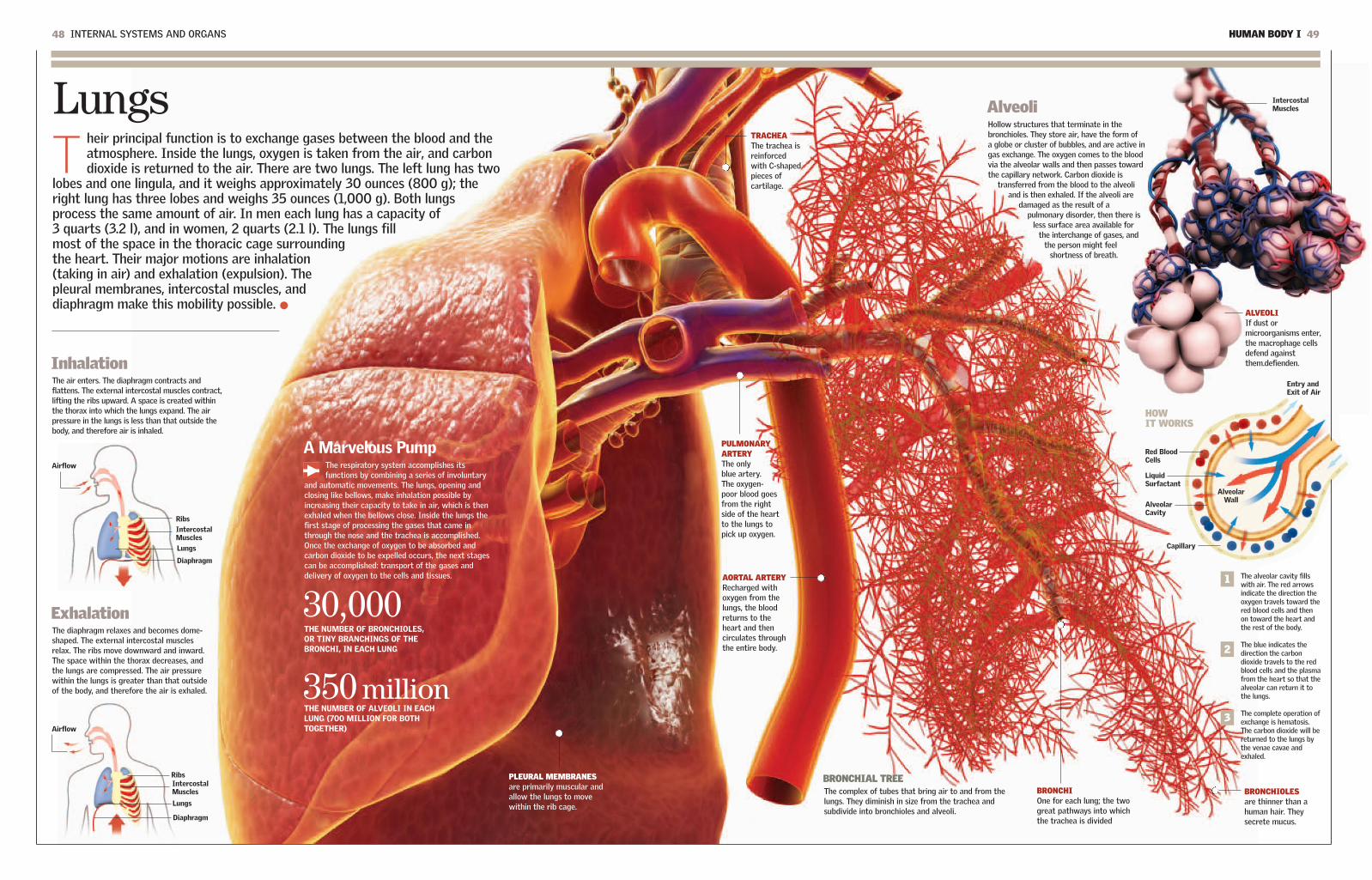
HUMAN BODY I 4948 INTERNAL SYSTEMS AND ORGANS
LungsT
heir principal function is to exchange gases between the blood and theatmosphere. Inside the lungs, oxygen is taken from the air, and carbondioxide is returned to the air. There are two lungs. The left lung has two
lobes and one lingula, and it weighs approximately 30 ounces (800 g); theright lung has three lobes and weighs 35 ounces (1,000 g). Both lungsprocess the same amount of air. In men each lung has a capacity of3 quarts (3.2 l), and in women, 2 quarts (2.1 l). The lungs fillmost of the space in the thoracic cage surroundingthe heart. Their major motions are inhalation(taking in air) and exhalation (expulsion). Thepleural membranes, intercostal muscles, anddiaphragm make this mobility possible.
InhalationThe air enters. The diaphragm contracts andflattens. The external intercostal muscles contract,lifting the ribs upward. A space is created withinthe thorax into which the lungs expand. The airpressure in the lungs is less than that outside thebody, and therefore air is inhaled.
ExhalationThe diaphragm relaxes and becomes dome-shaped. The external intercostal musclesrelax. The ribs move downward and inward.The space within the thorax decreases, andthe lungs are compressed. The air pressurewithin the lungs is greater than that outsideof the body, and therefore the air is exhaled.
AlveoliHollow structures that terminate in thebronchioles. They store air, have the form of a globe or cluster of bubbles, and are active ingas exchange. The oxygen comes to the bloodvia the alveolar walls and then passes towardthe capillary network. Carbon dioxide is
transferred from the blood to the alveoliand is then exhaled. If the alveoli are
damaged as the result of apulmonary disorder, then there is
less surface area available forthe interchange of gases, and
the person might feelshortness of breath.
A Marvelous PumpThe respiratory system accomplishes itsfunctions by combining a series of involuntary
and automatic movements. The lungs, opening andclosing like bellows, make inhalation possible byincreasing their capacity to take in air, which is thenexhaled when the bellows close. Inside the lungs thefirst stage of processing the gases that came inthrough the nose and the trachea is accomplished.Once the exchange of oxygen to be absorbed andcarbon dioxide to be expelled occurs, the next stagescan be accomplished: transport of the gases anddelivery of oxygen to the cells and tissues.
30,000THE NUMBER OF BRONCHIOLES, OR TINY BRANCHINGS OF THEBRONCHI, IN EACH LUNG
350 millionTHE NUMBER OF ALVEOLI IN EACHLUNG (700 MILLION FOR BOTHTOGETHER)
BRONCHIAL TREEThe complex of tubes that bring air to and from thelungs. They diminish in size from the trachea andsubdivide into bronchioles and alveoli.
BRONCHIOLESare thinner than ahuman hair. Theysecrete mucus.
BRONCHIOne for each lung; the twogreat pathways into whichthe trachea is divided
TRACHEAThe trachea isreinforcedwith C-shapedpieces ofcartilage.
PULMONARYARTERYThe onlyblue artery.The oxygen-poor blood goesfrom the rightside of the heartto the lungs topick up oxygen.
AORTAL ARTERYRecharged withoxygen from thelungs, the bloodreturns to theheart and thencirculates throughthe entire body.
ALVEOLIIf dust ormicroorganisms enter,the macrophage cellsdefend againstthem.defienden.
PLEURAL MEMBRANESare primarily muscular andallow the lungs to movewithin the rib cage.
3
2
1 The alveolar cavity fillswith air. The red arrowsindicate the direction theoxygen travels toward thered blood cells and thenon toward the heart andthe rest of the body.
The blue indicates thedirection the carbondioxide travels to the redblood cells and the plasmafrom the heart so that thealveolar can return it tothe lungs.
The complete operation ofexchange is hematosis.The carbon dioxide will bereturned to the lungs bythe venae cavae andexhaled.
HOW IT WORKS
Airflow
IntercostalMuscles
Ribs
Lungs
Diaphragm
Airflow
IntercostalMuscles
Alveolar Wall
Entry andExit of Air
Red BloodCells
LiquidSurfactant
AlveolarCavity
Capillary
IntercostalMuscles
Ribs
Lungs
Diaphragm

50 INTERNAL SYSTEMS AND ORGANS HUMAN BODY I 51
Digestive SystemT
he digestive system is the protagonist of a phenomenaloperation that transforms food into fuel for the entirebody. The process begins with ingestion through the
mouth and esophagus and continues with digestion in thestomach, the small intestine, and the large intestine, fromwhich the feces are evacuated by the rectum and anus. Bythen the task will have involved important chemicalcomponents, such as bile, produced by the liver, and otherenzymes, produced by the pancreas, by which the food isconverted into nutrients. Separating the useful from theuseless requires the filtering of the kidneys, whichdiscard the waste in urine. 00:00:10
00:00:00
03:00:00
08:00:00
Between 20 and 44 hours afterhaving entered the mouth asfood, the residue that wasconverted into semisolid fecesin the previous stage arrivesat the rectum. The waste willbe evacuated through theanus as excrement.
24:00
The First Step: IngestionThe digestive process begins with themouth, the entry point to the large tract
that changes in form and function and ends atthe rectum and anus. The tongue and teeth arethe first specialists in the task. The tongue is incharge of tasting and positioning the food,
which is cut and ground by the teeth. Thissynchronized activity includes the maxillarybones, which are controlled by theircorresponding muscles. The palate, in the upperpart of the mouth, prevents food from passinginto the nose. The natural route of the food isdown the esophagus to the stomach.
TeethThere are 32 teeth, and they are extremely hard, a conditionnecessary for chewing food. There are eight incisors, fourcanines, eight premolars, and 12 molars. Humans developtwo sets of teeth, a provisional or temporary set (the babyteeth) and a permanent set (adult teeth). The firsttemporary teeth appear between six and 12 months of age.At 20 years of age the process of replacement that beganat about age five or six is complete.
Digestion ChronologyThe process that converts food into nutrients begins a fewseconds after the food is raised to the mouth and chewingbegins. The average digestion time is about 32 hours,though digestion can range from 20 to 44 hours.
Enzymes and HormonesThe complex chemical processes that transform food areessentially accomplished by enzymes and hormones. Bothtypes of substances are secreted by various glands of thedigestive system, such as the salivary glands. Enzymes aresubstances that act as catalysts. Hormones are substancesthat regulate processes such as growth metabolism,reproduction, and organ function.
ESOPHAGUSIts muscles force thebolus toward thestomach. Theesophagus and stomachare separated by asphincter.
PHARYNXThe muscles in thewalls of the pharynxcontract, forcing thebolus of chewed foodinto the esophagus.
ROOTEntirely
buried in themaxillary
bone
PULPSoft tissue that
is the core ofthe tooth
THE INSIDE OF A TOOTHTHE MOUTH
A SET OF TEETH
ENAMELhas no feelingand is thehardestsubstance inthe body.
DENTINSensitive tocold and heat,it is strongerthan bonetissue.
NERVESpassinformationabout thetooth to thenervoussystem.
CEMENTThe thin layer
that covers theroots and
anchors them inthe jawbone
CANINES
PREMOLARS
MOLARS
INCISORS
06:00:00
32A PERSON
NORMALLY HASTHIS NUMBER
OF TEETH.
TractThe muscular
movement calledperistalsis pushes
the food along.That is why it ispossible to eatupside down or
duringweightlessness, as
astronauts do.
TONGUEIts notable flexibilitymakes eatingpossible. It alsotastes the food.
THE HARD PALATEThe “roof” of the oralcavity. It is made ofbone.
THE SOFT PALATEAlso called the velarpalate, the palate keepsthe food from goinginto the nose.
1
3
4
5
6
2
2
1
3
4
5
6
About 10 seconds after chewing hasbegun, the food is transformed into amoist alimentary bolus that makes itsway through the pharynx to theesophagus and then to the stomach,where other changes will take place.
Three hours after its arrival, thefood leaves the stomach, which hasaccomplished its function. The firstphase of digestion is over. The bolusnow has a liquid and creamyconsistency.
Three hours later, the food that hasbeen digested in the stomach arrivesat the midpoint of the smallintestine. At this point it is ready tobe absorbed.
Two hours later, the non-digested,watery residue arrives at thejunction of the small and largeintestines. The useless materialrejected by the body's chemicalselectors continues its course, and itis now prepared to be expelled fromthe organism in the form of feces.
The process begins when the food reaches the mouth.The entire organism is involved in the decision, but it isthe digestive system that plays the main role. The firststeps are taken by the teeth and the tongue, aided bythe salivary glands, which provide saliva to moisten thealimentary bolus. The morsels are chewed so that theycan pass through the esophagus.
20:00:00The alimentary residueremains in the large intestinebetween 12 and 28 hours. Inthis part of the process theresidue is converted intosemisolid feces.
10 inches (25 cm)IS THE LENGTH OFTHE ESOPHAGUS.