absolute beachfront. total luxury. - sea sentosa, echo beach
TRANSCRIPT

Neurology Case Presentation
Rawan Albadareen, MD
12/20/13

Case presentation
� A 49 y.o. female presented to the ED after an episode of zigzagging w a jagged bright light crossing through her Rt visual field early that morning that lasted 15 min then resolved.
� Later that afternoon she heard a sudden loud noise in her ears associated with Rt LE weakness (transient) along with Rt visual field deficit(persisted). She complained of a throbbing HA earlier that resolved by the time of presentation.
� Pt has Hx of classic migraines with visual aura and field defect that can last up to 24 hours.
� Her migraines in the past were related to birth control pill use that she stopped 5 years ago followed by resolution of her HA.
� This summer she began using estrogen injections to control her hot flashes, her most recent injection was day prior to presentation
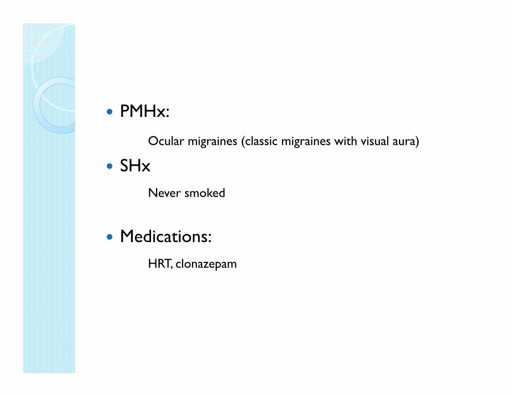
� PMHx:
Ocular migraines (classic migraines with visual aura)
� SHx
Never smoked
� Medications:
HRT, clonazepam

Neurological Exam
Mental status: alert, oriented to person/place/time
Speech: fluent with no dysarthria
Cranial Nerves: II-XII intact except for Rt homonomoushemianopsia (mainly upper quadrantopia)
Muscle/motor: 5/5 all over
Sensation: intact to LT/PP/ vibration
Coordination: intact FTN and HTS
Reflexes: 2/4 all over with down going toes

Ophthalmological Exam
� Near Visual Acuity: 20/20 OU
� Pupils: PERRL, no APD
� CVF: right homonymous hemianopia
� Motility: EOMI
� DILATED FUNDUS EXAM:
� Optic Nerve: 0.2 elevated nerve with refractiledrusen, no edema OU

� Where?
� What?


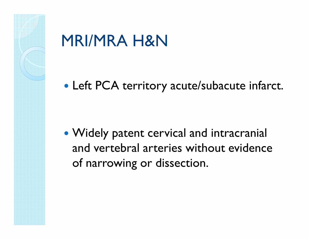
MRI/MRA H&N
� Left PCA territory acute/subacute infarct.
� Widely patent cervical and intracranial and vertebral arteries without evidence of narrowing or dissection.

� Hypercoagulability work up –ve◦ ANA
◦ B2glycoprotein
◦ Cardiolpin
◦ Protein C
◦ Protein S
◦ Anti-thrombin 3
◦ ESR
◦ CRP
◦ Factor 2 mutation
� LDL 88� HbA1c 4.9� Echo: Nl EF and no cardiac thrombus

Visual Pathway

Homonymous Hemianopia
� Lesions in the retrochiasmatic pathways (affecting the optic tract, lateral geniculate body, optic radiations, or occipital lobe).
� Lesions of the tract and lateral geniculate body tend to be incongruous, but the more posterior the lesion, the greater the congruity of the defect in either field.
� In general, tumors produce sloping field defects, whereas vascular lesions produce sharp field defects.

Optic Tract Lesions
� Complete unilateral optic tract lesions cause a complete macular splitting homonymous hemianopia, usually without impaired visual acuity
� Partial optic tract lesions are more common than complete ones and result in an incongruous field defect that may be scotomatous.
� Optic tract lesions are often associated with a relative afferent pupillary defect (RAPD) in the eye with temporal field loss (contralateral to the side of the lesion)
� This reflects the difference in light sensitivity between the intact temporal and nasal hemifield
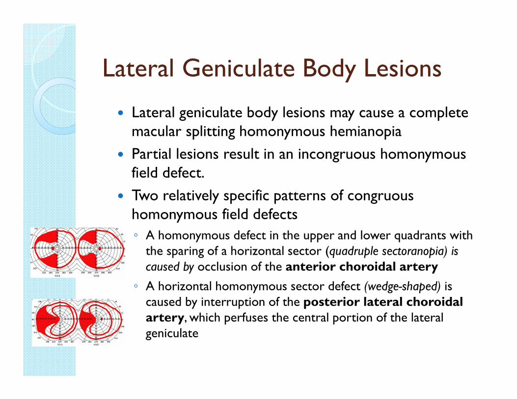
Lateral Geniculate Body Lesions
� Lateral geniculate body lesions may cause a complete macular splitting homonymous hemianopia
� Partial lesions result in an incongruous homonymous field defect.
� Two relatively specific patterns of congruous homonymous field defects
◦ A homonymous defect in the upper and lower quadrants with the sparing of a horizontal sector (quadruple sectoranopia) is caused by occlusion of the anterior choroidal artery
◦ A horizontal homonymous sector defect (wedge-shaped) is caused by interruption of the posterior lateral choroidal artery, which perfuses the central portion of the lateral geniculate

Optic Radiation Lesions
� Superior homonymous quadrantic defects (“pie-in-the-sky” field defects) may result from a lesion in the temporal (Meyer’s) loop of the optic radiations or in the inferior bank of the calcarine fissure
� For a quadrantic defect the lesion has to be quite extensive; small lesions result in scotomata
� Certain fibers from the ipsilateral eye travel more anteriorly and laterally in Meyer’s loop and supports the hypothesis that visual field defects due to anterior retrogeniculate lesions are incongruous because of anatomic differences in the afferent pathway
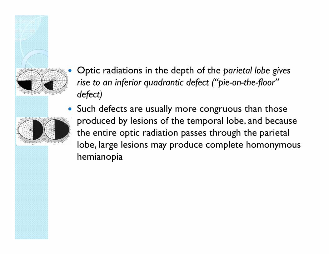
� Optic radiations in the depth of the parietal lobe gives rise to an inferior quadrantic defect (“pie-on-the-floor” defect)
� Such defects are usually more congruous than those produced by lesions of the temporal lobe, and because the entire optic radiation passes through the parietal lobe, large lesions may produce complete homonymous hemianopia

Occipital Lesions
� Lesion of the extrastriate cortex (areas V2 and V3) has sharp horizontal edge because V2 and V3 are divided along the horizontal meridian into separate halves flanking the striate (V1) cortex
� Medial occipital lesions cause highly congruous homonymous hemianopias
� When both the upper and the lower calcarine cortices are affected, a complete homonymous hemianopia, usually with macular sparing, develops
� Macular sparing is common with occipital lesions, due to a combination of a large macular representation (as much as 50%–60%) and dual blood supply
� Anterior lesions adjacent to the parieto-occipital fissure affect the monocular temporal crescent of the contralateral visual field (temporal crescent or half-moon syndrome)

� Bilateral occipital lobe lesions may occur from a single or from consecutive events and may cause bilateral homonymous scotomas, usually with some macular sparing (“ring” scotomas), “keyhole” fields.
� Posterior lesions located in the posterior 50% to 60% of the striate cortex, including the occipital pole and operculum, and affect macular vision
� Bilateral homonymous hemianopia (double hemianopia) may result in cortical blindness

Clinical Features
� In patients with occipital lesions, the field defects often occurs in isolation, while other localizing signs of parietal involvement are evident in patients with parietal lesions
� Patients with purely occipital lesions are partially or fully aware of the hemianopia, whereas patients with larger or more anterior lesions, affecting parietal regions may be unaware of their defecit
� Hemianopic anosognosia occurs predominantly in right-sided lesions
� Spontaneous improvement of homonymous hemianopia is seen in at least 50% of patients first seen within 1 month of injury. In most cases, the improvement occurs within the first 3 months from injury.

Association between HRT and subsequent arterial and venous vascular events:
a meta-analysisSare, Gray, and Bath ( Eur Heart J. 2008)
� HRT is associated with an increased risk of stroke, stroke severity, and VTE, but not of CHD events.
� Although most trials studied older patients, increased risk was not related to age.
� Combined HRT increases the risk of VTE compared with estrogen monotherapy.

THANK
YOU!!