abstract title of thesis: evolutionary transitions …
TRANSCRIPT

ABSTRACT
Title of Thesis: EVOLUTIONARY TRANSITIONS BETWEEN STATES OF STRUCTURAL AND DEVELOPMENTAL CHARACTERS AMONG THE ALGAL CHAROPHYTA (VIRIDIPLANTAE).
Jeffrey David Lewandowski, Master of Science, 2004
Thesis Directed By: Associate Professor, Charles F. Delwiche, Cell Biology and Molecular Genetics
The Charophyta comprise green plant representatives ranging from familiar
complex-bodied land plants to subtle and simple forms of green algae, presumably
the closest phylogenetic relatives of land plants. This biological lineage provides a
unique opportunity to investigate evolutionary transition series that likely facilitated
once-aquatic green plants to colonize and diversify in terrestrial environments. A
literature review summarizes fundamental structural and developmental transitions
observed among the major lineages of algal Charophyta. A phylogenetic framework
independent of morphological and ontological data is necessary for testing hypotheses
about the evolution of structure and development. Thus, to further elucidate the
branching order of the algal Charophyta, new DNA sequence data are used to test
conflicting hypotheses regarding the phylogenetic placement of several enigmatic
taxa, including the algal charophyte genera Mesostigma, Chlorokybus, Coleochaete,

and Chaetosphaeridium. Additionally, technical notes on developing RNA methods
for use in studying algal Charophyta are included.

EVOLUTIONARY TRANSITIONS BETWEEN STATES OF STRUCTURAL AND DEVELOPMENTAL CHARACTERS AMONG THE ALGAL CHAROPHYTA
(VIRIDIPLANTAE).
By
Jeffrey David Lewandowski
Thesis submitted to the Faculty of the Graduate School of the University of Maryland, College Park, in partial fulfillment
of the requirements for the degree ofMaster of Science
2004
Advisory Committee:Associate Professor Charles F. Delwiche, ChairProfessor Todd J. CookeAssistant Professor Eric S. Haag

© Copyright byJeffrey D. Lewandowski
2004

ii
Preface
The following are examples from several of the works that have provided philosophical inspiration for this document.
“Nothing in biology makes sense except in the light of evolution.”Theodosius DobzhanskyAmerican Biology Teacher, volume 35: 125-129, 1973
“Body form evolves through geologic time, but it also arises in each generation through development. The time frames and apparent processes couldn't be more different, but explanations of the evolution of form have to consider how form is generated, and they must account for both underlying stability and immense change in design.”
Rudolf A. Raff The Shape of Life: Genes, Development, and the Evolution of Animal Form, 1996
“There is a parallel, long appreciated, between the developmental changes that convert an egg into an adult, and the evolutionary changes that, on an enormously longer time scale, have converted simple single-celled ancestors into the existing array of multicellular animals and plants.”
John Maynard SmithShaping Life: Genes, Embryos and Evolution, 1999
“Developmental biologists must come to appreciate diversity, the comparative method, and the importance of phylogeny. Evolutionary biologists need to understand and incorporate the underlying rules of developing systems into a more comprehensive theory of evolution.”
Rudolf A. Raff The Shape of Life: Genes, Development, and the Evolution of Animal Form, 1996
“The body of a plant can best be understood in terms of its long history and, in particular, in terms of the evolutionary pressures involved in the transition to land.”
Peter H. Raven, Ray F. Evert, and Susan E. EichhornBiology of Plants, 1999

iii
Dedication
This work is dedicated to my mother, Nancy, and my father, Jerome, who
have supported my education and always encouraged me to pursue my love of
science. It is also dedicated in loving memory to Jane Kulas, my grandmother, and to
the memory of Dr. Tzong-Yow “T.Y.” Lee, one of the finest and most dedicated
teachers I have encountered.

iv
Acknowledgements
Dr. Charles F. DelwicheDr. Todd CookeDr. Eric Haag
Dr. Charles MitterDr. Richard McCourt
Dr. Steven WolniakDr. Elisabeth GanttDr. Lindley Darden
Dr. W. John Hayden
Dr. Angela CainesDonna Hammer
Dr. Tsetso BachvaroffDr. Matthew CiminoDr. Kenneth Karol
Dr. Malin KerrDr. Tanya Marushak
John HallMaria Virginia Sanchez Puerta
Jill Marie RickerOther members of the Delwiche Lab, past and present
Dr. Michael DandenaultDr. Christopher Desjardins
Corey GonzalezDamien Ober
Natalie Kahla
My family and many friends who have loved, supported, and encouraged me throughout my life.
Research funded by:National Science Foundation (PEET; Deep Green-GPPRCG)

v
Table of Contents
Preface........................................................................................................................... iiDedication .................................................................................................................... iiiAcknowledgements...................................................................................................... ivTable of Contents.......................................................................................................... vList of Figures .............................................................................................................. viChapter 1: Introduction........................................................................................... 1Chapter 2: The Algal Charophyta......................................................................... 10
2.1 Mesostigmatales.......................................................................................... 102.2 Chlorokybales ............................................................................................. 142.3 Klebsormidiales .......................................................................................... 192.4 Zygnematales .............................................................................................. 242.5 Coleochaetales ............................................................................................ 282.6 Charales....................................................................................................... 33
Chapter 3: Testing Phylogenetic Hypotheses ....................................................... 393.1 Introduction................................................................................................. 393.2 Materials and Methods................................................................................ 423.3 Results......................................................................................................... 43
3.3.1 Chloroplast Transfer RNA Gene Introns ............................................ 433.3.2 Actin Gene Sequence Analysis ............................................................ 46
3.4 Discussion ................................................................................................... 463.4.1 Phylogenetic Position of Scaly Biflagellate Mesostigma ................... 463.4.2 On the Monophyly of the Coleochaetales ........................................... 483.4.3 Other Phylogenetic Inference ............................................................. 50
3.5 Conclusions................................................................................................. 51Appendices.................................................................................................................. 53
Appendix A Isolation of total RNA from algal charophytes........................... 53Literature Cited ........................................................................................................... 55
vi
List of Figures
1. Chloroplast transcribed spacer structure in green algae 44
2. Actin gene phylogeny in the Viridiplantae 47

1
Chapter 1: Introduction
Consider the 4.5 billion-year history of planet Earth. Each extant species on
our planet is the product of more than 3,500 million years of evolutionary change
acting upon generations of life forms, all presumed to share a common ancestor.
Thus, for most of the course of Earth's history, the dynamical processes of physical
change and organismal evolution have shared an intimate linkage; geologic
transformation has played a part in shaping biological forms and, likewise, biological
evolutionary change has had a significant impact on physical geography. Some
evolutionary events of inherently botanical nature—the origin of photosynthesis, the
acquisition of primary plastids through endosymbiosis, and the colonization of
terrestrial environments by plants—have had especially profound effects on the
biosphere of our planet. A thorough understanding of the evolutionary origin of land
plants, chronologically the most recent of these historical examples, requires an
examination of the morphologically simpler green algal forms most closely related to
the more familiar land plant lineage.
Due to their awesome environmental, ecological, and economic importance,
arguably no group of multicellular organisms is more vital for humans to understand
than the land plants, also called embryophytes. Both labels—'embryophytes' and
'land plants'—possess the potential to confound the biology of the group they identify
(Graham, 1993). For the sake of this document, both terms, land plants and
embryophytes, will be used interchangeably as synonyms, defined as the group
including the bryophytes plus the tracheophytes. A number of studies utilizing

2
different lines of evidence support the monophyly of the land plant clade (Mishler et
al., 1994; Waters and Chapman, 1996; Lewis et al., 1997).
To even the casual observer or amateur naturalist, a close relationship between
land plants and green algae lies well within the realm of imagination. Predictions
about the exact nature of such a relationship have existed for over a century (Bower,
1908). Fossils representing the oldest known embryophyte remains indicate the
colonization of land by plants occurred more than 470 million years ago (Gray et al.,
1982). To date, no discovery of older embryophyte fossils has been made, so there
exists no direct physical evidence revealing the relationship between green algae and
land plants (Graham, 1993; Graham and Wilcox, 2000). Thus, hypotheses about
evolutionary relationships, both between algae and land plants and among the aquatic
and terrestrial groups comprising this monophyletic clade, must be drawn by
application of the comparative method to data collected from extant green algal and
embryophyte entities.
Comparative studies of morphology have been used to infer phylogeny in
various biological groups. Homoplasy, such as convergence of forms through
parallel evolution, is quite prevalent in the green algae, and in the algae in general
(Graham and Wilcox, 2000). This phenomenon confounds attempts to infer
phylogeny based on external morphology alone (Pickett-Heaps and Marchant, 1972;
Pickett-Heaps, 1975; Graham and Wilcox, 2000). Numerous biochemical (Frederick
et al., 1973; Syrett and al-Houty, 1984; DeJesus et al., 1989; Jacobshagen and
Schnarrenberger, 1990; Gross, 1993) and ultrastructural (Pickett-Heaps and
Marchant, 1972; Pickett-Heaps, 1975; Cook et al., 1998) investigations have

3
contributed to the resolution of evolutionary relationships between green algae and
land plants. Based on consideration of ultrastructural, biochemical, and
morphological characters, Mattox and Stewart (1984) named a new class of green
algae, the Charophyceae, grouping the lineages hypothesized to be most closely
related to embryophytes.
The Charophyceae sensu Mattox and Stewart (1984) include five orders; they
are, in likely order of evolutionary divergence, the Chlorokybales, Klebsormidiales,
Zygnematales, Coleochaetales, and Charales. A problem with this taxonomic group,
however, is that it does not include the land plants. Thus, if the embryophyte lineage
is assumed to have diverged from within the charophycean green algae, the
Charophyceae sensu Mattox and Stewart (1984) is paraphyletic, an unnatural group
that does not reflect evolutionary relationships. Bremer (1985) recognized this
dilemma and advised recognition of the charophycean algae plus embryophytes as a
novel division, the Streptophyta, distinct from all other green algae. A number of
phylogenetic analyses of molecular sequence data (Chapman and Buchheim, 1991;
McCourt et al., 1996; Karol et al., 2001; Turmel et al., 2002b) and several genome
architectural characters (Baldauf et al., 1990; Manhart and Palmer, 1990; Starke and
Gogarten, 1993) support Bremer's (1985) proposed classification. Additionally, actin
gene sequence data (Bhattacharya et al., 1998) support the inclusion of a scaly,
biflagellate, unicellular alga, Mesostigma viride, at the base of the Streptophyta
lineage. Utilizing a multi-gene dataset representing nuclear, chloroplast, and
mitochondrial DNA characters, molecular sequence analyses by Karol et al. (2001)
corroborate this finding; authors of the study proposed the name Charophyta for the

4
phylogenetic lineage comprising land plants, charophycean green algae (the
Charophyceae sensu Mattox and Stewart, 1984), and Mesostigma.
Based on all evidence to date, the "green plant" lineage, Viridiplantae sensu
Cavalier-Smith, (1981), splits into two major branches; the Charophyta sensu Karol et
al. (2001) include land plants and their green algal relatives, and the Chlorophyta
comprise the ulvophycean, trebouxiophycean, chlorophycean, and most of the yet-to-
be classified flagellate (prasinophyte) green algae. This taxonomic scheme, with one
exception, will be exercised for the remainder of this treatment. Due to conflicting
data on the phylogenetic placement of the enigmatic genus Mesostigma, and the
importance of considering all possible evolutionary hypotheses, the author will
refrain from assuming inclusion of this genus in the Charophyta. Here, the genus
Mesostigma will be treated as a currently unallied entity. Forthcoming discussion
will use the term Charophyta, or the charophytes, to refer to land plants plus the
closely related green algae of the Charophyceae sensu Mattox and Stewart (1984).
Furthermore, to avoid confusion associated with naming paraphyletic groups, the
lineages of green algae traditionally termed charophyceans (Graham, 1993; Graham
and Wilcox, 2000; Graham et al., 2000), or the Charophyceae (sensu Mattox and
Stewart, 1984) will be referred to here as the algal Charophyta, algal charophytes,
algal members of Charophyta, and so forth.
According to Raven et al. (1999) "the body of a plant can best be understood
in terms of its long history and, in particular, in terms of the evolutionary pressures
involved in the transition to land." These conditions for comprehension of the plant
body require consideration of both algal members and land plant representatives of

5
the Charophyta. Regarding evolutionary history, molecular systematic analyses of a
four-gene DNA sequence dataset have revealed phylogenetic relationships among
algal groups within Charophyta at an unprecedented level of resolution (Karol et al.,
2001). Utilizing data from each of the three genomes of the green plant cell, Karol et
al. (2001) found the Charales to be the extant algal group most closely related to
embryophytes. Additionally, the phylogeny presented bolsters support for many of
the other relationships among the algal Charophyta. Some of these relationships are
congruent with findings based on analysis of chloroplast small- and large-subunit
ribosomal DNA (Turmel et al., 2002b). These results are exciting and contribute
much to our understanding of evolution in the Charophyta. However, because
phylogenetic problems can never be deterministically solved, trees generated by such
analyses should be treated as hypotheses that are to be tested and retested with new
data and ever-improving probabilistic methods. Two analyses included in this report
utilize previously unavailable data to test phylogenetic hypotheses, specifically those
involving the enigmatic genera Mesostigma and Chaetosphaeridium.
In examining adaptations necessary for the transition of green plants to
terrestrial environments, consideration of the algal Charophyta is inherently vital. At
all levels of land plant body organization—macroscopic to microscopic,
physiological, cellular, and molecular—adaptations to terrestrial life have been
investigated extensively (Kenrick and Crane, 1997). Some of the most fundamental
innovations associated with colonization of the land originated in, and thus were
likely inherited from, the algal ancestors of modern land plants (Graham, 1993). Yet,
relatively few investigators have studied the evolution of specialized body features

6
and cellular mechanisms in the algal Charophyta, the lineage that would include algal
ancestors of embryophytes (if such ancestors were present today) and does comprise
the extant taxa most likely to resemble those ancestors. Due to the generally
microscopic nature of green algal features, such characters are sublime relative to
many of the traditionally studied land plant body parts. Because characters relevant
to the invasion of land are plesiomorphic with respect to the embryophyte clade,
describing the evolution of many character states is not possible without
consideration of the algal Charophyta. Admittedly, botanists have utilized green algal
experimental systems in pursuit of a better understanding of land plant form and
function. For many decades Acetabularia, Chlamydomonas, and Chlorella have been
used in investigations of plant cytology, morphology, and development. Because
these and other commonly studied genera of green algae are now known to have
diverged from the Chlorophyta lineage of the Viridiplantae, structures that have
evolved in these algae are not homologous to similar features found in land plants.
While these studies have contributed to the advancement of knowledge about
evolution among chlorophyte green algae, it is illogical to extend findings across
branches of the tree of life. Thus, such results are not informative to those inquiring
about the evolution of morphology and development in the embryophyte and algal
representatives of the Charophyta. Understanding of morphology and development in
land plants is therefore incomplete without consideration of the green algal members
of the Charophyta. Future investigators should seriously consider more extensive
utilization of algal charophyte experimental systems in studies of the evolution and
development of land plants and related organisms.

7
Incorporation of information about the algal Charophyta is clearly a key to
understanding the plant body. The evolution of morphology and development across
the algal Charophyta is also intrinsically fascinating. Although the green algae lack
the extraordinary morphological complexity and diversity of land plants, the algal
Charophyta exhibit an impressive array of structural and mechanical characteristics
with an exceptionally interesting pattern of phylogenetic distribution (Graham, 1993;
Graham and Wilcox, 2000). Mesostigma viride is a scaly, unicellular biflagellate that
likely diverged from the base of the Charophyta (Melkonian and Surek, 1995;
Bhattacharya et al., 1998; Karol et al., 2001) or at the base of the Viridiplantae, prior
to the split distinguishing the Charophyta and Chlorophyta lineages (Lemieux et al.,
2000; Turmel et al., 2002a; Turmel et al., 2002b). Members of the Charales and
Coleochaetales exhibit multicellular parenchyma, phragmoplastic cytokinesis,
plasmodesmata, oogamous sexual reproduction, differentiated reproductive
structures, apical growth, and a number of other highly derived characteristics
reminiscent of fundamental land plant features. Other groups of algal Charophyta,
diverging between the Mesostigmatales and Charales branches, exhibit a variety of
forms—colonial or sarcinoid, simple filamentous, branched filamentous and
parenchymatous—and numerous types of specially differentiated cells with distinct
functions. In contemplating the evolution of morphological transitions between
lineages of the algal Charophyta, from unicellular to more and more complex
multicellular forms, it becomes difficult to ignore a striking similarity between those
evolutionary changes and the ontogenetic stages observed in more derived examples
of algal charophyte thallus development. Unicellular, often flagellate stages develop

8
into simple filaments, and then into branched filaments or parenchyma before some
cells differentiate for specialized functions. The Charophyta thereby represent a
valuable "model lineage" within which the intimately related phenomena of evolution
and development may be explored.
This concept for approaching investigation of the Charophyta is actually a
fresh reinvention of an old idea, a synthesis of evolutionary and developmental
biology. Evolutionary developmental biology, or evo-devo, refers to the combination
of two historically distinct disciplines in biology in order to pursue a grander
understanding of evolution. Morphological and mechanical features of organisms
originate and then change over long periods of geologic time through the process of
biological evolution. Those features, including every existing variation, also arise via
developmental mechanisms in each and every life-history generation of the organisms
in which they evolved. Raff (1996) describes this close relationship between
evolutionary and developmental processes, and recommends several directions for
intertwining the two long-distinct biological disciplines. According to Raff (1996),
developmental biologists need to value and understand biodiversity, the comparative
method, and the importance of testable phylogenies; likewise, evolutionary biologists,
in order to generate more complete evolutionary hypotheses, must come to
understand how developing systems function. The literature review, experimental
results, and technical methodology to follow strive to incorporate the spirit of this
endeavor, the synthesis of evolutionary and developmental thought, into an
investigation of the evolution of form and function in the Charophyta, one of the most
important and interesting branches on the tree of life.

9
This thesis has three main objectives: a review of cytology, morphology, and
development in representatives of the algal Charophyta; the reporting of results that
contribute new data to the task of resolving some of the more controversial
phylogenetic relationships among the green algae of the Charophyta lineage; and
description of technical progress in developing RNA methods for eventual
examination of molecular mechanisms that likely underlie the morphological and
developmental transitions observed in the Charophyta.

10
Chapter 2: The Algal Charophyta
In order to elucidate the evolutionary transitions between lineages of the algal
Charophyta, the following literature review aims to describe the cytological,
morphological, and developmental features that characterize each of the five
recognized orders of algal charophytes, as well as the related taxon, Mesostigma.
Complexity at all levels generally increases from earlier to later diverging lineages in
the Charophyta. This justifies the following approach, in which discussion of taxa
with simpler morphology will include more details about cytological features, with
emphasis on characters that define the Charophyta in general. Likewise, the more
derived thalli of later-diverging taxa will provide ample opportunity for description of
complex morphology and the development of specialized cells and multicellular
structures. To best demonstrate evolutionary and developmental trends, organisms
will be treated in the order of phylogenetic divergence, as it is currently best
understood (sensu Karol et al., 2001).
2.1 Mesostigmatales
Mesostigma viride Lauterborn is the only representative of the monogeneric
order Mesostigmatales that has been studied with modern methods. Mesostigma had
traditionally been classified among the morphologically simple prasinophycean green
algae, unicellular flagellates classified into a group that is now recognized as
unnatural. Based on ultrastructural data, Rogers et al. (1981) first predicted a close
phylogenetic relationship between Mesostigma and the charophyte lineage.
Subsequent ultrastructural analyses (Melkonian, 1982; 1984; 1989) supported the

11
hypothesis including Mesostigma among the algal charophytes. With advances in
molecular methods for collecting nucleic acid sequence data and systematic methods
for conducting phylogenetic analyses of such datasets, a way to test this evolutionary
hypothesis using characters independent of the morphology and ultrastructure in
question became possible. Yet, the exact phylogenetic position of the
Mesostigmatales is still controversial. Inclusion of Mesostigma within the
Charophyta clade has been supported by analyses of sequence data from nuclear
small subunit ribosomal RNA (SSU rRNA) genes (Melkonian and Surek, 1995;
Marin and Melkonian, 1999), nuclear actin genes (Bhattacharya et al., 1998), and a
combined analysis of nuclear SSU rRNA, chloroplast, and mitochondrial genes
(Karol et al., 2001). Contrary to these results, analyses of sequence data from
chloroplast (Lemieux et al., 2000; Turmel et al., 2002b) and mitochondrial (Turmel et
al., 2002a) genomes support placement of the Mesostigmatales at the base of the
Viridiplantae, prior to the divergence of the two major lineages, Chlorophyta and
Charophyta, sensu Karol et al. (2001). Incongruence between various molecular
phylogenetic studies may be due to inadequate taxon sampling (Hillis, 1998; Qiu and
Lee, 2000; Karol et al., 2001; Pollock et al., 2002). Despite the current debate over
its phylogenetic position, there is little doubt that Mesostigma likely diverged early in
the Charophyta lineage or at the base of the Viridiplantae, before the splitting of the
two major groups of green plants. In either case, Mesostigma is likely to share some
ancestral character states with members of the charophyte lineage. Likewise, many of
the putative characteristics of a green flagellate ancestor of the charophytes proposed
by Graham (1993) are present in Mesostigma. For these reasons, any treatment of

12
morphology and ontogeny in the algal Charophyta is well served by an early
discussion of Mesostigma.
Mesostigma is presumed to be a motile unicell primarily encountered in
freshwater habitats (Graham and Wilcox, 2000). Cells of Mesostigma take the form
of warped disks with two flagella emerging laterally from a deep flagellar pit opening
(Sym and Pienaar, 1993). Lacking a cell wall, Mesostigma cells are covered by at
least three distinct scale layers. The outermost layer consists of large, ornate, basket-
like scales; middle layer scales are smaller, flat, oval in shape, and decorated with pits
(Manton and Ettl, 1965). Most similar to the scales observed in flagellate cells of the
reproductive stages of many charophytes (Chlorokybus, Coleochaete,
Chaetosphaeridium) are innermost scales covering Mesostigma—small, flat, and
ranging from square- to maple leaf-shaped (Marin and Melkonian, 1999; Graham and
Wilcox, 2000). Between the innermost scale layer and the cell is a substance
associated with both the scales and the cell membrane. This glue-like material has
been hypothesized to represent an evolutionary precursor to charophyte cell walls
(Rogers et al., 1980).
Mesostigma cells contain a single chloroplast, shaped like a shallow bowl with
regions at the margin thicker than in the center (Sym and Pienaar, 1993). Plastids
contain several pyrenoids and, unlike typical charophyte chloroplasts, an eyespot
composed of several globular pigment layers, usually located near the flagellar basal
bodies. Associated with the basal bodies and situated between the plastid and
flagellar apparatus is a lobed microbody, a structure also referred to as a peroxisome
(Rogers et al., 1981). Each basal body is associated with two microtubular roots.

13
The “s” root consists only of microtubules, but the “d” root is composed of several
layers—an array of microtubules, a thin series of parallel plates, several amorphous
regions, and an arrangement of smaller tubules. Such multilayered structures (MLSs)
are typical of roots in the flagellate cells of both algal and land plant representatives
of the charophytes, although each Mesostigma cell contains two MLSs and flagellate
cells of charophytes possess only one. Basal bodies in Mesostigma are linked by
connecting fiber, material that appears densely stained in transmission electron
microscopy (TEM). Flagella are approximately equal in length to the cell and are
covered by small polygonal scales similar to those that make up the innermost layer
of cell body scales.
At present, Mesostigma appears to be the most sensible experimental system
for studying features of the presumed green flagellate ancestor of the charophyte
lineage. In addition to the ultrastructural, biochemical, and phylogenetic studies that
have been conducted, complete DNA sequences of the Mesostigma chloroplast and
mitochondrial genomes have recently been published (Lemieux et al., 2000; Turmel
et al., 2002a). However, a number questions about this mysterious organism remain
unanswered.
Although most reported collections of Mesostigma specify a freshwater
habitat, a recent environmental sampling study of coral reefs in the southern
Caribbean reported a puzzling result. Utilizing 16S chloroplast SSU rDNA markers,
two clones from the marine samples resulted in sequences that when BLAST-
searched return 89% and 92% identities to the Mesostigma viride chloroplast DNA
sequence (Frias-Lopez et al., 2002). With members of the Charophyta currently

14
known only to inhabit freshwater and terrestrial environments, an obvious question
arises: Are marine examples of the Mesostigmatales or closely related taxa awaiting
discovery and description?
Description of sexual reproduction in Mesostigma has not been published, yet
distinct mating types are listed in one of the mainstream culture collections (NIES).
Non-motile Mesostigma cells are common in culture (Lewandowski, unpublished
results), but whether these represent yet-to-be described life history stages or
responses to environmental pressures is currently unknown. Published accounts of
Mesostigma life history are generally incomplete and inconsistent. Much work
remains to be done in this area.
Furthermore, the phylogenetic relationships between Mesostigma and other
flagellate green algae and protists are not well understood, as relatively few
unicellular eukaryotes have been included with Mesostigma in molecular
phylogenetic studies.
2.2 Chlorokybales
The order Chlorokybales (Mattox and Steward, 1984) is composed of a single
described species, Chlorokybus atmophyticus. Chlorokybus is currently thought to
represent the earliest diverging lineage of algal charophytes exhibiting a non-motile,
multicellular vegetative thallus life history stage (Graham and Wilcox, 2000). This
conjecture is supported by recent analyses of combined DNA sequences from the
chloroplast, mitochondrial, and nuclear genomes of algae representing most of the
major charophyte lineages (Karol et al. 2001). While little is known about
15
Chlorokybus ecology, its habitat is presumed to be terrestrial or freshwater and it can
be grown in culture using either solid agar or liquid medium (Lewandowski,
unpublished results). Despite reports that Chlorokybus has only rarely been collected
and isolated (Rogers et al., 1980), strains that have not yet been studied with modern
methods seem to be available from reputable culture collections.
Sexual reproduction has not been observed in Chlorokybus, but asexual
propagation occurs via zoosporogenesis. Each cell of the vegetative thallus may
produce a single biflagellate zoospore. Interestingly, some characteristics of
Chlorokybus zoospores, as revealed by an ultrastructural study conducted by Rogers
et al. (1980), are similar to those of Mesostigma cells and the motile cells of other
charophytes. The ovoid zoospores of Chlorokybus, like cells of Mesostigma, feature
two laterally inserted flagella and a flagellar groove. Flagella are scaly, like those of
Mesostigma, and also possess hairs similar to those of other algal charophyte flagella.
Such flagellar hairs are not found in Mesostigma (Marin and Melkonian, 1999;
Graham and Wilcox, 2000). Like the flagellate cells of other charophytes, the
Chlorokybus zoospore is wall-less, but instead is covered by small flat scales,
reminiscent of those found in the innermost layer of the Mesostigma cell covering.
Zoospores contain a single cup-shaped chloroplast. Within the chloroplast are
two distinct types of pyrenoid. Embedded within the plastid is a very large pyrenoid
characterized by traversing thylakoids. Found at the periphery of the chloroplast are
one or more smaller pyrenoids that lack thylakoids. These enigmatic structures are
sometimes termed “pseudopyrenoids.” Distinct from Mesostigma, yet similar to
motile cells of other algal charophytes, the Chlorokybus zoospore possesses a plastid
16
lacking an eyespot. Zoospores also contain a single cup-shaped microbody
(peroxisome), attached at its narrow base to the flagellar apparatus and surrounding a
portion of the nucleus with the broader, hollowed end. The two mitochondria
observed in Chlorokybus zoospores are also attached to the flagellar apparatus. The
flagellar apparatus includes two basal bodies, linked by connecting fiber, and a
microtubular root associated with an MLS. The MLS of Chlorokybus zoospores is
similar to those found in Mesostigma and the motile cells of some charophyte genera,
yet is distinct in several ways (Rogers et al., 1980). Unlike other MLS roots found in
this lineage, the segmented layer extends beyond the rest of the root. The entire root
generally extends a relatively short distance into the cell, rarely reaching the depth of
the nucleus. Additionally, the Chlorokybus MLS has been described as narrow,
composed of only 10-11 microtubules, although there is some evidence that the
number of root microtubules may be variable.
After swimming for approximately an hour, zoospores cease motion.
Germination initiates as flagella are retracted at the point of insertion and the cell
becomes more spherical. Completing the solitary vegetative cell, a cell wall is
secreted between the cell membrane and layer of scales. The scaly layer is eventually
shed. Internal cytology of vegetative Chlorokybus cells is quite similar to that of the
zoospores (Lockhorst et al., 1988). Interphase cells feature a nucleus, with central
nucleolus, lying between a single large plastid and a lone microbody. The parietal
chloroplast lines nearly half the inner surface of the cell and contains two distinct
pyrenoids—one large central pyrenoid, coated with starch granules and traversed by
single thylakoids, and a smaller peripheral “pseudopyrenoid,” lacking thylakoids and
17
starch coating (Lockhorst et al., 1988). Mitochondria and dictyosomes are sparsely
distributed throughout the cytoplasm; ribosomes and endoplasmic reticula are quite
abundant. Closely associated with the nucleus, the microbody is also attached to a
pair of centrioles. Developmental precursors of flagellar basal bodies, the two
centrioles are linked by a structure resembling the connecting fiber that link basal
bodies in the zoospore flagellar apparatus.
To form the sarcinoid thallus characteristic of Chlorokybus, solitary vegetative
cells undergo a series of cell divisions. In cell division of true unicellular algae, new
cell wall material is deposited completely surrounding the entire surface of each
daughter cell. Contrary to this, Chlorokybus is considered to incorporate true
vegetative cell division (Bold and Wynne, 1978; Rogers, 1980) because cytokinesis
involves the deposition of a septum-like cross wall that divides the parental cell into
two compartments. Early events of cell division include cleavage of the pyrenoids
and division of the plastid at a plane that is marked by cell membrane invagination,
indicative of the future site of cytokinesis. At prophase the centrioles migrate along
arrays of microtubules from their early-mitotic position, associated with the nucleus
and microbody at the plane of division, to the poles of the forthcoming spindle.
Mitosis is open, with disruption of the nuclear membrane, from prometaphase
through telophase. Chromosome separation is seemingly facilitated by drastic
elongation of the spindle during anaphase, and spindle microtubules are persistent
until completion of telophase, presumably aiding in the separation of the two
telophase daughter nuclei (Lockhorst et al., 1988). Early mitotic furrowing of the cell
membrane, open mitosis, and spindle elongation and persistence are all characters of
18
mitosis observed in other algal charophytes, but not found in members of the
Chlorophyta lineage (Graham and Wilcox, 2000). Completion of the membrane
partitioning the two daughter cells occurs with fusion of vesicles and continued
furrowing along a transverse array of microtubules. A septum-like cell wall common
to the two daughter cells is secreted, completing cytokinesis. Plasmodesmata are not
present in the Chlorokybus cross-wall.
Division of a solitary Chlorokybus vegetative cell forms a 2-celled
arrangement, shaped like a short cylinder with rounded ends. The next division
creates a four-celled array like a square (2 x 2) with rounded corners. Most
Chlorokybus thalli are composed of 2-4 cells, yet older, larger arrays of cells are
possible through additional divisions. For example, a third serial division results in a
blunt-cornered, cube-shaped (2 x 2 x 2) packet of eight cells. Clearly, the term
“sarcinoid,” loosely defining thalli with vegetative growth not resulting in a filament
or parenchyma (Bold and Wynn, 1978; Rogers et al., 1980), can refer to thalli of
various shapes and sizes. All vegetative configurations are encased in a thick sheath
of extracellular matrix material, or mucilage.
Each cell of the Chlorokybus thallus is capable of producing a single
zoospore. After the protoplast is transformed into a scaly zoospore, flagella
commence beating while still within the confines of the parental cell wall. Crude
dissolution of the cell wall allows for release of the zoospore, thus renewing the cycle
of asexual propagation and dispersal.

19
2.3 Klebsormidiales
Following Chlorokybus on the phylogenetic lineage of algal Charophyta, the
Klebsormidiales are the next diverging order of morphologically simple green algae.
Klebsormidium (formerly Hormidium) was once allied with members of the
Ulotrichales (Chlorophyta) based on superficial similarities observed at the light
microscope level of resolution. This hypothesized relationship was challenged due to
striking differences between patterns of mitosis and cytokinesis in the two genera,
Klebsormidium and Ulothrix (Mattox and Bold, 1962; Pickett-Heaps, 1975).
Furthermore, the simple filamentous genera Raphidonema and Stichococcus have
been included in the Klebsormidiales on the basis of gross morphology and cell
division characteristics (Mattox and Stewart, 1984), but these genera lack centrioles
and flagellate life history stages and have not been included in comprehensive
molecular phylogenetic analyses (Graham, 1993; Graham and Wilcox, 2000). These
examples provide more evidence of thallus morphology, in this case unbranched
filaments, evolving independently in phylogenetically unrelated lineages of green
algae. For this reason, consideration of cytological and developmental characters is
worthy of discussion
Recent molecular analyses (Karol et al., 2001; Turmel et al., 2002b) and
investigation of cellular characteristics at the LM, SEM, and TEM levels (Cook,
2004) support the inclusion of two genera, Klebsormidium and Entransia, in the
Klebsormidiales lineage. Although a description of isogamous sexual reproduction in
Klebsormidium is referred to in several publications (Mattox and Bold, 1962; Pickett-

20
Heaps, 1975), sex is generally not believed to occur in Klebsormidium (Graham and
Wilcox, 2000); comprehensive descriptions of flagellate stages in this genus are
limited to zoospores, a mode of asexual reproduction (Marchant et al., 1973; Pickett-
Heaps, 1975). Likewise, zoosporogenesis has recently been documented in Entransia
(Cook, 2004), refuting previous hypotheses allying Entransia with the Zygnematales,
a later-diverging charophyte lineage that lack centrioles and flagellate cells.
Pickett-Heaps and Marchant (1972) predicted Klebsormidium zoospore
ultrastructure would be more like known charophyte motile cells (Coleochaete
zoospores and Chara spermatozoids) than Ulotrichalean zoospores. This conjecture
was upheld when zoospore structure in Klebsormidium was documented with light
and electron micrographs (Marchant et al., 1973; Pickett-Heaps, 1975). Zoospores of
Entransia have not been directly observed (Cook, 2004), but are presumably similar
to those of Klebsormidium and other algal charophytes. Within the walls of the
parental vegetative cell, a single zoospore develops from the protoplast (Pickett-
Heaps, 1975). The chloroplast bulges to form a papilla in the parental cell wall,
terminal vacuoles are reduced, the microbody dissociates from the nucleus, centrioles
move to the periphery of the cell and flagella form. A contractile vacuole appears
near the basal bodies, which also develop an associated MLS. The mature zoospore
emerges, chloroplast leading and flagella trailing, from a pore created by localized
disintegration of the papilla structure; this mechanism of release is considered a
derived state relative to the general dissolution of the parental cell wall in
Chlorokybus (Graham and Wilcox, 2000). Predictably, the free zoospore is ovoid in
shape with attachment of two flagella described as lateral or subapical. Unlike
21
Mesostigma and motile charophyte cells, both the body and flagella of the wall-less
Klebsormidium zoospores are naked, lacking scales and hairs (Rogers et al., 1980;
Graham and Wilcox, 2000). Zoospore covering in Entransia is unknown, but
revelation of these characters would allow testing of the hypothesis that loss of scales
and hairs occurred shortly after the divergence of the Klebsormidiales from the
Charophyta lineage (Graham and Wilcox, 2000). Zoospores contain a single parietal
chloroplast. The plastid partially cups the nucleus, includes a single starch-
surrounded pyrenoid, and lacks an eyespot. Flagellar basal bodies are attached to a
microtubular band that extends the length of the cell and is associated with a MLS up
to several times as broad as that found in Chlorokybus zoospores. In contrast to
Mesostigma and Chlorokybus, the flagellar apparatus in Klebsormidium is not closely
associated with the microbody. After the zoospore swims for about an hour, flagella
are folded back along the cell and apparently absorbed directly through the membrane
and into the cytoplasm (Pickett-Heaps, 1975; Rogers et al., 1980). Flagella seem to
beat for a short period after absorption, as wave-like movements of the membrane
appear to travel around the cell from the site of flagellar attachment (Marchant et al.,
1973). Without forming a holdfast, the cell rounds up and secretes a cell wall.
Germination in Entransia sometimes results in the terminal cell generating a spine-
like projection and attaching to a substrate via a shapeless adhesive of unknown
composition (Cook, 2004).
A series of linear divisions of the single Klebsormidium vegetative cell
produce an unbranched filament, consisting of cylindrical cells generally somewhat
greater in length than diameter. Thalli of Entransia take the same form, but cell size

22
can be larger and more variable than that of Klebsormidium. Intercalary growth is
responsible for filament elongation in both genera. Although straight filaments are
most prevalent, in Entransia (Cook, 2004) and some species of Klebsormidium
(Lockhorst, 1996), spiraling of the filaments—individually, in pairs, or as groups of
many—can occur. Klebsormidium and Entransia cells contain a single parietal
plastid that covers a minor portion of the inner cell surface. Klebsormidium
chloroplasts are basically plate-like and contain a single, land plant-like pyrenoid with
traversing thylakoids and associated starch grains. In contrast, chloroplasts of
Entransia are fimbriate—featuring lobes, fingers, or drip-like projections—and
enclose multiple pyrenoids, structurally similar to those found in Klebsormidium
plastids. Along the plastid-free lateral wall of Klebsormidium lies the nucleus, with a
single nucleolus. Sandwiched between the plastid and nucleus is a small microbody;
close between the microbody and the chloroplast, as well as synchronized cleavage of
the two organelles during cell division, is also described in Coleochaete. Persistent
centrioles are generally found at one edge of the microbody. Terminal vacuoles are
present at both ends of the Klebsormidium cell, but vegetative cells of Entransia
generally feature a single large vacuole (Cook, 2004).
Cell division in Klebsormidium (Floyd et al., 1972; Pickett-Heaps, 1972;
Pickett-Heaps, 1975) and Entransia (Cook, 2004) is generally similar to the mitotic
and cytokinetic processes observed in Chlorokybus. Mitosis is open and centric. In
preprophase Klebsormidium cells elongate and cleavage of the plastid and pyrenoid
begins. With prophase, cortical microtubule arrays disperse and centrioles replicate
and migrate, establishing the poles of the forthcoming spindle. As the spindle forms

23
both the nucleus and microbody elongate drastically. By metaphase the microbody is
split in two and disruption of the nuclear envelope is complete. In the anaphase
interzone two small vacuoles appear and then fuse into a single vacuole that expands
as the original terminal vacuoles shrink. The distance from chromosome to spindle
pole is constant as the interzone expands. Some evidence suggests that a single
vacuolar network is formed by contiguous terminal and interzonal vacuoles (Pickett-
Heaps, 1975). Interzonal microtubules persist throughout telophase and cytokinesis,
maintaining a great distance between the two daughter nuclei. Cytokinesis initiates as
a diaphragm-like invagination of the cell membrane, first appearing during
metaphase, contracts, cutting the interzonal vacuole into two terminal vacuoles, one
for each of the newly formed daughter cells. Cross-walls, often with lateral
extensions that form an H-shape, are secreted. The arrangement of organelles in each
daughter cell returns to interphase conditions—microbody and centrioles between the
nucleus and chloroplast with terminal vacuoles capping each end of this complex.
Irregularities and protuberances, or even interruptions through which the nucleus can
pass from one cell to an adjoining cell, have been observed in the central cross wall of
some Entransia cells (Cook, 2004). How these structures form is not clear, but Cook
(2004) has hypothesized a possible evolutionary connection between these structures
and potential innovations related to the origin of phragmoplastic cytokinesis.
Entransia cells, as well as those of Klebsormidium, lack plasmodesmata.
In addition to propagation through zoosporogenesis, Klebsormidium
(Lockhorst, 1996) and Entransia (Cook, 2004) have both been reported to produce
non-motile aplanospores. Immature aplanospores have the developmental potential to

24
become zoospores, but, instead of forming flagella, secrete a second cell wall within
the walls of the parental vegetative cell. Germination initiates while aplanospores are
still associated with the vegetative thallus and results in typical filaments of
cylindrical cells. Furthermore, both genera are also capable of propagation through
fragmentation of the vegetative thallus. Klebsormidium and Entransia are
characterized by interlocking, H-shaped cell wall segments that can pull apart,
dividing a single filament into two or more smaller thalli with collar-like termini. In
Entransia very short dead cells seem to be specialized for facilitation of thallus
fragmentation (Cook, 2004). It is currently unknown whether or not this phenomenon
is homologous to other instances of programmed cell death, a mechanism important
to the morphogenesis of numerous algal thalli and land plant bodies.
2.4 Zygnematales
Relative to earlier-diverging charophyte lineages, the Zygnematales are a
morphologically diverse group comprising at least a thousand described species.
Traditional classification schemes of the Zygnematales define three distinct groups.
The family Zygnemataceae (sensu Bold and Wynne, 1985) includes unbranched
filaments. Some examples are members of the genera Mougeotia, Spirogyra, and
Zygnema (for which the order and family are named). The thalli of these algae are
morphologically similar to Klebsormidium and Entransia, except that they do not
have collar-like H-shaped cell walls and the corresponding tendency for frequent
filament fragmentation. Also distinguishing the Zygnemataceae from the algae of the
Klebsormidiales, some filamentous members of the Zygnematales feature terminal
25
cells that serve as holdfasts and develop into elaborate lobed shapes. In fact, a
diverse collection of intricately shaped cells characterizes both filamentous and
single-celled thalli of zygnematalean algae. Saccoderm desmids, algae of the
Mesotaeniaceae (sensu Bold and Wynne, 1985), are unicells with pore-less walls of
homogeneous composition. Genera comprising this family include Cylindrocystis,
Mesotaenium, Netrium, and Spirotaenia. Algae classically placed in the two families
Zygnemataceae and Mesotaeniaceae possess chloroplasts with a wide range of
morphologies, from plate-like parietal to highly lobed or fimbriate, and even
spiraling, as in the case of the familiar Spirogyra. Analyses of DNA sequence data
(McCourt et al., 1995) indicate that organismal phylogeny may be more accurately
reconstructed based on plastid form than thallus complexity. McCourt et al. (1995)
demonstrated that unicellular and filamentous algae sharing similar plastids group
together to form monophyletic groups (McCourt et al., 1995). Such results demand
reevaluation of the Zygnemataceae and Mesotaeniaceae, as this study suggests the
two families are not natural groups. The Desmidiaceae, or placoderm desmids, are
algae that feature unique morphologies. Defining this group are two characters—
thalli constructed of two semicells connected by a thin isthmus, and a specialized cell
wall with distinctive pores. Representative organisms include unicells—the highly
ornate Micrasterias and Staurastrum, as well as the simpler Closterium and
Cosmarium—and pseudofilaments of loosely connected cells, like Desmidium.
Placoderm desmids often possess chloroplasts that are similar to cell shape. So far,
taxonomic and phylogenetic hypotheses that incorporate this three-family system of
classification have not stood up well to the rigorous testing of molecular analyses

26
(McCourt et al., 1995; 2000; Karol et al., 2001). Additional work is needed to resolve
the phylogenetic relationships among algae of this very large and complex order.
Such studies are currently underway (John Hall, personal communication).
Despite the tremendous diversity of the Zygnematales, members of this
lineage have several unique characteristics in common (Graham and Wilcox, 2000).
Centrioles and flagellate life history stages are absent in all known members of the
Zygnematales. This lineage is also characterized by a specialized mode of sexual
reproduction, conjugation. Cell shapes, chloroplast structure, and zygote morphology
vary greatly across the algae of the Zygnematales. The overwhelming diversity, lack
of flagellate cells, and sex via conjugation distinguish the algae of this lineage from
other charophytes. Yet, analyses of morphology, ultrastructure, biochemistry, and
molecular sequence data have all confirmed this monophyletic clade of green algae
diverged from within the Charophyta lineage.
Even with great variation in cell and thallus morphology, algae of the
Zygnematales share a number of cytological features, both among members of the
order and with other charophytes. Cells are generally mononucleate and possess
peroxisomes (microbodies) typical of other algal charophytes and land plants.
Chloroplasts, although variable in shape and size, contain one or many pyrenoids,
similar to those known in other lineages of the Charophyta.
Primary walls of cells in filamentous forms and related saccoderm desmids
contain both cellulose and other carbohydrates in a combination similar to that found
in the genus Klebsormidium (Hotchkiss et al., 1989). These cells may also feature a
thick secondary wall and a layer of extracellular matrix material, often composed of
27
hemicelluloses and calcium pectate. Placoderm desmids develop a secondary wall
and distinctive outer layer, both of which commonly feature complex patterns of
bumps, ridges, spines, and other ornamentations. The primary wall of these cells is
often lost during the development of the secondary wall. Alternatively, bits of
primary wall may remain in pseudofilamentous placoderm desmids. This retained
material may play a role in holding individual cells together in long, linear arrays
(Krupp and Lang, 1985). A pectin-rich mucilage is released from pores in the
secondary walls of placoderm species. This process of mucilage extrusion has been
hypothesized as a mechanism that explains a gliding-type of cell motility observed in
some desmids (Hader and Hoiczyk, 1992).
Plasmodesmata have not been reported in algae of the Zygnematales.
Cytokinesis in some filamentous members of the Zygnematales is thought to
incorporate a combination of typical centripetal furrowing and centrifugal
development involving a unique structure that resembles a small phragmoplast
(Graham, 1993; Graham and Wilcox, 2000). This unique process has been proposed
as a possible intermediate state in the evolutionary transition between furrowing cell
division, typical in Chlorokybus and Klebsormidium, and phragmoplastic cytokinesis,
present in land plants and the algal orders Coleochaetales and Charales.
Lacking zoospores, algae of the Zygnematales can reproduce asexually
through fragmentation of filaments, mitotic and cytokinetic division of unicells, or
production of dormant cells—aplanospores, akinetes, or parthenospores—that feature
specialized walls containing resistant compounds. Without flagellate isogametes or
motile sperm cells, members of this order undergo sexual reproduction by the process

28
of conjugation. Conjugation involves the pairing of individual unicells or filaments,
often resulting in two thalli sharing a common mucilaginous enclosure, and a physical
connection through which protoplast-derived gametes meet and subsequently fuse
and then develop into ornate and morphologically diverse zygotes with resistant walls
(Bold and Wynne, 1985). Although flagellate cells are not present in algae of the
Zygnematales, the process of development from single cell to multicellular thallus
occurs in the process of germination of specialized dormant cells and zygotes.
2.5 Coleochaetales
The order Coleochaetales is named for the genus Coleochaete, algal
charophytes that exhibit a diverse array of thallus morphologies and cytological
characters often hypothesized to be homologous to the specialized features of land
plants. Coleochaete possesses distinct seta cells, characterized by sheathed hairs
thought to be unique to this lineage of green algae. Based on similarities in thallus
and zoospore morphology, and especially due to the presence of sheathed hairs, the
genus Chaetosphaeridium is also likely a member of the order Coleochaetales. This
relationship is currently somewhat controversial, as some molecular analyses have
resulted in phylogenies that support this hypothesis while other studies suggest
alternative placements of Chaetosphaeridium. Other genera bearing morphological
and cytological resemblance to Coleochaete have not yet been included in molecular
phylogenetic examinations of this order.
Asexual reproduction in Coleochaete occurs via zoospore production and
propagation. Features of zoospores (Sluiman, 1983) are similar to flagellate cells
29
found in other algal charophytes; they are wall-less but scaly cells, spherical to ovoid
in shape, possessing two flagella inserted subapically or laterally, a single chloroplast
with a starch-coated pyrenoid but lacking an eyespot, and a flagellar apparatus
including connecting fiber-linked basal bodies and two root structures, one composed
of few microtubules and a second broader root that incorporates a single MLS.
Zoospores of Chaetosphaeridium are also characterized by these features (Moestrup,
1974).
Graham and McBride (1979) investigated the spermatozoids of the discoid
species Coleochaete scutata and found that they are more specialized than, but also
similar to Coleochaete zoospores. About one-quarter the size of zoospores,
spermatozoids are scaly biflagellates, spherical to ovoid in shape. Relative to
zoospores, both the contents of the nucleus and cytoplasm in spermatozoids have
increased density and decreased volume. The spherical nucleus inhabits most of the
central region of the spermatozoid with a portion extending to the flagellar apparatus
in the apex of the cell. Clusters of small mitochondria, potentially homologous to the
large mitochondrion found in land plant male gametes, are also found near the basal
bodies in the anterior portion of the specialized sex cell. In the posterior region of the
spermatozoid lies a single plastid, reduced and packed with large starch granules,
much like the amyloplasts found in spermatozoids of charalean algae and bryophytes.
Spermatozoids cells are wall-less, covered with flat body scales, unlike the three-
dimensional conical or pyramid-shaped scales of Coleochaete zoospores. Subapically
or laterally inserted flagella are covered with hairs and flat, polygonal scales. A
flagellar apparatus root structure is composed of a band of microtubules that extends

30
along the periphery of the cell. At the anterior end of the cell, close to the flagellar
basal bodies, the microtubular band terminates with an MLS, smaller than but similar
in organization to those found in Coleochaete zoospores. Graham and McBride
(1979) compare the morphology and cytological organization of Coleochaete
spermatozoids to those of Lycopodium, a genus of land plants generally categorized
as fern allies. Although the round spermatozoid differs from the elongate, helical-
shaped flagellate sex cells of charalean algae and bryophytes, Delwiche et al. (2002)
have observed the elongation of Coleochaete spermatozoids as they approach sessile
egg cells.
In Coleochaete sexual reproduction is oogamous. Both homothallic and
heterothallic species of Coleochaete have been described (Graham and Wilcox,
2000). Sperm are produced in specialized antheridial cells. In some species
antheridia form small extensions of translucent cells that develop in close proximity
to egg cells (Graham and Wedemayer, 1984); other species feature antheridia
development by asymmetric divisions of cells grouped in concentric rings halfway
between the center and perimeter of the discoid, parenchymatous thallus (Graham,
1993). Egg cells are retained by the Coleochaete thallus and are characterized by a
cell wall protuberance, the trichogyne. Both antheridia and trichogynes exhibit
localized cell wall disintegration, to allow spermatozoid release and fertilization of
the egg, respectively. Zygotes are retained by the parental thallus, and in some
species corticating vegetative cells grow around or completely surround the
developing zygote. Such corticating cells sometimes feature intricate cell wall
ingrowths (Graham and Wilcox, 1983), hypothesized to facilitate the flow of nutrients

31
from vegetative cells to zygotes. Graham et al. (1991) speculated that corticating
cells with wall ingrowths in Coleochaete might be homologous to the placental
transfer cells found in embryophytes. Zygotes enlarge due to accumulation of
nutrients. Both zygotes and corticating cells are known to feature linings composed
of resistant materials similar to protective compounds found in land plants (Delwiche
et al., 1989). Zygotic meiosis and a series of subsequent mitotic divisions (Hopkins
and McBride, 1976) result in the release of haploid meiospores, scaly biflagellates
that develop flagellar apparatus featuring an MLS root and other characteristics
similar to Mesostigma and the charophyte motile cells described above (Graham,
1993; Graham and Wilcox, 2000). Meiospores swim, settle, and develop into new
vegetative thalli. Sexual reproduction in Chaetosphaeridium has also been described
(Thompson, 1969), but the phenomenon has not been documented photographically.
Development of the Coleochaete vegetative thalli from single-celled
zoospores or meiospores proceeds via apical growth, as all morphological variants of
the genus feature terminal or marginal meristem cells (Graham, 1993). After
flagellate cells cease motion, the cells become round, settle, and attach to the
substrate. A transverse cell division creates an asymmetrical stack of two cells. The
smaller upper cell differentiates into a seta cell by developing a sheathed hair. The
lower cell begins a series of divisions, variable in pattern across the species of
Coleochaete (Mathew Cimino, personal communication), resulting in development of
the vegetative thallus.
Various thallus morphologies are found in the genera Coleochaete and
Chaetosphaeridium, but all are examples of branched filaments with apical or

32
peripheral growth (Graham, 1993; Graham and Wilcox, 2000). Several Coleochaete
species, as defined in recent molecular analyses (Delwiche et al., 2002), possess
prostrate, branched filaments that form by Y-shaped cell divisions. In such cell
divisions, branching occurs when a vegetative cell develops a protuberance that
elongates before it is eventually divided from the parental cell by cytokinesis. Other
forms include prostrate and erect filaments, both featuring Y-shaped cell division.
While most species generally grow into circular or hemispherical forms, some are
specialized for epiphytic growth, following the pattern of cell boundaries on aquatic
embryophyte substrates, or endophytic growth, between the cell walls of fellow algal
charophyte Nitella (Cimino and Delwiche, 2002). Distances separating adjacent
filaments in round thallus forms also vary from species to species. Due to very
tightly packed arrangements of branched filaments, forms featuring Y-shaped
division may misleadingly appear to be discoid, “parenchymatous” thalli; such forms
are considered pseudoparenchymatous (Graham, 1993). Alternatively, when
filaments are extremely tightly arranged due to a series of circumferential and radial
divisions (cytokinesis occurring in two perpendicular planes), the thallus takes on a
true disc-like appearance. Discoid species featuring these perpendicular cell divisions
(T-shaped cell division sensu Delwiche et al., 2002) exhibit a growth pattern
reminiscent of parenchymatous tissue organization within the embryophytes
(Graham, 1993). Although this parenchymatous organization appears in tissues of the
Coleochaetales, Charales, and land plants—presumably the three clades composing
the monophyletic group at the apex of the Charophyta lineage—because parenchyma
is not found in the more basal members of the Coleochaetales, and is not known to

33
have been lost by these less-derived algae, parenchyma in Coleochaete is not
homologous to parenchyma found in the Charales and land plants. Also similar to
cells of the Charales and land plants, cross walls in discoid species of Coleochaete are
developed via cytokinesis featuring a phragmoplast (Brown et al., 1994).
Plasmodesmata have also been described in discoid thallus forms (Graham, 1993;
Graham and Wilcox, 2000). Although documentation has not yet been published,
observations of phragmoplastic cytokinesis and plasmodesmata have been made in
non-discoidal forms of Coleochaete and Chaetosphaeridium (Martha Cook, personal
communication). If further studies confirm these observations, homology of
phragmoplasts and plasmodesmata found in the Coleochaetales, Charales, and
embryophytes would appear a likely hypothesis. Only several percent of Coleochaete
cells develop sheathed hairs; generally every cell of the Chaetosphaeridium thallus
generates this distinctive structure.
2.6 Charales
Genera comprising the living members of the order Charales are Chara,
Lamprothamnium, Lycnothamnus, Nitella, Nitellopsis, and Tolypella. Additionally, a
vast record of charalean fossils has been described. This lineage includes many more
extinct than extant forms, a potentially problematic cause of sampling error that may
affect attempts to construct phylogenetic hypotheses from molecular data (McCourt et
al, 1996).
Members of the Charales are considered to be among the most complex algae
in terms of vegetative morphology, reproductive systems, and developmental

34
processes (Graham, 1993; Graham and Wilcox, 2000). Thalli of some species may
attain lengths of well over one meter. Vegetative morphology is organized into a type
of nodal arrangement. Both the tremendous size and nodal organization of the
charalean thallus may cause some observers to confuse these algae with aquatic
embryophytes. However, there are several important differences between
morphology in algae of the Charales and the bodies of land plants. Nodal
organization in the Charales is a feature of the haploid (gametophyte) thallus, yet it is
generally, to the exception of mosses and leafy liverworts, the diploid sporophyte
generation of land plants that possesses a nodal arrangement. Furthermore, in the
Charales the “nodes,” “internodes,” and “branches” of the thallus are each composed
single celled sections. Generally, each component of the nodal structure in
embryophytes is multicellular.
Algae of the Charales are fundamentally quite similar to those of the
Coleochaetales in that thallus forms in both orders are essentially branched filaments
of cells, some of which may differentiate to perform specialized functions. Many
times larger than those of the Coleochaetales, charalean thalli also feature a more
complex body that can be described as several distinct regions distributed along a
main axis of cells.
Erect portions of the charalean thallus, superficially resembling the shoot
system of land plant bodies, extend out of and above the sandy substrate of benthic
habitats. The single, most distal cell of the “shoot” portion is specialized to function
as an apical meristem. In contrast to the tissue-producing meristematic cells of land
plants, which have multiple cutting faces, the apical cell in algae of the Charales
35
divides only transversely from the lower cutting surface. Division of the meristem
produces a derivative directly below the apical cell. This cell is commonly called a
segment cell.
The remainder of the charalean shoot develops from the segment cell (Cook et
al., 1998). First, the segment cell divides transversely, resulting in a stack of two
cells, each shaped like a disc or short cylinder. The upper of the two cells develops
into a node cell; the lower cell becomes the internode.
In development of the node complex, initial nodal cells first divide in half
longitudinally, creating two adjacent semicircular node cells, as viewed in cross-
section. These two cells then alternate turns of directed, asymmetrical cell divisions
that give rise to a peripheral whorl of small cells, called branch initials. The
coordinated division of branch initials has been compared to the development of
antheridial initials in discoid species of Coleochaete (Graham 1996; Graham and
Wilcox, 2000). Like the apical cell of the main axis, branch initials of the nodal
complex serve as meristematic cells that generate lateral branches. Branches of the
thallus are also organized into a nodal configuration, similar in structure and
development to the main axis, with alternating internode and node cells, of which the
latter may generate smaller branchlets.
An internode cell develops from the lower derivative of the event dividing the
segment cell transversely. Internode cells grow from disc-like into extremely
elongate cylindrical cells. Single internode cells may attain lengths between 10 and
20 cm. In species of the genus Chara, the outer surface of internode cells may be
36
covered by a layer of corticating cells. Corticating filaments, columnar lines of cells,
grow from the nodal complexes at both termini of the long internode cell.
The apical cell and alternating system of nodal complexes (node cells,
branches, branchlets) and internode cells make up the erect, shoot-like portion of
typical charalean thalli. Attached to solid substrates or beneath the level of penetrable
surfaces is the most basal part of the thallus, a system of rhizoids. Rhizoids are very
long, colorless cells that elongate via tip growth. Cell polarity and distinct,
specialized zones within the cells of Chara rhizoids have been described (Kiss and
Staehelin, 1993). Functionality of such zones—including vacuole, nuclear, plastid,
clear, statolith, and apical—is not well understood and requires more investigation.
Modes of geotropism and asexual reproduction may be related to components of the
rhizoid.
Zoospores are not produced by algae of the Charales, but other variations of
asexual reproduction have been reported. New thalli may be generated directly from
nodal complexes, rhizoids, or severed parts of existing thalli. Also, some species of
the Charales may produce bulbils on rhizoidal surfaces. Bulbils can be spherical or
ovoid in shape, although sometimes are more ornate, and are generally light in color.
These structures can develop into new thalli and presumably serve as a dispersal
mechanism.
A variety of interesting cytological characters are present in the different types
of cells that make up thalli of the Charales. During cell division in vegetative cells,
mitosis is open, a trait typical of charophytes. Unlike the centric mitosis described in
other algal members of the lineage, however, cells of charalean algae lack centrioles

37
at the poles of mitotic spindles. Cytokinesis in the Charales involves a land plant-like
phragmoplast (Pickett-Heaps, 1975; Cook et al., 1997). Plasmodesmata are also
present in cells of the Charales, although these structures have been both compared
and contrasted to the plasmodesmata found in land plants (Cook et al., 1997).
Internodal cells are multinucleate, containing as many as several thousand nuclei.
Plastids in rhizoidal cells evidently lack pigment and function in the storage of starch,
like amyloplasts in land plants.
Sexual reproduction in the Charales is oogamous and characterized by
complex gametangia that feature protective coverings of corticating vegetative cells.
Both male and female gametangia form at nodal cells of branchlets. Antheridia
develop according to a complex program of divisions that results in sperm-producing
reproductive cells enclosed by an outer layer of shield cells, arranged in groups of
eight (Pickett-Heaps, 1975). From internal reproductive cells, unbranched filaments
of tiny cells develop. Each sperm filament cell can produce a single, helically twisted
spermatozoid, and mature spermatozoids are released by the separation of shield
cells. Development of oogonia commences as an initial cell undergoes two
consecutive transverse divisions; the upper cell becomes the egg, the lower cell is
designated a basal stalk cell, and the middle cell undergoes a series of divisions that
generates five peripheral tube cells (Pickett-Heaps, 1975). As the egg cell enlarges,
tube cells elongate and extend around it in a counter-clockwise helical pattern,
essentially corticating and protecting the precious sex cell. The tip of each tube cell,
upon reaching the apex of the egg, divides transversely once or twice, producing a
crown of coronal cells. When oogonia mature, gaps form between tube cells in order
38
to allow fertilization to occur. Zygotes form thick walls that incorporate resistant
materials. Zygotes are retained on the parental thallus until maturity, at which time
they may separate and disperse. Members of the Charales are thought to undergo
zygotic meiosis, although this is based on indirect observations (Graham and Wilcox,
2000). Germination of a zygote occurs when the protonema, a translucent filament,
emerges from a crack in the zygote wall. A new vegetative thallus develops from the
protonema, initiating the beginning of a new life history cycle.

39
Chapter 3: Testing Phylogenetic Hypotheses
3.1 Introduction
Understanding evolutionary transitions between lineages of the algal
Charophyta is made possible when structural and developmental characters can be
mapped onto trees representing hypothesized phylogenetic relationships. Using
ultrastructural, morphological, and developmental characters to generate hypotheses
of phylogeny (such as in Graham et al., 1991) is a worthwhile exercise in that it
facilitates consideration of many possible transition series hypotheses. However,
mapping and analyzing structure and development onto trees that have been
reconstructed from structural and developmental characters is a circular and thus
uninformative, tautological method of investigating evolutionary trends (Raff, 1996).
Ideally, characters independent of structure and development should be utilized to
build phylogenies intended for use in polarizing transition series of morphological
and developmental character states. The advent of molecular methods has resulted in
vast amounts of DNA sequence data that can be used as characters, independent of
structure and development, in phylogenetic studies. Large datasets composed of
collections of aligned, homologous DNA base pairs can be analyzed with systematic
methods that test many different phylogenetic hypotheses objectively, and even
probabilistically. Furthermore, knowledge of genome architectural character states—
variations in how DNA markers are arranged within a genome—can provide another
type of data, independent of physical structure and development, for testing
alternative hypotheses of organismal evolutionary relationships.
40
Recent phylogenetic analyses of DNA sequence data (Bhattacharya et al.,
1998; Karol et al., 2001; Turmel et al., 2002b) have supported a monophyletic clade,
the Charophyta, that includes land plants and the green algal orders discussed above.
These studies have also generally supported the same order of divergence for the
lineages of the Charophyta. Current understanding of the phylogenetic relationships
between orders of the Charophyta indicate a likely sequence of evolutionary
branching to be, from earliest- to latest-diverging, the Chlorokybales,
Klebsormidiales, Zygnematales, Coleochaetales, Charales, and embryophytes.
Although these features of Charophyta phylogeny are generally congruent among
analyses of various types of data, the phylogenetic placement of several genera
remains controversial.
Mesostigma, the motile, scaly, biflagellate unicell is one such enigmatic taxon.
Analysis of actin gene sequence data (Bhattacharya et al., 1998) and the four-gene
analyses of Karol et al. (2001) both support the placement of Mesostigma within the
Charophyta clade as the earliest-diverging lineage among taxa sampled. Analyses of
characters from sequenced chloroplast (Lemieux et al., 2000) and mitochondrial
genomes (Turmel et al., 2002a) support an alternative placement of Mesostigma at the
base of the Viridiplantae prior to the divergence between the two major green
lineages, Chlorophyta and Charophyta. Furthermore, based on DNA sequence
analysis of nuclear ribosomal RNA small subunit (SSU, 18S) data, Marin and
Melkonian (1999) proposed the naming of a new algal class, the
Mesostigmatophyceae, including Mesostigma and Chaetosphaeridium as sister taxa.
41
Traditionally, morphological characters—possession of Coleochaete-like
sheathed hairs and a branched filamentous thallus—have supported a sister
relationship between Coleochaete and Chaetosphaeridium within the order
Coleochaetales (Graham et al., 1991; Graham, 1993; Graham and Wilcox, 2000).
Despite this, other analyses of nuclear ribosomal RNA SSU gene data (Sluiman and
Guihal, 1999; Cimino et al., 2000; Sluiman, 2000) support the inclusion of
Chaetosphaeridium at the base of the Charophyta clade, and not allied with
Coleochaete.
Before phylogenetic analysis of DNA characters became commonplace,
Manhart and Palmer (1990) presented one of the first bits of molecular evidence
supporting the monophyletic alliance of land plants and members of the green algal
orders Zygnematales, Coleochaetales, and Charales. The transcribed spacer region is
the segment of chloroplast genomes typically flanked by the genes coding for
chloroplast ribosomal RNA small- (16S) and large-subunits (23S). Within this
region, nearly all land plants sampled were known to have two chloroplast transfer
RNA (tRNA) genes, tRNA-Ala and tRNA-Ile, interrupted by introns. On the
phylogeny of primary plastids, organisms basal to the Viridiplantae—Rhodophyta,
Glaucocystophyta, and cyanobacteria—lack introns within chloroplast tRNA-Ala and
tRNA-Ile genes. Looking for introns, homologous to those known in land plants, in
the algal charophytes, Manhart and Palmer (1990) isolated and sequenced portions of
the transcribed spacer region containing the two tRNA genes from Spirogyra maxima
(Zygnematales), Coleochaete orbicularis (Coleochaetales), and Nitella axillaris
(Charales). The results of this study support a monophyletic group composed of

42
charophyte green algae and the land plants, with the Charales and Coleochaetales
sharing the closest relationships with embryophytes.
Here, using the genome architectural characters explored by Manhart and
Palmer (1990), hypothesized relationships involving the genera Mesostigma and
Chaetosphaeridium have been tested. Furthermore, due to the confounding results of
previous analyses of charophyte nuclear rRNA SSU genes, a different nuclear gene,
encoding the actin protein, was characterized from strains of Chaetosphaeridium and
Coleochaete. Sequences from these taxa were not included in previous analyses of
charophyte actin genes (Bhattacharya, 1998; An et al., 1999). Analyses of chloroplast
genome architectural data and nuclear protein coding DNA characters have shed
additional light on the phylogenetic placement of the genera Mesostigma and
Chaetosphaeridium.
3.2 Materials and Methods
Algal strains used in this study were obtained from UTEX, SAG, and
Delwiche Lab (CFD) culture collections. Cultures were grown in liquid medium,
Guillard’s Woods Hole MBL or DY III. Cycles of 16 hours light and 8 hours dark at
a temperature of 20°C were controlled within a culture chamber. DNA was isolated
using Nucleon Phytopure kits from Amersham Biological. Manufacturer’s protocol
was modified by inclusion of a second chloroform extraction in order to further
eliminate contaminating cellular debris and mucilage. Amplification of chloroplast
transcribed spacer regions and actin genes was achieved through the use of
polymerase chain reaction. DNA primers were based on published primer sequences
43
(Bhattacharya et al., 1998) or were designed by investigator Lewandowski using
Amplify v. 1.2 software. DNA sequencing reactions were completed using Big Dye
terminator v. 2.0 (ABI Technology) kits. Reactions were read on ABI 3100 capillary
systems. Sequence data was compiled with Sequencher v. 3.1 software (GeneCodes).
Alignments were manipulated using MacClade. Distance matrices for estimation of
intron sequence similarity were estimated using the software package PAUP*
(Swofford, 2001).
Phylogenetic analyses of the actin dataset were conducted in PAUP*
(Swofford, 2001). Optimality criterion was set to maximum likelihood using the
general time reversible nucleotide substitution model (GTR+I+Γ). A full heuristic
search was conducted with tree bisection and reconnection (TBR) branch swapping.
Bootstrapping of the dataset (203 replicates) was conducted using the nearest
neighbor interchange (NNI) type of branch swapping. Additional chloroplast
transcribed spacer region and nuclear actin gene sequences were obtained from
GenBank (NCBI).
3.3 Results
3.3.1 Chloroplast Transfer RNA Gene Introns
Amplification and sequencing of the transcribed spacer region of chloroplast
DNA isolated from several algal charophyte genera has revealed the following
results.
Both the tRNA-Ala and tRNA-Ile chloroplast genes of Mesostigma viride
were found to be continuous, uninterrupted by introns (Fig. 1). This finding was

44
Figure 1. Illustrations of the structure of chloroplast transcribed spacer regions in various examples of green algae from the two major lineages of the Viridiplantae, the Chlorophyta and Charophyta. Data for Chaetosphaeridium were generated in this study, and data for Mesostigma and Coleochaete were collected in order to confirm previously published results. Illustrations representing chloroplast genome structure in other pictured genera were drawn from existing data, available in GenBank (NCBI). Furthermore, the chloroplast transcribed spacer region was also sequenced for Chlorokybus atmophyticus (UTEX); both genes, for tRNA-Ala and tRNA-Ile, were found to be continuous, not interrupted by introns (not pictured).
45
corroborated by the publication of the complete chloroplast genome of Mesostigma
(Lemieux et al., 2000).
Confirming the results of Manhart and Palmer (1990), both tRNA-Ala and
tRNA-Ile chloroplast genes of Coleochaete orbicularis (UTEX 2651) were found to
contain introns (Fig. 1). Some differences between the DNA sequences produced by
Manhart and Palmer (1990) and those of this study were observed and are possibly
explained by errors frequently encountered in the manual sequencing methods
utilized by Manhart and Palmer (1990). Yet, the base composition of both gene and
intron sequences determined by this author are fundamentally similar to those
previously published by Manhart and Palmer (1990). Intron insertion position was
the same in sequences determined by both studies. For both the tRNA-Ala and
tRNA-Ile genes, intron length in Coleochaete orbicularis is approximately 570 base
pairs. Variation in PCR product length, possibly due to template DNA secondary
structures, has made for difficulty in determining exact intron lengths.
In two distinct strains of the genus Chaetosphaeridium, SAG 26.98 and CFD
5c1, chloroplast genes for tRNA-Ala and tRNA-Ile were both found to be interrupted
by introns (Fig. 1). The intron contained in the gene for tRNA-Ala is approximately
660 base pairs in length. Sequence similarity (60-75%, depending on alignment)
between this intron and the intron found in tRNA-Ala of Coleochaete is the basis for
hypothesizing homology between these two introns. Insertion position within the
tRNA-Ala gene is identical in Coleochaete and both strains of Chaetosphaeridium.
Compared to the intron found in the tRNA-Ile gene of Coleochaete, the tRNA-Ile
intron found in both Chaetosphaeridium strains is similar in length (approximately

46
570 base pairs) and sequence (70-80%, depending on alignment), and is identical in
position of insertion.
3.3.2 Actin Gene Sequence Analysis
Four new DNA sequences were determined for the actin gene of algal
charophyte strains, Chaetosphaeridium globosum (SAG 26.98), Chaetosphaeridium
sp. (CFD 1c11), Chaetosphaeridium sp. (CFD 5c1), and Coleochaete soluta (CFD
32d1). The new sequences were combined with previously existing data to form a
775-base pair dataset, including 22 Viridiplantae representatives and one outgroup
taxon. Heuristic search with tree bisection and reconnection branch-swapping
identified a best tree with likelihood –ln L = 9100.4149. This phylogeny (Fig. 2)
supports the monophyly of the Coleochaetales (54% bootstrap support), including the
three Chaetosphaeridium and two Coleochaete samples. Placement of Mesostigma
within the Charophyta clade, to the exclusion of Chlorophyta taxa and Cyanophora,
was also supported (91%). Monophyly of algal and land plant charophyte taxa (not
including Mesostigma) was also supported (76% bootstrap) by this phylogenetic
analysis.
3.4 Discussion
3.4.1 Phylogenetic Position of Scaly Biflagellate Mesostigma
The lack of introns in Mesostigma chloroplast tRNA genes supports either of
two hypotheses; both placements, Mesostigma diverging at the base of the
Charophyta or at the base of the Viridiplantae, are congruent with this genome
architectural data. These data do not favor one hypothesis over the other, but are
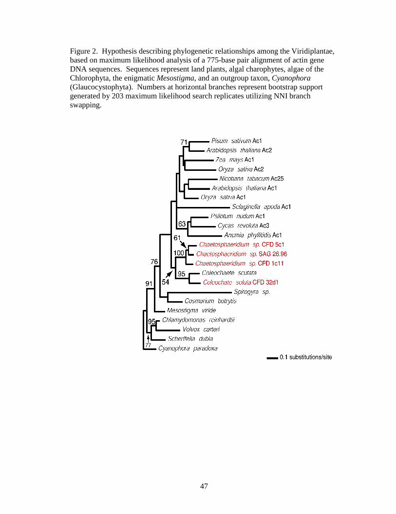
47
Figure 2. Hypothesis describing phylogenetic relationships among the Viridiplantae, based on maximum likelihood analysis of a 775-base pair alignment of actin gene DNA sequences. Sequences represent land plants, algal charophytes, algae of the Chlorophyta, the enigmatic Mesostigma, and an outgroup taxon, Cyanophora (Glaucocystophyta). Numbers at horizontal branches represent bootstrap support generated by 203 maximum likelihood search replicates utilizing NNI branch swapping.

48
also inconsistent with other hypotheses, such as those allying Mesostigma with more
morphologically complex algal charophytes, such as Chaetosphaeridium.
Likewise, the phylogeny of nuclear actin genes in the Charophyta supports the
hypothesis including Mesostigma within the Charophyta (91% bootstrap) at the base
of the lineage. Previous analysis of Mesostigma actin gene sequence (Bhattacharya et
al., 1998) did not include Chaetosphaeridium in the range of taxa sampled. In this
analysis, the partitioning of Mesostigma from Chaetosphaeridium and the other
morphologically complex algal charophytes is supported by 76% bootstrap support.
These data are not consistent with the hypothesis and taxonomic scheme proposed by
Marin and Melkonian (1999). However, due to short branch lengths and incomplete
taxon-sampling in this study, alternative placements of Mesostigma outside the
Charophyte clade cannot be ruled out entirely.
3.4.2 On the Monophyly of the Coleochaetales
The discovery of introns in both Coleochaete and Chaetosphaeridium
chloroplast tRNA-Ala and tRNA-Ile genes supports a monophyletic order,
Coleochaetales that includes the genera Coleochaete plus Chaetosphaeridium. These
results were later independently generated and published by another group of
investigators (Turmel et al., 2002b; Turmel et al., 2002c). It may be argued that
introns in Coleochaete chloroplast tRNA genes are homologous to those of
Chaetosphaeridium; this hypothesis is supported by high levels of sequence similarity
(greater than 70%) and identical intron insertion positions within the two chloroplast
tRNA genes.
49
Phylogeny of charophyte actin genes (Fig. 2) also supports a monophyletic
Coleochaetales, with 54% bootstrap uniting the order consisting of Coleochaete plus
Chaetosphaeridium. The addition of three new Chaetosphaeridium taxa and a new
Coleochaete strain distinguishes this study from prior work by Bhattacharya et al.
(1998). The addition of Chaetosphaeridium to the actin phylogeny allows for
contrasting of this nuclear gene analysis against that of the nuclear rRNA SSU
sequence data analyzed by Marin and Melkonian (1999).
Phylogenetic analysis of actin gene sequence data including the three genera
Mesostigma, Coleochaete, and Chaetosphaeridium supports a monophyletic
Coleochaetales clade composed of Coleochaete plus Chaetosphaeridium.
Furthermore, this analysis supports phylogenetic placement of Mesostigma at the base
of the Charophyta clade, contrary to the alliance of Mesostigma and
Chaetosphaeridium suggested by the results of Marin and Melkonian (1999). Basal
placement of Mesostigma and a monophyletic Coleochaetales is a phylogenetic
hypothesis that is also bolstered by the finding of Coleochaete-like introns within the
chloroplast tRNA genes of Chaetosphaeridium (Lewandowski, unpublished results;
Turmel et al., 2002b; Turmel et al., 2002c). More recent phylogenetic analyses of
chloroplast, mitochondrial and nuclear ribosomal DNA sequence data also support
the monophyly of the Coleochaetales (Karol et al., 2001; Turmel et al., 2002b;
Delwiche et al., 2002) and the basal placement of Mesostigma (Karol et al., 2001;
Turmel et al., 2002a).
Testing of enigmatic phylogenetic hypotheses is necessary if structural and
developmental character states are to be polarized and analyzed. Both chloroplast

50
genome architecture characters and nuclear actin gene base pair characters are very
likely to be independent from most structural and developmental characters of interest
among algal Charophytes. Intron insertion and deletion events are considered to be
evolutionarily conservative, making them useful for testing of hypothesized
relationships based on other forms of data (Manhart and Palmer, 1990; Graham,
1993; Qui and Lee, 2000). Nuclear genes coding for actin are conserved and
ubiquitous across eukaryotes. These features make actin gene base pair data useful
for phylogenetic analyses (An et al., 1999). Beyond ribosomal RNA genes, the use of
nuclear genes for phylogenetic analysis of the algal Charophyta has been extremely
limited. Both sources of data, chloroplast introns and actin gene DNA sequences,
support the relationships among algal charophytes found by Karol et al. (2001) and
Turmel et al. (2002b).
3.4.3 Other Phylogenetic Inference
Investigation of tRNA-Ala and tRNA-Ile genes in the transcribed spacer
region of chloroplast genomes has revealed a lack of interrupting introns in
cyanobacteria, likely closely related to the symbiotically acquired primary plastids
(Delwiche, 1997). Furthermore, these introns are missing from the tRNA genes
examined in the chloroplast genomes of the algal Chlorophyta. New sequences
produced in this study reveal the absence of both introns from the chloroplast tRNA
genes of Chlorokybus. Recently published results (Turmel et al., 2002b) demonstrate
that the tRNA-Ala gene is also interrupted by group II introns in members of the
charophyte orders Klebsormidiales (Klebsormidium, Entransia), Zygnematales
(Cosmarium, Mesotaenium, Spirogyra, Staurastrum, Zygnema), Coleochaetales

51
(Coleochaete, Chaetosphaeridium), and Charales (Chara, Nitella). In all of these
taxa except genera of the Zygnematales an intron is also known to be present in the
chloroplast tRNA-Ile gene (Turmel et al., 2002b). It is possible that a genome
rearrangement event has affected the chloroplast DNA sequence in Zygnematales, as
it is not clear that the transcribed spacer region organization demonstrated in each of
the other orders is conserved in the algae of the Zygnematales. This distribution,
introns present in the later-diverging lineages and the absent in both outgroup taxa
and Chlorokybus, supports the placement of Chlorokybus near the base of the
Charophyta clade.
3.5 Conclusions
Analyses of these new data support the view of evolutionary transitions
presented in the literature review above. Character states found in the scaly
biflagellate unicell, Mesostigma, are likely ancestral conditions among the
charophytes. Branched filamentous thalli, like those in the Charales and
Coleochaetales, including the genus Chaetosphaeridium, are likely derived conditions
resulting from a set of advanced cellular and developmental capabilities possessed by
algal lineages more closely related to the embryophytes. Continued testing of
phylogenetic hypotheses will result in an even more refined understanding of the
evolutionary relationships among algal members of the Charophyta. New
observations of structural features, as well as discoveries of developmental
mechanisms through the application of molecular genetics methodology, will add to
the growing matrix of characters that can be mapped onto the tree of green life. This

52
perpetual process should lend to a cascade of increasingly precise hypotheses
explaining the evolution of this fascinating and important group, the Charophyta.

53
Appendices
Appendix A Isolation of total RNA from algal charophytes
These methods are based on the Sigma Tri Reagent protocol, and modified by the author according to Sambrook and Russell (2000) and experience gained while working on algae of the Charophyta.
1. Harvest thalli and remove excess culture medium. Record mass of the algal material.
2. To a 50-mL PPCO Oak Ridge centrifuge tube, add 1 mL of Sigma Tri Reagent per 50-100 mg thallus material. Close the tube and coat entire interior surface area of the tube by manual agitation followed by gentle rocking for ~5 minutes.
3. Add algal thallus material to tube containing Tri Reagent. Volume of algal material should not exceed 20% the volume of Tri Reagent. Homogenize thalli and Tri Reagent using the Polytron for ~1 minute at 80-100% power. Homogenized samples can be stored at -70°C for up to 1 month.
4. Allow sample to stand at room temperature for ~5 minutes. Centrifuge the homogenate at 12,000 x g at 4°C for 10 minutes. Transfer the translucent supernatant to a fresh tube. The supernatant contains RNA and protein. The pellet should contain high molecular weight DNA.
5. Add 0.2 mL pure chloroform per 1 mL of Tri Reagent used in step 2. Cover and shake the sample vigorously for 15 seconds. Allow the sample to stand at room temperature for 2-15 minutes.
6. Centrifuge the mixture at 12,000 x g at 4°C for 15 minutes. Separation should result in three phases—a colorless upper aqueous phase containing RNA, an interphase containing DNA, and a red organic phase containing protein. Interphase and organic phases can be stored temporarily at 4°C for subsequent isolation of DNA and protein, as per the Sigma Tri Reagent protocol.
7. Exercising care not to disturb the DNA-containing interphase, transfer the aqueous phase containing RNA to a fresh tube. For each 1 mL Tri Reagent used in step 2, add 0.25 mL isopropanol and 0.25 mL RNA Precipitation Solution. Mix thoroughly and allow the sample to stand at room temperature for 10 minutes.
8. Centrifuge the sample at 12,000 x g at 4°C for 10 minutes. Precipitated RNA will form a pellet on the side and bottom of the tube.

54
9. Remove supernatant and wash the RNA pellet by adding at least 1 mL 75% ethanol for each 1 mL Tri Reagent used in step 2. Vortex the sample and centrifuge at 7,500 x g at 4°C for 5 minutes. RNA sample can be stored in the ethanol solution at 4°C for up to 1 week and at -20°C for up to one year.
10. Repeat the wash step described in step 9.
11. Remove ethanol from the pellet and allow to air dry on the bench top for 5-10 minutes. Do not allow the pellet to dry completely.
12. Resuspend the pellet in an appropriate RNase-free storage buffer, or resuspend in 1.5 mL Binding Buffer and proceed on to poly(A)+ selection protocol (Sambrook and Russell, 2000; Eric Haag, personal communication).

55
Literature Cited
An, S. S., B. Mopps, K. Weber, and D. Bhattacharya. 1999. The origin and evolution of green algal and plant actins. Molecular Biology and Evolution16(2): 275-285.
Baldauf, S. L., J. R. Manhart, and J. D. Palmer. 1990. Different fates of the chloroplast tufA gene following its transfer to the nucleus in green algae. Proceedings of the National Academy of Sciences of the United States of America 87(14): 5317 -5321.
Bhattacharya, D., K. Weber, S. S. An, and W. Berning-Koch. 1998. Actin phylogeny identifies Mesostigma viride as a flagellate ancestor of the land plants. Journal of Molecular Evolution 47(5): 544-550.
Bold, H. C. and M. J. Wynne. 1978. Introduction to Phycology. Englewood Cliffs, NJ: Prentice Hall.
Bold, H. C. and M. J. Wynne. 1985. Introduction to Phycology. Englewood Cliffs, NJ: Prentice Hall.
Bower, F. O. 1908. The Origin of a Land Flora: A Theory Based upon the Facts of Alternation. London, United Kingdom: Macmillan and Co.
Bremer, K. 1985. Summary of green plant phylogeny and classification. Cladistics1: 369-385.
Brown, R. C., B. E. Lemmon, and L. E. Graham. 1994. Morphogenetic plastid migration and microtubule arrays in mitosis and cytokinesis in the green alga Coleochaete orbicularis. American Journal of Botany 81: 127-133.
Cavalier-Smith, T. 1981. Eukaryote kingdoms: Seven or nine. BioSystems 14: 461-481.
Chapman, R. L. and M. A. Buchheim. 1991. Ribosomal RNA gene sequences: Analysis and significance in the phylogeny and taxonomy of green algae. Critical Reviews in Plant Science 10: 343-368.
Cimino, M. T. and C. F. Delwiche. 2002. Molecular and morphological data identify a cryptic species complex in endophytic members of the genus ColeochaeteBreb. (Charophyta: Coleochaetaceae). Journal of Phycology 38: 1213-1221.

56
Cimino, M. T., K. G. Karol, and C. F. Delwiche. 2000. An artifact in the small subunit rDNA sequence of Chaetosphaeridium globosum (Charophyceae, Streptophyta). Journal of Phycology 36: 440-442.
Cook, M. E. 2004. Structure and asexual reproduction of the enigmatic charophycean green alga Entransia fimbriata (Klebsormidiales, Charophyceae). Journal of Phycology 40: 424-431.
Cook, M. E., L. E. Graham, C. E. J. Botha, and C. A. Lavin. 1997. Comparative ultrastructure of plasmodesmata of Chara and selected bryophytes: Toward an elucidation of the evolutionary origin of plant plasmodesmata. American Journal of Botany 84: 1169-1178.
Cook, M. E., L. E. Graham, and C. A. Lavin. 1998. Cytokinesis and nodal anatomy in the charophycean green alga Chara zeylanica. Protoplasma 203: 65-74.
DeJesus, M. D., F. Tabatabai, and D. J. Chapman. 1989. Taxonomic distribution of copper-zinc superoxide dismutase in green algae and its phylogenetic importance. Journal of Phycology 25: 767-772.
Delwiche, C. F. and J. D. Palmer. 1997. The origin of plastids and their spread via secondary endosymbiosis. The Origins of Algae and Their Plastids. D. Bhattacharya, ed. Springer-Verlag, Vienna: 53-86.
Delwiche, C. F., L. E. Graham, and N. Thomson. 1989. Lignin-like compounds and sporopollenin in Coleochaete, an algal model for land plant ancestry. Science245: 399-401.
Delwiche, C. F., K. G. Karol, M. T. Cimino, and K. J. Sytsma. 2002. Phylogeny of the genus Coleochaete (Coleochaetales; Charophyta) and related taxa inferred by analysis of the chloroplast gene rbcL. Journal of Phycology 38: 394-403.
Dobzhansky, T. 1973. Nothing in biology makes sense except in the light of evolution. American Biology Teacher 35: 125-129.
Floyd, G. L., K. D. Stewart, and K. R. Mattox. 1972. Cellular organization, mitosis, and cytokinesis in the ulotrichalean alga, Klebsormidium. Journal of Phycology 8: 176-184.
Frederick, S. E., P. J. Gruber, and N. E. Tolbert. 1973. The occurrence of glycolate dehydrogenase and glycolate oxidase in green plants. Plant Physiology 52: 318-323.

57
Frias-Lopez, J., A. L. Zerkle, G. T. Bonheyo, and B. W. Fouke. 2002. Partitioning of bacterial communities between seawater and healthy, black band diseased, and dead coral surfaces. Applied and Environmental Microbiology 68(5): 2214-2228.
Graham, L. E. 1993. Origin of Land Plants. New York, NY: John Wiley & Sons, Inc.
Graham, L. E. 1996. Green algae to land plants: An evolutionary transition. Journal of Plant Research 109: 241-251.
Graham, L. E. and G. E. McBride. 1979. The occurrence and phylogenetic significance of a multilayered structure in Coleochaete scutata spermatozoids. American Journal of Botany 66: 887-894.
Graham, L. E. and G. J. Wedemayer. 1984. Spermatogenesis in Coleochaete pulvinata (Charophyceae): Sperm maturation. Journal of Phycology 20 : 302-309.
Graham, L. E. and L. W. Wilcox. 1983. The occurrence and phylogenetic significance of putative placental transfer cells in the green alga Coleochaete. American Journal of Botany 70: 113-120.
Graham, L. E., M. E. Cook, and J. S. Busse. 2000. The origin of plants: Body plan changes contributing to a major evolutionary radiation. Proceedings of the National Academy of Sciences of the United States of America 97(9): 4535-4540.
Graham, L. E., C. F. Delwiche, and B. D. Mishler. 1991. Phylogenetic connections between the 'green algae' and the 'bryophytes'. Advances in Bryology 4: 213-244.
Graham, L. E. and L. W. Wilcox. 2000. Algae. Upper Saddle River, NJ: Prentice Hall.
Gray, J., D. Massa, and A. J. Boucot. 1982. Caradocian land plant microfossils from Libya. Geology 10: 197-201.
Gross, W. 1993. Peroxisomes in algae: their distribution, biochemical function and phylogenic [phylogenetic] importance. Progress in Phycological Research 9: 47-78.
Hader, D. -P. and E. Hoiczyk. 1992. Gliding motility. Algal Cell Motility. M. Melkonian, ed. Chapman and Hall, London, United Kingdom: 1-38.
58
Hillis, D. M. 1998. Taxonomic sampling, phylogenetic accuracy, and investigator bias. Systematic Biology 47: 3-8.
Hopkins, A. W. and G. E. McBride. 1976. The life history of Coleochaete scutata(Chlorophyceae) studied by a Feulgen microspectrophotometric analysis. Journal of Phycology 12: 29-35.
Hotchkiss, A. T., M. R. Gretz, K. C. Hicks, and R. M. Brown. 1989. The composition and phylogenetic significance of the Mougeotia (Charophyceae) cell wall. Journal of Phycology 25: 646-654.
Jacobshagen, S. and C. Schnarrenberger. 1990. Two class I aldolases in Klebsormidium flaccidum (Charophyceae): An evolutionary link from chlorophytes to higher plants. Journal of Phycology 26: 312-317.
Karol, K. G., R. M. McCourt, M. T. Cimino, and C. F. Delwiche. 2001. The closest living relatives of land plants. Science 294: 2351-2353.
Kenrick, P. and P. R. Crane. 1997. The Origin and Early Diversification of Land Plants: A Cladistic Study. Washington, DC: Smithsonian Institution Press.
Kiss, J. Z. and L. A. Staehelin. 1993. Structural polarity in the Chara rhizoid: A reevaluation. American Journal of Botany 80: 273-282.
Krupp, J. M. and N. J. Lang. 1985. Cell division and filament formation in the desmid Bambusina brebissonii (Chlorophyta). Journal of Phycology 21 : 16-25.
Lemieux, C., C. Otis, and M. Turmel. 2000. Ancestral chloroplast genome in Mesostigma viride reveals an early branch of green plant evolution. Nature403: 649-652.
Lewis, L. A., B. D. Mishler, and R. Vilgalys. 1997. Phylogenetic relationships of the liverworts (Hepaticae), a basal embryophyte lineage, inferred from nucleotide sequence data of the chloroplast gene rbcL. Molecular Phylogenetics and Evolution 7: 377-393.
Lockhorst, G. M., H. J. Sluiman, and W. Star. 1988. The ultrastructure of mitosis and cytokinesis in the sarcinoid Chlorokybus atmophyticus (Chlorophyta, Charophyceae) revealed by rapid freeze fixation and freeze substitution. Journal of Phycology 24: 237-248.
Lockhorst, G. M. 1996. Comparative taxonomic studies on the genus Klebsormidium (Charophyceae) in Europe. Cryptogamic Studies 5: 1-132.

59
Manhart, J. R. and J. D. Palmer. 1990. The gain of two chloroplast transfer RNA introns marks the green algal ancestors of land plants. Nature 345(6272): 268-270.
Manton, I. and H. Ettl. 1965. Observations on the fine structure of Mesostigma viride Lauterborn. Botanical Journal of the Linnean Society 59:175-184.
Marchant, H. J., J. D. Pickett-Heaps, and K. Jacobs. 1973. An ultrastructural study of zoosporogenesis and the mature zoospore of Klebsormidium flaccidum. Cytobios 8: 95-107.
Marin, B. and M. Melkonian. 1999. Mesostigmatophyceae, a new class of streptophyte green algae revealed by SSU rRNA sequence comparisons. Protist 150(4): 399 -417.
Mattox, K. R. and H. C. Bold. 1962. Phycological Studies III. The Taxonomy of Certain ulotrichalean Algae. University of Texas Publication No. 6222. Austin, TX: University of Texas.
Mattox, K. R. and K. D. Stewart. 1984. Classification of the Green Algae: A Concept Based on Comparative Cytology. Systematics of the Green Algae—(The Systematics Association special volume; no. 27). D. E. G. Irvine and D. M. John, eds. Academic Press, Inc., London, United Kingdom: 29-72.
Maynard Smith, J. 1998. Shaping Life: Genes, Embryos and Evolution. New Haven, CT: Yale University Press.
McCourt, R. M. 1995. Green algal phylogeny. Trends in Ecology and Evolution 10: 159-163.
McCourt, R. M., K. G. Karol, S. Kaplan, and R. W. Hoshaw. 1995. Using rbcLsequences to test hypotheses of chloroplast and thallus evolution in conjugating green algae (Zygnematales, Charophyceae). Journal of Phycology 31: 989-995.
McCourt, R. M., K. G. Karol, M. Guerlesquin, and M. Feist. 1996. Phylogeny of extant genera in the family Characeae (Charales; Charophyceae) based on rbcL sequences and morphology. American Journal of Botany 83: 125-131.
Melkonian, M. 1982. Structural and evolutionary aspects of the flagellar apparatus in green algae and land plants. Taxon 31: 255-265.
Melkonian, M. 1984. Flagellar apparatus ultrastructure in relation to green algal classification. Systematics of the Green Algae—(The Systematics Association special volume; no. 27). D. E. G. Irvine and D. M. John, eds. Academic Press, Inc., London, United Kingdom: 73-120.

60
Melkonian, M. 1989. Flagellar apparatus ultrastructure in Mesostigma viride(Prasinophyceae). Plant Systematics and Evolution 164: 93-122.
Melkonian, M. and B. Surek. 1995. Phylogeny of the Chlorophyta: congruence between ultrastructural and molecular evidence. Bulletin de la Societe Zoologique de France 120(2): 191-208.
Mishler, B. D., L. A. Lewis, M. A. Buchheim, K. S. Renzaglia, D. J. Garbary, C. F. Delwiche, F. W. Zechman, T. S. Kantz, and R. L. Chapman. 1994. Phylogenetic relationships of the "green algae" and "bryophytes". Annals of the Missouri Botanical Garden 81(3): 451-483.
Moestrup, O. 1974. Ultrastructure of the scale-covered zoospores of the green alga Chaetosphaeridium, a possible ancestor of the higher plants and bryophytes. Biological Journal of the Linnean Society 6: 111-125.
Pickett-Heaps, J. D. 1975. Green Algae: Structure, Reproduction and Evolution in Selected Genera. Sunderland, MA: Sinauer Associates, Inc.
Pickett-Heaps, J. D. 1972. Cell division in Klebsormidium subtilissimum (formerly Ulothrix subtilissima) and its possible phylogenetic significance. Cytobios 6: 167-183.
Pickett-Heaps, J. D. and H. J. Marchant. 1972. The phylogeny of green algae: a new proposal. Cytobios 6: 255-264.
Pollock, D. D., D. J. Zwickl, J. A. McGuire, and D. M. Hillis. 2002. Increased taxon sampling is advantageous for phylogenetic inference. Systematic Biology51(4): 664-671.
Qui, Y. –L. and J. Lee. 2000. Transition to a land flora: a molecular phylogenetic perspective. Journal of Phycology 36: 799-802.
Raff, R. A. 1996. The Shape of Life: Genes, Development, and the Evolution of Animal Form. Chicago, IL: The University of Chicago Press.
Raven, P. H., R. F. Evert, and S. E. Eichhorn. 1999. Biology of Plants. New York, NY: W. H. Freeman and Company.
Rogers, C. E., D. S. Domozych, K. D. Stewart, and K. R. Mattox. 1981. The flagellar apparatus of Mesostigma viride (Prasinophyceae): multilayered structures in a scaly green flagellate. Plant Systematics and Evolution 138: 247-258.

61
Rogers, C. E., K. R. Mattox, and K. D. Stewart. 1980. The zoospore of Chlorokybus atmophyticus, a charophyte with sarcinoid growth habit. American Journal ofBotany 67(5): 774-783.
Sambrook, J. and D. Russell. 2000. Molecular Cloning: A Laboratory Manual. Cold Spring Harbor, NY: Cold Spring Harbor Laboratory Press.
Sluiman, H. J. 1983. The flagellar apparatus of the zoospore of the filamentous green alga Coleochaete pulvinata: Absolute configuration and phylogenetic significance. Protoplasma 115: 160-175.
Sluiman, H. J. 2000. Molecular phylogeny of Chaetosphaeridium revisited. Journal of Phycology 36: 443-444.
Sluiman, H. J. and C. Guihal. 1999. Phylogenetic position of Chaetosphaeridium (Chlorophyta), a basal lineage in the Charophyceae inferred from 18S rDNA sequences. Journal of Phycology 35: 395-402.
Starke, T. and J. P. Gogarten. 1993. A conserved intron in the V-ATPase A subunit genes of plants and algae. Federation of European Biological Societies 315: 252-258.
Swofford, D. L. 2001. PAUP* Phylogenetic Analysis Using Parsimony (*and Other Methods). Version 4.0b8. Sinauer Associates: Sunderland, MA.
Sym, S. D. and R. N. Pienaar. 1993. The class Prasinophyceae. Progress in Phycological Research 9: 281-376.
Syrett, P. J. and F. A. A. al-Houty. 1984. The phylogenetic significance of the occurrence of urease/urea amidolyase and glycolate oxidase/glycolate dehydrogenase in green algae. British Phycological Journal 19: 11-21.
Thompson, R. H. 1969. Sexual reproduction in Chaetosphaeridium (Nordst.) Klebahn (Chlorophyceae) and description of a species new to science. Journal of Phycology 5: 285-290.
Turmel, M., C. Otis, C. Lemieux. 2002a. The complete mitochondrial DNA sequence of Mesostigma viride identifies this green alga as the earliest green plant divergence and predicts a highly compact mitochondrial genome in the ancestor of all green plants. Molecular Biology and Evolution 19(1): 24-38.
Turmel, M., M. Ehara, C. Otis, and C. Lemieux. 2002b. Phylogenetic relationships among streptophytes as inferred from chloroplast small and large subunit rRNA gene sequences. Journal of Phycology 38: 364-375.

62
Turmel, M., C. Otis, and C. Lemieux. 2002c. The chloroplast and mitochondrial genome sequences of the charophyte Chaetosphaeridium globosum: Insights into the timing of the events that restructured organelle DNAs within the green algal lineage that led to land plants. Proceedings of the National Academy of Sciences of the United States of America 99(17): 11275-11280.
Waters, D. A. and R. L. Chapman. 1996. Molecular phylogenetics and the evolution of green algae and land plants. Cytology, Genetics and Molecular Biology of Algae. B. R. Chaudhary and S. B. Agrawal, eds. SPB Publishing, Amsterdam, Netherlands: 337-349.