accaddeemm icc sscieencceess - international journal of ... · solvents and subjected for...
TRANSCRIPT

Research Article
ANTIOXIDANT AND CYTOTOXIC STUDIES ON TWO KNOWN COMPOUNDS ISOLATED FROM HYPTIS SUAVEOLENS LEAVES
SUBRAMANIAN DEEPIKA PRIYADHARSHINI, VENUGOPAL SUJATHA*
*Department of Chemistry, Periyar University, Salem 636011, Tamil Nadu, India. Email: [email protected]
Received: 11 July 2013, Revised and Accepted: 22 Aug 2013
ABSTRACT
Objective: In the present study, it is intended to investigate the antioxidant profile of Hyptis suaveolens leaves, through numerous in vitro biochemical assays and to isolate useful constituents. This plant has been extensively researched for its essential oils till date.
Methods: Various solvent extracts (n-hexane, ethyl acetate, acetone, methanol) were assessed for total content of phenolic, flavonoid, carbohydrate, protein, in vitro antioxidant assays viz. DPPH, superoxide, hydrogen peroxide- radical scavenging assays, Ferric reducing power activity and inhibition lipid peroxidation (TBARS) assays. Bioassay-guided fractionation on ethyl acetate crude was performed using column chromatography which revealed two known compounds characterized through supportive and evidencing spectral data like 1D & 2D-NMR, DEPT and HR-MS.
Results: All the performed in vitro assays have revealed the excellence of ethyl acetate extract in contrast to other fractions. Its SC50/ IC50/ EC50 values calculated have recorded significant impact with reference to the standard. Significantly, the estimations viz. phenolic, flavonoid, carbohydrate and protein seem to be aligned with the in vitro antioxidant assays. Thus from the ethyl acetate crude, two bioactive compounds isolated were identified to be menthol and linalool from the spectral evidences. Cytotoxicity (MTT assay) for the two isolates showed moderate cell viability with respect to its concentration, using Cisplatin as positive control.
Conclusion: The significance of Hyptis suaveolens in traditional medicine and the importance of its chemical constituents shall improve its role against free radical production.
Keywords: Cytotoxic activity, DPPH Radical Scavenging Ability, Ferric Reducing Power, Linalool, Menthol, Total Phenol Content.
INTRODUCTION
Phytochemistry reports thousands of newer organic molecules or compounds every year. Pharmacological testing, modifying, derivatising and research on natural products represent a major strategy for discovering and developing new drugs [1,2]. The use of medicinal plants for the treatment of human diseases is well- known and practiced in Ayurveda since ancient times. The plant is a biosynthetic laboratory, not only for chemical compounds, but also a multitude of bioactive compounds which exert physiological and therapeutic effect. Traditionally, medicinal plants have already provided leads for potential antioxidant, antibacterial, antifungal compounds including flavonoids, coumarins, tannins, terpenoids, alkaloids, steroids etc. Such compounds and their modified derivatives may be studied for pharmacological properties and may form a basis for novel drugs. Recently, interest has increased considerably in finding naturally occurring antioxidants for use in foods, cosmetics or medicinal materials to replace synthetic antioxidants which are restricted due to their carcinogenicity [3]. The interest in analysis and identification of the antioxidant compounds and their derivatives present in plant extracts rises not only from the need to find new sources, but from the relationships between structures of the compounds and the extract uses in traditional medicine.
The present investigation has been focused on one such antioxidant propensities of the species, Hyptis suaveolens. It is an annual, aromatic, medium sized shrub, belonging to the family Lamiaceae and commonly called ‘American mint’, ‘Vilaiti tulasi’, ‘Bush mint’ etc. An ethnobotanically important plant, also considered as an obnoxious weed found to be distributed throughout the sub-tropics of India. Their leaves find applications as a stimulant, carminative, wound healing, sudorific, galactogogue etc., [4]. It has been used as a traditional medicine against various illnesses like cold, pain, fever, skin diseases, cramps etc. The essential oils of Hyptis were researched for their anticonvulsant [5], antimicrobial [6-8], anticancer [9], tumerogenic properties [10] etc. To kindle the area of phytochemistry with well designed, systematic and objective research, which would benefit people to cure their ailments who lack technological and economic resources with their orthodox medicine, is considered.
This work has been attempted in search of compounds that are biologically active with a low profile of side effects. Attention has therefore been directed towards the isolation of natural antioxidants from botanical sources, especially edible plants. Investigation on the fundamental scientific basis regarding the in vitro antioxidant and free radical scavenging properties of various solvent extracts of the leaf extracts of Hyptis suaveolens have been performed. Furthermore, two known compounds viz. menthol and linalool were isolated by normal column chromatography directly from the plant crude to study their cytotoxic activity.
MATERIALS AND METHODS
Chemicals
1,1-diphenyl-2-picrylhydrazyl (DPPH), butylated hydroxyanisole (BHA), gallic acid, ascorbic acid, (+)-catechin and solvents were purchased from Sigma Chemicals Pvt. Ltd.
Plant selection and authentification
Identification, extraction and the phytochemical screening were based on their antioxidant activity, derived primarily from a random selection of commonly occurring native plants. Fresh and healthy leaves of Hyptis suaveolens Poit. were collected in and around the University campus and at the time of collection [October- November], a pressed specimen was prepared and authenticated by a Botanist [BSI/SRC/5/23/2010-11/Tech-1482] by Botanical Survey of India, Southern Circle, TNAU, Coimbatore.
Extraction procedure
The old, insect-damaged, fungus-infected leaves were removed, deprived of dusts and the rest were washed well with dechlorinated water prior to distilled water. The air-dried, powdered leaf samples were subjected to Soxhelation using each of the following solvents in increasing polarity: n-hexane, ethyl acetate, acetone and methanol. Then the solvent was distilled off, till the extract turns off into a syrupy consistency. All the crude solvent extracts were stored at 4–5 ˚C until further use and the following assays were performed from 2 mg/ml stock solution.
International Journal of Pharmacy and Pharmaceutical Sciences
ISSN- 0975-1491 Vol 5, Issue 4, 2013
AAccaaddeemmiicc SScciieenncceess

Sujatha et al. Int J Pharm Pharm Sci, Vol 5, Issue 4, 283-290
284
Phytochemical screening
The leaf extracts of Hyptis suaveolens were diluted in their respective solvents and subjected for qualitative preliminary phytochemical screening to identify the presence of the secondary metabolites according to the standard methods [11]. From the intensity of the color inferred for the tests, they will be rated for their presence.
Total phenolic content
Contents of total phenolics in the extracts were estimated by a colorimetric assay based on standard procedures [12]. Gallic acid was used for constructing the standard curve (0.01–0.4 mM; y= 2.94848 x -0.09211; R2 = 0.99914) and the results were expressed as μg of gallic acid equivalents (GAEs).
Total flavonoid content
Flavonoid contents in the extracts were determined by a colorimetric method [12]. (+)-Catechin was used to calculate the standard curve (0.250–2.500 mM; Y = 0.2903; R2 = 1.0000) and the results were expressed as μg of catechin equivalents (CEs) per mg of extract.
Estimation of carbohydrate content
Total carbohydrate contents were estimated by anthrone method [13]. Glucose was used to calculate the standard curve (20-120 µg/ml, Y= 0.0263x +0.0532, R2= 0.9992) and the results were expressed has µg of glucose equivalents per mg of extract.
Estimation of protein content
Total proteins were estimated by Lowry’s method [14]. Bovine serum albumin was used to calculate the standard curve (20-160 µg/ml, Y = 0.0159x + 0.0319, R2 = 0.9569) and the results were expressed as µg of bovine serum albumin equivalents per mg of extract.
DPPH radical-scavenging activity
The radical scavenging capacity of the extracts using DPPH radicals is analyzed using the method described by Berreira et al., [12]. RSA was calculated as a percentage of DPPH discoloration, using the equation: %RSA= [(ADPPH -AS) /ADPPH] x 100, where AS is the absorbance of the solution when the sample extract is added at a particular level and ADPPH is the absorbance of the DPPH solution. Ascorbic acid was used as standard.
Superoxide anion radical scavenging assay
The assay for superoxide anion radical scavenging activity was based on riboflavin – light –NBT system described by Beauchamp and Fridovich et al., [15]. Ascorbic acid was used as standard. The percent inhibition of superoxide anion generation was calculated using the following formula:
Scavenging activity (%) = 1- absorbance of sample x 100 absorbance of control
Hydrogen peroxide scavenging assay
The ability of the extracts to scavenge hydrogen peroxide was determined according to the method of Ruch et al., [16]. Ascorbic acid was used as standard. The abilities to scavenge the hydrogen peroxide were calculated using the following equation:
Hydrogen peroxide scavenging activity = 1- absorbance of sample x 100 absorbance of control
Reducing power
This assay was performed as suggested by Berreira et al., [12]. Different aliquots of ascorbic acid were treated as standard and results were expressed in terms of mg/ ascorbic acid equivalents.
Inhibition of lipid peroxidation using thiobarbituric acid reactive substances
The procedure of Lam and Ng (2009) using a Fenton reaction-induced lipid peroxidation has been adapted for this assay [17]. α-Tocopherol was used as positive control.
Isolation
Ethyl acetate extract was subjected to fractionation on column chromatography packed with silica gel [60-120 mesh] using hexane and ethyl acetate. Initially the column was run with hexane and the fractions eluted were grouped after TLC analysis. By increasing the polarity of the eluant using ethyl acetate about 6 compounds were obtained among which 2 compounds were stable with good yield. Compound 1 [90.3 mg at 60:40 of hexane: ethyl acetate] as white crystalline solid and Compound 2 [7 ml at 20:80 of hexane: ethyl acetate] as colorless liquid were repeatedly chromatographed for purification and characterization. IR, 1H, 13C NMR, COSY, HMBC, HSQC and HR-MS were used for identifying the structure of the isolates.
In vitro cytotoxicity assay for isolated compounds
MTT Assay: Cytotoxicity studies [18] of the compound were carried out on human breast cancer cell line (MCF 7) which was obtained from National Centre for Cell Science, Pune, India. Cell was grown in EMEM containing 10% FBS and maintained at 37 0C, 5% CO2, 95% air and 100% relative humidity for 24 h prior to addition of compound. The monolayer cells were detached with trypsin-ethylenediamine tetraacetic acid (EDTA) to make single cell suspensions and viable cells were counted using a hemocytometer and diluted with medium containing 5% FBS to give final density of 1x10⁵ cells/ml. one hundred microlitres per well of cell suspension were seeded into 96-well plates at plating density of 10,000 cells/well and incubated to allow for cell attachment at 37 0C, 5% CO2, 95% air and 100% relative humidity. After 24 h the cells were treated with serial concentrations of the test samples. They were initially dissolved in neat DMSO and diluted to twice the desired final maximum test concentration with serum free medium. Aliquots of 100 µl of these different sample dilutions were added to the appropriate wells already containing 100 µl of medium, resulted the required final sample concentrations. Following drug addition the plates were incubated for an additional 48 h at 37 0C, 5% CO2, 95% air and 100% relative humidity. The medium containing without samples were served as control and triplicate was maintained for all concentrations. After 48 h of incubation, 15 µl of MTT (5 mg/ml) in PBS was added to each well and incubated at 37 0C for 4h. The medium with MTT was then flicked off and the formed formazan crystals were solubilized in 100 µl of DMSO and then measured the absorbance at 570 nm using micro plate reader. The % cell viability was determined using the following formula.
% Cell viability = Abs (sample)/Abs (control) x100
Statistical analysis
All the experiments were carried out in triplets and the results are expressed as mean values and standard error or standard deviation (SD).
RESULTS
Phytochemical screening
Table.1 shows the list of constituents present in various solvent fractions, tested using standard procedures. Their presence was rated from intensely present to moderate to absence.
Total phenolic and flavonoid content
Intense phenolic content was recorded by ethyl acetate extract whereas flavonoid content by methanol extract, in contrast to other extracts (Table 2). These contents were analyzed using gallic acid and catechin as equivalents.
Total carbohydrate and protein content
Acetone fraction produced significant results for both these contents, in terms of glucose and bovine serum albumin equivalents. Next to acetone hexane fraction showed richer contents whereas the high polar methanol extract produced the least content.
Radical scavenging activities
DPPHo: Fig. 1 shows the amount of each extract required for 50% inhibition/ scavenging (SC50). The highest synergistic antioxidant activity
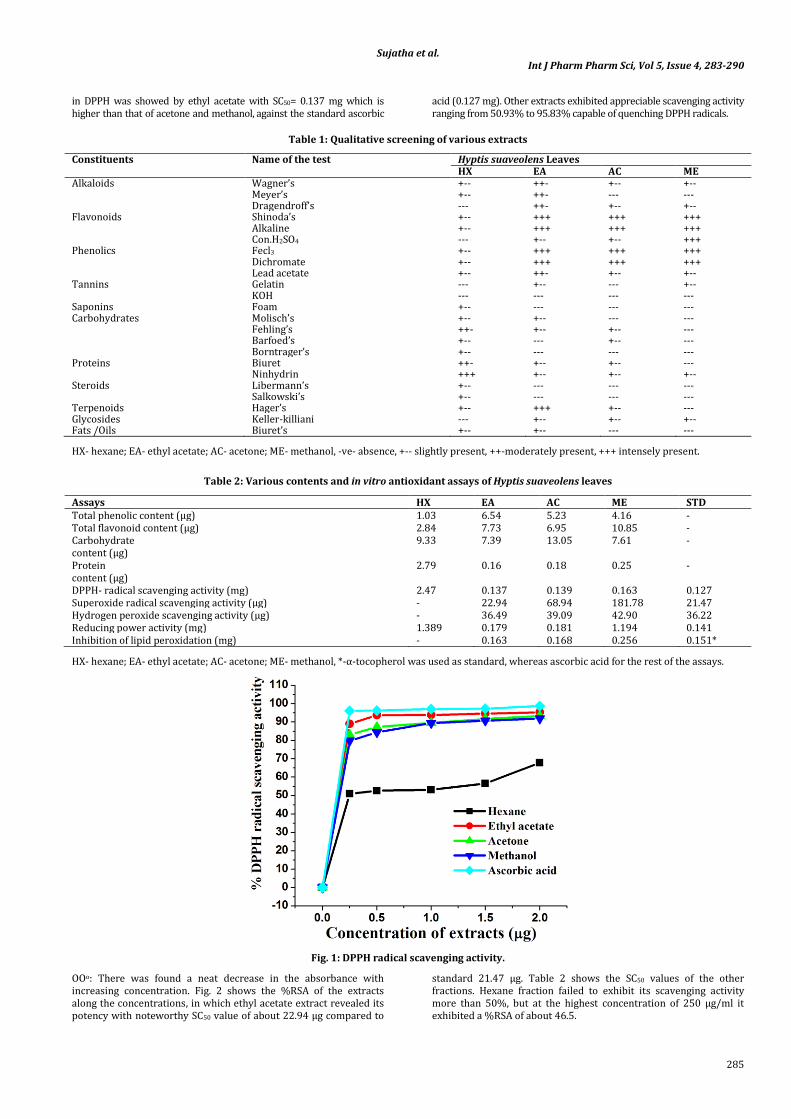
Sujatha et al. Int J Pharm Pharm Sci, Vol 5, Issue 4, 283-290
285
in DPPH was showed by ethyl acetate with SC50= 0.137 mg which is higher than that of acetone and methanol, against the standard ascorbic
acid (0.127 mg). Other extracts exhibited appreciable scavenging activity ranging from 50.93% to 95.83% capable of quenching DPPH radicals.
Table 1: Qualitative screening of various extracts
Constituents Name of the test Hyptis suaveolens Leaves HX EA AC ME
Alkaloids Wagner’s +-- ++- +-- +-- Meyer’s +-- ++- --- --- Dragendroff’s --- ++- +-- +--
Flavonoids Shinoda’s +-- +++ +++ +++ Alkaline +-- +++ +++ +++ Con.H2SO4 --- +-- +-- +++
Phenolics Fecl3 +-- +++ +++ +++ Dichromate +-- +++ +++ +++ Lead acetate +-- ++- +-- +--
Tannins Gelatin --- +-- --- +-- KOH --- --- --- ---
Saponins Foam +-- --- --- --- Carbohydrates Molisch’s +-- +-- --- ---
Fehling’s ++- +-- +-- --- Barfoed’s +-- --- +-- --- Borntrager’s +-- --- --- ---
Proteins Biuret ++- +-- +-- --- Ninhydrin +++ +-- +-- +--
Steroids Libermann’s +-- --- --- --- Salkowski’s +-- --- --- ---
Terpenoids Hager’s +-- +++ +-- --- Glycosides Keller-killiani --- +-- +-- +-- Fats /Oils Biuret’s +-- +-- --- ---
HX- hexane; EA- ethyl acetate; AC- acetone; ME- methanol, -ve- absence, +-- slightly present, ++-moderately present, +++ intensely present.
Table 2: Various contents and in vitro antioxidant assays of Hyptis suaveolens leaves
Assays HX EA AC ME STD Total phenolic content (μg) 1.03 6.54 5.23 4.16 - Total flavonoid content (μg) 2.84 7.73 6.95 10.85 - Carbohydrate content (µg)
9.33 7.39 13.05 7.61 -
Protein content (µg)
2.79 0.16 0.18 0.25 -
DPPH- radical scavenging activity (mg) 2.47 0.137 0.139 0.163 0.127 Superoxide radical scavenging activity (µg) - 22.94 68.94 181.78 21.47 Hydrogen peroxide scavenging activity (µg) - 36.49 39.09 42.90 36.22 Reducing power activity (mg) 1.389 0.179 0.181 1.194 0.141 Inhibition of lipid peroxidation (mg) - 0.163 0.168 0.256 0.151*
HX- hexane; EA- ethyl acetate; AC- acetone; ME- methanol, *-α-tocopherol was used as standard, whereas ascorbic acid for the rest of the assays.
Fig. 1: DPPH radical scavenging activity.
OOo: There was found a neat decrease in the absorbance with increasing concentration. Fig. 2 shows the %RSA of the extracts along the concentrations, in which ethyl acetate extract revealed its potency with noteworthy SC50 value of about 22.94 µg compared to
standard 21.47 µg. Table 2 shows the SC50 values of the other fractions. Hexane fraction failed to exhibit its scavenging activity more than 50%, but at the highest concentration of 250 µg/ml it exhibited a %RSA of about 46.5.

Sujatha et al. Int J Pharm Pharm Sci, Vol 5, Issue 4, 283-290
286
Fig.2: Superoxide radical scavenging activity.
H2O2: In this assay too, ethyl acetate extract recorded its excellence against peroxides and its SC50 value was 36.49 µg which is very closer to ascorbic acid ie., 36.22 µg. For a comparison, the next
significance was presented by acetone of about 39.09 µg as SC50 value. Fig. 3 evidences the neat scavenging effects rendered by all the extracts.
Fig. 3: Peroxide radical scavenging activity.
Fig. 4: Reducing power activity.

Sujatha et al. Int J Pharm Pharm Sci, Vol 5, Issue 4, 283-290
287
Reducing power
In this assay there was observed a neat increase in the absorbance along the increasing concentration as seen in Fig. 4. In contrary to the above assays, hexane fraction exhibited its EC50 value of about 1.389 mg. On the other hand, it was ethyl acetate fraction being significant with EC50 value of 0.179 mg, against the standard (0.141 mg) which was the striking evidence for the antioxidant property of Hyptis suaveolens leaves.
Inhibition of lipid peroxidation
In TBARS assay, the extracts lowered the degree of lipid peroxidation induced by hydroxyl radical generated by an iron/ascorbate system as observed in Fig. 5. Obviously it was ethyl acetate extract which recorded highest SC50 value of about 0.163 mg. There was a strong competence provided by the acetone fraction whose SC50 value was 0.168 mg.
Fig. 5: Inhibition of lipid peroxidation using thiobarbituric acid reactive substances.
Bioassay- Guided Fractionation and Characterization of Compounds (I) & (II)
From the above discussed facts, it can be made clear that the ethyl acetate extract has afforded its potent antioxidant and antibacterial activity. Hence by suspecting this extract to collectively contain immense number of constituents, a work on column chromatography has let out two compounds.
Compound I: Colorless needle like crystals (Melting point- 45°C). UV (MeOH) λmax (log Ɛ) nm: 200, 220, 270; IR (neat) ѵmax cm-1: 3262 (hydroxyl), 2956, 2926, 2871, 2723, 2647, 1455, 1375, 1312, 1048, 997, 878, 775, 671, 596, 428. For 1H spectrum (600 MHz, CD3OD), the peak at δ 4.067 (1H d, J= 5Hz) indicated the presence of hydroxyl group. The doublet of doublet peak appears at δ 0.688 and 1.099 which denotes the presence of three tertiary methyl groups. The C8 proton peak which appeared at 1.248 is a multiplet. The cyclohexane ring of this compound was assigned from the 1H NMR signals at δ 1.063 (1H, C1H, M), δ 0.850 (2H, C2H, M), δ 3.130 (1H, C3H, M), δ 0.901 (1H, C4H, M), δ 1.586 (2H, C5H, M) and δ 1.063 (2H, C6H, M). In agreement with the molecular formula, the 13C NMR spectrum
(150 MHz, CD3OD); with DEPT experiments indicated 10 carbon
resonances that can be attributed to six carbons at cyclohexane and three methyl groups present. ESIMS m/z 281 [M+H]+, 156 [M+Na]+; HRESIMS m/z 156.7 (calcd for C18H16O3Na, 156.75). 2D-NMR [COSY, HMBC, HSQC] studies have provided a great evidence supporting the positions of the substituents (Fig. 6). Hence the compound was identified as menthol (C10H20O) which is in agreement with previous report [19].
Compound II: Colourless oily liquid (Boiling point- 198 °C).UV (EtOH) λmax (log Ɛ) nm: 240; IR (neat) ѵmax cm-1: 3405, 2969, 2975, 2359, 1637, 1451, 1411, 1375, 1112, 994, 918, 688, 589; The IR spectrum showed that the presence of hydroxyl group due to a broad peak was observed at 3405 cm−1. The 1H-NMR, 13C-NMR and 2D NMR spectra (Fig.7) of this compound is in agreement with previously published reports [20] and identified as linalool (C10H18O). 13C- NMR data (100 MHz) and 1H-NMR (400 MHz) showed the presence of two secondary methyl group at δ 1.627, 1.548 (6H, s), one tertiary methyl group (δ 1.417, 3H, s), and hydroxyl group (δ 1.979, 1H, d). ESIMS m/z 249 [M+H]+, 154 [M+Na]+; HRESIMS m/z 154.5 (calcd for C13H12O5Na, 154.25). DEPT analysis and 2D-NMR has provided a neat support.
A

Sujatha et al. Int J Pharm Pharm Sci, Vol 5, Issue 4, 283-290
288
Fig. 6: 1H(A) & 13C(B) NMR spectra of Compound I.
Fig. 7: 1H(A) & 13C(B) NMR spectra of Compound II.
Hence the structures of the isolated compounds were identified as menthol and linalool as shown in Fig. 8.
OH
OH
(A) (B)
Fig. 8: Structure of menthol (A) and linalool (B).
B
A
B

Sujatha et al. Int J Pharm Pharm Sci, Vol 5, Issue 4, 283-290
289
Fig. 9: Cytotoxic effect of menthol (A) and linalool (B) on human breast cancer cell line (MCF-7). Cell viability after treatment with different concentrations of menthol (A) and linalool (B) (18.5, 37.5, 75, 150 and 300 µg/ml) was assessed by MTT assay.
Cytotoxic study on compounds I and II
Cytotoxic studies on compounds I and II had rendered potent effects on cancer cells (MCF-7) as shown in Fig. 9A and B. The growth of the cell lines was inhibited in a concentration-dependent manner for 72 h exposure to the test samples. This was evidenced by the decrease in cell viability with the increasing concentration of the compound, with Cisplatin as positive control.
DISCUSSION
Despite the great successes already achieved in natural products chemistry and drug development, we have barely begun to tap the potential of our molecular diversity [21]. The versatile propensities of Hyptis suaveolens illustrated using assays like DPPH, hydrogen peroxide, superoxide- radical scavenging activities, reducing power activities concerned with antioxidant assays and antimicrobial activities has been of interest. With the above implications, it is worth discussing the straight correlation between the estimated contents and in vitro assays. Though there were variations among the contents, there was found a uniformity of the order between the solvent extracts at the in vitro assays.
Ethyl acetate and acetone extracts had deposited good contents except for the flavonoid content. The reason behind the methanol fraction witnessed its significant flavonoid content may be due to its polariy. On the other hand, the high polar constituents present in the leaf extracts which escaped from the ethyl acetate and acetone, would have got trapped at methanol. A contrary with phenolic and flavonoid contents, carbohydrates and protein contents were found abundant in acetone and hexane respectively, adding credit to the antioxidant activity. A closer, but less intense value was given by all the other extracts equally ranging from 1.057 to 2.098 mg. These evidences have opened-up a third vision for the leaves being used as a traditional medicine11 and encourage further dietary applications. A less significant phenolic and flavonoid contents were presented by hexane extract of about 1.03 µg, 2.84 µg respectively. Whatsoever, ethyl acetate being rich in phenolics in combinations with other antioxidants, has extracted at its polarity range had played a vital role in this study, as discussed below.
All the crude fractions had high-powered scavenging ability, but exhibited less activity than ethyl acetate extract. There was found a great uniformity between the extracts in all the antioxidant assays. In other words, ethyl acetate extract was found to be significant in all the assays compared to other extracts, though it procured richer phenolics alone. There was a systematic scavenging profile against the radicals DPPH°, 2OH°, OO°, with decrease in absorbance along the increasing concentration. Among which, hexane extract has not recorded SC50 value, because of its least efficiency of constituents got extracted, in all these assays. In hydrogen peroxide scavenging activity, except hexane extract, others gave their competence, which were normally above 70% scavenging activity. For instance, at 250
µg/ml, %inhibition recorded were: ascorbic acid (93%) > ethyl acetate (85.71%) > acetone (77.63%) > methanol (72.51%) > hexane (48.29%). Next to ethyl acetate, acetone fractions provide better SC50 values, though being less polar than methanol. There was a concentration- dependant activity and the results are indicated in the inhibition rate as shown in Figs. 1-3.
Antioxidant activities of phenolic compounds are often associated with their redox properties, which allow them to act as reducing agents [22,23]. Roughly, for a correlation, there is an ascertained reducing activity where an extract possess significant phenolic and flavonoid content. Besides carbohydrates and protein contents, these both content are identified as a superfluous reason for reducing power activity. It is worth mentioning that the reducing power activities of ethyl acetate extract were beyond the excellence with respect to their absorption values, as it produced a remarkable activity ahead the standard. In contrast, its EC50 value (0.179 mg) was not much significant than the standard (0.141 mg), but it does with the other extracts. A uniform and racing increase in the absorption with increase in the concentration was observed (Fig. 4) due to the electron donating capacity of plant extract. This activity was found in correlation with TP contents. Thus an intense value of about 0.654 mg of TPC in 2 mg of the extract reduces/ suppresses a free radical with 50% inhibition of about 0.179 mg in ethyl acetate extract. It was convincing to note that; hexane fraction (1.389 mg) exhibited its 50% inhibition in this assay.
As far as lipid peroxidation assay is concerned, all crude extracts had good antioxidant activity (IC50) as shown in Table 2, among which the noteworthy was about 0.163 mg for ethyl acetate followed by acetone and methanol, against the α-tocopherol (0.151 mg). These effects are due to the presence of antioxidant compounds which are vital substances that possess the ability to protect body from damage by free radical- induced oxidative stress [24]. Alike, the above scavenging assays, TBARS have also deposited nil IC50 value for hexane extract, but at the same time it was surprising to witness its SC50 value at DPPH activity.
Coming to the isolation part, in this study two known compounds menthol and linalool were isolated for the first time from Hyptis genus using standard analytical methods. Spectral analyses have clearly indicated the structure and substituents of these compounds I and II. For instance, the –OH groups present at both the compounds may also add to the electron donating capacity of the ethyl acetate fraction. Moreover, menthol is found to be present in mint plants, and so is Hyptis suaveolens. Menthol acts as an analgesic, expectorant, antibronchitic etc., and thus evidencing the therapeutic applications of Hyptis against cold, fever, bronchitis etc., [25]. Linalool commonly used as an antiviral and expectorant has opened up the possibility to evaluate the antiviral property of Hyptis suaveolens leaves for a scientific approach. Furthermore, constituents like linalool, and sometimes menthol [26] were

Sujatha et al. Int J Pharm Pharm Sci, Vol 5, Issue 4, 283-290
290
identified tentatively from the essential oils through GC-MS analysis. Exceptional reports are only available on the separation of menthol and linalool directly from the plant crudes, through ordinary silica packed column chromatogram without any pre-treatments like distillations.
It encouraged assessing the cytotoxicity of the isolates influenced from the positive results of antioxidant and antibacterial studies. With cisplatin as positive control, the cell viability of the human breast cancer cells [MCF-7] under the influence of the constituents was measured. As shown in Fig. 9, there was measured a steep decrease in the %viability as the concentration (18.5, 37.5, 75, 150 and 300 µg/ml) of menthol and linalool increased. At highest concentration of 300 µg/ml, menthol exhibited a viability of 59%, whereas linalool exhibited 78% on the cancer cells. Though there were moderate %cell viability observed, this study has proved that menthol and linalool has cytotoxic effects on human breast cancer cell lines. To be specific, menthol was efficient compared to linalool. Reports on the antitumor mechanism of the essential oils showed that the cytotoxic activity could be due to the liposolubility which could affect membrane fluidity, dissolve, or destroy plasma membrane [27]. Nowadays attention has focused on to natural antioxidants because the use of synthetic antioxidant has been failing off due to their suspected action as cancer inducer [28]. Further testing is needed to determine the in vivo bioactivity and therapeutic applications of these isolated compounds.
CONCLUSION
The present study is thus discussed in light of free radical scavenging effects and the cytotoxic activity of the two isolated compounds is the highlight. The bioassay-guided fractionation on the ethyl acetate fraction has produced good outputs, and the interest on other solvent crudes for the separation and characterization will be of the future work.
ACKNOWLEDGEMENT
Financial assistance rendered by the ‘University Grants Commission’ through ‘Rajiv Gandhi National Fellowship’ (UGC-RGNF) [F1-17.1/2011-12/12195/SA-III/website] to S. Deepika, is gratefully acknowledged.
REFERENCES
1. Ferriola PC, Cody Y, Middleton E. Protein kinase inhibition by plant flavonoids. Kinetic mechanisms and structure- activity relationships. Biochem Pharmac, 1989; 30: 1617-1624.
2. Kayser O, Kidderlen AF, Croft SL. Natural product as potential anti-parasitic drugs. Acta Tropica, 2000; 77: 307-314.
3. Sasaki YF, Kawaguchi S, Kamaya A et al. The comet assay with 8 mouse organs: results with 39 currently used food additives. Mutat Res/ Genet Toxicol Environmental Mutagen, 2002; 519: 103-109.
4. The wealth of India. Vol V, Council of Scientific and Industrial Research, CSIR, New Delhi, 1964; 159.
5. Akah PA, Nwambie AI. Nigerian plants with anticonvulsant properties. Fitoter, 1993; 64: 42-44.
6. Singh G, Upaphyay RK, Rao GP. Fungitoxic activity of the volatile oil of Hypits suaveolens. Fitoter, 1992; 63: 462-465.
7. Asekun OT, Ekundayo O, Adeniyi BA. Antimicrobial activity of the essential oil of Hyptis suaveolens leaves. Fitoter, 1999; 70: 440-442.
8. Malele RS, Mutayabarwa CK, Mwangi JW et al. Essential oil of Hyptis suaveolens from Tanzania: Composition and antifungal activity. J Essent Oil Res, 2003; 15: 438-440.
9. Mudgal V, Khanna KK, Hazra PK. Flora of Madhya Pradesh II Botanical Survey of India, 1997; p. 403-404.
10. Peerzada N. Chemical composition of the essential oil of Hyptis suaveolens. Molecules, 1997; 2: 165-168.
11. Trease GE, Evans, WCA. Pharmacognosy. 12th edn, English Language Book Society, Baillire, Tindall, 1985; 394.
12. Barreira CM, Ferreira CFR, Beatriz M et al. Antioxidant activities of the extracts from chestnut flower, leaf, skins and fruit. Food Chem, 2008; 107: 1106-1113.
13. Hedge, Hofreiter BT, Whistler RL et al. Carbohydrate chemistry. 17th edn., Academic press, New York, NY, 1962; 11-12.
14. Lowery OH, Rosebrough NJ, Farr AL et al. Protein measurement with the folin phenol reagent. J Biol Chem, 1951; 193: 265.
15. Beauchamp C, Fridovich L. Superoxide dismutase: Improved assay and an assay applicable to acryl amide gels. Anal Biochem, 1971; 44: 276-277.
16. Ruch RJ, Cheng SJ, Kalunig JE. Prevention of cytotoxicity and inhibition of inter cellular communication by antioxidant catechins isolated from Chinese green tea. Carcinogen, 1989; 10: 1003-1008.
17. Lam SK, Ng TB. A protein with antiproliferative, antifungal and HIV-1 reverse transcriptase inhibitory activities from Caper (Capparis spinnosa) seeds. Phytomed, 2009a; 16: 444-450.
18. Sheldrick, G. M. A short history of SHELX. Acta Crystallogr, 2007; 64: 112–122.
19. Eugene E. Kwan, Shaw G et al. Structural elucidation with NMR spectroscopy: Practical strategies for organic chemists. Eur J Org Chem, 2008; 16: 2671-2688.
20. Wallin I, Narbonne C, Wahlberg I et al. Two new acyclic diterpenoids from Nicotiane sylvestris. Acta Chem Scand, 1980; 34: 391-396.
21. Deepika priyadharshini S and Sujatha V. Antioxidant profile and GC-MS analysis of Solanum erianthum leaves and stem- A Comparison. Int J Pharm Pharm Sci, 2013; 5 Suppl 3.
22. Wong CC, Li HB, Cheng KW et al. A systematic survey of antioxidant activity of 30 Chinese medicinal plants using the ferric reducing antioxidant power assay. Food Chem, 2006; 97: 705-711.
23. Witayapan Nantitanon, Sombat Chowwanapoonpohn, Siriporn Okonogi. Antioxidant and antimicrobial activities of Hyptis suaveolens essential oil. Sci Pharmaceut, 2007; 75: 35-46.
24. Yildirim A, Mavi A, Oktay M et al. Comparison antioxidant and antimicrobial activities of Tilia, Sage (Salvia tribola) and Bark Tea (Camellia sinensis) extracts. J Agric Food Chem, 2000; 48: 5030-5034.
25. Iwu, MM. Handbook of African medicinal plants. CRC Press Inc., Boca Raton, USA; 1993.
26. Liu K, Chen Q, Liu Y et al. Isolation and biological activities of decanal, linalool, valencene and octanal from sweet orange oil. J Food Sci, 2012; 77(11): 1156-61.
27. Wink M. Evolutionary advantage and molecular modes of action of multi-component mixtures used in phytomedicine. Curr Drug Metab, 2008; 9: 996–1009.
28. Shirmila Jose G and Radhamany PM. Invitro antioxidant activities, total phenolics and flavonoid of wild edible mushroom Macrolepiota mastoidea (fr.) Singer. Int J Pharm Pharm Sci, 2013, 5 Suppl 2, 161-166.