accelerated neuronal differentiation toward motor neuron lineage from human embryonic stem cell line...
TRANSCRIPT

Accelerated Neuronal Differentiation Toward Motor NeuronLineage from Human Embryonic Stem Cell Line (H9)
David Lu, MS,1,* Eric Y.T. Chen, PhD,1–3,* Philip Lee, BS,1 Yung-Chen Wang, BS,1
Wendy Ching, BS,1 Christopher Markey, BS,1 Chase Gulstrom, BS,1
Li-Ching Chen, PhD,4 Thien Nguyen, MD, PhD,5 and Wei-Chun Chin, PhD1
Motor neurons loss plays a pivotal role in the pathoetiology of various debilitating diseases such as, but notlimited to, amyotrophic lateral sclerosis, primary lateral sclerosis, progressive muscular atrophy, progressivebulbar palsy, pseudobulbar palsy, and spinal muscular atrophy. However, advancement in motor neuron re-placement therapy has been significantly constrained by the difficulties in large-scale production at a cost-effective manner. Current methods to derive motor neuron heavily rely on biochemical stimulation, chemicalbiological screening, and complex physical cues. These existing methods are seriously challenged by extensivetime requirements and poor yields. An innovative approach that overcomes prior hurdles and enhances the rateof successful motor neuron transplantation in patients is of critical demand. Iron, a trace element, is indis-pensable for the normal development and function of the central nervous system. Whether ferric ions promoteneuronal differentiation and subsequently promote motor neuron lineage has never been considered. Here, wedemonstrate that elevated iron concentration can drastically accelerate the differentiation of human embryonicstem cells (hESCs) toward motor neuron lineage potentially via a transferrin mediated pathway. HB9 ex-pression in 500 nM iron-treated hESCs is approximately twofold higher than the control. Moreover, irontreatment generated more matured and functional motor neuron-like cells that are *1.5 times more sensitive todepolarization when compared to the control. Our methodology renders an expedited approach to harvest motorneuron-like cells for disease, traumatic injury regeneration, and drug screening.
Introduction
Motor neuron lesions occur in conditions such asstroke, traumatic brain injury, and motor neuron dis-
eases, which can result in weakness, immobility, and mor-tality.1,2 Unfortunately, the paucity of effective medicinaltreatment and the inability to repair motor axons renders thepath toward regeneration seemingly insurmountable. Ham-pered by limited therapeutic options, current strategies aimto reestablish damaged connections/functions and transplantwith derived motor neurons.2–4 As a result, stem cell therapyis being developed as a potential new avenue to amelioratethe pathology. Traditionally, motor neuron derivation pro-tocols call for multistep procedures from human embryonicstem cells (hESCs) in more than 30 days. These complicatedprocesses necessitate generation of neuralepithelial cells in*2 weeks followed by 4–6 weeks of motor neuron differ-
entiation. Reinforcing the cumbersome nature, long-termincubation with a myriad of chemicals, growth factors andexcessive transcriptional activators such as, but not limitedto, purmorphamine, cAMP, retinoic acid (RA), sonic hedge-hog (SHH), fibroblast growth factors (FGFs), nerve growthfactor (NGF), brain-derived neurotrophic factor (BDNF),glial-derived neurotrophic factor, and insulin-like growthfactor-1 (IGF-1) are indispensable, in addition to modifyingoxygen level during culture.5–10 It does, however, appearthat biochemical contributions have reached a plateau inadvancing motor neuron differentiation, which has subse-quently spawned an array of investigations on alternativestimulations.
In recent years, a growing body of evidence has attributedmotor neuron differentiation and maturation to physicalsignals. Recently, attention has been directed toward micro-and nano-architectures such as geometry of extracellular
1Bioengineering Program, School of Engineering, University of California, Merced, California.2Biomedical Engineering Research Center, Chang Gung University, Tao-Yuan, Taiwan.3MicroBase Technology Corp, Bade City, Taoyuan, Taiwan.4Taipei Medical University–Shuang Ho Hospital, Ministry of Health and Welfare, Division of Gastroenterology and Hepatology, Taipei
Medical University, Taipei, Taiwan.5Department of Neurology, The Johns Hopkins University, Baltimore, Maryland.*These authors equally contributed to this work.
TISSUE ENGINEERING: Part CVolume 00, Number 00, 2014ª Mary Ann Liebert, Inc.DOI: 10.1089/ten.tec.2013.0725
1
matrices, patterns, grooves, and alignments that promotesurvival, growth, processes of neuritogenesis, and polarityformation of motor neurons. For instance, both aligned andrandomly orientated poly-L-lactic acid (PLLA) electrospunnanofibers significantly accelerated the spinal motor neuronneuritogenesis and major neurite development comparedwith flat surface without engineered topography.11 Otherfactors influencing motor neuron growth includes surfacefunctionalization since polyanionic film had been demon-strated to enhance neuritic arborization and length of motorneurons.12 Despite the positive effects of biophysical cues inmotor neuron differentiation and development, costly small-scale fabrication severely hinders stem cell-derived motorneuron transplantation as a feasible therapy or a potentialclinical application. Therefore, more economical and effi-cient strategies to accelerate motor neuron differentiationand functional maturity are very much in need.
In addition to both biological and topographical stimu-lations, various ions had been demonstrated to criticallyinfluence cellular proliferation, homeostasis, and degenera-tion.13–18 The question of whether a simple modification inionic concentration can modulate regeneration by promot-ing neuronal differentiation toward motor neuron lineagehas not, heretofore, been considered. Iron is fundamentallyrequired by all mammalian cells for DNA synthesis, DNArepair, proliferation, oxygen transport, electron transfer,neurotransmitter metabolism, and mitochondrial energyproduction.19,20 Iron concentrations in the cerebral cortex,cerebellum-pons, and midbrain are at their highest imme-diately after birth. It is crucial for myelination, neurotrans-mitter synthesis, synaptogenesis, and neuronal developmentand function.21 While ferric and ferrous forms are widelydistributed in the rat brain, they are concentrated in areassuch as the globus pallidus, basal ganglia, amygdaloid body,cerebellar nuclei, red nucleus, vestibular nuclei, and tri-geminal motor nucleus.21 Iron rapidly accumulates in theseareas during neural development, suggesting its participa-tion in behavioral organization and motor activities. In fact,some neurons specifically involved in the motor systemcontain high level of ferric and ferrous ions, and iron reg-ulatory proteins in axons, cytoplasm, and lysosomes.21 Co-incidentally, iron deficiency is strongly correlated withadverse effects on brain function including cognitive im-pairments, learning impairments, and motor deterioration ininfants and young children.22,23 For instance, iron insuffi-cient rats traveled significantly slower in the elevated plusmaze and fell off rotarod faster than control rats indicatingdamaged motor functions.24 Iron-deficient humans (espe-cially during childhood) also show decreased physical ac-tivity, weakened skeletal motor performance, and motorimpairments such as restless legs syndrome.24–26 In additionto modulating motor activities, iron regulates cellular energymetabolism and ATP production.27 Cellular energy is a criti-cal controller of proliferation rate, self-renewal, and differ-entiation. Neurons and axons maintain high levels of iron tosupport a continuous demand for high metabolic activities.Experiments have also shown that iron treatment increasedgeneration of oligodendrocytes from glial precursors.27
Additionally, during neuronal differentiation, mitochondrialbiogenesis coupled with ATP production support neuriteoutgrowth.28 Hence, increasing iron availability may be pro-viding the energy needed for neuronal differentiation; how-
ever, whether iron can promote motor neuron differentiationremains an enigma.
Currently available motor neuron differentiation ap-proaches rely on stimulations from biochemical and physi-cochemical means, little is known whether iron may provideadditional improvement for the existing methods. Since ironexerts crucial influence on neural physiology and the lack ofwhich leads to morbidity, we examined the ability of iron tofacilitate neural differentiation toward motor neuron lineageand the mechanism involved.
Materials and Methods
hESC culture
hESC lines H9 from Wicell (passage 30–50) were culturedin 20% knockout (KO) serum replacement medium on mito-mycin C (Sigma-Aldrich)-treated mouse embryonic fibro-blasts (MEFs).29–32 The standard 20% KO serum replacementmedium contained 20% KO serum replacement (Invitrogen),1% nonessential amino acids (Invitrogen), 1 mM L-glutamine(Invitrogen), Dulbecco’s modified Eagle’s medium (DMEM/F12; Invitrogen), 0.1 mM b-mercaptonethanol (Sigma-Al-drich), and 4 ng/mL FGF-2 (Sigma-Aldrich). The mediumwas changed every day and hESCs were passed every 7 days.
Embryoid body formation
hESC colonies were treated with dispase (0.5 mg/mL;Invitrogen) to remove colonies from MEF feeder layer. Thecolonies were cultured in an ultra-low attachment dish(Costar, Fisher) for 5 days in 20% KO serum replacementmedium without FGF-2 to form embryoid bodies (EBs).
Iron supplement
To allow attachment, EBs were transferred to wells coatedwith PLO and Laminin30,32 and subsequently incubated withN2 medium supplement consisting of DMEM/F12, non-essential amino acids, sodium pyruvate (Invitrogen), N2supplement (Invitrogen), 0.1 mM b-mercaptonethanol (Sigma-Aldrich), and FGF-2 (8 ng/mL). For experimental purpose,EBs were treated with N2 medium containing iron (III)chloride at various concentrations (100 nM, 500 nM, and1mM) (Sigma-Aldrich) for a maximum of 14 days. At day5 of differentiation, N2 medium was replaced with Neural-basal media containing 2 M L-glutamine, 2% B27 serum-freesupplement, and 25 ng/mL NGF to further the maturation ofneural-like colonies for an additional 3 days.33 Samples werethen fixed for immunofluorescent staining or collected forwestern blot analysis.
Immunocytochemistry
Adherent cells were rinsed with Hank’s solution and fixedwith 4% paraformaldehyde (Sigma-Aldrich) for 20 min.Cells were treated with 0.1% Triton X-100 (Sigma-Aldrich)and 1% bovine serum albumin for 30 min before they wereincubated at 4�C overnight in primary antibodies b-III tu-bulin (1:500; Millipore), Peripherin (1:200; Santa Cruz),choline acetyltransferase (ChAT, 1:200; Millipore), microtubule-associated protein 2 (MAP2, 1:500; Millipore), NeuN (1:200;Millipore), neurofilament (NF, 1:1000; Millipore), synapsin(1:500; Millipore), and HB9 (motor neuron-specific transcription
2 LU ET AL.

factor, 1:500; Santa Cruz). Secondary fluorescent antibodieswere used at 1:500 for 1 h at room temperature (AlexaFluor488 and Cy3; Invitrogen), cell nuclei were stained with DAPI(1:5000). Cell imaging obtained using same exposure timefor image acquisitions in each group. Images were acquiredby Nikon Eclipse TE2000-U fluorescent microscope (NikonEclipse TE2000-U).
Image analysis
Quantification of the axonal length. Axonal length wasquantified in accord to our protocol published previously.30
In brief, more than 20 fluorescently stained axons were mea-sured at random from each of *10 EBs. Axons were mea-sured (in pixels using NIS-Elements software) from the tip ofaxons to the end and averaged. Micron beads (F13838; In-vitrogen) were used for calibration converting pixel to length(mm) of axons. The results were obtained using the NIS-ELEMENTS software (Nikon Instruments).
Quantification of the axonal density. Axonal density wasquantified in accordance with our previously publishedprotocol.30 In short, axonal density was assessed by quan-tifying the total fluorescence intensity of axons using ran-dom placement of more than 30 defined regions of interest.Axonal density was measured from more than 10 EBs inpixels and analyzed with NIS-ELEMENTS software. Finalratios were derived by dividing total intensity of axonscultured with iron additive by that of the controls. Becausethe cells were subjected to the same staining conditions andassessment procedures, potential inconsistencies in fluores-cence intensity could be counteracted.
Western blot
Neuronal differentiated stem cells were harvested andlysed. A total of 50 mg of protein was loaded into each welland subsequently resolved on 10% SDS-PAGE gel with a5% stacking SDS-PAGE. Proteins were transferred ontoImmobilon-P Membrane (PVDF; Millipore) overnight at4�C. The membranes were blocked in 5% nonfat milk andhybridized with primary antibodies including b-III tubulin(1:1000; Millipore), peripherin (1:1000; Santa Cruz Bio-technology, Inc.), NeuN (1:1000; Millipore), and GAPDH(1:10,000; Millipore). Membranes were then rinsed with TBST,incubated with secondary antibodies (goat anti-mouse, don-key anti-goat, and goat anti-rabbit HRP; Millipore) anddeveloped with SuperSignal West Pico Chemiluminesent Sub-strate (Thermo Scientific). Images were taken with Chemi-Doc XRS (Bio-Rad Laboratories, Inc.) and analyzed withQuantity One software (Bio-Rad Laboratories, Inc.).
Neural functional assay
Neural functional assay was measured by testing for de-polarizing-dependent synaptic vesicle recycling in accord toour previously established protocol.30 In brief, cells werecarefully rinsed with DPBS, loaded with FM1-43 dye(2 mM) (kEx = 510 nm and kEm = 626) (Invitrogen) for 5 min,and were carefully rinsed again. Exocytosis of synapticvesicles was triggered by 50mM glutamic acid, 200mMacetylcholine (ACh), or depolarizing solution (high K + ) (inmM: 20.9 NaCl, 100 KCl, 1.2 MgCl2, 1.2 NaH2PO4, 1.2
Na2SO4, 2.5 CaCl2, 25 NaHCO3, and 10 glucose, pH 7.3;Sigma-Aldrich). Data were collected with Nikon EclipseTE2000-U microscope with heated stage controlled at 37�C.
Acquisition of granular pH
Granular pH of EBs was monitored by loading EBs with1 mM of LysoSensor� Green DND-189 (kEx = 443 nm andkEm = 505) (Invitrogen) for 30 min.30 LysoSensor Green DND-189 was carefully rinsed with Tyrode solution. Granularfluorescence was captured using Nikon Eclipse TE2000-U.The images were analyzed with Simple PCI software (Compix,Inc., Imaging Systems).
Iron uptake disruption
A modification from prior published protocol was used todisrupt transferrin acidification.34 Methylamine hydrochlo-ride (100mM) was added to the culture medium and culturedin the same fashion with varying iron concentrations. Me-thylamine is a known blocker of transferrin-iron releasethrough their lysosomotropic properties.
Statistical analysis
The data were presented as mean – SD. Each experimentwas performed independently at least three times. Statisticalsignificance was determined using a Student’s t-test analysiswith p-values of < 0.005 (Microsoft Excel and GraphPadPrism 4.0; GraphPad Software, Inc.).
Results
Elevated iron concentration acceleratedneuronal differentiation from hESCs
We examined whether higher iron concentration can ac-celerate neuronal differentiation and maturation. Conven-tional protocol uses basal neural differentiation (N2) medium(with existing 1.5mM of basal iron) to promote differentia-tion.5,31,32 To test the hypothesis, hESC EB was supple-mented with 0.1–1mM FeCl3 in N2 medium. Iron-instigatedaccelerated differentiation toward neural lineage was assessedby measuring axonal extension (length) and density of fluor-escently labeled b-III tubulin (Fig. 1) and peripherin anti-bodies (Fig. 2). These two biomarkers signify matured axonaldevelopment during neuronal differentiation.7,8,35 Elevatingiron concentrations increased axonal length and density fromday 2 to 8 of differentiation (Fig. 1). Major differences be-tween the control and iron-enriched groups were observedafter 5 days of differentiation. The axonal lengths were en-hanced by *1.52 · , 1.61 · , and 1.76 · when compared withthe control, at 0.1, 0.5, and 1mM of FeCl3, respectively (Fig.1B). Simultaneously, augmenting iron levels distinctly im-proved axonal density by *1.35-, 1.5-, and 1.48-folds at0.1, 0.5, and 1mM of FeCl3, respectively (Fig. 1C). Axonalgrowth reached a plateau after 11 days of differentiation(Figs. 1 and 2). A commensurate increase in axonal lengthand density was also independently confirmed with periph-erin staining (Fig. 2). Our data suggested that higher ironconcentrations can accelerate neuronal differentiation by en-hancing axonal sprouting and elongation in a dose-dependentfashion throughout 2–8 days of differentiation.
ACCELERATED NEURONAL DIFFERENTIATION 3

In addition to morphological changes, western blottingwas conducted to quantitatively assess key neuronal bio-markers for accelerated differentiation. Analysis of globalcellular expression of b-III tubulin and peripherin showedthat the optimal neuronal differentiation occurred at day 5.
After 5 days of differentiation, b-III tubulin expressionlevels were notably elevated by about 1.6-, 3.0-, and 2.0-folds at 0.1, 0.5, and 1 mM of FeCl3, respectively (Fig. 1D,E). After 11 days of differentiation, both neuronal markersrevealed no significant differences between iron-enriched
FIG. 1. Human embryonic stem cells (hESCs)-derived neuronal progenitor with varying additive Fe3 + . (A) Immuno-fluorescence of b-III tubulin staining of the control (nonadditive Fe3 + ), 100 nM, 500 nM, and 1mM Fe3 + at 2, 5, 8, and 11days of differentiation (red; scale bar = 200mm). Analysis of (B) axonal length (mm) and (C) density of b-III tubulin withvarying concentrations of Fe3 + at 2, 5, 8, and 11 days of differentiation (n ‡ 100, **p < 0.005). (D) Western blot of b-IIItubulin with (E) quantitative analysis of b-III tubulin protein expression fold of increase for 5 and 11 days of differentiation(n ‡ 3, ***p < 0.001). Color images available online at www.liebertpub.com/tec
FIG. 2. hESC-derived neuronal progenitor with varying additive Fe3 + . (A) Immunofluorescent of peripherin stainingcontrol (nonadditive Fe3 + ), 100 nM, 500 nM, and1 mM Fe3 + at 2, 5, 8, and 11 days of differentiation (scale bar = 200mm).Analysis of (B) axonal length (mm) and (C) density of peripherin with varying concentrations of Fe3 + at 2, 5, 8, and 11 daysof differentiation (n ‡ 100, **p < 0.005). Color images available online at www.liebertpub.com/tec
4 LU ET AL.
and the control samples (Fig. 1E). Figure 1E is also wellcorroborated by the peripherin data from Figure 2B and C.
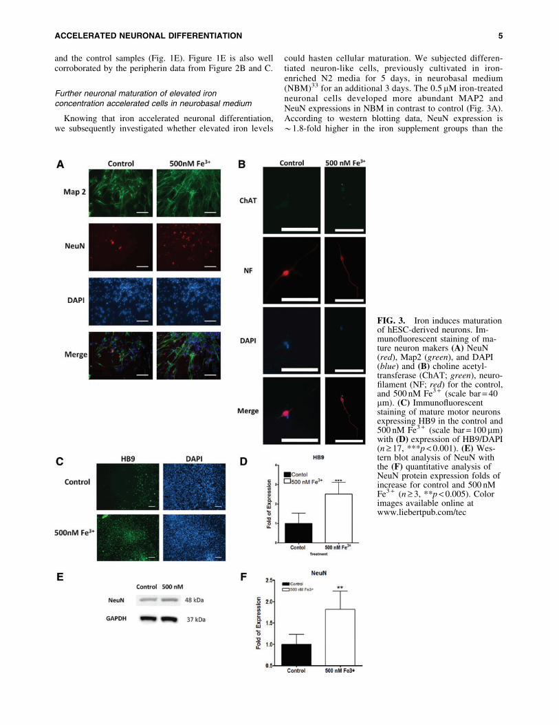
Further neuronal maturation of elevated ironconcentration accelerated cells in neurobasal medium
Knowing that iron accelerated neuronal differentiation,we subsequently investigated whether elevated iron levels
could hasten cellular maturation. We subjected differen-tiated neuron-like cells, previously cultivated in iron-enriched N2 media for 5 days, in neurobasal medium(NBM)33 for an additional 3 days. The 0.5 mM iron-treatedneuronal cells developed more abundant MAP2 andNeuN expressions in NBM in contrast to control (Fig. 3A).According to western blotting data, NeuN expression is*1.8-fold higher in the iron supplement groups than the
FIG. 3. Iron induces maturationof hESC-derived neurons. Im-munofluorescent staining of ma-ture neuron makers (A) NeuN(red), Map2 (green), and DAPI(blue) and (B) choline acetyl-transferase (ChAT; green), neuro-filament (NF; red) for the control,and 500 nM Fe3 + (scale bar = 40mm). (C) Immunofluorescentstaining of mature motor neuronsexpressing HB9 in the control and500 nM Fe3 + (scale bar = 100mm)with (D) expression of HB9/DAPI(n ‡ 17, ***p < 0.001). (E) Wes-tern blot analysis of NeuN withthe (F) quantitative analysis ofNeuN protein expression folds ofincrease for control and 500 nMFe3 + (n ‡ 3, **p < 0.005). Colorimages available online atwww.liebertpub.com/tec
ACCELERATED NEURONAL DIFFERENTIATION 5

control (Fig. 3E, F). Our data indicated that higher ironconcentrations fostered maturation of differentiated neuronswithin 8 days of differentiation. With establishment of iron-enriched acceleration in neuronal differentiation and matu-ration, we further investigated whether it preferentiallypromoted a motor neuron lineage. After cultured in NBMfor 3 days, cells previously treated with 0.5 mM iron werepositively stained with biomarkers, including ChAT, NF,and HB9,8 characteristics of matured motor neurons (Fig.3B–D). Quantitative immunofluorescence measurement ofHB9 expression also unveiled *2.5-fold upregulation iniron-enriched cultures comparing with the control (Fig. 3D).
Neuronal functional assay
With Figure 4A showing positive colocalized Synapsin Iand b-III tubulin expression, our next step examined thefunctionality of the derived motor neurons with FM 1–43dye to label active synaptic vesicles.36 High potassium (K + )buffer was used to depolarize motor neurons and facilitatedye uptake. Physiological relevant agonists such as ACh,glutamic acid, and high potassium depolarization bufferwere used to induce synaptic depolarization36 (Fig. 4B, C).
Synaptic depolarization stimulated with ACh, glutamic acid,and high K + was drastically apparent in the 0.5 mM Fe3 +
group. Our results suggest that higher iron levels led to moremature and functional motor neuron-like cells with activesynaptic vesicles.
Differential role of Fe3 + and Fe2 + in acceleratingneuronal differentiation
Both ferric and ferrous forms can coexist in nature and indifferentiation media. To explore the role of each ionicspecies that contributes to the acceleration, Fe3 + (0.5 mM),Fe2 + (0.5 mM), and the combination of the two (0.25 mM ofeach) were examined separately. Fe3 + treatment clearlyaccelerated axonal sprouting by *1.25-fold and enhancedaxonal density by *1.4-fold in comparison with the control(Fig. 5). On the contrary, Fe2 + seemed to foster inhibitoryeffects on axonal growth and slightly promoted axonal den-sity (Fig. 5). The combinatory treatment of Fe3 + and Fe2 +
substantially increased both sprouting density and axonallength (Fig. 5). Therefore, while both ionic species coexistin solution, Fe3 + ions played a more predominant role in theacceleration effect observed in this study.
FIG. 4. Functional proper-ties of hESC-derived moto-neuron. (A) Confocal imagesof synapsin I and b-III tubulincolocalize staining (scalebar = 40 mm). (B) Synapticvesicles loaded with FM1-43Dye of the control and 500 nMFe3 + (scale bar = 80 mm). (C)Cytocyolic fluorescence of thecontrol (red circle) and500 nM Fe3 + (blue circle)treatment groups loaded withFM1-43 dye and stimulatedwith 50 mM glutamic acid,200 mM acetylcholine, andpotassium (90 mM) solution toobserve cell synaptic re-cycling responses to differentnatural agonist (n ‡ 3,**p < 0.005). Photo bleachingwas tested to normalize dif-ferences (black circle). Colorimages available online atwww.liebertpub.com/tec
6 LU ET AL.

Reactive oxidative species involvementin iron-induced acceleration
We then analyzed the partake of reactive oxidative spe-cies (ROS)37 in determining accelerated differentiation.38
N-acetyl-L-cysteine (NAC; 1 mM) was added to Fe+ 3-treatedhESCs to block generation of ROS.39 We found NACtreatment was not able to reduce acceleration or differenti-ation (Fig. 6A). The result suggested that ROS might not beinvolved in accelerating differentiation and that an alterna-tive mechanism must be engaged. The data led to the pos-tulation of an iron-mediated neuronal differentiation.
Transferrin mediated iron-induced acceleration
We postulated that an iron directed mechanism maybe attributed to faster differentiation. By inhibiting granu-lar acidification with methylamine chloride, transferrin-
mediated iron intake can be blocked.34,40 Figure 6B showedthat blocking transferrin pathway significantly attenuatedaxonal sprouting and density (Fig. 3). LysoSensor GreenDND-189 (Invitrogen) was used to verify the effect ofmethylamine on vesicular acidification.34,41 Methylamineeffectively diminished the fluorescence of the LysoSensorGreen DND-189 by disrupting granular pH (Fig. 7). Ourdata suggested that accelerated neuronal differentiation isclearly related to a transferrin-mediated mechanism.
Discussion
Motor neuron diseases are a group of life-threateningneurological disorders that are characterized by progres-sive motor neuron degeneration.42–44 Recent studies havesuggested that stem cell-derived motor neuron transplantationmay be a promising therapy. This emergent strategy has been
FIG. 5. Immunofluorescent staining of (A) b-III tubulin for hESC-differentiated neurons with Fe2 + (0.5 mM), Fe3 +
(0.5 mM), and a combination of both solution (0.25 mM of Fe2 + /3 + respectfully) (scale bar = 200mm). Analysis of the (B)axonal length (mm) (n ‡ 100, ***p < 0.001) and (C) axonal density on varying iron concentrations. Color images availableonline at www.liebertpub.com/tec
FIG. 6. Reactive oxidativespecies and transferrin inhi-bition in iron induced hESC-derived neurons. (A) hESCcultured with N-acetyl-L-cysteine (NAC; 1 mM) inboth control and 500 nMFe3 + groups stained with b-III tubulin (green) (scalebar = 200mm) and (B) me-thylamine treated (100 mM)hESC expressing b-III tubu-lin (green) (scale bar = 80mm). Color images availableonline at www.liebertpub.com/tec
ACCELERATED NEURONAL DIFFERENTIATION 7

authenticated by solid in vivo efficacy and bears potentialclinical relevance. Despite the potential as an effectivetherapy, the usage of costly neurotrophic factors seriouslyrestricts motor neuron accessibility and expandability, andconsequently hampers translational capacity. Thus, a cost-effective derivation method is desperately needed. Ourprevious discoveries reported how biochemical and physicalcues could successfully promote neural differentiation. Herewe presented the unexpected finding that increasing ferricavailability to hESC hastened neuronal-specific motor neu-ron differentiation and enhanced the final yield.
To date, generation of motor neurons from hESC ne-cessitates a continuous incubation with multiple doses oftranscription activators and growth factors over a monthto express matured motor neuron markers, that is,HB9.5,8,10,45 Contrary to conventional methodology, ourinnovative protocol generated HB9 positive motor neu-rons in under 2 weeks, without RA or SHH, by increasingthe iron concentration. According to immunofluorescencestaining, 500 nM Fe3 + significantly raised HB9 expression(terminally differentiated motor neuron marker) by * 2.5-folds compared with control (Fig. 3C, D). Higher ferricconcentration also engendered more matured motor neuronmarkers including synapsin and FM1-43-positive vesicleswhen compared with control (Fig. 4A, B). Colocalizedstaining of Synapsin I with bIII tubulin and ChAT with NFfurther suggested the presence of neurotransmitter vesicles(Figs. 3B, 4A, and 4B). Besides the signature HB9 expres-sion, we further assessed the function of derived motorneurons. Figure 4C showed that 500 nM Fe3 + treatmentgroups were drastically more responsive to active synapticdepolarization than control, when stimulated with physio-logical agonists including ACh, glutamic acid, and high K + .Our findings are corroborated by previous studies showingthat iron could encourage proliferation and differentiation ofepidermal keratinocytes, dermal fibroblasts, and epidermaland dermal melanocytes in the skin of newborn mice.46 Inaddition, higher ferric concentrations instigated oligoden-drocyte differentiation from glial-restricted precursor cells.27
Interestingly, our data authenticated for the first time that
increasing iron availability accelerated motor neuron dif-ferentiation, maturity, and functionality.
To understand the mechanism involved in acceleratedmotor neuron differentiation, we investigated the rate ofneuronal differentiation from hESC under elevated ironconcentration. The iron concentration range used in thestudy (0.1–1mM) did not cause significant cellular toxicity(Figs. 1 and 2). By augmenting iron level in differentiationmedia, a concentration-dependent increase in bIII tubulinand peripherin expressions, axonal length, and density waswitnessed (Figs. 1 and 2) where most of the increase oc-curred relatively early between 5–8 days. Moreover, 500 nMFe3 + significantly amplified matured neuronal makers suchas NeuN and Map2 (Fig. 3A). These phenomena indicatedthat raising ferric concentration considerably acceleratedhESC differentiation by rapidly committing to neural line-age, which precedes motor neuron derivation. Among var-ious molecular mechanisms proposed, one of the prominentpathways involves generation of ROS and changing redoxstate of cells. Redox active iron may catalyze the formationof intracellular hydroxyl radicals through Fenton reaction inmany cells including epithelium and neurons.37,44,47–49 In-tracellular ROS level and signaling closely modulate earlyneural development and can promote O-2A progenitor cellsdifferentiating into oligodendrocytes.37,44,49,50 We surmisedthat the iron-instigated accelerated neuronal differentiationcould follow a similar pathway. Surprisingly, treatment withNAC, a common antioxidant, failed to inhibit acceleratedneuronal differentiation and subsequent motor neuron de-velopment (Fig. 6A). The evidence suggested that iron-mediated ROS does not play a major role in acceleratingneuronal differentiation and motor neuron maturation.Hence, an alternative mechanism may be involved.
We then hypothesized that iron could directly influenceneuronal differentiation via transferring-mediated pathway.After binding to iron, diferric holo-transferrin attaches to trans-ferrin receptor forming a complex, which is subsequently en-docytosed into early endosome.51 The acidic vesicular with pH5.5 then induces a conformational change between the complex,releasing iron from transferrin into vesicles.51 To validate the
FIG. 7. Differentiated hESCloaded with LysoSensor� GreenDND-189 to observed granular pHwithin the cell. The control and500 nM Fe3 + treatment groupswere treated with methylaminehydrochloride (100mM) to observethe increase in granular pH, reduc-ing the florescence of the Lyso-Sensor Green DND-189 (scalebar = 12 mm). Color images avail-able online at www.liebertpub.com/tec
8 LU ET AL.

involvement of transferrin pathway, 500 nM Fe + treatment es-calated the number of low pH vesicles as indicated by thebrightly fluorescent spots (Fig. 7), implying increased formationof transferrin complex in endosomes.52,53 Raising the vesicularpH with methylamine, disturbed the transferrin mechanismand subsequently disabled neuronal differentiation and signifi-cantly eliminated axonal sprouting and motor neuron formation(Fig. 7). While there are other types of iron chelators/inhibitorsthat can block cellular iron uptake, methylamine is commonlyused to inhibit intercellular iron transport.34,54–56 Our results arefurther supported by the well-established transferrin-ferritin in-tracellular iron transport and storage mechanism.53 Additionalevidences showed that the predominant proteins, upregulatedduring neural stem cell differentiation, involved in iron storage(ferritin L chain) and cytoskeleton.57 In particular, ferritin canbind to microtubules, using them to transport and distribute cy-tosolic iron for further metabolism that is vital for cytoskeletalrearrangement.58 Ferritin is also associated with the secretion ofBDNF known to promote motor neuron differentiation.59,60
Since elevating extracellular iron concentration can increasecytosolic ferritin,61 it is implicative that increasing iron avail-ability accelerates neuronal differentiation of hESC into motorneuron by increasing ferritin and BDNF. Conversely, perinataliron deficiency in rats decreases BDNF and IGF levels, whichmay lead to reduced neuronal proliferation, differentiation,dendritic complexity, and impaired synaptic plasticity.23 Suchinsufficiencies place a developmental brake on neural structuresand underscore the importance of iron homeostasis in modulat-ing neurotrophic factors critical for early-life neural differenti-ation.23 It would be of interest to understand whether iron-accelerated hESC differentiation to motor neuron involves fer-ritin, BDNF, and IGF, and the possible downstream cellularsignaling pathways.
In addition to stimulating the secretion of growth factors,iron homeostasis is intimately coupled to energy metabolismand neural developments.62,63 Iron facilitates the generationof energy (ATP) by acting as a cofactor for cytochromes andFe-sulfur complexes during oxidative phosphorylation.27
High energy-dependent processes, such as neuronal differ-entiation, neurotransmission, and neuronal dendrite develop-ments, are significantly compromised under iron deprivation.64,65
Since transferrin-mediated iron transport may be a vitalmechanism, we analyzed transferrin’s role in shuttling ex-tracellular ferrous ions, ferric ions, and a mixture of both.Ferric iron was significantly more effective in acceleratingaxonal extension and density, and motor neuron differenti-ation than ferrous alone or mixture (Fig. 5). These resultslent support to the possible notion that transferrin transportsferric ions to hasten neuronal differentiation from hESCtoward motor neurons. Other reports aligned well with ourdiscovery documenting that ferrous ions can be more toxicto neuronal cells than ferric form by possibly inducingmembrane peroxidation through ROS,66 and it may reduceproliferation of differentiating neuroprogenitor cells.67
Moreover, ferric ionic state successfully promoted Schwanncell and osteoclast differentiation.68,69 These phenomenamay help explain why transferrin and ferric ions play in-dispensable roles in driving motor neuron differentiation.
Conclusion
In this study, we investigated the role of iron in motorneuron differentiation from hESC. We have demonstrated
that increasing the iron availability accelerated motor neu-ron-like differentiation and maturity. The H9-derived cellsdisplay some motor neuron characteristics and are func-tional that exhibits synaptic recycling capacities. This cost-effective protocol capitalizing on altering ionic concentrationssignificantly expedites the mass production of differentiatedneurons for patients in need of neuronal regenerative ther-apy and can be potentially modified to accelerate the yieldof multiple neuronal lineages.
Acknowledgments
This study was supported in part by grants from theMuscular Dystrophy Association (MDA) and Linkou ChangGung Memorial Hospital (CMRPD1C0031).
Disclosure Statement
No competing financial interests exist.
References
1. Shelton, F.N., and Reding, M.J. Effect of lesion location onupper limb motor recovery after stroke. Stroke 32, 107, 2001.
2. Arvidsson, A., Collin, T., Kirik, D., Kokaia, Z., and Lind-vall, O. Neuronal replacement from endogenous precursorsin the adult brain after stroke. Nature Med 8, 963, 2002.
3. Kondziolka, D., Wechsler, L., Goldstein, S., Meltzer, C.,Thulborn, K., Gebel, J., et al. Transplantation of culturedhuman neuronal cells for patients with stroke. Neurology55, 565, 2000.
4. Lindvall, O., and Kokaia, Z. Stem cells for the treatment ofneurological disorders. Nature 441, 1094, 2006.
5. Hu, B.Y., and Zhang, S.C. Differentiation of spinal motorneurons from pluripotent human stem cells. Nat Protoc 4,1295, 2009.
6. Stacpoole, S.R., Bilican, B., Webber, D.J., Luzhynskaya,A., He, X.L., Compston, A., et al. Efficient derivation ofNPCs, spinal motor neurons and midbrain dopaminergicneurons from hESCs at 3% oxygen. Nat Protoc 6, 1229,2011.
7. Erceg, S., Ronaghi, M., and Stojkovic, M. Human embry-onic stem cell differentiation toward regional specificneural precursors. Stem Cells 27, 78, 2009.
8. Li, X.J., Du, Z.W., Zarnowska, E.D., Pankratz, M., Hansen,L.O., Pearce, R.A., et al. Specification of motoneuronsfrom human embryonic stem cells. Nat Biotechnol 23, 215–221, 2005.
9. Axell, M.Z., Zlateva, S., and Curtis, M. A method for rapidderivation and propagation of neural progenitors fromhuman embryonic stem cells. J Neurosci Methods 184, 275,2009.
10. Wichterle, H., Lieberam, I., Porter, J.A., and Jessell, T.M.Directed differentiation of embryonic stem cells into motorneurons. Cell 110, 385, 2002.
11. Gertz, C.C., Leach, M.K., Birrell, L.K., Martin, D.C.,Feldman, E.L., and Corey, J.M. Accelerated neuritogenesisand maturation of primary spinal motor neurons in responseto nanofibers. Dev Neurobiol 70, 589, 2010.
12. Vodouhe, C., Schmittbuhl, M., Boulmedais, F., Bagnard,D., Vautier, D., Schaaf, P., et al. Effect of functionalizationof multilayered polyelectrolyte films on motoneurongrowth. Biomaterials 26, 545, 2005.
13. Moolenaar, W.H., Tsien, R.Y., Van der Saag, P.T., and DeLaat, S.W. Na · /H · exchange and cytoplasmic pH in the
ACCELERATED NEURONAL DIFFERENTIATION 9

action of growth factors in human fibroblasts. Nature 304,645, 1983.
14. Quinn, D.A., Dahlberg, C.G., Bonventre, J.P., Scheid, C.R.,Honeyman, T., Joseph, P.M., et al. The role of Na + /H +exchange and growth factors in pulmonary artery smoothmuscle cell proliferation. Am J Respir Cell Mol Biol 14, 139,1996.
15. Hennings, H., Michael, D., Cheng, C., Steinert, P., Hol-brook, K., and Yuspa, S.H. Calcium regulation of growthand differentiation of mouse epidermal cells in culture. Cell19, 245, 1980.
16. Sage, H., Vernon, R.B., Funk, S.E., Everitt, E.A., andAngello, J. SPARC, a secreted protein associated withcellular proliferation, inhibits cell spreading in vitro andexhibits Ca + 2-dependent binding to the extracellularmatrix. J Cell Biol 109, 341, 1989.
17. MacDonald, R.S. The role of zinc in growth and cell pro-liferation. J Nutr 130, 1500S, 2000.
18. Beyersmann, D., and Haase, H. Functions of zinc in sig-naling, proliferation and differentiation of mammaliancells. Biometals 14, 331, 2001.
19. Hirobe, T. Ferrous ferric chloride stimulates the prolifera-tion of human skin keratinocytes, melanocytes, and fibro-blasts in culture. J Health Sci 55, 447, 2009.
20. Cazzola, M., Bergamaschi, G., Dezza, L., and Arosio, P.Manipulations of cellular iron metabolism for modulatingnormal and malignant cell proliferation: achievements andprospects. Blood 75, 1903, 1990.
21. Meguro, R., Asano, Y., Odagiri, S., Li, C., and Shoumura,K. Cellular and subcellular localizations of nonheme ferricand ferrous iron in the rat brain: a light and electron mi-croscopic study by the perfusion-Perls and -Turnbullmethods. Arch Histol Cytol 71, 205, 2008.
22. Sadrzadeh, S.M., and Saffari, Y. Iron and brain disorders.Am J Clin Pathol 121 Suppl, S64, 2004.
23. Tran, P.V., Carlson, E.S., Fretham, S.J., and Georgieff,M.K. Early-life iron deficiency anemia alters neurotrophicfactor expression and hippocampal neuron differentiation inmale rats. J Nutr 138, 2495, 2008.
24. Li, Y., Kim, J., Buckett, P.D., Bohlke, M., Maher, T.J., andWessling-Resnick, M. Severe postnatal iron deficiency al-ters emotional behavior and dopamine levels in the pre-frontal cortex of young male rats. J Nutr 141, 2133, 2011.
25. O’Keeffe, S.T., Gavin, K., and Lavan, J.N. Iron status andrestless legs syndrome in the elderly. Age Ageing 23, 200,1994.
26. Krieger, J., and Schroeder, C. Iron, brain and restless legssyndrome. Sleep Med Rev 5, 277, 2001.
27. Todorich, B., Pasquini, J.M., Garcia, C.I., Paez, P.M., andConnor, J.R. Oligodendrocytes and myelination: the role ofiron. Glia 57, 467, 2009.
28. Cheng, A., Hou, Y., and Mattson, M.P. Mitochondria andneuroplasticity. ASN Neuro 2, e00045, 2010.
29. Chao, T.I., Xiang, S., Chen, C.S., Chin, W.C., Nelson, A.J.,Wang, C., et al. Carbon nanotubes promote neuron differ-entiation from human embryonic stem cells. BiochemBiophys Res Commun 384, 426, 2009.
30. Chen, E.Y.T., Wang, Y.-C., Mintz, A., Richards, A., Chen,C.-S., Lu, D., et al. Activated charcoal composite bioma-terial promotes human embryonic stem cell differentiationtoward neuronal lineage. J Biomed Mater Res Part A 100A,2006, 2012.
31. Zhang, S.C., Wernig, M., Duncan, I.D., Brustle, O., andThomson, J.A. In vitro differentiation of transplantable
neural precursors from human embryonic stem cells. NatBiotechnol 19, 1129, 2001.
32. Pankratz, M.T., Li, X.J., LaVaute, T.M., Lyons, E.A.,Chen, X., and Zhang, S.C. Directed neural differentiationof human embryonic stem cells via an obligated primitiveanterior stage. Stem Cells 25, 1511, 2007.
33. Nguyen, T., Mehta, N.R., Conant, K., Kim, K.-J., Jones,M., Calabresi, P.A., et al. Axonal protective effects of themyelin-associated glycoprotein. J Neurosci 29, 630, 2009.
34. Swaiman, K.F., and Machen, V.L. Iron uptake by mam-malian cortical neurons. Ann Neurol 16, 66, 1984.
35. Dyson, S.E., and Jones, D.G. Synaptic remodelling duringdevelopment and maturation: junction differentiation andsplitting as a mechanism for modifying connectivity. DevBrain Res 13, 125, 1984.
36. Ryan, T.A., Reuter, H., Wendland, B., Schweizer, F.E.,Tsien, R.W., and Smith, S.J. The kinetics of synaptic ves-icle recycling measured at single presynaptic boutons.Neuron 11, 713, 1993.
37. Zecca, L., Youdim, M.B.H., Riederer, P., Connor, J.R., andCrichton, R.R. Iron, brain ageing and neurodegenerativedisorders. Nat Rev Neurosci 5, 863, 2004.
38. Suzukawa, K., Miura, K., Mitsushita, J., Resau, J., Hirose,K., Crystal, R., et al. Nerve growth factor-induced neuronaldifferentiation requires generation of Rac1-regulated reac-tive oxygen species. J Biol Chem 275, 13175, 2000.
39. Zafarullah, M., Li, W.Q., Sylvester, J., and Ahmad, M.Molecular mechanisms of N-acetylcysteine actions. CellMol Life Sci 60, 6, 2003.
40. Qian, Z.M., and Shen, X. Brain iron transport and neuro-degeneration. Trends Mol Med 7, 103, 2001.
41. Barg, S., Huang, P., Eliasson, L., Nelson, D.J., Obermuller,S., Rorsman, P., et al. Priming of insulin granules forexocytosis by granular Cl- uptake and acidification. J CellSci 114, 2145, 2001.
42. Leigh, P.N., and Ray-Chaudhuri, K. Motor neuron disease.J Neurol Neurosurg Psychiatry 57, 886, 1994.
43. Mulder, D.W., Kurland, L.T., Offord, K.P., and Beard,C.M. Familial adult motor neuron disease: amyotrophiclateral sclerosis. Neurology 36, 511, 1986.
44. Altamura, S., and Muckenthaler, M.U. Iron toxicity indiseases of aging: Alzheimer’s disease, Parkinson’s diseaseand atherosclerosis. J Alzheimer Dis 16, 879, 2009.
45. Miles, G.B., Yohn, D.C., Wichterle, H., Jessell, T.M., Ra-fuse, V.F., and Brownstone, R.M. Functional propertiesof motoneurons derived from mouse embryonic stem cells.J Neurosci 24, 7848, 2004.
46. Hirobe, T. Ferrous ferric chloride induces the differentia-tion of cultured mouse epidermal melanocytes additionallywith herbal medicines. J Health Sci 55, 86, 2009.
47. Dringen, R., Bishop, G.M., Koeppe, M., Dang, T.N., andRobinson, S.R. The pivotal role of astrocytes in the metab-olism of iron in the brain. Neurochem Res 32, 1884, 2007.
48. Britton, R.S., Leicester, K.L., Bacon BR. Iron toxicity andchelation therapy. Int J Hematol 76, 219, 2002.
49. Todorich, B., Zhang, X., and Connor, J.R. H-ferritin is themajor source of iron for oligodendrocytes. Glia 59, 927,2011.
50. Smith, J., Ladi, E., Mayer-Proschel, M., and Noble, M.Redox state is a central modulator of the balance betweenself-renewal and differentiation in a dividing glial precursorcell. Proc Natl Acad Sci U S A 97, 10032, 2000.
51. Zhang, D., Lee, H.F., Pettit, S.C., Zaro, J.L., Huang, N., andShen, W.C. Characterization of transferrin receptor-mediated
10 LU ET AL.

endocytosis and cellular iron delivery of recombinant humanserum transferrin from rice (Oryza sativa L.). BMC Bio-technol 12, 92, 2012.
52. Dautry-Varsat, A., Ciechanover, A., and Lodish, H.F. pHand the recycling of transferrin during receptor-mediatedendocytosis. Proc Natl Acad Sci U S A 80, 2258, 1983.
53. Dautry-Varsat, A. Receptor-mediated endocytosis: the in-tracellular journey of transferrin and its receptor. Biochimie68, 375, 1986.
54. Paterson, S., Armstrong, N.J., Iacopetta, B.J., McArdle,H.J., and Morgan, E.H. Intravesicular pH and iron uptakeby immature erythroid cells. J Cell Physiol 120, 225, 1984.
55. Levi, A., Shechter, Y., Neufeld, E.J., and Schlessinger, J.Mobility, clustering, and transport of nerve growth factor inembryonal sensory cells and in a sympathetic neuronal cellline. Proc Natl Acad Sci U S A 77, 3469, 1980.
56. Bomford, A., Young, S.P., and Williams, R. Release of ironfrom the two iron-binding sites of transferrin by culturedhuman cells: modulation by methylamine. Biochemistry24, 3472, 1985.
57. Skalnikova, H., Halada, P., Vodicka, P., Motlik, J., Re-hulka, P., Horning, O., et al. A proteomic approach tostudying the differentiation of neural stem cells. Proteomics7, 1825, 2007.
58. Hasan, M.R., Koikawa, S., Kotani, S., Miyamoto, S., andNakagawa, H. Ferritin forms dynamic oligomers to asso-ciate with microtubules in vivo: implication for the role ofmicrotubules in iron metabolism. Exp Cell Res 312, 1950,2006.
59. Schonberg, D.L., Goldstein, E.Z., Sahinkaya, F.R., Wei, P.,Popovich, P.G., and McTigue, D.M. Ferritin stimulatesoligodendrocyte genesis in the adult spinal cord and canbe transferred from macrophages to NG2 cells in vivo.J Neurosci 32, 5374, 2012.
60. Koliatsos, V.E., Clatterbuck, R.E., Winslow, J.W., Cayouette,M.H., and Price, D.L. Evidence that brain-derived neuro-trophic factor is a trophic factor for motor neurons in vivo.Neuron 10, 359, 1993.
61. Casey, J.L., Hentze, M.W., Koeller, D.M., Caughman,S.W., Rouault, T.A., Klausner, R.D., et al. Iron-responsiveelements: regulatory RNA sequences that control mRNA
levels and translation. Science (New York, NY) 240,924, 1988.
62. Rouault, T.A. The role of iron regulatory proteins inmammalian iron homeostasis and disease. Nat Chem Biol2, 406, 2006.
63. Hentze, M.W., Muckenthaler, M.U., and Andrews, N.C.Balancing acts: molecular control of mammalian iron me-tabolism. Cell 117, 285, 2004.
64. Beard, J.L. Iron biology in immune function, muscle me-tabolism and neuronal functioning. J Nutr 131, 568S, 2001.
65. Benarroch, E.E. Brain iron homeostasis and neurodegen-erative disease. Neurology 72, 1436, 2009.
66. Singh, A.V., Vyas, V., Montani, E., Cartelli, D., Parazzoli,D., Oldani, A., et al. Investigation of in vitro cytotoxicityof the redox state of ionic iron in neuroblastoma cells.J Neurosci Rural Pract 3, 301, 2012.
67. Mazur-Kolecka, B., Cohen, I.L., Jenkins, E.C., Flory, M.,Merz, G., Ted Brown, W., et al. Sera from children withautism alter proliferation of human neuronal progenitorcells exposed to oxidation. Neurotox Res 16, 87, 2009.
68. Salis, C., Davio, C., Usach, V., Urtasun, N., Goitia, B.,Martinez-Vivot, R., et al. Iron and holotransferrin inducecAMP-dependent differentiation of Schwann cells. Neu-rochem Int 61, 798, 2012.
69. Jia, P., Xu, Y.J., Zhang, Z.L., Li, K., Li, B., Zhang, W.,et al. Ferric ion could facilitate osteoclast differentiationand bone resorption through the production of reactiveoxygen species. J Orthop Res 30, 1843, 2012.
Address correspondence to:Wei-Chun Chin, PhD
Bioengineering ProgramSchool of Engineering
University of California5200 North Lake Road
Merced, CA 95343
E-mail: [email protected]
Received: December 3, 2013Accepted: July 10, 2014
Online Publication Date: August 22, 2014
ACCELERATED NEURONAL DIFFERENTIATION 11