accuracy and precision evaluation of international
TRANSCRIPT
Research ArticleAccuracy and Precision Evaluation of International StandardSpherical Model by Digital Dental Scanners
Hong Xin Cai,1 Qi Jia,1 HaoYu Shi,1 Yujie Jiang,1 Jingnan Xue,1 ChunXu Chen,1
Haotian Gong,1 Jie Liu,1 Eui-Seok Lee ,2 and Heng Bo Jiang 1
1Stomatological Materials Laboratory, School of Stomatology, Shandong First Medical University & Shandong Academy ofMedical Sciences, Tai’an, Shandong 271016, China2Department of Oral and Maxillofacial Surgery, Graduate School of Clinical Dentistry, Korea University,Seoul 08308, Republic of Korea
Correspondence should be addressed to Eui-Seok Lee; [email protected] and Heng Bo Jiang; [email protected]
Received 10 June 2020; Revised 30 July 2020; Accepted 12 August 2020; Published 9 December 2020
Guest Editor: Ying Zhao
Copyright © 2020 Hong Xin Cai et al. This is an open access article distributed under the Creative Commons Attribution License,which permits unrestricted use, distribution, and reproduction in any medium, provided the original work is properly cited.
With the popularization of digital technology and the exposure of traditional technology’s defects, computer-aided design andcomputer-aided manufacturing (CAD/CAM) has been widely used in the field of dentistry. And the accuracy of the scanningsystem determines the ultimate accuracy of the prosthesis, which is a very important part of CAD/CAM, so we decided toevaluate the accuracy of the intraoral and extraoral scanners. In this study, we selected the sphere model as the scanning objectand obtained the final result through data analysis and 3D fitting. In terms of trueness and precision, the scanner of SHININGwas significantly different from that of others; however, there was no significant difference between TRIOS and CEREC.SHINING showed the lowest level of accuracy, with CEREC slightly lower than TRIOS. The sphere model has also been provento be scanned successfully.
1. Introduction
With the emergence of digitalization, CAD/CAM systems [1]have found an increasingly wide utilization in the field ofprosthodontics on account of its considerable strengths [2].Given the fact that the traditional prostheses gradually failto meet the needs of patients, the use of intraoral [3] andextraoral scanners in dentistry is becoming more and morecommon [4, 5].
By using the specific and intuitive model presented fromintraoral and extraoral scanners [6–9], we can immediatelyobtain detailed information of the patient’s oral cavity andits digital files. And the files will be imported into the com-puter to complete the design and production [10]. The extra-oral scanner scans the impression model, and the intraoralscanner directly scans the patient’s oral cavity [7]. Comparedto the traditional technology, the scanning technology isundoubtedly time-saving and efficient [11, 12].
There are many factors that affect the scanning resultsand data collection. Recent studies have shown that differentscanning strategies can affect the accuracy of results [13]. Ithas been reported that different scanning systems also con-tribute to different experimental results [14]. In addition,the selection of the impression materials and operation timewill affect the accuracy of extraoral scanners, while the scan-ning range, light [15], and oral tissue will affect intraoralscanners [16, 17].
Considering the above factors, it is of clinical significanceto evaluate the accuracy of intraoral and extraoral scanners[9]. The early Flügge’s study claimed that the extraoral scan-ners performed better than intraoral scanners under the oralenvironment [13]. However, as the algorithms and scanningtechniques evolve, Tomita et al. concluded that the intraoralscanners had higher accuracy than extraoral scanners as theywere studying their self-manufactured denture modelin vitro [18]. Further, the performance of the intraoral
HindawiScanningVolume 2020, Article ID 1714642, 6 pageshttps://doi.org/10.1155/2020/1714642

scanner is significantly influenced by the geometry of thescanned object, for the fact that it accumulates the scannedimage and records the scanning path of the object to obtainthe complete image. Especially, the uniformity of the modelwill also reduce the performance of the intraoral scanner.Therefore, this study is based on an international standardmodel to evaluate the accuracy of intraoral and extraoral scan-ners [19, 20].
After looking up a lot of literature, we found that therewere few papers based on the sphere model [19], so thisexperiment adopted the ISO standard sphere model [21].According to the definition of accuracy regulated in ISO5725 including trueness and precision, the trueness refersto the degree of uniformity between a measurement resultand the reference value, while the latter one maintains theuniformity between independent measurement results.According to the standard in ISO 3290-2, we chose asphere with Grade 20 to conduct the experiment. And theerror of diameter and surface less than 0.001mm can beignored in this grade [22].
2. Materials and Methods
2.1. Fabrication of Models. According to the clinical practice,the theoretical value of the diameter of the sphere modelbased on the ISO 12836 [21] was set as 8mm, as shown inFigure 1(a). The 3D file of the model was drawn bycomputer-aided design software (AutoCAD 2018, Autodesk,USA), which was exported in stereolithography (STL)
Sphere
Simulated root
(a)
(b)
Figure 1: Design drawings of the (a) sphere model, and (b) opticalimage.
0Experimental groups
0.010.020.030.040.050.060.070.08
Abso
lute
mea
n de
viat
ion
0.09
SHINING
ISO-Sphere Model-trueness
CERECTRIOS
(a)
ISO-Sphere Model-Precision
00.010.020.030.040.050.060.070.08
Abso
lute
mea
n de
viat
ion
0.090.1
Experimental groups
SHININGCERECTRIOS
(b)
Figure 2: Boxplots of absolute mean trueness (a) and precisionvalues (b) of the sphere model. The horizontal line of eachBoxplot from top to bottom represents the upper edge, upperquartile, median, lower quartile, and lower edge of the value,respectively, in addition the round dots represent outliers, and thecrosses represent averages.
Table 1: Mean trueness/precision values ± standard deviation (SD).
Test group Trueness (mm) Precision (mm)
SHINING 0:061 ± 0:018 0:059 ± 0:023
CEREC 0:010 ± 0:010 0:014 ± 0:010
TRIOS 0:006 ± 0:003 0:006 ± 0:003True value 7.908
Table 2: Relative errors of the trueness and precision of the spheremodel (diameter).
Test group Trueness (mm) Precision (mm)
SHINING 0.008 0.008
CEREC 0.002 0.002
TRIOS 0.001 0.001
2 Scanning
format. The test model was made of stainless steel, and weused computer numerical controlled (CNC) milling to fabri-cate it. Moreover, the model was sandblasted with a powdersize of 80μm as specified by the international standard.
2.2. Creation of Gold Standard Values. Thirty measurementsof the sphere model’s diameter were taken by Vernier Cali-pers, the average of which was taken as the truth value in thispaper. One of the measurements was shown in Figure 1(b).The true value was set as the gold standard for evaluation,and the 3D file based on the value was created as the goldstandard file for 3D fitting.
2.3. Scanning Process. One extraoral scanner (SHININGD200+, CHN) and two intraoral scanners (CEREC ACD3492, Sirona, GER and TRIOS T12A, 3Shape, DEN),respectively, performed 30 scans of the sphere model andsaved them as STL files. The model was scanned by the sameskilled technician who followed the scanning method recom-mended by the different instrument manufacturers to elimi-nate interference and improve feasibility [7].
2.4. Data Acquisition and ImageMatching. The scanned orig-inal files were converted into STL files, which were thenimported into a reverse engineering software (GeomagicControl X 2018, 3D SYSTEMS, USA). The test indicator(diameter) was measured and recorded through the software,and the 3D files of test groups were compared with the goldstandard files for 3D fitting. Meanwhile, root mean squareerror (RMSE) values were recorded.
2.5. Mathematical Analysis. The formula for accuracy assess-ment is as follows [23]:
Trueness = dR − dMð Þj j, ð1Þ
Precision = dA − dMð Þj j, ð2ÞΔdM = dR − dMð Þ/dRj j, ð3Þ
ΔS dMð Þ = S dMð Þ/dRj j,dR :The standard reference value for the
diameter of themodel,
dM : Measured value for the diameter of themodel,
dA :Average of themeasured value forthe diameter of themodel,
ΔdM : Relative error of trueness,
ΔS dMð Þ : Relative error of precision,S dMð Þ : Standard deviation of themeasured
value for the diameter of themodel:ð4Þ
Perform statistical analysis on scanned data using SPSSv.20.0 (IBM, USA). The data conform to a normal distribu-tion, but they are not conformable in the homogeneity ofthe variance. The Nonparametric Kruskal-Wallis test ana-lyzed the difference in parameters and the result of p < 0:01attested statistically significant differences.
3. Results
3.1. Absolute Mean Trueness and Precision of the SphereModel. The value of absolute mean trueness and precisionof the sphere model is enumerated in Table 1. Figure 2(a)shows boxplots of the absolute mean trueness values, andFigure 2(b) shows boxplots of the absolute mean precision
7.87.827.847.867.88
7.97.927.947.967.98
8
1 2 3 4 5 6 7 8 9 10 11 12 13 14 15 16 17 18 19 20 21 22 23 24 25 26 27 28 29 30
Dia
met
er (m
m)
Scanning times
SHININGCEREC
TRIOSGOLDEN STANDARD
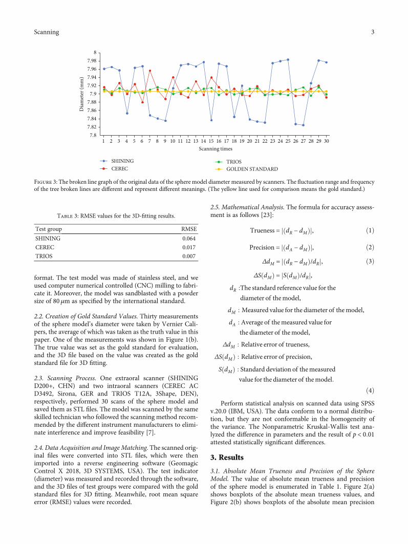
Figure 3: The broken line graph of the original data of the sphere model diameter measured by scanners. The fluctuation range and frequencyof the tree broken lines are different and represent different meanings. (The yellow line used for comparison means the gold standard.)
Table 3: RMSE values for the 3D-fitting results.
Test group RMSE
SHINING 0.064
CEREC 0.017
TRIOS 0.007
3Scanning
values of the sphere model. Regarding diameter errors of thesphere model, the TRIOS reveals the highest trueness andprecision, respectively. The comparison between theintraoral scanners and the extraoral scanners shows thatcompared to the CEREC and SHINING, the mean truenessvalue of TRIOS is much lower. The mean precision value ofTRIOS is significantly lower than CEREC and SHINING.
3.2. Relative Errors of Sphere Model. Table 2 shows the rela-tive errors of the trueness and precision of the sphere model.The relative errors of the sphere model are less than0.008mm, respectively. This is an ideal result that when com-paring the relative errors of trueness and precision betweenthese scanners, the trend is similar to that of the absolutemean deviation of trueness and precision.
3.3. Broken Line Graph and RMSE. The broken line graph ofthe original data of the sphere model diameter measured byscanners is shown in Figure 3. The yellow line is the goldstandard. It is obvious that SHINING’s values are not con-centrated and far from the yellow line. We found that thevalues of CEREC and TRIOS were very close to the gold stan-dard, also highly centralized and accurately combined withRMSE in Table 3.
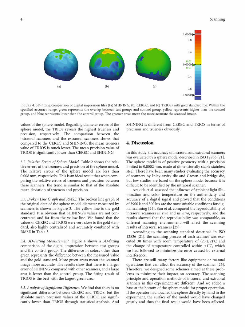
3.4. 3D-Fitting Measurement. Figure 4 shows a 3D-fittingcomparison of the digital impression between test groupsand the control group. The difference in colors other thangreen represents the difference between the measured valueand the gold standard. More green areas mean the scannedimage more accurate. The results show that there is a largererror of SHINING compared with other scanners, and a largearea is lower than the control group. The fitting result ofTRIOS is the best with the largest green area.
3.5. Analysis of Significant Difference.We find that there is nosignificant difference between CEREC and TRIOS, but theabsolute mean precision values of the CEREC are signifi-cantly lower than TRIOS through statistical analysis. And
SHINING is different from CEREC and TRIOS in terms ofprecision and trueness obviously.
4. Discussion
In this study, the accuracy of intraoral and extraoral scannerswas evaluated by a sphere model described in ISO 12836 [21].The sphere model is of positive geometry with a precisionlimited to 0.0002mm, made of dimensionally stable stainlesssteel. There have been many studies evaluating the accuracyof scanners by Inlay-cavity die and Grown-and-bridge die,but few studies are based on the sphere model because it isdifficult to be identified by the intraoral scanner.
Arakida et al. assessed the influence of ambient light illu-mination and color temperature on the authenticity andaccuracy of a digital signal and proved that the conditionsof 3900 k and 500 lux are the most suitable conditions for dig-ital scanning [24]. Sun et al. compared the reproducibility ofintraoral scanners in vivo and in vitro, respectively, and theresults showed that the reproducibility was comparable, sodifferent scanning environment will affect the scanningresults of intraoral scanners [25].
According to the scanning standard described in ISO12836 [21], the scanning process of each scanner was exe-cuted 30 times with room temperature of (23 ± 2)°C andthe change of temperature controlled within ±1°C, whichwe had followed to minimize the error caused by externalinterference.
There are still many factors like equipment or manualoperations that can affect the accuracy of the scanner [26].Therefore, we designed some schemes aimed at these prob-lems to minimize their impact on accuracy. The scanningprinciple and operation methods of intraoral and extraoralscanners in this experiment are different. And we added abase at the bottom of the sphere model for proper operation.If the operator had touched the sphere directly by hand in theexperiment, the surface of the model would have changedgreatly and thus the final result would have been affected.
–1.0000
(a) (b) (c)–0.8
–0.4
0.0000
0.4
0.8
1.0000
–0.01
0.01
Figure 4: 3D-fitting comparison of digital impression files ((a) SHINING, (b) CEREC, and (c) TRIOS) with gold standard file. Within thespecified accuracy range, green represents the overlap between test groups and control group, yellow represents higher than the controlgroup, and blue represents lower than the control group. The greener areas mean the more accurate the scanned image.
4 Scanning
Thus, it is necessary to add the base which can also fix thesphere model to make scanning easier.
Reproducibility and repeatability are also importantcomponents of the evaluation [27]. Reproducibility can bedefined as the consistency of the measured results of the sameobject under different conditions, while repeatability isaffected by operating, timing, equipment, etc. Therefore, thisexperiment was carried out by a skilled operator in strictaccordance with the methods specified by the scanner manu-facturers to reduce the influence of operation.
There is no significant difference between values obtainedin the experiment and the standard values created from themodel, so it is feasible to scan the sphere model with thesethree scanners.
There are limitations to our experiments as well. The longoperation of the scanner may result in the degradation of theperformance, which may affect the scanning results of the lat-ter part. And the choice of a single model has some limita-tions in the evaluation of scanners. In terms of the clinic,due to the complex oral environment, like saliva, blood,and soft tissue, there will be deviations in the actual operation[28, 29]. Additionally, the level of analysis and measurementsoftware used also affects the results.
According to the study of Burzynski et al., with the prog-ress of intraoral scanning technology, the shrinking of thecamera, and the acceleration of acquisition time, patientsmay show a greater preference for digital impression [30].Through investigation, Ahmed et al. found that with thedevelopment of digitization and the improvement of scanneraccuracy, people’s acceptance of using CAD/CAM for oraldisease treatment has gradually increased [31]. Alghazzawiargued that the coming trend for most practitioners wouldbe the use of an acquisition camera attached to a computerthat was equipped with appropriate software and the capabil-ity of forwarding the image to the laboratory [32].
Our experiment compared one extraoral scanner (SHIN-ING) with two intraoral scanners (CEREC, TRIOS), whoseresults showed that the accuracy of CEREC was betweenthe other two. It indicated that the intraoral scanners werebetter than the extraoral one. Since no controversy wasshown in the results of this experiment, the results of clinicaltrials of the extraoral and intraoral scanners need furtherdiscussion.
5. Conclusions
The accuracy of the intraoral scanners in this experimentwas greater than that of the extraoral scanner. The intraoralscanner has certain reliability in practical application and isworth to trial in clinics. The sphere model is achievable as ascanning object, but it requires further research andimprovement.
Data Availability
The data used to support the findings of this study areincluded within the article.
Conflicts of Interest
We declare that we have no financial and personal relation-ships with other people or organizations that can inappropri-ately influence our work.
Authors’ Contributions
HongXin Cai, Qi Jia, and HaoYu Shi contributed equally tothis work.
Acknowledgments
The authors thank the CONVERSATIONALIST club inShandong First Medical University School of Stomatology.This study was funded by the Shandong First Medical Uni-versity & Shandong Academy of Medical Sciences of PhDStart-up Project (Jiang-161839).
References
[1] M. J. Peluso, S. D. Josell, S. W. Levine, and B. J. Lorei, “Digitalmodels: an introduction,” Seminars in Orthodontics, vol. 10,no. 3, pp. 226–238, 2004.
[2] M. Santoro, S. Galkin, M. Teredesai, O. F. Nicolay, and T. J.Cangialosi, “Comparison of measurements made on digitaland plaster models,” American Journal of Orthodontics andDentofacial Orthopedics, vol. 124, no. 1, pp. 101–105, 2003.
[3] M. Imburgia, S. Logozzo, U. Hauschild, G. Veronesi,C. Mangano, and F. G. Mangano, “Accuracy of four intraoralscanners in oral implantology: a comparative in vitro study,”BMC Oral Health, vol. 17, no. 1, p. 92, 2017.
[4] R. Nedelcu, P. Olsson, I. Nyström, J. Rydén, and A. Thor,“Accuracy and precision of 3 intraoral scanners and accuracyof conventional impressions: a novel in vivo analysis method,”Journal of Dentistry, vol. 69, pp. 110–118, 2018.
[5] S. Shimizu, A. Shinya, S. Kuroda, and H. Gomi, “The accuracyof the cad system using intraoral and extraoral scanners fordesigning of fixed dental prostheses,” Dental Materials Jour-nal, vol. 36, no. 4, pp. 402–407, 2017.
[6] L. O. L. Bohner, G. De Luca Canto, B. S. Marció, D. C. Laganá,N. Sesma, and P. T. Neto, “Computer-aided analysis of digitaldental impressions obtained from intraoral and extraoral scan-ners,” The Journal of Prosthetic Dentistry, vol. 118, no. 5,pp. 617–623, 2017.
[7] G. H. Park, K. Son, and K. B. Lee, “Feasibility of using anintraoral scanner for a complete-arch digital scan,” The Jour-nal of Prosthetic Dentistry, vol. 121, no. 5, pp. 803–810, 2019.
[8] A. P. Keating, J. Knox, R. Bibb, and A. I. Zhurov, “A compar-ison of plaster, digital and reconstructed study model accu-racy,” Journal of Orthodontics, vol. 35, no. 3, pp. 191–201,2008.
[9] S. B. Patzelt, A. Emmanouilidi, S. Stampf, J. R. Strub, andW. Att, “Accuracy of full-arch scans using intraoral scanners,”Clinical Oral Investigations, vol. 18, no. 6, pp. 1687–1694,2014.
[10] S. Logozzo, E. M. Zanetti, G. Franceschini, A. Kilpelä, andA. Mäkynen, “Recent advances in dental optics – part i: 3dintraoral scanners for restorative dentistry,” Optics and Lasersin Engineering, vol. 54, pp. 203–221, 2014.
5Scanning
[11] S. B. Patzelt, S. Bishti, S. Stampf, and W. Att, “Accuracy ofcomputer-aided design/computer-aided manufacturing-generated dental casts based on intraoral scanner data,” Jour-nal of the American Dental Association, vol. 145, no. 11,pp. 1133–1140, 2014.
[12] R. van Noort, “The future of dental devices is digital,” DentalMaterials, vol. 28, no. 1, pp. 3–12, 2012.
[13] T. V. Flugge, S. Schlager, K. Nelson, S. Nahles, andM. C. Metz-ger, “Precision of intraoral digital dental impressions withitero and extraoral digitization with the itero and a modelscanner,” American Journal of Orthodontics and DentofacialOrthopedics, vol. 144, no. 3, pp. 471–478, 2013.
[14] D. T. Chandran, D. C. Jagger, R. G. Jagger, and M. E. Barbour,“Two- and three-dimensional accuracy of dental impressionmaterials: effects of storage time andmoisture contamination,”Bio-medical Materials and Engineering, vol. 20, no. 5, pp. 243–249, 2010.
[15] B. Giménez, M. Özcan, F. Martínez-Rus, and G. Pradíes,“Accuracy of a digital impression system based on active trian-gulation technology with blue light for implants: effect of clin-ically relevant parameters,” Implant Dentistry, vol. 24, no. 5,pp. 498–504, 2015.
[16] T. Grunheid, S. D. McCarthy, and B. E. Larson, “Clinical use ofa direct chairside oral scanner: an assessment of accuracy,time, and patient acceptance,” American Journal of Orthodon-tics and Dentofacial Orthopedics, vol. 146, no. 5, pp. 673–682,2014.
[17] J. Maeng, Y. J. Lim, B. Kim, M.-J. Kim, and H.-B. Kwon, “Anew approach to accuracy evaluation of single-tooth abutmentusing two-dimensional analysis in two intraoral scanners,”International Journal of Environmental Research and PublicHealth, vol. 16, no. 6, article 1021, 2019.
[18] Y. Tomita, J. Uechi, M. Konno, S. Sasamoto, M. Iijima, andI. Mizoguchi, “Accuracy of digital models generated by con-ventional impression/plaster-model methods and intraoralscanning,” Dental Materials Journal, vol. 37, no. 4, pp. 628–633, 2018.
[19] F. Emir and S. Ayyildiz, “Evaluation of the trueness and preci-sion of eight extraoral laboratory scanners with a complete-arch model: a three-dimensional analysis,” Journal of Prostho-dontic Research, vol. 63, no. 4, pp. 434–439, 2019.
[20] J. L. Porter, C. K. Carrico, S. J. Lindauer, and E. Tüfekçi, “Com-parison of intraoral and extraoral scanners on the accuracy ofdigital model articulation,” Journal of Orthodontics, vol. 45,no. 4, pp. 275–282, 2018.
[21] International Standard (ISO) 12836, Dentistry —DigitizingDevices for CAD/CAM Systems for Indirect Dental Restora-tions— Test Methods for Assessing Accuracy, InternationalOrganization for Standardization, Geneva, Switzerland, 2ndedition, 2015.
[22] R. G. Nedelcu and A. S. Persson, “Scanning accuracy and pre-cision in 4 intraoral scanners: an in vitro comparison based on3-dimensional analysis,” The Journal of Prosthetic Dentistry,vol. 112, no. 6, pp. 1461–1471, 2014.
[23] ADA/ANSI Standard No. 132, Scanning Accuracy of DentalChairside and Laboratory CAD/CAM System, American Den-tal Association, Chicago, IL, USA, 2015.
[24] T. Arakida, M. Kanazawa, M. Iwaki, T. Suzuki, andS. Minakuchi, “Evaluating the influence of ambient light onscanning trueness, precision, and time of intra oral scanner,”Journal of Prosthodontic Research, vol. 62, no. 3, pp. 324–329, 2018.
[25] L. Sun, J. S. Lee, H. H. Choo, H. S. Hwang, and K. M. Lee,“Reproducibility of an intraoral scanner: a comparisonbetween in-vivo and ex-vivo scans,” American Journal ofOrthodontics and Dentofacial Orthopedics, vol. 154, no. 2,pp. 305–310, 2018.
[26] P. Gonzalez de Villaumbrosia, F. Martinez-Rus, A. Garcia-Orejas, M. P. Salido, and G. Pradíes, “In vitro comparison ofthe accuracy (trueness and precision) of six extraoral dentalscanners with different scanning technologies,” The Journalof Prosthetic Dentistry, vol. 116, no. 4, pp. 543–550.e1, 2016.
[27] H. B. Jacob, G. D. Wyatt, and P. H. Buschang, “Reliability andvalidity of intraoral and extraoral scanners,” Progress in Ortho-dontics, vol. 16, no. 1, p. 38, 2015.
[28] J. Abduo and M. Elseyoufi, “Accuracy of intraoral scanners: asystematic review of influencing factors,” The European Jour-nal of Prosthodontics and Restorative Dentistry, vol. 26, no. 3,pp. 101–121, 2018.
[29] F. S. Andriessen, D. R. Rijkens, W. J. van der Meer, and D. W.Wismeijer, “Applicability and accuracy of an intraoral scannerfor scanning multiple implants in edentulous mandibles: apilot study,” The Journal of Prosthetic Dentistry, vol. 111,no. 3, pp. 186–194, 2014.
[30] J. A. Burzynski, A. R. Firestone, F. M. Beck, H. W. Fields Jr.,and T. Deguchi, “Comparison of digital intraoral scannersand alginate impressions: time and patient satisfaction,” Amer-ican Journal of Orthodontics and Dentofacial Orthopedics,vol. 153, no. 4, pp. 534–541, 2018.
[31] K. E. Ahmed, T. Wang, K. Y. Li, W. K. Luk, and M. F. Burrow,“Performance and perception of dental students using threeintraoral CAD/CAM scanners for full-arch scanning,” Journalof Prosthodontic Research, vol. 63, no. 2, pp. 167–172, 2019.
[32] T. F. Alghazzawi, “Advancements in CAD/CAM technology:options for practical implementation,” Journal of Prosthodon-tic Research, vol. 60, no. 2, pp. 72–84, 2016.
6 Scanning