ace personal trainer manual, 4th...
TRANSCRIPT
1
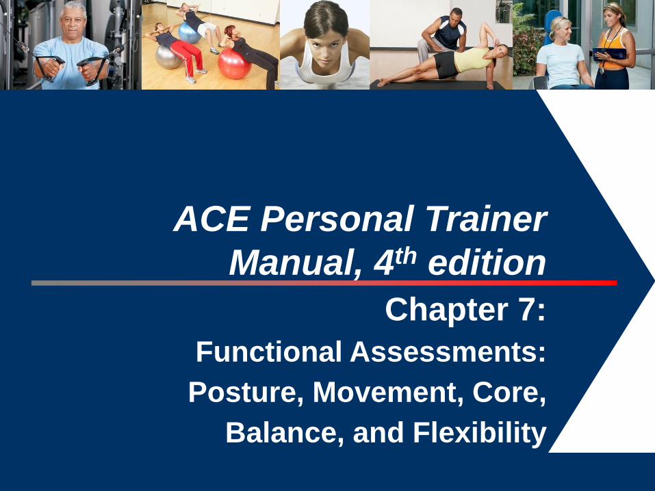
ACE Personal Trainer
Manual, 4th edition
Chapter 7:
Functional Assessments:
Posture, Movement, Core,
Balance, and Flexibility

Learning Objectives
This session, which is based on Chapter 7 of the ACE
Personal Trainer Manual (4th ed.), explains the
importance of various functional assessments and
outlines how to properly perform each.
After completing this session, you will have a better
understanding of:
– How to set up a plumb line to conduct a basic postural
assessment.
– How to identify five key postural deviations.
– How to conduct various movement screens, including clearing
tests.
– How to conduct key flexibility (muscle-length) assessments.
– How to evaluate balance and core function.
Introduction
Sequencing a client’s assessments involves
consideration of protocol selection and timing of the
assessments.
The physiological assessments must be consistent with
the client’s goals and desires, and with the discoveries
made during the needs assessment.
One primary objective of all training programs should be
to improve functionality (movement efficiency).
Movement Efficiency
Movement efficiency is the ability to generate
appropriate levels of force and movement at desired
joints while stabilizing the entire kinetic chain against
reactive and gravity-based forces.
– All movement begins and ends from a static base, ideally a
position where all body segments are optimally aligned.
– Since movement originates from this base, a postural
assessment should be conducted to evaluate body-segment
alignment.
– Additionally, movement screens that evaluate how posture
impacts the ability to move should be incorporated.

Static Posture
Static posture represents the alignment of the body’s
segments.
– Holding a proper postural position involves the actions of
postural muscles.
Good posture is a state of musculoskeletal alignment
that allows muscles, joints, and nerves to function
efficiently.
– If a client exhibits poor static posture, this may reflect muscle-
endurance issues in the postural muscles and/or potential
imbalances at the joints.
Since movement begins from a position of static posture,
the presence of poor posture is an indicator that
movement may be dysfunctional.
Static Postural Assessment
A static postural assessment may offer valuable insight
into:
– Muscle imbalance at a joint and the working relationships of
muscles around a joint
– Altered neural action of the muscles moving and controlling the
joint
– Potentially dysfunctional movement
Tight or shortened muscles are often overactive and
dominate movement at the joint, potentially disrupting
healthy joint mechanics.
– Personal trainers should consider conducting a static postural
assessment on their clients as an initial assessment.

Muscle Imbalance and
Postural Deviation Factors
Muscle imbalance and postural deviations can be attributed to many
factors that are both correctible and non-correctible.
Correctible factors:
– Repetitive movements
– Awkward positions and movements
– Side dominance
– Lack of joint stability or mobility
– Imbalanced strength-training programs
Non-correctible factors:
– Congenital conditions
– Some pathologies
– Structural deviations
– Certain types of trauma

Neural Activity
Proper postural alignment promotes optimal neural activity of the
muscles controlling a joint.
– When joints are correctly aligned, the length-tension relationships and
force-coupling relationships function efficiently.
– Good posture facilitates proper joint mechanics.
Muscle
Balance Normal Length-Tension Relationship
Normal Force-coupling
Relationships
Proper Joint Mechanics
(Arthrokinematics)
Efficient Force
Acceptance and
Generation Promotes Joint Stability
and Joint Mobility Movement Efficiency
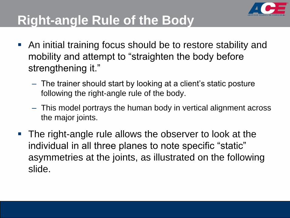
Right-angle Rule of the Body
An initial training focus should be to restore stability and
mobility and attempt to “straighten the body before
strengthening it.”
– The trainer should start by looking at a client’s static posture
following the right-angle rule of the body.
– This model portrays the human body in vertical alignment across
the major joints.
The right-angle rule allows the observer to look at the
individual in all three planes to note specific “static”
asymmetries at the joints, as illustrated on the following
slide.
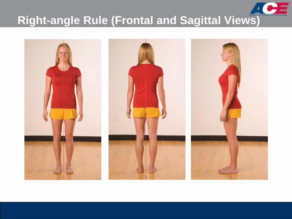
Right-angle Rule (Frontal and Sagittal Views)

Line of Gravity
Good posture is observed when the body parts are
symmetrically balanced around the body’s line of gravity.
– While the right-angle rule can identify potential muscle
imbalances, there are limitations in using this model.
Line of Gravity

Plumb Line Instructions
The objective of this assessment is to observe the
client’s symmetry against the plumb line.
– Using a length of string and an inexpensive weight, trainers can
create a plumb line that suspends from the ceiling to a height 0.5
to 1 inch (1.3 to 2.5 cm) above the floor.
– A solid, plain backdrop or a grid pattern with vertical and
horizontal lines that offer contrast against the client is
recommended.
– Clients should assume a normal, relaxed position.
– Personal trainers should focus on the obvious, gross imbalances
and avoid getting caught up in minor postural asymmetries.

Plumb Line Positions: Anterior View
For the anterior view, position the client between the plumb line and
a wall.
With good posture, the plumb line will pass equidistant between the
feet and ankles, and intersect the:
– Pubis
– Umbilicus
– Sternum
– Manubrium
– Mandible (chin)
– Maxilla (face)
– Frontal bone (forehead)
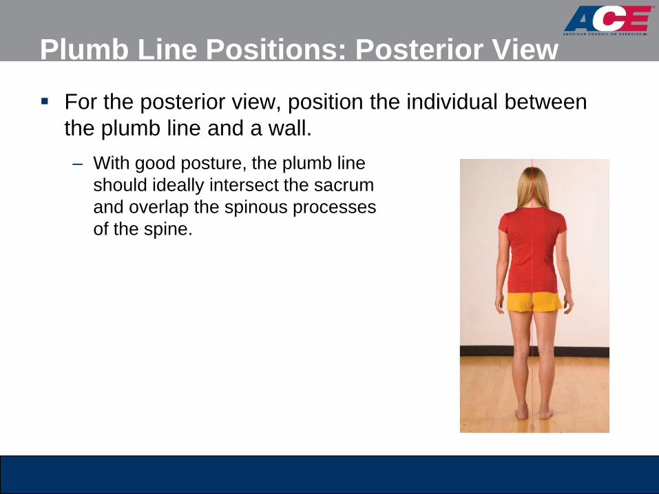
Plumb Line Positions: Posterior View
For the posterior view, position the individual between
the plumb line and a wall.
– With good posture, the plumb line
should ideally intersect the sacrum
and overlap the spinous processes
of the spine.

Plumb Line Positions:
Sagittal/Transverse Views
Position the individual between the plumb line and the wall, with the
plumb line aligned immediately anterior to the lateral malleolus.
With good posture, the plumb line should ideally pass through:
– The anterior third of the knee
– The greater trochanter of the femur
– The acromioclavicular (A-C) joint
– Slightly anterior to the mastoid process of
the temporal bone of the skull
All transverse views of the limbs and torso
are performed from frontal- and
sagittal-plane positions.
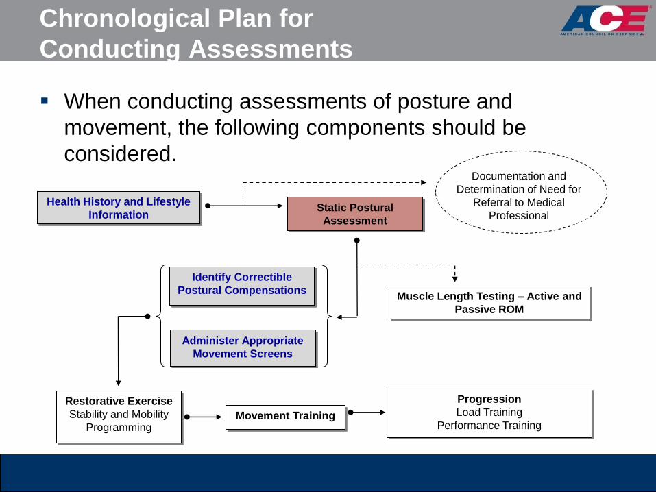
Chronological Plan for
Conducting Assessments
When conducting assessments of posture and
movement, the following components should be
considered.
Health History and Lifestyle
Information Static Postural
Assessment
Muscle Length Testing – Active and
Passive ROM
Administer Appropriate
Movement Screens
Restorative Exercise
Stability and Mobility
Programming
Progression
Load Training
Performance Training
Documentation and
Determination of Need for
Referral to Medical
Professional
Identify Correctible
Postural Compensations
Movement Training

Deviation 1: Ankle Pronation/Supination
Both feet should face forward in parallel
or with slight (8 to 10 degrees) external
rotation.
– Toes pointing outward from the midline,
as the ankle joint lies in an oblique plane
with the medial malleolus slightly anterior
to the lateral malleolus
The toes should be aligned in the same
direction as the feet.

Ankle Pronation and Tibial
and Femoral Rotation
The body is one continuous kinetic chain.
Barring structural differences in the skeletal system, a
pronated ankle typically forces internal rotation of the
tibia and faster, greater internal rotation of the femur.
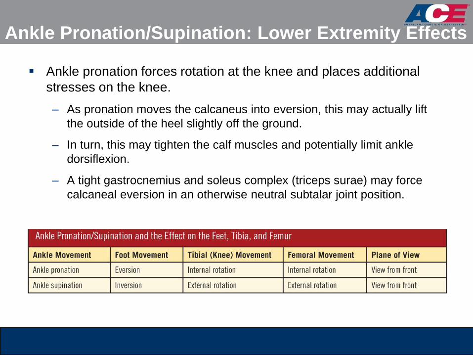
Ankle Pronation/Supination: Lower Extremity Effects
Ankle pronation forces rotation at the knee and places additional
stresses on the knee.
– As pronation moves the calcaneus into eversion, this may actually lift
the outside of the heel slightly off the ground.
– In turn, this may tighten the calf muscles and potentially limit ankle
dorsiflexion.
– A tight gastrocnemius and soleus complex (triceps surae) may force
calcaneal eversion in an otherwise neutral subtalar joint position.
Deviation 2: Hip Adduction
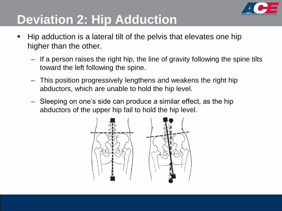
Hip adduction is a lateral tilt of the pelvis that elevates one hip
higher than the other.
– If a person raises the right hip, the line of gravity following the spine tilts
toward the left following the spine.
– This position progressively lengthens and weakens the right hip
abductors, which are unable to hold the hip level.
– Sleeping on one’s side can produce a similar effect, as the hip
abductors of the upper hip fail to hold the hip level.

Alignment of the Pelvis
Relative to the Plumb Line
To evaluate the presence of hip adduction with a client, a
personal trainer must identify the alignment of the pelvis
relative to the plumb line.

Hip Adduction Screen
The plumb line should pass through:
– The pubis in the anterior view
– The middle of the sacrum in the posterior view
Positioning a dowel or lightly weighted bar across the
iliac crests can help determine whether the iliac crests
are parallel with the floor.

Deviation 3: Hip Tilting (Anterior or Posterior)
Anterior tilting of the pelvis frequently occurs in individuals with tight
hip flexors.
– With standing, a shortened hip flexor pulls the pelvis into an anterior tilt.
– An anterior pelvic tilt rotates the superior, anterior portion of the pelvis
forward and downward.
– A posterior tilt rotates the superior, posterior portion of the pelvis
backward and downward.

Pelvic Rotation An anterior pelvic tilt will increase lordosis in the lumbar spine,
whereas a posterior pelvic tilt will reduce the amount of lordosis in
the lumbar spine.
– Tight hip flexors are generally coupled with tight erector spinae muscles,
producing an anterior pelvic tilt.
– Tight rectus abdominis muscles are generally coupled with tight
hamstrings, producing a posterior pelvic tilt.
– This coupling relationship between tight hip flexors and erector spinae is
defined as the lower-cross syndrome.
– With ankle pronation and accompanying internal femoral rotation, the
pelvis may tilt anteriorly to better accommodate the head of the femur.

Pelvic Tilt Screen: ASIS and PSIS
To evaluate the presence of a pelvic tilt, a trainer can
use a consensus of four techniques:
– The relationship of the anterior superior iliac spine (ASIS) and
the posterior superior iliac spine (PSIS) (two bony landmarks on
the pelvis)
– The appearance of lordosis in the lumbar spine
– The alignment of the pubic
bone to the ASIS
– The degree of flexion or
hyperextension in the knees
Deviation 4: Shoulder Position
and Thoracic Spine
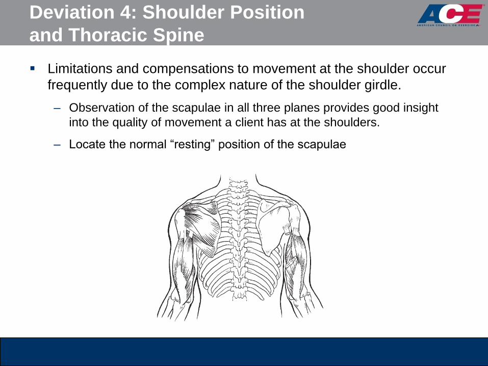
Limitations and compensations to movement at the shoulder occur
frequently due to the complex nature of the shoulder girdle.
– Observation of the scapulae in all three planes provides good insight
into the quality of movement a client has at the shoulders.
– Locate the normal “resting” position of the scapulae

Shoulder Screen: Level Shoulders
Determine whether the shoulders are level.
– If the shoulders are not level, trainers need to identify potential
reasons.
Shoulders: Torso/Shoulders
Relative to Line of Gravity
Determine whether the torso and shoulders are symmetrical relative
to the line of gravity.
– A torso lean would shift the alignment of the sternum and spine away
from the plumb line and create tightness on the flexed side of the trunk.
– However, if the hips are level with the floor and the spine is aligned with
the plumb line, but the shoulders are not level with the floor, this may
represent muscle imbalance within the shoulder complex itself.
– An elevated shoulder may present with an overdeveloped or tight upper
trapezius muscle.
– A depressed shoulder may present with more forward rounding of the
scapula.
– The shoulder on a person’s dominant side may hang lower than the
non-dominant side.

Shoulders: Rotation of the
Scapulae and/or Arms
Determine whether the scapulae and/or arms are internally rotated.
Anterior view
– If the knuckles or the backs of the client’s hands are visible when the
hands are positioned at the sides, this generally indicates internal rotation
of the humerus or scapular protraction.
Posterior view – If the vertebral/inferior angles of the scapulae protrude
outward, it indicates an inability of the scapulae
stabilizers to hold the scapulae in place.
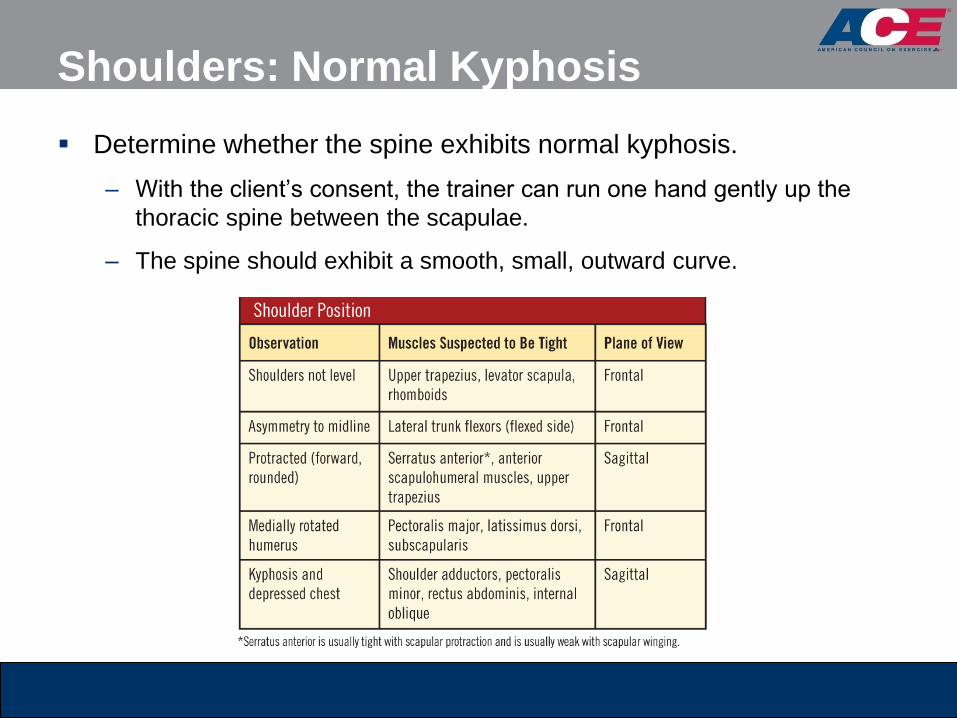
Shoulders: Normal Kyphosis
Determine whether the spine exhibits normal kyphosis.
– With the client’s consent, the trainer can run one hand gently up the
thoracic spine between the scapulae.
– The spine should exhibit a smooth, small, outward curve.

Deviation 5: Head Position
With good posture, the earlobe should align approximately over the
acromion process.
A forward-head position is very common.
– This altered position does not tilt the head downward, but simply shifts it
forward.
– The earlobe appears significantly forward of the acromioclavicular (AC)
joint.

Forward-head Position Screen
In the sagittal view, align the plumb
line with the AC joint, and observe its
position relative to the ear.
A forward-head position represents
tightness in the cervical extensors and
lengthening of the cervical flexors.
With good posture, the cheek bone
and the collarbone should almost be in
vertical alignment with each other.

Movement Screens
Observing active movement is an effective method to
identify movement compensations.
When compensations occur, it is indicative of altered
neural action.
These compensations normally manifest due to muscle
tightness or an imbalance between muscles acting at the
joint.

Five Primary Movements
Movement can essentially be broken down and
described by five primary movements that people
perform during many daily activities:
– Bending/raising and lifting/lowering movements (e.g., squatting)
– Single-leg movements
– Pushing movements
– Pulling movements
– Rotational movements
ADL are essentially the integration of one or more of
these primary movements.
Movement Screens and the Kinetic Chain
Movement screens must be skill- and conditioning-level
appropriate, and be specific to the client’s needs.
– Screens generally challenge clients with no recognized
pathologies to perform basic movements.
– This can help the personal trainer evaluate a client’s stability and
mobility throughout the entire kinetic chain.
Clearing Tests
Prior to administering any movement screens, trainers
should screen for pain by using basic clearing tests.
– These tests may uncover issues that the individual did not know
existed.
– Trainers should select clearing tests according to the
movements that require evaluation.
– The objective when conducting clearing tests is to ensure that
pain is not exacerbated by movement.
Any client who exhibits pain during a clearing test
should:
– Be referred to his or her physician
– Not perform additional assessments for that part of the body

Clearing Test: Cervical Spine
The client performs the following movements in a seated
position while the personal trainer monitors for any
indication of pain:
– Move the chin to touch the chest.
– Tilt the head back until the face lies approximately parallel or
near parallel to the floor.
– Drop the chin left and right to rest on, or within 1 inch (2.5 cm) of,
the shoulder or collarbone.

Clearing Test: Shoulder Impingement
The client performs the following movement in a seated
position while the personal trainer monitors for any
indication of pain:
– Reach one arm across the chest to rest upon the opposite
shoulder and slowly elevate the elbow as high as possible.

Clearing Test: Low Back
The client performs the following movements from a prone position
while the personal trainer monitors for any indication of pain:
– Slowly move into a trunk-extension position, producing lumbar
extension and compression in the vertebrae and shoulder joint.
– Move into a quadruped position and slowly sit back on the heels with
outstretched arms, producing lumbar and hip flexion.
Bend and Lift Screen: Objective
To examine symmetrical lower-extremity mobility and
stability, and upper-extremity stability during a bend-and-
lift movement
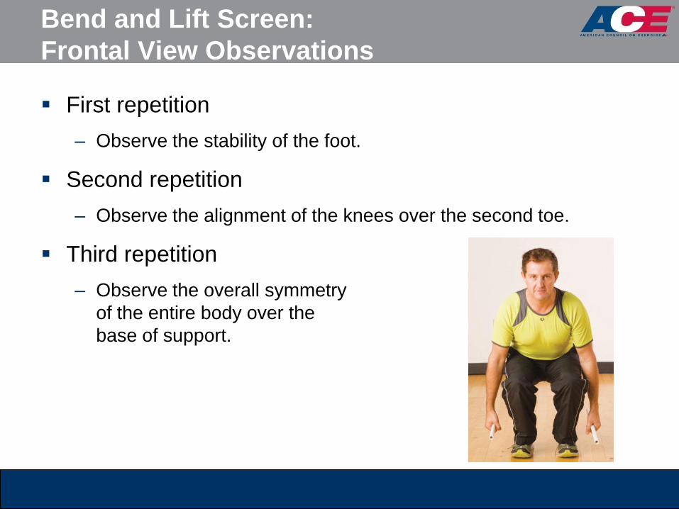
Bend and Lift Screen:
Frontal View Observations
First repetition
– Observe the stability of the foot.
Second repetition
– Observe the alignment of the knees over the second toe.
Third repetition
– Observe the overall symmetry
of the entire body over the
base of support.
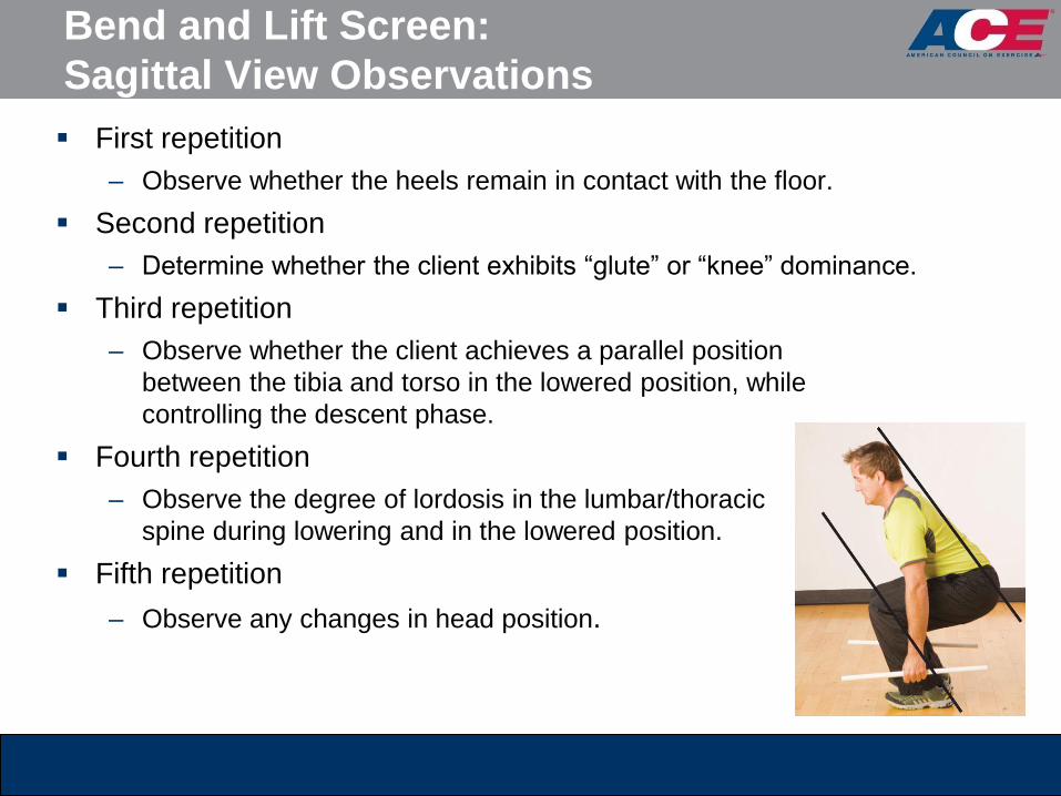
Bend and Lift Screen:
Sagittal View Observations
First repetition
– Observe whether the heels remain in contact with the floor.
Second repetition
– Determine whether the client exhibits “glute” or “knee” dominance.
Third repetition
– Observe whether the client achieves a parallel position
between the tibia and torso in the lowered position, while
controlling the descent phase.
Fourth repetition
– Observe the degree of lordosis in the lumbar/thoracic
spine during lowering and in the lowered position.
Fifth repetition
– Observe any changes in head position.

Bend and Lift Screen:
Potential Compensations

Hurdle Step Screen: Objective
To examine simultaneous mobility of one limb and
stability of the contralateral limb while maintaining both
hip and torso stabilization under a balance challenge of
standing on one leg

Hurdle Step Screen: Frontal View Observations
First repetition
– Observe the stability of the foot.
Second repetition
– Observe the alignment of the stance-leg knee over the foot.
Third repetition
– Watch for excessive hip adduction greater than 2 inches (5.1 cm) as
measured by excessive stance-leg adduction or downward hip-tilting
toward the opposite side.
Fourth repetition
– Observe the stability of the torso.
Fifth repetition
– Observe the alignment
of the moving leg.

Hurdle Step Screen:
Sagittal View Observations
First repetition
– Observe the stability of the torso and stance leg.
Second repetition
– Observe the mobility of the hip.
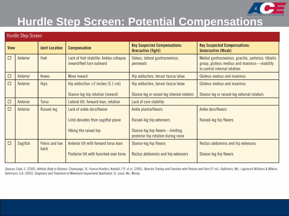
Hurdle Step Screen: Potential Compensations
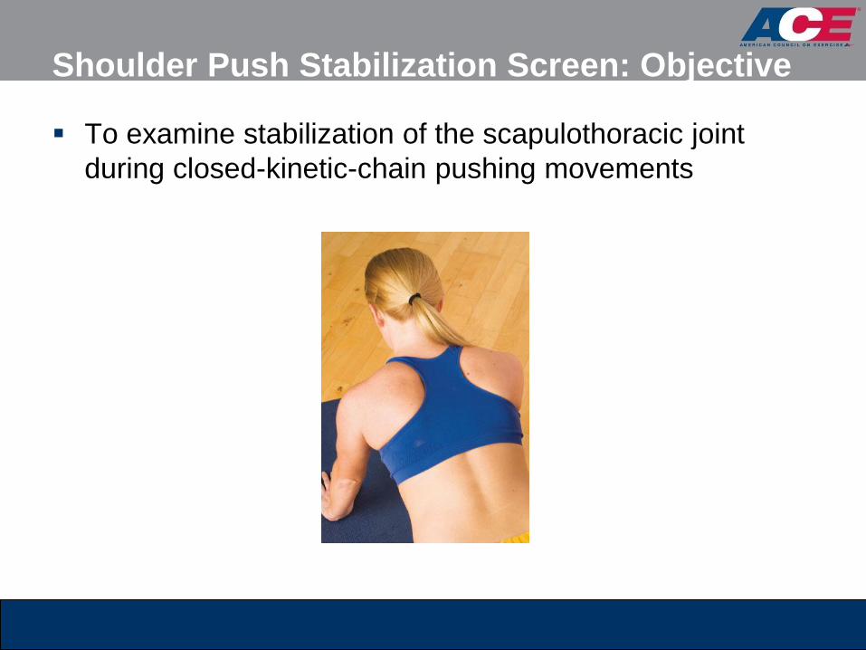
Shoulder Push Stabilization Screen: Objective
To examine stabilization of the scapulothoracic joint
during closed-kinetic-chain pushing movements

Shoulder Push Stabilization Screen:
Observations
Observe any notable changes in the position of the
scapulae relative to the ribcage at both end-ranges of
motion.
Observe for lumbar hyperextension in the press position.

Should Push Screen: Potential Compensations
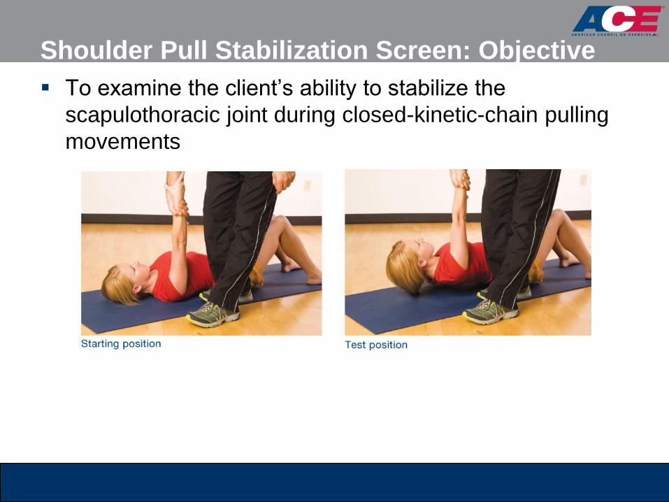
Shoulder Pull Stabilization Screen: Objective
To examine the client’s ability to stabilize the
scapulothoracic joint during closed-kinetic-chain pulling
movements
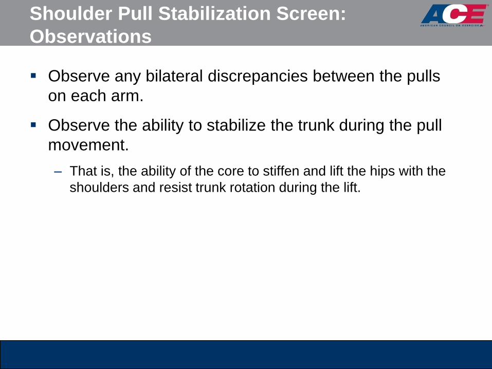
Shoulder Pull Stabilization Screen:
Observations
Observe any bilateral discrepancies between the pulls
on each arm.
Observe the ability to stabilize the trunk during the pull
movement.
– That is, the ability of the core to stiffen and lift the hips with the
shoulders and resist trunk rotation during the lift.
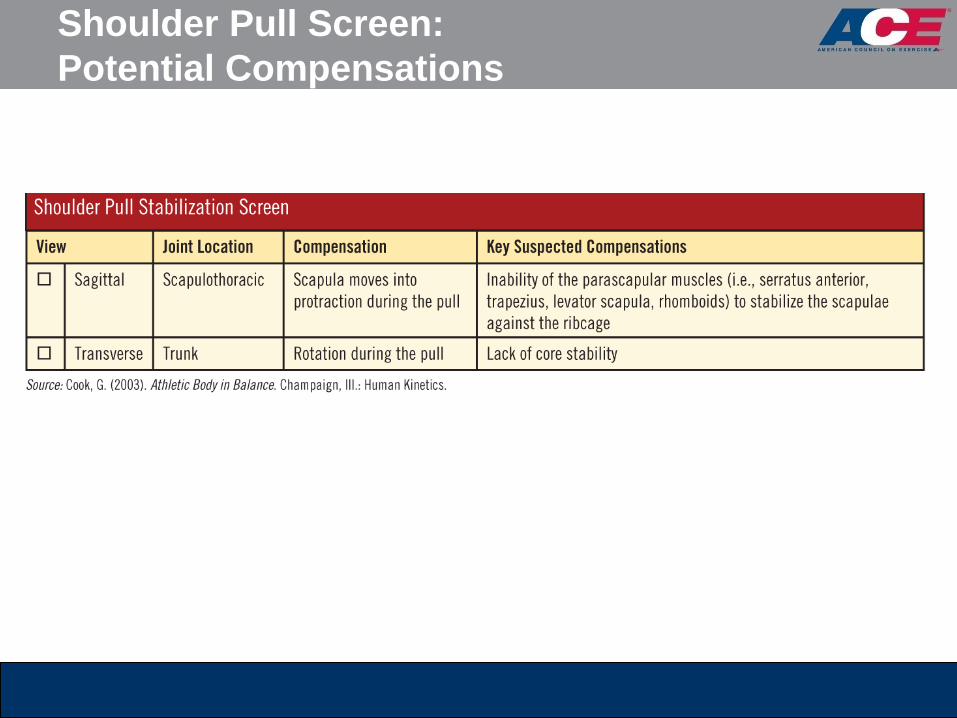
Shoulder Pull Screen:
Potential Compensations

Thoracic Spine Mobility Screen: Objective
To examine bilateral mobility of the thoracic spine
Lumbar spine rotation is considered insignificant, as it
only offers approximately 15 degrees of rotation.
T-Spine Mobility Screen: General Interpretations
Observe any bilateral discrepancies between the
rotations in each direction.
– Identify the origin(s) of movement limitation or compensation.
– This screen evaluates trunk rotation in the transverse plane.
– Evaluate the impact on the entire kinetic chain.
– The lumbar spine generally exhibits limited rotation of
approximately 15 degrees, with the balance of trunk rotation
occurring through the thoracic spine.
– If thoracic spine mobility is limited, the body strives to gain
movement in alternative planes within the lumbar spine.

Thoracic Spine Screen:
Potential Compensations
Flexibility and Muscle-length Testing
A personal trainer may opt to assess the flexibility of
specific muscle groups.
Specific muscle groups that frequently demonstrate
tightness or limitations to movement are discussed in
this section.
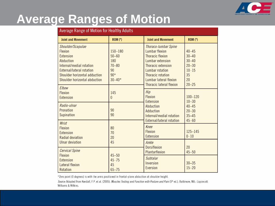
The table on the following slide provides normal ranges
of motion for healthy adults at each joint.
Average Ranges of Motion
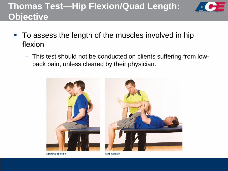
Thomas Test—Hip Flexion/Quad Length:
Objective
To assess the length of the muscles involved in hip
flexion
– This test should not be conducted on clients suffering from low-
back pain, unless cleared by their physician.

Thomas Test—Hip Flexion/Quad Length:
Observations
Observe whether the back of the lowered thigh touches
the table (hips positioned in 10 degrees of extension).
Observe whether the knee of the lowered leg achieves
80 degrees of flexion.
Observe whether the knee remains aligned straight or
falls into internal or external rotation.

Thomas Test: General Interpretations

Passive Straight-leg (PSL) Raise: Objective
To assess the length of the hamstrings

Passive Straight-leg (PSL) Raise: Observations
Note the degree of movement attained from the table or
mat that is achieved before the spine compresses the
hand under the low back or the opposite leg begins to
show visible signs of lifting off the table or mat.
– The mat or table represents 0 degrees.
– The leg perpendicular to the mat or table represents 90 degrees.

Passive Straight-leg Raise:
General Interpretations
Shoulder Mobility Assessment
Apley’s scratch test involves multiple and simultaneous
movements of the scapulothoracic and glenohumeral
joints in all three planes.
– To identify the source of the limitation, trainers can first perform
various isolated movements in single planes to locate potentially
problematic movements.
Consequently, the scratch test is completed in
conjunction with:
– The shoulder flexion-extension test
– An internal-external rotation test of the humerus

Apley’s Scratch Test—Shoulder Mobility:
Objective
To assess simultaneous movements of the shoulder girdle (primarily
the scapulothoracic and glenohumeral joints)
Movements include:
– Shoulder extension and flexion
– Internal and external rotation of the humerus at the shoulder
– Scapular abduction and adduction
Apley’s Scratch Test—Shoulder Mobility:
Observations
Note the client’s ability to touch the medial border of the
contralateral scapula or how far down the spine he or
she can reach with shoulder flexion and external
rotation.
Note the client’s ability to touch the opposite inferior
angle of the scapula or how far up the spine he or she
can reach with shoulder extension and internal rotation.
Observe any bilateral differences between the left and
right arms in performing both movements.
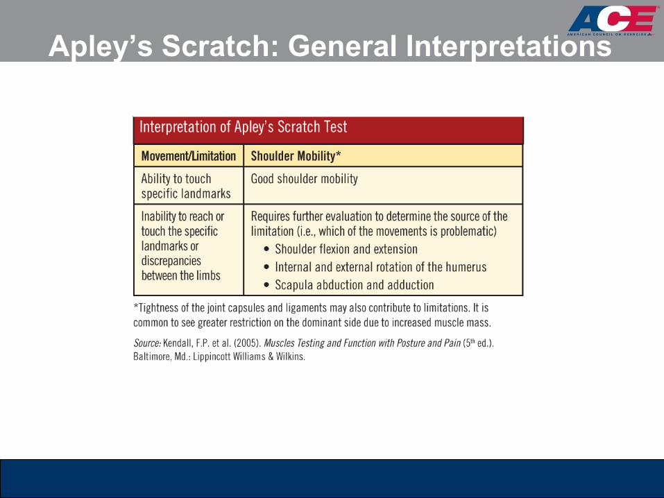
Apley’s Scratch: General Interpretations
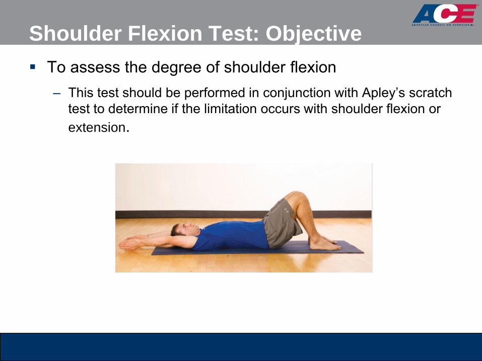
Shoulder Flexion Test: Objective
To assess the degree of shoulder flexion
– This test should be performed in conjunction with Apley’s scratch
test to determine if the limitation occurs with shoulder flexion or
extension.

Shoulder Extension Test: Objective
To assess the degree of shoulder extension
– This test should be performed in conjunction with Apley’s scratch
test to determine if the limitation occurs with shoulder flexion or
extension.
Shoulder Flexion/Extension Tests:
Observations
Measure the degree of movement in each direction.
Note any bilateral differences between the left and right
arms in performing both movements.

Shoulder Flexion/Extension:
General Interpretations
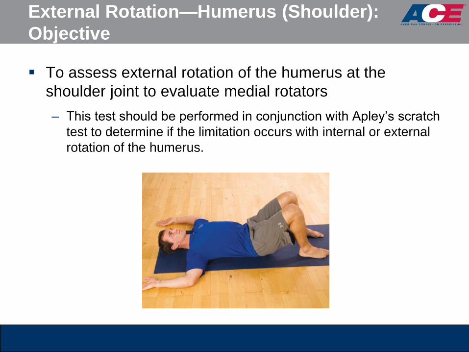
External Rotation—Humerus (Shoulder):
Objective
To assess external rotation of the humerus at the
shoulder joint to evaluate medial rotators
– This test should be performed in conjunction with Apley’s scratch
test to determine if the limitation occurs with internal or external
rotation of the humerus.
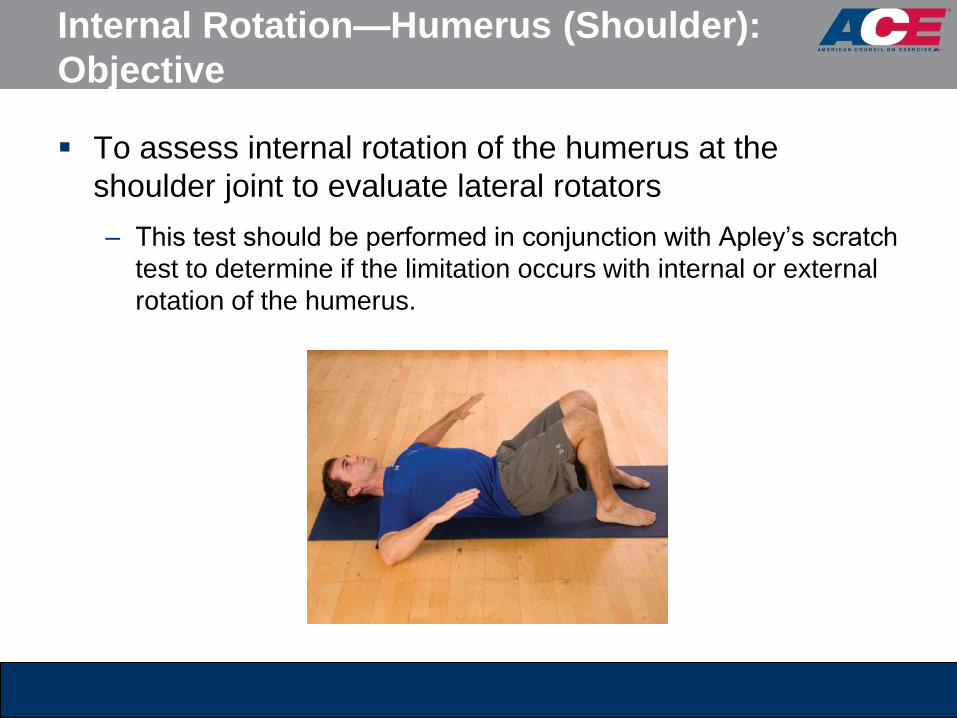
Internal Rotation—Humerus (Shoulder):
Objective
To assess internal rotation of the humerus at the
shoulder joint to evaluate lateral rotators
– This test should be performed in conjunction with Apley’s scratch
test to determine if the limitation occurs with internal or external
rotation of the humerus.

Internal/External Rotation—Humerus: Observations
Measure the degree of movement in each direction.
Note any bilateral differences between the left and right
arms in performing both movements.

Internal/External Rotation—Humerus: Interpretation
Balance and the Core
Balance and core baseline assessments evaluate the
need for comprehensive balance training and core
conditioning.
Dynamic balance tests are generally movement-specific
and quite complex.
– Trainers should aim to first evaluate the basic level of static
balance that a client exhibits by using the sharpened Romberg
test or the stork-stand test.

Sharpened Romberg Test: Objective
To assess static balance by standing with a reduced
base of support while removing visual sensory
information

Sharpened Romberg Test: Observations
Continue to time the client’s performance until one of the
following occurs:
– The client loses postural control and balance
– The client’s feet move on the floor
– The client’s eyes open
– The client’s arms move from the folded position
– The client exceeds 60 seconds with good postural control

Sharpened Romberg Test:
General Interpretations
The client needs to maintain his or her balance with
good postural control (without excessive swaying) and
not exhibit any of the test-termination criteria for 30 or
more seconds.
The inability to reach 30 seconds is indicative of
inadequate static balance and postural control.

Stork-stand Balance Test: Objective
To assess static balance by standing on one foot in a
modified stork-stand position

Stork-stand Balance Test: Observations
Timing stops when any of the following occurs:
– The hand(s) come off the hips
– The stance or supporting foot inverts, everts, or moves in any
direction
– Any part of the elevated foot loses contact with the stance leg
– The heel of the stance leg touches the floor
– The client loses balance

Stork-stand Balance Test:
General Interpretation
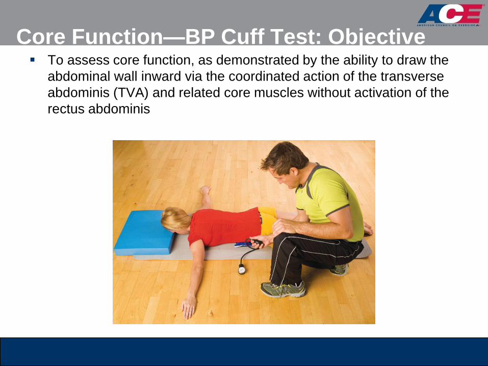
Core Function—BP Cuff Test: Objective To assess core function, as demonstrated by the ability to draw the
abdominal wall inward via the coordinated action of the transverse
abdominis (TVA) and related core muscles without activation of the
rectus abdominis

Core Function—BP Cuff Test: Observations
While the client attempts the contraction, carefully
monitor for any movement of the hips, ribcage, or
shoulders.
Clients must avoid any movement at the ankles
(dorsiflexion) or pushing from the elbows that would be
used as leverage to raise the torso.
Core Function—BP Cuff Test:
General Interpretation
A good indicator of TVA function is the ability to reduce
the pressure in the cuff by 10 mmHg during the
contraction.
– If a client lacks effective core function, he or she usually recruits
the rectus abdominis muscle instead to achieve the desired
movement.
– No change or a change <10 mmHg does not necessarily
represent a lack of core function.

Summary
Trainers should adhere to the principle of “straightening
the body before strengthening it.”
Trainers should consider performing the assessments in
Chapter 7 of the ACE Personal Trainer Manual (4th ed.),
in the sequence presented.
This session covered:
– Static postural assessment
– Movement screens
– Flexibility and muscle-length testing
– Shoulder mobility assessment
– Balance and the core