acetylation-induced transcription is required for active dna demethylation in methylation
TRANSCRIPT
MOLECULAR AND CELLULAR BIOLOGY, Nov. 2007, p. 7462–7474 Vol. 27, No. 210270-7306/07/$08.00�0 doi:10.1128/MCB.01120-07Copyright © 2007, American Society for Microbiology. All Rights Reserved.
Acetylation-Induced Transcription Is Required for Active DNADemethylation in Methylation-Silenced Genes�
Ana C. D’Alessio, Ian C. G. Weaver, and Moshe Szyf*Department of Pharmacology and Therapeutics, McGill University, Montreal, Quebec H3G 1Y6, Canada
Received 23 June 2007/Returned for modification 5 July 2007/Accepted 9 August 2007
A hallmark of vertebrate genes is that actively transcribed genes are hypomethylated in critical regulatorysequences. However, the mechanisms that link gene transcription and DNA hypomethylation are unclear.Using a trichostatin A (TSA)-induced replication-independent demethylation assay with HEK 293 cells, weshow that RNA transcription is required for DNA demethylation. Histone acetylation precedes but is notsufficient to trigger DNA demethylation. Following histone acetylation, RNA polymerase II (RNAP II) interactswith the methylated promoter. Inhibition of RNAP II transcription with actinomycin D, �-amanitin, orCDK7-specific small interfering RNA inhibits DNA demethylation. H3 trimethyl lysine 4 methylation, a markerof actively transcribed genes, was associated with the cytomegalovirus promoter only after demethylation.TSA-induced demethylation of the endogenous cancer testis gene GAGE follows a similar sequence of eventsand is dependent on RNA transcription as well. These data suggest that DNA demethylation follows ratherthan precedes early transcription and point towards a novel function for DNA demethylation as a memory ofactively transcribed genes.
The epigenome programs gene expression profiles of verte-brate cells. The epigenome is composed of chromatin structureand DNA methylation. The basic unit of chromatin, the nucleo-some, is composed of histones, which undergo an assortment ofcovalent modifications in their N-terminal tails, including phos-phorylation, acetylation, methylation, and ubiquitination (57, 66).These modifications affect the structural dynamics of the nucleo-some by forming a “histone code” that regulates chromatin func-tion through the recruitment of specific interacting proteins thatrecognize a single or conformational set of modifications.
The other component of the epigenome, DNA methylation,is a covalent modification of cytosines residing at CpG dinucle-otides in vertebrate genomes (47). Actively transcribed chro-matin regions are distinguished by hypomethylated DNA (46,48), whereas many inactive genes, such as parentally imprintedgenes and tumor suppressor genes in cancer, are hypermethylatedin critical regions and associate with hypoacetylated lysine9-methylated histone tails (21). A recent high-resolution anal-ysis of DNA methylation patterns and histone H3 and H4acetylation patterns in the HOXA cluster region revealed noacetylated histones in the hypermethylated regions, demon-strating a reciprocal relationship between DNA methylationand histone H3 and H4 acetylation (23).
Although DNA methylation is associated with silencing ofseveral genes, such as tumor suppressor genes in cancer (2),the relationship between DNA methylation and gene tran-scription is complex. A recent comprehensive analysis of thehuman methylome revealed that CG-rich islands are almostuniformly unmethylated, irrespective of their state of expres-sion, while CG-poor genes are generally methylated (63). The
fact that CG islands are unmethylated, even in the absence oftranscription, seems to suggest that a mechanism other thantranscription “per se” is responsible for the hypomethylatedstate.
Interestingly, the same authors identified an inverse corre-lation between methylation and expression in promoters withintermediate CG density; this suggests that a mechanism existsto coordinate transcription and DNA demethylation, at least inthis fraction of the promoters in the human genome. Thesepromoters were for germ line-specific genes, which are meth-ylated in somatic cells, hypomethylated in the testis (63), andinduced by DNA-demethylating agents (55). We therefore fo-cused this study on the cytomegalovirus (CMV) promoter as aprobe to study replication-independent demethylation of anintermediate CG promoter and demethylation of GAGE, atestis-specific gene. The remaining issue is determining themechanisms which lead to demethylation of genes and tomaintenance of their unmethylated state.
Several lines of evidence suggest that the “histone code”regulates the level of DNA methylation. This epigenetic crosstalk has been studied extensively for chromatin silencing.Firstly, DIM-5 from the fungus Neurospora crassa and krypto-nite from Arabidopsis thaliana, two histone methyltransferasesfor lysine 9, were shown to be important for normal DNAmethylation (24, 25, 59). Secondly, targeted deletion of Lsh, amember of the SNF2/helicase family involved in chromatinremodeling, produces a substantial loss of CpG methylationthroughout the genome (65). In addition, EZH2, a member ofthe multiprotein Polycomb complex PRC2, which methylateshistone H3 at K27, is important for the establishment of newmethylation patterns (61).
Work from our laboratory demonstrated that there is also anepigenetic activation cross talk (7, 13). We showed that inhi-bition of histone deacetylases (HDACs) by trichostatin A(TSA) triggers active DNA demethylation of an in vitro meth-ylated nonreplicating plasmid introduced into HEK 293 cells.
* Corresponding author. Mailing address: Department of Pharma-cology and Therapeutics, McGill University, 3655 Promenade Sir Wil-liam Osler, Montreal, Quebec H3G 1Y6, Canada. Phone: (514) 398-7107. Fax: (514) 487-1834. E-mail: [email protected].
� Published ahead of print on 20 August 2007.
7462
on April 5, 2019 by guest
http://mcb.asm
.org/D
ownloaded from

In addition, blockage of histone acetylation by the expressionof inhibitors of histone acetylation proteins inhibited demethyl-ation by TSA, supporting the hypothesis that histone acetyla-tion rather than side effects of the HDAC inhibitor triggereddemethylation (6). A recent comprehensive analysis of humanpromoters revealed a correlation between lack of methylationand H3K4 dimethylation, leading the authors to suggest thatchromatin modification might protect CG-rich islands frommethylation (63).
Here we test the hypothesis that chromatin activation “per se”leads to DNA demethylation. To gain insights into the mecha-nisms linking chromatin modification, gene activation, and DNAdemethylation, we delineated their temporal relationship follow-ing histone hyperacetylation induced by TSA. Our findings sug-gest that DNA demethylation is a stepwise, highly coordinatedprocess that involves histone acetylation and, importantly, RNApolymerase II (RNAP II) transcription. To our surprise, ourfindings indicate that DNA demethylation follows rather thanprecedes early transcription. Following DNA demethylation,histones associated with the CMV-green fluorescent protein(CMV-GFP) plasmid are trimethylated on lysine 4 (H3K4me3).H3K4me3, a memory of actively transcribed genes (38, 50), isselectively associated with unmethylated copies of the CMV pro-moter. We show that RNA transcription by RNAP II is requiredfor DNA demethylation, suggesting that the epigenetic repro-gramming of genes requires an initial state of transcription. Ourresults are consistent with the hypothesis that DNA demethyla-tion plays a role in the stable memory of RNAP II-mediatedtranscription, since early transcription of a methylated promoterresults in its demethylation, which enhances and stabilizes itsactive state.
MATERIALS AND METHODS
Plasmids. pEGFP-C2 (pCMV-GFP) was purchased from Clontech (GenBankaccession number U55763). A CDK7 short hairpin RNA-expressing plasmid waspurchased from Upstate Biotechnology. The negative control, which doesnot target any sequence contained in the human genome, was provided by apSilencer 2.1-U6 neo kit from Ambion.
In vitro methylation of plasmid DNA. pCMV-GFP was methylated in vitro asdescribed previously (7).
Cell culture and transient transfections. HEK 293 cells were plated at adensity of 0.5 � 106 in a 10-cm plate and transiently transfected with 500 ng ofmethylated pCMV-GFP, using the calcium phosphate precipitation method.Three hundred nanomolar TSA was added at 24 h posttransfection. Cells wereharvested at different times after initiation of TSA treatment. Each experimentwas performed in triplicate.
Bisulfite mapping. Bisulfite mapping was performed as described previously,with minor modifications (9). Five micrograms of sodium bisulfite-treated DNAsample was subjected to PCR amplification using a first set of primers, and thePCR products were used as templates for a subsequent PCR utilizing nestedprimers. The primers used for the CMV promoter (pCMV-GFP) (Clontech)were CMV-outside-sense (5�-GTT TAG TAT ATG ATT TTA TGG G-3�),CMV-outside-antisense (5�-CCA AAA TAA ACA CCA CCC C-3�), CMV-nested-sense (5�-ATT TTT TTA TTT GGT AGT ATA TTTA-3�), and CMV-nested-antisense (5�-CCC TTA CTC ACC ATA ATA AC-3�); the primers forthe GFP coding region were GFP-outside-sense (5�-GTT ATT ATG GTG AGTAAG GG-3�), GFP-outside-antisense (5�-TAT AAC TAT TAT AAT TAT ACTCCA-3�), GFP-nested-sense (5�-GGG GTG GTG TTT ATT TTG G-3�), andGFP-nested-antisense (5�-CTT ATA CCC CAA AAT ATT ACC-3�); theprimers for the simian virus 40 (SV40) polyadenylation site were PA-outside-sense (5�-TTA TTG TAT TTT AGT TGT GGT-3�), PA-outside-antisense(5�-AAA ACT CCC TTT AAA ATT CC-3�), PA-nested-sense (5�-TTG TTTAAA TTT ATT AAT GTA TTT AA-3�), and PA-nested-antisense (5�-CAAAAA ACT TAA TTA AAA TAA TTC-3�); the primers for the GAGEpromoter were GAGEprom-outside-sense (5�-GTT TAT ATT GAA TAA
TTT TTT TTT G-3�), GAGEprom-outside-antisense (5�-ATC TCA ATAAAA AAA AAA AAA TCC-3�), GAGEprom-nested-sense (5�-GAA TAGGTT GTT ATT TTT GTT T-3�), and GAGEprom-nested-antisense (5�-TACCTC ACA ACT CCC TAA C-3�); and the primers for the first GAGE exonwere GAGEcoding-outside-sense (5�-TTA GGG AGT TGT GAG GTAG-3�), GAGEcoding-outside-antisense (5�-CCC CAC TCA CAA CAA ATTTA-3�), GAGEcoding-nested-sense (5�-ATT TAT TTG GTA GGT GTGTAG-3�), and GAGEcoding-nested-antisense (5�-CCT TAC AAT ACT TCTCAC TC-3�). The PCR products of the second reaction were then subclonedusing an Invitrogen TA cloning kit following the manufacturer’s protocol.The clones were sequenced using a T7 sequencing kit as recommended by themanufacturer (procedure C; Amersham Pharmacia Biotech).
ChIP assays. Chromatin immunoprecipitation (ChIP) assays (12) were doneusing a ChIP assay kit protocol (Upstate Biotechnology). HEK 293 cells weretransfected with 500 ng of methylated pCMV-GFP, using the calcium phosphateprecipitation method as described below. Three hundred nanomolar TSA wasadded at 24 h posttransfection. Formaldehyde was added to the culture mediumat a final concentration of 1% at different times after initiation of TSA treatment,and the cells were incubated for 10 min at 37°C. Chromatin was immunopre-cipitated using the following antibodies: anti-acetylated histone H3 (UpstateBiotechnology), anti-polymerase II (anti-Pol II; Santa Cruz), anti-trimethylatedK4 H3 (Abcam), and normal rabbit immunoglobulin G (IgG; Santa Cruz).One-tenth of the lysate was kept to quantify the amount of DNA present in thedifferent samples before immunoprecipitation. PCRs on DNAs purified fromnonimmunoprecipitated and immunoprecipitated samples were quantified intriplicate by real-time PCR, using PowerSYBR green PCR master mix (AppliedBiosystems). Input samples were diluted 1/40, whereas antibody and IgG sampleswere diluted 1/10. The following primers were used: CMV sense, 5�-CAC CAAAAT CAA CGG GAC TTT C-3�; CMV antisense, 5�-TAC ACG CCT ACCGCC CAT T-3�; GFP sense, 5�-GAC GGA AAC GGC CAC AAG TTC-3�; GFPantisense, 5�-TTG CCG GTG GTG CAG AT-3�; GAGE prom sense, 5�-TACTTG AGT CCC AGA GGC ATA GG-3�; GAGE prom antisense, 5�-CCT CCATGC CCA TCC TCA T-3�; GAGE exon1 sense, 5�-TTT TTC CTC TAC TGAGAT TCA TCT GGT A-3�; GAGE exon1 antisense, 5�-CTC CAC CCT CACTCA CAC TTC A-3�; GLOBIN Delta sense, 5�-CTG TCT GTG AAT GAAAAG AAG GAA AT-3�; GLOBIN Delta antisense, 5�-CTA CGT TCA TTGCCA GAT CCA A-3�; LACZ sense, 5�-CCC ATC TAC ACC AAC GTA ACCTAT C-3�; and LACZ antisense, 5�-GAG TAA CAA CCC GTC GGA TTC T-3�.Amplifications were performed with an ABI 7900 HT sequence detection system,using the following protocol: 50°C for 2 min, 95°C for 10 min, and 40 cycles of95°C for 15 s and 60°C for 1 min.
Northern and Southern blot analysis. For Northern blot analysis, approxi-mately 2 �g of RNA was electrophoresed in a 1.2% denaturing agarose gel andthen transferred with 10� SSC (1� SSC is 0.15 M NaCl plus 0.015 M sodiumcitrate) to a Hidrobond-N� membrane (Amersham Pharmacia Biotech). South-ern blot analysis was performed as described previously (7). The levels of ex-pression of the different mRNAs in the Northern blots were quantified bydensitometric scanning of the relevant autoradiograms. Each experiment wasnormalized for the amount of total RNA by hybridization with a 32P-labeled 18SrRNA oligonucleotide probe.
Western blot analyses. Twenty-five micrograms of proteins was loaded into a10% sodium dodecyl sulfate-polyacrylamide gel. After the proteins were trans-ferred to a nitrocellulose membrane and blocked with 5% skim milk, CDK7protein was detected using rabbit polyclonal IgG (Abcam) at a 1:500 dilution,followed by peroxidase-conjugated anti-rabbit IgG (Sigma) at 1:5,000. ActinWestern blots were performed using a 1:5,000 dilution of actin monoclonalantibody (Sigma) followed by the mouse secondary antibody at 1:20,000 (JacksonImmunoResearch). An enhanced chemiluminescence kit (Amersham Bio-sciences) was used for all Western blots.
Microarray analysis. Human embryonic kidney HEK 293 cells (ATCC CRL1573) were plated at a density of 0.5 � 106 cells in a 10-cm plate. Twenty-fourhours later, 300 nM TSA was added for 72 h. Total RNA was extracted withRNeasy (QIAGEN). Microarray analysis was performed as previously described(18). Briefly, 20 �g of RNA was used for cDNA synthesis, followed by in vitrotranscription with a T7 promoter primer containing a poly(T) tail. The resultingproduct was hybridized to a HuGeneFL DNA microarray containing oligonu-cleotides specific for 20,000 human transcripts and then processed with a Gene-Chip system (Affymetrix). Data analysis, average difference, and expression foreach feature on the chip were computed using the Affymetrix GeneChip analysissuite, version 3.3, with default parameters. Each experiment was performedin duplicate. The Montreal Genome Centre performed the gene expressionanalysis.
VOL. 27, 2007 MECHANISM OF ACTIVE DNA DEMETHYLATION 7463
on April 5, 2019 by guest
http://mcb.asm
.org/D
ownloaded from

Reverse transcription-PCR (RT-PCR). Total RNA was extracted using thestandard guanidinium isothiocyanate method. cDNA was synthesized in a 20-�lreaction volume containing 5 �g of total RNA, 40 units of Moloney murineleukemia virus reverse transcriptase (MBI), 5 �M of random primers (RocheMolecular Biochemicals), a 1 mM concentration of each of the four de-oxynucleoside triphosphates, and 40 units of RNase inhibitor (Roche MolecularBiochemicals). mRNA was denatured for 5 min at 70°C, the random primerswere annealed for 10 min at 25°C, and mRNA was reverse transcribed for 1 h at37°C. The reverse transcriptase was heat inactivated at 70°C, and the productswere stored at �20°C until use. PCR was performed in a 30-�l reaction mixturecontaining 2 �l of synthesized cDNA product, 3 �l of 10� PCR buffer, 2 mMMgCl2, a 0.2 mM concentration of each deoxynucleoside triphosphate, 1 unit Taqpolymerase (all from MBI), and 0.5 �M of each primer.
Quantitative RT-PCR. Quantitative PCRs were performed as described previ-ously, using PowerSYBR green PCR master mix (Applied Biosystems) and thefollowing primers: GAGE sense, 5�-GCT GCT CAG AAG GGA GAG GAT-3�;GAGE antisense, 5�-CTG TTC CTG GCT ATG AGC TTC A-3�; GFP sense,5�-GAC GGA AAC GGC CAC AAG TTC-3�; and GFP antisense, 5�-TTG CCG GTGGTG CAG AT-3�. For glyceraldehyde-3-phosphate dehydrogenase (GAPDH) geneamplification, a human GAPD endogenous control (FAM/MGB probe; non-primer-limited TaqMan predeveloped assay reagents [Applied Biosystems]) was used withTaqMan Universal PCR master mix (Applied Biosystems). For CGA, SPANAX1, andARC amplifications, a universal probe library set from Roche was used (for CGA,probe 61 was used; for SPANAX1, probe 68 was used; and for ARC, probe 21 wasused) with FastStart TaqMan probe master mix from Roche. All amplificationswere performed with an ABI 7900 HT sequence detection system, using thefollowing protocol: 50°C for 2 min, 95°C for 10 min, and 40 cycles of 95°C for 15 sand 60°C for 1 min.
RESULTS
Time course of demethylation of ectopically methylatedCMV promoter following TSA treatment. DNA demethylationin a replicating cell can be either an active or a passive repli-cation-dependent process. To distinguish between these twopossibilities, we took advantage of a previously developed ac-tive demethylation assay using living HEK 293 cells (7). In thissystem, pharmacological induction of histone acetylation byHDAC inhibition with TSA triggers active DNA demethyla-tion of ectopically methylated pCMV-GFP. We have previ-ously shown, using the DpnI restriction enzyme, which digestsonly DNA that maintains its bacterial methylation (GmATC)pattern, that pCMV-GFP did not replicate in HEK 293 cellsduring the course of the experiment (7). This was also repeatedhere to verify that the demethylation events studied were in-deed active (data not shown). In this study, we used ChIPassays with pertinent histone modification antibodies andbisulfite mapping to study the temporal and causal relationshipbetween chromatin and active DNA demethylation.
We first measured the time course of demethylation of theCMV promoter (Fig. 1A; also see Fig. 3, left panel). Bisulfitemapping of transfectants treated with 300 nM TSA for differ-ent times showed that DNA demethylation increases over timeand is a slow process, starting at 18 h and being completed onlyafter 48 h of TSA treatment.
Histone acetylation is not sufficient to trigger DNA de-methylation of the ectopically methylated CMV promoter. Totest the hypothesis that histone acetylation per se is the onlychange required for DNA demethylation, we analyzed the timecourse of histone H3 acetylation at the CMV promoter follow-ing TSA treatment, using ChIP analysis. We found that histoneacetylation (Fig. 1B) precedes DNA demethylation (Fig. 1A;also see Fig. 3). These data suggest that there is a lag betweenhistone acetylation and demethylation. To directly assess whetherhistone acetylation triggers immediate DNA demethylation, we
measured the state of methylation of pCMV-GFP DNA asso-ciated with acetylated histones early after TSA induction byusing a combination of ChIP and bisulfite mapping. Input,anti-acetyl-H3 (H3K9Ac), and IgG ChIP DNAs prepared fromtransfectants treated with TSA for different times were sub-jected to bisulfite PCR (Fig. 1C) and mapping (Fig. 1D). So-dium bisulfite DNA amplification was observed in the inputand immunoprecipitated H3K9Ac samples but not in the im-munoprecipitated IgG or water control sample (Fig. 1C), in-dicating that the bisulfite-sequenced material represents DNAassociated with H3K9Ac histones. If histone acetylation werethe only rate-limiting step for demethylation, we would expectthat as soon as CMV-GFP is associated with acetylated histoneH3, it would be demethylated straight away, even at the early6-h time point. However, we observed that the molecules as-sociated with H3K9Ac were methylated at 6 h and only became
FIG. 1. Histone acetylation precedes DNA demethylation. In vitro-methylated pCMV-GFP plasmid was transfected into HEK 293 cellsand treated with TSA at 24 h posttransfection. Cells were harvested atdifferent time points of TSA treatment. (A) Time course of activeDNA demethylation. DNA was subjected to bisulfite methylation map-ping analysis. The CMV region was amplified, subcloned, and se-quenced as described in Materials and Methods. Each bar representsthe average for 10 different clones at each site. (B) ChIP assay using anantibody against either H3K9Ac or control IgG. Samples were spikedwith LacZ prior to immunoprecipitation to rule out nonspecific DNAprecipitation with the antibody. The CMV and negative control LacZsequences were amplified from purified DNA by quantitative PCR.(C) Representative bisulfite PCR amplification from input DNA andDNAs purified from immunoprecipitated acetyl histone H3 and IgGsamples. (D) The DNAs used for panel C were subjected to bisulfitemapping analysis. Each line represents one clone. Filled circles, meth-ylated CG dinucleotides within the CMV promoter; open circles, de-methylated CG dinucleotides.
7464 D’ALESSIO ET AL. MOL. CELL. BIOL.
on April 5, 2019 by guest
http://mcb.asm
.org/D
ownloaded from
demethylated 24 h after the addition of TSA (Fig. 1D), indi-cating that other events in addition to H3 acetylation might berequired for DNA demethylation following TSA treatment.
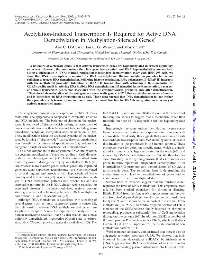
Time course of DNA demethylation and transcription. IfDNA demethylation plays a causal role in gene expression,then it is expected that demethylation would precede transcrip-tion. To address this, we studied the time course of GFPtranscription and CMV demethylation after TSA treatment(Fig. 2C and 3). RNA transcription started between 3 h and12 h after TSA treatment (Fig. 2C); however, no DNA de-methylation was observed at these time points (Fig. 1A and 3).DNA demethylation was first detected at the 18-h time point(Fig. 1A and 3). This is consistent with the notion that tran-scription preceded DNA demethylation.
A ChIP experiment with an anti-RNAP II antibody revealed
that occupancy of the CMV promoter with RNAP II followingTSA treatment is detectable 12 h following TSA treatment(Fig. 2A). Transcription of genes by RNAP II is a complexprocess (11, 32, 36, 40, 41) and, as such, requires a highlycoordinated and multistep process utilizing a large number ofbasal and transactivating factors. Furthermore, there exists adynamic association of mRNA processing factors with differ-ently modified forms of the polymerase throughout the tran-scription cycle (31). More specifically, the phosphorylation ofthe C-terminal domain (CTD) in RNAP II at serine 5 has beenassociated with transcription initiation. Using a ChIP experi-ment against this phosphorylated form of RNAP II, we wereable to control for initiation of the transcription process. Anexcellent correlation was observed between GFP expression(Fig. 2C) and serine 5 phosphorylation of the CTD of RNAPII (Fig. 2B). The small discrepancy between the initiation ofbinding of RNAP II and its serine 5-phosphorylated formcould be due to differences in antibody sensitivity and dynamicrange.
RNAP II binds the methylated CMV promoter during RNAtranscription. We resorted again to a combination of ChIP andbisulfite mapping to determine whether demethylation is aprerequisite for binding of RNAP II. If demethylation were aprecondition for RNAP II interaction, then CMV promotersbound to RNAP II would be demethylated even at the earliesttime point when RNAP II binding to CMV is detected. How-ever, if demethylation follows RNAP II interaction with meth-ylated DNA, RNAP II would initially bind to methylated CMV
FIG. 2. RNAP II transcribes and binds the methylated CMV pro-moter. In vitro-methylated pCMV-GFP was transfected into HEK 293cells, and at 24 h posttransfection, cells were treated with TSA. Cellswere harvested at different time periods of TSA treatment. (A and B)Cells were subjected to ChIP assay using an anti-RNAP II antibody(A) or an antibody against the serine 5-phosphorylated form of theCTD of RNAP II (B) or were processed with IgG. The CMV sequencewas amplified from purified DNA by quantitative PCR. As a negativecontrol, a region 5 kb upstream of the globin delta gene was amplifiedby quantitative PCR. No amplification of the globin delta gene wasobserved in the RNAP II ChIP samples. (C) Kinetics of GFP expres-sion after TSA treatment. Quantitative real-time RT-PCR of the GFPand GAPDH genes was performed. The ratios of the GFP to GAPDHsignals were plotted. No signal was observed in the no-RT control.(D) Representative bisulfite PCR amplification from input DNA andDNAs purified from immunoprecipitated RNAP II and IgG samples.(E) The DNAs used for panel D were subjected to bisulfite mappinganalysis. Each line represents one clone. Filled circles, methylated CGdinucleotides within the CMV promoter; open circles, demethylatedCG dinucleotides.
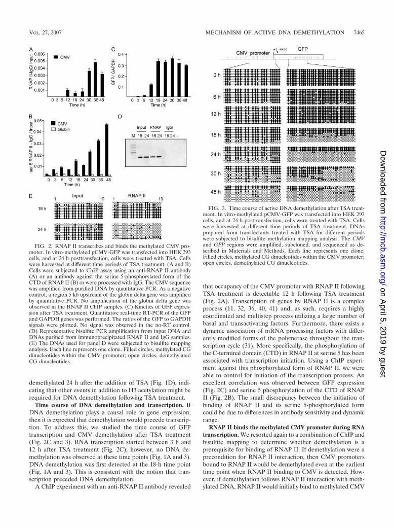
FIG. 3. Time course of active DNA demethylation after TSA treat-ment. In vitro-methylated pCMV-GFP was transfected into HEK 293cells, and at 24 h posttransfection, cells were treated with TSA. Cellswere harvested at different time periods of TSA treatment. DNAsprepared from transfectants treated with TSA for different periodswere subjected to bisulfite methylation mapping analysis. The CMVand GFP regions were amplified, subcloned, and sequenced as de-scribed in Materials and Methods. Each line represents one clone.Filled circles, methylated CG dinucleotides within the CMV promoter;open circles, demethylated CG dinucleotides.
VOL. 27, 2007 MECHANISM OF ACTIVE DNA DEMETHYLATION 7465
on April 5, 2019 by guest
http://mcb.asm
.org/D
ownloaded from

promoters. Sodium bisulfite DNA amplification was observedin the input and immunoprecipitated RNAP II samples but notin the immunoprecipitated IgG or water control sample (Fig.2D). DNAs amplified from anti-RNAP II ChIP and inputsamples were subjected to bisulfite mapping. Interestingly, ourdata show that CMV promoter sequences bound to RNAP IIwere methylated at 18 h (Fig. 2E), implying that RNAP IIinitially binds to methylated CMV promoters. At 24 h, RNAPII-bound CMV promoters were enriched with unmethylatedcopies in comparison with the input DNA, suggesting thatCMV promoters bound to RNAP II are targeted for demethyl-ation. Promoters that are not loaded with RNAP II and are notactively transcribed remain methylated.
Since RNAP II binds a methylated CMV promoter, thequestion raised is whether demethylation starts at the pro-moter or downstream of the transcription initiation site. Atime course analysis of demethylation of the CMV promoterand the GFP coding sequence showed that demethylation ofthe CMV promoter lags behind demethylation of the GFPsequence (Fig. 3). This is consistent with the hypothesis thatdemethylation initiates along the path of transcription ofRNAP II and then spreads to the promoter sequence.
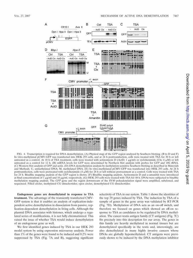
RNA transcription is required for DNA demethylation. Totest the hypothesis that transcription elongation is required fordemethylation, we used two well-characterized pharmacologi-cal inhibitors of RNAP II transcription activity, namely,�-amanitin and actinomycin D. Actinomycin D is a DNA-intercalating agent which is believed to stall the progression ofRNAP II but does not inhibit transcription initiation (29).�-Amanitin interacts with the “funnel,” “cleft,” and “bridge�-helix” regions of the Rbp1 subunit of RNAP II and is be-lieved to inhibit elongation at each translocation step of theRNA-DNA hybrid (5, 19). Methylation-sensitive Southern blotanalysis of the GFP coding region showed that actinomycin Dinhibits DNA demethylation (Fig. 4D). On the other hand, theprotein synthesis inhibitor cycloheximide did not inhibit DNAdemethylation, suggesting that the effect that inhibition oftranscription had on demethylation was not caused by a changein the cellular levels of a “high-turnover” demethylation factor.In addition, pretreatment of the cell with cycloheximide priorto TSA treatment had no effect on DNA demethylation (Fig.4E), further excluding the possibility that our actinomycintreatment inhibited DNA demethylation through blocking thesynthesis of a labile high-turnover protein required for DNAmethylation.
Our data suggest that RNA transcription, but not proteinsynthesis, is required for demethylation. Figure 4B shows thatactinomycin D, but not cycloheximide, inhibited GFP mRNAas expected; however, cycloheximide inhibited GFP proteinsynthesis to an even larger extent than the inhibition by acti-nomycin D (Fig. 4C). Bisulfite mapping of the promoter andtranscribed GFP regions showed that actinomycin D inhibitedDNA demethylation both at the GFP transcribed sequenceand at the promoter (Fig. 4F). If progression of RNAP II wereindeed required for DNA demethylation, inhibition of tran-script elongation by RNAP II with �-amanitin should have thesame effect as actinomycin D. As expected, �-amanitin inhib-ited DNA demethylation at the GFP transcribed region (Fig.4F). To further prove that DNA demethylation requires pro-gression of RNAP II, we mapped the state of methylation of
CG sites residing downstream of the SV40 polyadenylation siteat the 3� end of the GFP coding sequence. If DNA demeth-ylation is linked to transcription, DNA demethylation wouldstop at the point where transcription terminated. As expected,CGs downstream of the polyadenylation signal remainedmethylated at 48 h of TSA treatment, whereas CGs upstreamof the point of transcription termination were demethylated(Fig. 4G). Thus, transcription is required for DNA demeth-ylation.
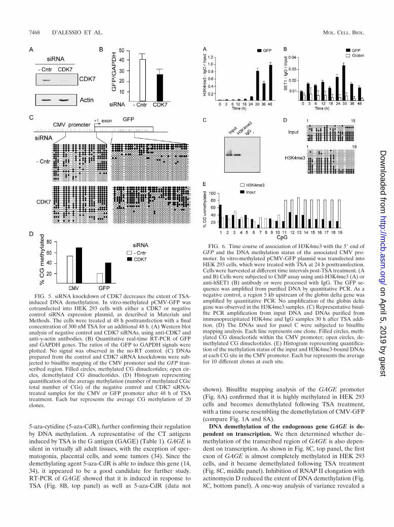
siRNA depletion of CDK7 inhibits DNA demethylation. Inorder to test the relationship between transcription and DNAdemethylation, we performed small interfering RNA (siRNA)knockdown of CDK7, a protein kinase which is found at thetranscription initiation complex and phosphorylates the CTDof RNAP II (53, 54), regulating the step between transcriptioninitiation and elongation. A Western blot analysis with anti-CDK7 shows CDK7 knockdown by the siRNA treatment (Fig.5A). Supporting the hypothesis that transcription is requiredfor demethylation, CDK7 knockdown resulted in decreasedGFP expression (Fig. 5B) as well as a reduction in the extent ofDNA demethylation of the GFP transcribed sequence and theCMV promoter (Fig. 5C). In summary, different manipulationsthat inhibit the progression of RNAP II by distinct mechanismsinhibit DNA demethylation, suggesting a causal relationshipbetween passage of RNAP II and DNA demethylation. CDK7knockdown had a smaller effect on DNA demethylation thandid inhibition of transcription with small molecules. This prob-ably reflects a more effective blockage of transcription by in-hibitors of RNAP II progression than that by CDK7 knock-down.
The GFP coding region associates with H3K4me3 after theCMV promoter is demethylated. H3K4me3 is a hallmark ofgenes programmed to be active (22, 38, 49, 50). SET1, thehistone methyltransferase for lysine 4, associates with thenewly initiated RNAP II following phosphorylation of serine 5of the CTD (38).
If DNA demethylation indeed sets the stage for stable pro-gramming of gene activity, we predict that H3K4me3 will occurafter DNA demethylation and that CMV promoters associatedwith H3K4me3 will be enriched in unmethylated DNA. To testthis hypothesis, we followed the time course of H3K4me3 asso-ciation with the GFP gene following TSA treatment, using ChIPanalysis. H3K4me3 (Fig. 6A) occurred after DNA demethylation(Fig. 1A). The human homolog of yeast SET1 is one of theenzymes responsible for H3K4 methylation. Analysis of the timecourse of SET1 occupancy of the CMV promoter revealed acorrelation between SET1 occupancy and H3K4me3 at the GFPexon (Fig. 6B).
Furthermore, we determined the state of methylation ofCMV promoter DNA associated with H3K4me3 at the earliesttime point by using a combination of ChIP and bisulfite map-ping analysis. Sodium bisulfite DNA amplification was ob-served in the input and immunoprecipitated H3K4me3 sam-ples but not in the immunoprecipitated IgG or water controlsample (Fig. 6C). H3K4me3-bound CMV-GFP contained pro-moter DNA that was significantly more demethylated than theinput DNA (Fig. 6D and E). This interesting pattern of hy-pomethylation suggests a selection for unmethylated DNA as-sociated with H3K4me3 histones.
7466 D’ALESSIO ET AL. MOL. CELL. BIOL.
on April 5, 2019 by guest
http://mcb.asm
.org/D
ownloaded from
Endogenous genes are demethylated in response to TSAtreatment. The advantage of the transiently transfected CMV-GFP system is that it enables an analysis of replication-inde-pendent active demethylation in dissociation from passive, rep-lication-dependent demethylation in living cells. Although theplasmid DNA associates with histones, which undergo a regu-lated series of modifications, it is not fully chromatinized. Thisraised the issue of whether TSA would induce demethylationof an endogenous gene as well.
We first identified genes induced by TSA in our HEK 293model system by using expression microarray analysis. Fewerthan 1% of the genes were found to be induced and 0.2% weresuppressed by TSA (Fig. 7A and B), suggesting significant
selectivity of TSA in our system. Table 1 shows the identities ofthe top 29 genes induced by TSA. The induction by TSA of asample of genes in the gene array was validated by RT-PCR(Fig. 7D). Methylation of DNA acts as an on-off switch, andtherefore we focused on genes which showed an off-on re-sponse to TSA as candidates to be regulated by DNA methyl-ation. The cancer-testis antigen family (CT antigens) (Fig. 7C)fits precisely into this description for our array. The genes inthis family are heavily methylated in normal tissues but aredemethylated specifically in the testis and, interestingly, arealso demethylated in many highly invasive cancers whoseDNAs are globally hypomethylated. CT antigens were previ-ously shown to be induced by the DNA methylation inhibitor
FIG. 4. Transcription is required for DNA demethylation. (A) Physical map of the GFP region analyzed by Southern blotting. (B to D and F)In vitro-methylated pCMV-GFP was transfected into HEK 293 cells, and at 24 h posttransfection, cells were treated with TSA for 30 h or leftuntreated as a control. At 18 h of TSA treatment, cells were treated with actinomycin D (ActD; 1 �g/ml) or cycloheximide (Clx; 4 �M) or leftuntreated as a control for 12 h. (B) mRNA levels for GFP were determined by Northern blot analysis with probes for GFP and 18S rRNA.(C) Western blot analysis of GFP and actin. (D) DNA demethylation analysis by methylation-sensitive Southern blotting as described in Materialsand Methods. U, unmethylated DNA; M, methylated DNA. (E) In vitro-methylated pCMV-GFP was transfected into HEK 293 cells. At 24 hposttransfection, cells were pretreated with cycloheximide (4 �M) for 28 h or left without pretreatment as a control. Cells were treated with TSAfor 24 h. Bisulfite mapping analysis of the GFP region is shown. (F) Bisulfite mapping analysis. Actinomycin D and �-amanitin were introducedat final concentrations of 1 �g/ml and 10 �g/ml, respectively. (G) HEK 293 cells were treated with TSA for 48 h. DNAs were subjected to bisulfitemethylation mapping analysis. The GFP gene and the region downstream of the SV40 polyadenylation signal were amplified, subcloned, andsequenced. Filled circles, methylated CG dinucleotides; open circles, demethylated CG dinucleotides.
VOL. 27, 2007 MECHANISM OF ACTIVE DNA DEMETHYLATION 7467
on April 5, 2019 by guest
http://mcb.asm
.org/D
ownloaded from
5-aza-cytidine (5-aza-CdR), further confirming their regulationby DNA methylation. A representative of the CT antigensinduced by TSA is the G antigen (GAGE) (Table 1). GAGE issilent in virtually all adult tissues, with the exception of sper-matogonia, placental cells, and some tumors (34). Since thedemethylating agent 5-aza-CdR is able to induce this gene (14,34), it appeared to be a good candidate for further study.RT-PCR of GAGE showed that it is induced in response toTSA (Fig. 8B, top panel) as well as 5-aza-CdR (data not
shown). Bisulfite mapping analysis of the GAGE promoter(Fig. 8A) confirmed that it is highly methylated in HEK 293cells and becomes demethylated following TSA treatment,with a time course resembling the demethylation of CMV-GFP(compare Fig. 1A and 8A).
DNA demethylation of the endogenous gene GAGE is de-pendent on transcription. We then determined whether de-methylation of the transcribed region of GAGE is also depen-dent on transcription. As shown in Fig. 8C, top panel, the firstexon of GAGE is almost completely methylated in HEK 293cells, and it became demethylated following TSA treatment(Fig. 8C, middle panel). Inhibition of RNAP II elongation withactinomycin D reduced the extent of DNA demethylation (Fig.8C, bottom panel). A one-way analysis of variance revealed a
FIG. 5. siRNA knockdown of CDK7 decreases the extent of TSA-induced DNA demethylation. In vitro-methylated pCMV-GFP wascotransfected into HEK 293 cells with either a CDK7 or negativecontrol siRNA expression plasmid, as described in Materials andMethods. The cells were treated at 48 h posttransfection with a finalconcentration of 300 nM TSA for an additional 48 h. (A) Western blotanalysis of negative control and CDK7 siRNAs, using anti-CDK7 andanti-�-actin antibodies. (B) Quantitative real-time RT-PCR of GFPand GAPDH genes. The ratios of the GFP to GAPDH signals wereplotted. No signal was observed in the no-RT control. (C) DNAsprepared from the control and CDK7 siRNA knockdowns were sub-jected to bisulfite mapping of the CMV promoter and the GFP tran-scribed region. Filled circles, methylated CG dinucleotides; open cir-cles, demethylated CG dinucleotides. (D) Histogram representingquantification of the average methylation (number of methylated CGs/total number of CGs) of the negative control and CDK7 siRNA-treated samples for the CMV or GFP promoter after 48 h of TSAtreatment. Each bar represents the average CG methylation of 20clones.
FIG. 6. Time course of association of H3K4me3 with the 5� end ofGFP and the DNA methylation status of the associated CMV pro-moter. In vitro-methylated pCMV-GFP plasmid was transfected intoHEK 293 cells, which were treated with TSA at 24 h posttransfection.Cells were harvested at different time intervals post-TSA treatment. (Aand B) Cells were subjected to ChIP assay using anti-H3K4me3 (A) oranti-hSET1 (B) antibody or were processed with IgG. The GFP se-quence was amplified from purified DNA by quantitative PCR. As anegative control, a region 5 kb upstream of the globin delta gene wasamplified by quantitative PCR. No amplification of the globin deltagene was observed in the H3K4me3 samples. (C) Representative bisul-fite PCR amplification from input DNA and DNAs purified fromimmunoprecipitated H3K4me and IgG samples 30 h after TSA addi-tion. (D) The DNAs used for panel C were subjected to bisulfitemapping analysis. Each line represents one clone. Filled circles, meth-ylated CG dinucleotide within the CMV promoter; open circles, de-methylated CG dinucleotides. (E) Histogram representing quantifica-tion of the methylation status of the input and H3K4me3-bound DNAsat each CG site in the CMV promoter. Each bar represents the averagefor 10 different clones at each site.
7468 D’ALESSIO ET AL. MOL. CELL. BIOL.
on April 5, 2019 by guest
http://mcb.asm
.org/D
ownloaded from

highly significant effect of treatment (F � 24.97; P 0.0001).Post hoc analysis showed that TSA treatment significantly (P 0.001 for TSA versus controls and TSA versus TSA-plus-acti-nomycin D treatment) decreased levels of cytosine methylationof CpG sites at the 5� end of the GAGE exon 1 sequence (i.e.,increased DNA demethylation). The effect of TSA treatmentwas blocked (P 0.05 for control versus TSA-plus-actinomy-cin D treatment) by concomitant treatment of the gene tran-scription inhibitor actinomycin D (Fig. 8D). These findings areconsistent with the idea that transcription is required for DNAdemethylation.
To examine the temporal relationship between chromatinmodifications and DNA demethylation, we performed ChIPanalysis. As shown in Fig. 8B, middle panel, histone H3 acet-ylation at the GAGE promoter started shortly after TSA treat-ment. DNA demethylation followed histone H3 acetylation, asit does with CMV-GFP (Fig. 1A). Following DNA demeth-ylation, H3K4me3 was associated with the 5� first exon in boththe endogenous gene (Fig. 8B, bottom panel) and the CMV-GFP reporter (Fig. 6A). Interestingly, however, the H3K4me3association with the first exon of GAGE was transient and wasreversed between 30 and 48 h after treatment. We do not have,as of yet, an explanation for this difference between GAGE andour pCMV-GFP construct. However, when we analyzed theoccupancy of SET1 on GAGE’s first exon, we found no binding
of SET1 (data not shown). Several H3K4 methylases present inmammalian cells, such as MLL, ALL-1, SET9/7, and ALR1-2(reviewed in reference 33), could be responsible for theH3K4me3 recruitment at the GAGE promoter. In addition, itis possible that the discrepancy in H3K4me3 recruitment be-tween the two genes analyzed, GFP and GAGE, is due todifferent kinetics of the different enzymes responsible forH3K4me3.
DISCUSSION
Although a hallmark of vertebrate genomes is the correla-tion between histone acetylation and DNA hypomethylation,the nature of this correlation is still unclear (45). It was dem-onstrated that DNA methylation silences gene expression andthat demethylation activates genes. Therefore, it was expectedthat demethylation should precede the initiation of transcrip-tion and gene expression. However, in this study, we found thattranscription is required for active DNA demethylation.
Histone acetylation can be induced either by targeting thegene with a transcription factor that recruits histone acetyl-transferases (HATs) or by a pharmacological agent such asTSA. Several studies from our lab as well as others have shownthat altering the chromatin by the addition of HDAC inhibitorsresults in a decrease in DNA methylation in both mammaliancells (15, 62) and Neurospora (52). Our replication-indepen-dent active DNA demethylation assay demonstrated, as ex-pected, that histone acetylation preceded DNA demethylation(Fig. 1A and B). Moderate levels of histone acetylation (Fig.1B) were associated with methylated DNA (Fig. 1D); higherlevels of histone acetylation were starting to be associated withunmethylated DNA 18 to 24 h after TSA treatment (Fig. 1Band D). These results imply that higher levels of histone acet-ylation could trigger events needed for DNA demethylation. Itis well established that pharmacological DNA demethylationactivates gene expression (26), implying a causal relationshipbetween demethylation and gene activation. UnmethylatedDNA would enable the interaction of certain transcriptionfactors prohibited from binding by DNA methylation (10). Ifthis model were true, then binding of the transcription machin-ery to the CMV promoter would follow demethylation, andCMV promoters associated with RNAP II would be unmeth-ylated at the earliest binding time point. However, we foundthat CMV promoters associated with RNAP II were methyl-ated early after the beginning of their association with RNAPII (18 h) (Fig. 2E). By 24 h, RNAP II was associated withhighly unmethylated promoters (Fig. 2E). This suggests thatthe interaction of a methylated CMV-GFP with RNAP IIfacilitates demethylation through unknown intermediate stepsand raises the provoking hypothesis that early transcriptionmight be the initial cause of DNA demethylation.
We confirmed this hypothesis by using two pharmacologicalagents, actinomycin D and �-amanitin, which inhibit transcrip-tion elongation through different mechanisms (Fig. 4). Wefurther tested this hypothesis by using siRNA knockdown ofCDK7, a protein kinase that phosphorylates the RNAP II CTDat serine 5 (17, 56), mediating the transition between transcrip-tion initiation and elongation (Fig. 5). The fact that inhibitionof transcription elongation by three different mechanisms leadsto inhibition of demethylation supports the conclusion that the
FIG. 7. Gene expression changes following 72 h of treatment ofHEK 293 cells with TSA. (A) Percentages of mRNA transcripts in-creased and decreased by TSA. (B) Pie chart presentation of thepercentages of mRNA transcripts increased, decreased, or unchangedby TSA. (C) Distribution of TSA-induced genes over different func-tional classes. (D) Quantitative real-time PCR validation of the genearray data. Quantitative real-time RT-PCR of the CGA, SPANAX1(SPAX1), ARC, and GAPDH genes was performed as indicated inMaterials and Methods.
VOL. 27, 2007 MECHANISM OF ACTIVE DNA DEMETHYLATION 7469
on April 5, 2019 by guest
http://mcb.asm
.org/D
ownloaded from

effects observed were not results of idiosyncrasies of theseagents but rather of their common activity, i.e., inhibition oftranscription. To exclude the possibility that inhibition of tran-scription could act indirectly by changing the mRNA level of ahigh-turnover factor required for DNA demethylation, wetreated the cells with cycloheximide. Cycloheximide blockedprotein synthesis and should have blocked the expression ofany high-turnover factor. However, cycloheximide did not in-hibit DNA demethylation (Fig. 4).
The idea that transcription, rather than just RNAP II bind-ing per se, is required for demethylation is supported by thedifference observed in the kinetics of demethylation of thetranscribed regions of the GFP gene and the CMV promoter.If binding of RNAP II were the rate-limiting step for demeth-ylation, we would expect the promoter, which was the landingpad of RNAP II, to be demethylated first. However, our datasuggest that demethylation at the promoter lags behind dem-ethylation of the transcribed region of the reporter gene (Fig.3). These data are consistent with the hypothesis that theprogression of transcription recruits demethylases to the geneand that this demethylation eventually spreads to the promoterregion. In addition, the fact that DNA demethylation does notproceed beyond the position where transcription is terminatedsupports the hypothesis that demethylation is linked to thepassage of the transcription machinery (Fig. 4G).
Interestingly, we found that DNA demethylation at the pro-moter is followed by H3K4me3 of histones associated with thetranscribed region of GFP (Fig. 6). H3K4me3 at the codingregion of genes has been postulated to function as a memory ofactive transcription in yeast (38) as well as higher organisms(50). Using a combination of ChIP and bisulfite mapping, weshowed that the DNA associated with H3K4me3 was hypo-methylated compared with the unbound population of CMVpromoters (Fig. 6). This is consistent with the hypothesis thatH3K4me3 tags on demethylation of DNA. In agreement, theloss of Lsh, a modulator of CG DNA methylation, results inincreased H3K4me3, and treatment with the DNA methylationinhibitor 5-aza-CdR also results in increased H3K4me3 (65).
If RNAP II can bind and transcribe methylated DNA, whatis the purpose of DNA demethylation? The idea that RNAP IIprogression along a gene might affect epigenetic programmingof gene expression has been suggested before and is supportedby other evidence. For example, RNAP II interacts with chro-matin remodeling enzymes, such as BRG1, a member of theSWI/SNF chromatin remodeling complex; with HATs, such asCBP (8, 37); and with chromatin-modifying enzymes, such asSET1 (38). MLL1, a human equivalent of yeast SET1, associ-ates with highly expressed transcripts (22). The passage ofRNAP II transcribing a gene was proposed to be coupled tomechanisms that propagate the breakdown and reprogram-
TABLE 1. Genes induced by TSA in HEK 293 cellsa
Accession no. Foldincrease
Array valueName Abbreviation
Control TSA
NM_000735.2 317 15 4,798 Glycoprotein hormones alpha polypeptide CGANM_013453.1 114 22 2,508 Sperm protein associated with nucleus X SPANAX1M25915.1 85 9 768 Clusterin (complement lysis inhibitor SP-4040-sulfated
glycoprotein 2)CLU
BC004490.1 72 11 790 v-Fos FBJ murine osteosarcoma viral oncogene homolog FOSAF193421.1 60 18 1,079 Activity-regulated cytoskeleton-associated protein ARCNM_002305.2 53 52 2,778 Lectin galactoside-binding soluble protein 1 (galectin 1) LGALS1NM_006183.2 32 59 1,902 Neurotensin NTSNM_005794.1 29 124 3,651 Dehydrogenase/reductase (SDR family) member 2 DHRS2NM_015675.1 29 224 6,412 Growth arrest and DNA-damage-inducible protein beta GADD45BBE271470 22 17 367 H2B histone family member A H2BFANM_022661.1 21 104 2,157 SPANX family member C SPANXCNM_031272.1 18 15 266 Testis-expressed sequence 14 TEX14NM_001477.1 18 42 766 G antigen 7B GAGE7BAA451996 16 1.287 20,934 H2A histone family member O H2AFONM_005345.3 15 40 604 70-kDa heat shock protein 1A HSPA1AN36408 14 16 225 FOS-like antigen 2 FOSL2NM_021123.1 14 49 666 G antigen 5 GAGE5NM_014330.2 14 90 1,263 Protein phosphatase 1 regulatory (inhibitor) subunit 15A PPP1R15ABC001003.2 13 31 401 Synovial sarcoma X breakpoint 1 SSX1NM_001473.1 13 56 729 G antigen 3 GAGE3NM_001472.1 13 41 517 G antigen 2 GAGE2NM_001476.1 11 97 1,051 G antigen 6 GAGE6AL021977 11 42 457 v-Maf musculoaponeurotic fibrosarcoma oncogene
homolog F (avian)MAFF
NM_005985.1 11 42 478 Snail homolog 1 (Drosophila) SNAI1AF133207.1 9 77 664 Protein kinase H11 H11NM_002426.1 9 58 521 Matrix metalloproteinase 12 (macrophage elastase) MMP12NM_000700.1 9 90 781 Annexin A1 ANXA1NM_001674.1 8 679 5,367 Activating transcription factor 3 ATF3NM_014178.1 7 67 465 Syntaxin binding protein 6 (amisyn) STXBP6
a HEK 293 cells were either treated with TSA (300 nM) for 72 h in duplicate or left untreated as a control. Total RNA was subjected to a differential expressionmicroarray analysis using HuGeneFL DNA microarrays containing oligonucleotides specific for approximately 25,000 human transcripts, as described in Materials andMethods. The table lists representative genes which showed consistent changes in both experiments.
7470 D’ALESSIO ET AL. MOL. CELL. BIOL.
on April 5, 2019 by guest
http://mcb.asm
.org/D
ownloaded from

ming of chromatin (40). Thus, it was proposed that RNAP IItransmits the change in promoter accessibility caused by tran-scription factor binding, recruitment of HAT, and histone acet-ylation down the gene, thus translating early changes in pro-moter activity to more stable changes in chromatin structure(40).
Figure 9 illustrates our current model for the mechanism ofactive DNA demethylation in response to a signal triggeringhistone acetylation. Histone acetylation enables initial recruit-ment of RNAP II to the methylated promoter. The early pro-gression of RNAP II along the gene at the time of transcriptioneither facilitates or directly recruits DNA demethylases, which
demethylate the transcribed region, followed by demethylationof the promoter. Demethylation is a prerequisite for robustgene expression. The demethylated promoter significantly in-creased its association with RNAP II and augmented the acet-ylation of histone tails, resulting in elevated levels of GFPexpression and stable epigenetic programming of the gene, asindicated by the H3K4me3 tails. It is also tempting to speculatethat transcription of a gene is required for maintaining anunmethylated and epigenetically programmed gene. It is pos-
FIG. 8. The GAGE promoter is demethylated in response to TSAtreatment. HEK 293 cells were treated with TSA and harvested afterdifferent times of TSA treatment. (A) Physical map of the GAGEpromoter region analyzed by bisulfite mapping. (B) (Top) Kinetics ofGAGE expression after TSA treatment. Quantitative real-time RT-PCR of the GAGE and GAPDH genes was performed. The x-foldinduction of the GAGE to GAPDH signals was plotted. (Middle)ChIP assay using anti-H3K9Ac antibody. Results of quantitative real-time PCR of the GAGE promoter are shown. (Bottom) ChIP assayusing anti-H3K4me3 antibody. Results of quantitative real-time PCRof the GAGE exon are shown. (C) HEK 293 cells were treated withTSA for 30 h or left untreated as a control. After 18 h of treatment,cells were either treated or not treated (control) with a final concen-tration of 1 �g/ml actinomycin D (ActD) for 12 h. DNAs were sub-jected to bisulfite mapping analysis. Filled circles, methylated CGdinucleotides within the CMV promoter; open circles, demethylatedCG dinucleotides. (D) Percentages of unmethylated CGs were calcu-lated by measuring the percent demethylation per clone per treatmentfor the experiments shown in panel C. Tukey’s multiple comparisontest was used to calculate the statistical significance.
FIG. 9. RNAP II transcription is required for DNA demethylation.Pharmacological induction of a gene with TSA triggers limited histoneacetylation by the enzymatic activity of HATs. Following limited his-tone acetylation, RNAP II binds the methylated promoters. Progres-sion of RNAP II along the gene facilitates DNA demethylation, whichresults in robust acetylation and transcription and in recruitment ofhistone methylases to methylate lysine 4 at the coding region of thegene. DNMT, DNA methyltransferases.
VOL. 27, 2007 MECHANISM OF ACTIVE DNA DEMETHYLATION 7471
on April 5, 2019 by guest
http://mcb.asm
.org/D
ownloaded from
sible, therefore, that cessation of transcription might lead toremethylation of the gene, which might explain many observa-tions showing remethylation of genes which were previouslysilenced in culture (58). Thus, transcription and epigeneticprogramming by demethylation might act coordinately in apositive feedback loop to maintain an active gene.
We have shown here the existence of transcription-depen-dent active DNA demethylation, but it remains to be seenwhether all DNA demethylation-methylation events are simi-larly controlled. The demethylation seen in the CMV promoterand the downstream transcribed GFP sequences creates anunmethylated CG-rich area which resembles CG-rich promot-ers and the first exon regions of several housekeeping genes.We show here, however, that TSA-triggered DNA demethyla-tion and H3K4me3 are not limited to ectopically methylatedviral promoters but also target the promoter and first exon ofthe CT antigen gene GAGE (Fig. 8). This gene is methylatedin all somatic tissues but is hypomethylated in the testis and incancer. The demethylation of this gene, like the demethylationof the CMV-driven reporter gene, is transcription dependentsince it is inhibited by actinomycin D. This class of genes wasfound in a recent comprehensive analysis of human promotersto be regulated dynamically by DNA methylation (63). Itstands to reason that RNA transcription is specifically involvedin demethylation of this group of genes. However, it is unclearwhether this mechanism applies to other genes, notablysparsely methylated and CG-poor genes or CG-rich islandscontaining promoters.
A recent comprehensive analysis of the human “methylome”revealed that promoters with sparsely distributed CGs aremethylated irrespective of their state of expression. Anotherinteresting group are the CG-rich islands, which are unmeth-ylated irrespective of their state of expression, suggesting thattranscription cannot be involved in demethylation and main-tenance of hypomethylation of this group of promoters. Inter-estingly, CG-rich islands are associated with a specific chroma-tin modification, H3K4 dimethylation, which usually marksactive chromatin regions. It is proposed that this histone mod-ification might be involved in demethylation of CG-rich islands(63). Future experiments are required to determine how H3K4dimethylation specifies an unmethylated sequence, whether itstimulates active demethylation, or whether it protects from denovo methylation.
Interestingly, a recent paper analyzing genomic methylationin Arabidopsis thaliana showed that DNA methylation is biasedaway from gene ends, suggesting a dependence on RNA poly-merase transit (69). Moreover, the authors grouped the genesaccording to their expression levels and showed that genicmethylation is strongly influenced by transcription: moderatelytranscribed genes are more likely to be methylated, whereashighly and weakly transcribed genes are less likely to be meth-ylated. In addition, this study showed that short methylatedgenes are poorly expressed and that a loss of methylation leadsto enhanced gene expression. The CMV-GFP reporter testedhere seems to follow a similar course, since it was demethyl-ated once the state of transcription was changed from basallevels to high transcription upon the addition of TSA.
There are convincing examples of active, replication-inde-pendent DNA demethylation during development. Genes thatare highly methylated in the paternal genome are rapidly de-
methylated in the zygote only hours after fertilization, beforethe first round of DNA replication commences (42). Examplesof gene-specific active DNA demethylation in differentiatingcells include the immunoglobulin gene locus during B-cell mat-uration (16, 30), the muscle-specific alpha-actin gene (44), andthe vitellogenin genes in chicken tissues upon estrogen stimu-lation (64). Active demethylation was reported for the myo-sin gene in differentiating myoblast cells (35), for the inter-leukin-2 gene upon T-cell activation (4), and for the gammainterferon gene upon antigen exposure of memory CD8 Tcells (28). For these examples, transcription factor interac-tion was suggested to be critical for DNA demethylation;however, a detailed time course analysis of the chromatinevents leading to active DNA demethylation was missing inthese studies. We might need to revisit these examples ofactive demethylation to define the requirements for tran-scription.
A recently published paper from our lab (43) shows thatTSA induces both global acetylation and global DNA de-methylation. However, certain sequences, such as repetitiveAlu sequences, showed no change in DNA methylation uponTSA treatment, suggesting that even though there are globalchanges in DNA methylation, they reflect changes in specificpromoters, not the entire genome, which would have happenedif demethylation were totally passive. This important observa-tion suggests that TSA induces DNA demethylation by anactive mechanism. Moreover, this study also showed that inhi-bition of DNA replication by hydroxyurea did not inhibit TSA-induced DNA demethylation under conditions where therewas no cell division and no DNA synthesis. In agreement withour suggestion here, it was shown that TSA as well as valproatecan induce DNA demethylation in nondividing tissue such asthe brain (15, 62). However, unlike the case with the nonrep-licating reporter plasmid, where demethylation must be active,it is hard to formally exclude the possibility that a passiveprocess demethylates endogenous genes. Nevertheless, we show agood correspondence between reporter gene demethylation andthat of the endogenous gene which is consistent with the hypoth-esis that similar processes are involved in both cases.
What is the mechanism that drives DNA demethylationupon transcription elongation? Two alternative mechanismscan explain this observation. Firstly, transcription has beenshown to set a platform for histone eviction (51), and this eventallows a more open configuration of the chromatin which, inturn, may favor demethylase access to the DNA. Alternatively,it is possible that RNAP II interacts with the DNA demethyl-ase. Future experiments are required to identify the demeth-ylase involved in this process and to determine whether itinteracts with RNAP II.
The mechanism of active DNA demethylation is challengingbecause it requires the disruption of a carbon-carbon bond be-tween the methyl group at position 5 and the cytosine ring. Onepotential mechanism avoiding this difficult chemical reaction isthrough the action of 5-methylcytosine DNA glycosylases (27).These enzymes remove the 5-methylcytosine, and then localDNA repair removes the abasic nucleotide and introduces anunmethylated cytidylate (60). G/T-mismatch-repair DNA glyco-sylases, such as the methyl CpG binding protein MBD4, werefound to have DNA glycosylase activity (67, 68). Genetic evidencein Arabidopsis supports these findings, since mutations in the
7472 D’ALESSIO ET AL. MOL. CELL. BIOL.
on April 5, 2019 by guest
http://mcb.asm
.org/D
ownloaded from
DNA glycosylase ROS1 cause DNA hypermethylation and tran-scriptional gene silencing (20). A recent report shows that ROS1has 5-methylcytosine DNA glycosylase activity against severalDNA substrates in vitro (1). However, transcription-dependentDNA demethylation through this mechanism might create a se-rious threat to the genome’s integrity.
Our laboratory has proposed a second mechanism of DNAdemethylation through direct removal of the methyl moietyfrom the DNA. The methyl CpG binding protein MBD2 wasreported to belong to a new class of enzymes that can directlydemethylate DNA (3). There was significant controversy re-garding the identification of MBD2 as a demethylase becauseof reported irreproducibility of the demethylation activity invitro (39). Future experiments are required to understandwhether MBD2 or other DNA demethylases are responsiblefor the transcription-dependent demethylation described here.These experiments are currently in progress.
Although the data presented here pose many new questions,they also present a new and unexpected paradigm of geneactivation whereby transcription by RNAP II plays a causalrole in epigenetic programming by DNA demethylation. Thesedata provide an explanation for the long-established correla-tion of gene activity and DNA hypomethylation.
ACKNOWLEDGMENTS
This study was supported by a grant from the National CancerInstitute of Canada to M.S. A.C.D. was supported by a CIHR traininggrant.
We thank Luciana Molinero for advice and Marianne Godbout,Isabelle Guillet, and Andre Ponton from Genome Quebec for techni-cal assistance with the real-time PCR quantifications.
REFERENCES
1. Agius, F., A. Kapoor, and J. K. Zhu. 2006. Role of the Arabidopsis DNAglycosylase/lyase ROS1 in active DNA demethylation. Proc. Natl. Acad. Sci.USA 103:11796–11801.
2. Baylin, S. B., M. Esteller, M. R. Rountree, K. E. Bachman, K. Schuebel, andJ. G. Herman. 2001. Aberrant patterns of DNA methylation, chromatinformation and gene expression in cancer. Hum. Mol. Genet. 10:687–692.
3. Bhattacharya, S. K., S. Ramchandani, N. Cervoni, and M. Szyf. 1999. Amammalian protein with specific demethylase activity for mCpG DNA. Na-ture 397:579–583.
4. Bruniquel, D., and R. H. Schwartz. 2003. Selective, stable demethylation ofthe interleukin-2 gene enhances transcription by an active process. Nat.Immunol. 4:235–240.
5. Bushnell, D. A., K. D. Westover, R. E. Davis, and R. D. Kornberg. 2004.Structural basis of transcription: an RNA polymerase II-TFIIB cocrystal at4.5 angstroms. Science 303:983–988.
6. Cervoni, N., N. Detich, S. B. Seo, D. Chakravarti, and M. Szyf. 2002. Theoncoprotein Set/TAF-1beta, an inhibitor of histone acetyltransferase, inhib-its active demethylation of DNA, integrating DNA methylation and tran-scriptional silencing. J. Biol. Chem. 277:25026–25031.
7. Cervoni, N., and M. Szyf. 2001. Demethylase activity is directed by histoneacetylation. J. Biol. Chem. 276:40778–40787.
8. Cho, H., G. Orphanides, X. Sun, X. J. Yang, V. Ogryzko, E. Lees, Y. Nakatani,and D. Reinberg. 1998. A human RNA polymerase II complex containingfactors that modify chromatin structure. Mol. Cell. Biol. 18:5355–5363.
9. Clark, S. J., J. Harrison, C. L. Paul, and M. Frommer. 1994. High sensitivitymapping of methylated cytosines. Nucleic Acids Res. 22:2990–2997.
10. Comb, M., and H. M. Goodman. 1990. CpG methylation inhibits proen-kephalin gene expression and binding of the transcription factor AP-2. Nu-cleic Acids Res. 18:3975–3982.
11. Conaway, J. W., A. Shilatifard, A. Dvir, and R. C. Conaway. 2000. Control ofelongation by RNA polymerase II. Trends Biochem. Sci. 25:375–380.
12. Crane-Robinson, C., F. A. Myers, T. R. Hebbes, A. L. Clayton, and A. W.Thorne. 1999. Chromatin immunoprecipitation assays in acetylation map-ping of higher eukaryotes. Methods Enzymol. 304:533–547.
13. D’Alessio, A. C., and M. Szyf. 2006. Epigenetic tete-a-tete: the bilateralrelationship between chromatin modifications and DNA methylation. Bio-chem. Cell Biol. 84:463–476.
14. De Backer, O., K. C. Arden, M. Boretti, V. Vantomme, C. De Smet, S.
Czekay, C. S. Viars, E. De Plaen, F. Brasseur, P. Chomez, B. Van den Eynde,T. Boon, and P. van der Bruggen. 1999. Characterization of the GAGE genesthat are expressed in various human cancers and in normal testis. CancerRes. 59:3157–3165.
15. Dong, E., A. Guidotti, D. R. Grayson, and E. Costa. 2007. Histone hyper-acetylation induces demethylation of reelin and 67-kDa glutamic acid de-carboxylase promoters. Proc. Natl. Acad. Sci. USA 104:4676–4681.
16. Frank, D., M. Lichtenstein, Z. Paroush, Y. Bergman, M. Shani, A. Razin,and H. Cedar. 1990. Demethylation of genes in animal cells. Philos. Trans.R. Soc. Lond. B 326:241–251.
17. Gebara, M. M., C. Drevon, S. A. Harcourt, H. Steingrimsdottir, M. R.James, J. F. Burke, C. F. Arlett, and A. R. Lehmann. 1987. Inactivation of atransfected gene in human fibroblasts can occur by deletion, amplification,phenotypic switching, or methylation. Mol. Cell. Biol. 7:1459–1464.
18. Golub, T. R., D. K. Slonim, P. Tamayo, C. Huard, M. Gaasenbeek, J. P.Mesirov, H. Coller, M. L. Loh, J. R. Downing, M. A. Caligiuri, C. D. Bloom-field, and E. S. Lander. 1999. Molecular classification of cancer: class dis-covery and class prediction by gene expression monitoring. Science 286:531–537.
19. Gong, X. Q., Y. A. Nedialkov, and Z. F. Burton. 2004. Alpha-amanitin blockstranslocation by human RNA polymerase II. J. Biol. Chem. 279:27422–27427.
20. Gong, Z., T. Morales-Ruiz, R. R. Ariza, T. Roldan-Arjona, L. David, andJ. K. Zhu. 2002. ROS1, a repressor of transcriptional gene silencing inArabidopsis, encodes a DNA glycosylase/lyase. Cell 111:803–814.
21. Guccione, E., F. Martinato, G. Finocchiaro, L. Luzi, L. Tizzoni, V. Dall’Olio,G. Zardo, C. Nervi, L. Bernard, and B. Amati. 2006. Myc-binding-site rec-ognition in the human genome is determined by chromatin context. Nat. CellBiol. 8:764–770.
22. Guenther, M. G., R. G. Jenner, B. Chevalier, T. Nakamura, C. M. Croce, E.Canaani, and R. A. Young. 2005. Global and Hox-specific roles for the MLL1methyltransferase. Proc. Natl. Acad. Sci. USA 102:8603–8608.
23. Hayashi, H., G. Nagae, S. Tsutsumi, K. Kaneshiro, T. Kozaki, A. Kaneda, H.Sugisaki, and H. Aburatani. 2007. High-resolution mapping of DNA meth-ylation in human genome using oligonucleotide tiling array. Hum. Genet.120:701–711.
24. Jackson, J. P., A. M. Lindroth, X. Cao, and S. E. Jacobsen. 2002. Control ofCpNpG DNA methylation by the KRYPTONITE histone H3 methyltrans-ferase. Nature 416:556–560.
25. Johnson, L., X. Cao, and S. Jacobsen. 2002. Interplay between two epige-netic marks. DNA methylation and histone H3 lysine 9 methylation. Curr.Biol. 12:1360–1367.
26. Jones, P. A. 1985. Effects of 5-azacytidine and its 2�-deoxyderivative on celldifferentiation and DNA methylation. Pharmacol. Ther. 28:17–27.
27. Jost, J. P., S. Schwarz, D. Hess, H. Angliker, F. V. Fuller-Pace, H. Stahl, S.Thiry, and M. Siegmann. 1999. A chicken embryo protein related to themammalian DEAD box protein p68 is tightly associated with the highlypurified protein-RNA complex of 5-MeC-DNA glycosylase. Nucleic AcidsRes. 27:3245–3252.
28. Kersh, E. N., D. R. Fitzpatrick, K. Murali-Krishna, J. Shires, S. H. Speck,J. M. Boss, and R. Ahmed. 2006. Rapid demethylation of the IFN-� geneoccurs in memory but not naive CD8 T cells. J. Immunol. 176:4083–4093.
29. Kimura, H., K. Sugaya, and P. R. Cook. 2002. The transcription cycle ofRNA polymerase II in living cells. J. Cell Biol. 159:777–782.
30. Kirillov, A., B. Kistler, R. Mostoslavsky, H. Cedar, T. Wirth, and Y. Bergman.1996. A role for nuclear NF-kappaB in B-cell-specific demethylation of theIgkappa locus. Nat. Genet. 13:435–441.
31. Komarnitsky, P., E. J. Cho, and S. Buratowski. 2000. Different phosphor-ylated forms of RNA polymerase II and associated mRNA processing factorsduring transcription. Genes Dev. 14:2452–2460.
32. Lemon, B., and R. Tjian. 2000. Orchestrated response: a symphony of tran-scription factors for gene control. Genes Dev. 14:2551–2569.
33. Li, B., M. Carey, and J. L. Workman. 2007. The role of chromatin duringtranscription. Cell 128:707–719.
34. Li, J., Y. Yang, T. Fujie, K. Baba, H. Ueo, M. Mori, and T. Akiyoshi. 1996.Expression of BAGE, GAGE, and MAGE genes in human gastric carci-noma. Clin. Cancer Res. 2:1619–1625.
35. Lucarelli, M., A. Fuso, R. Strom, and S. Scarpa. 2001. The dynamics ofmyogenin site-specific demethylation is strongly correlated with its expres-sion and with muscle differentiation. J. Biol. Chem. 276:7500–7506.
36. Naar, A. M., B. D. Lemon, and R. Tjian. 2001. Transcriptional coactivatorcomplexes. Annu. Rev. Biochem. 70:475–501.
37. Neish, A. S., S. F. Anderson, B. P. Schlegel, W. Wei, and J. D. Parvin. 1998.Factors associated with the mammalian RNA polymerase II holoenzyme.Nucleic Acids Res. 26:847–853.
38. Ng, H. H., F. Robert, R. A. Young, and K. Struhl. 2003. Targeted recruitmentof Set1 histone methylase by elongating Pol II provides a localized mark andmemory of recent transcriptional activity. Mol. Cell 11:709–719.
39. Ng, H. H., Y. Zhang, B. Hendrich, C. A. Johnson, B. M. Turner, H. Erdjument-Bromage, P. Tempst, D. Reinberg, and A. Bird. 1999. MBD2 is a transcriptionalrepressor belonging to the MeCP1 histone deacetylase complex. Nat. Genet.23:58–61.
VOL. 27, 2007 MECHANISM OF ACTIVE DNA DEMETHYLATION 7473
on April 5, 2019 by guest
http://mcb.asm
.org/D
ownloaded from
40. Orphanides, G., and D. Reinberg. 2000. RNA polymerase II elongationthrough chromatin. Nature 407:471–475.
41. Orphanides, G., and D. Reinberg. 2002. A unified theory of gene expression.Cell 108:439–451.
42. Oswald, J., S. Engemann, N. Lane, W. Mayer, A. Olek, R. Fundele, W. Dean,W. Reik, and J. Walter. 2000. Active demethylation of the paternal genomein the mouse zygote. Curr. Biol. 10:475–478.
43. Ou, J. N., J. Torrisani, A. Unterberger, N. Provencal, K. Shikimi, M. Karimi,T. J. Ekstrom, and M. Szyf. 2007. Histone deacetylase inhibitor trichostatinA induces global and gene-specific DNA demethylation in human cancer celllines. Biochem. Pharmacol. 73:1297–1307.
44. Paroush, Z., I. Keshet, J. Yisraeli, and H. Cedar. 1990. Dynamics of dem-ethylation and activation of the alpha-actin gene in myoblasts. Cell 63:1229–1237.
45. Razin, A. 1998. CpG methylation, chromatin structure and gene silencing—athree-way connection. EMBO J. 17:4905–4908.
46. Razin, A., and H. Cedar. 1977. Distribution of 5-methylcytosine in chroma-tin. Proc. Natl. Acad. Sci. USA 74:2725–2728.
47. Razin, A., and H. Cedar. 1984. DNA methylation in eukaryotic cells. Int.Rev. Cytol. 92:159–185.
48. Razin, A., A. Levine, T. Kafri, S. Agostini, T. Gomi, and G. L. Cantoni. 1988.Relationship between transient DNA hypomethylation and erythroid differ-entiation of murine erythroleukemia cells. Proc. Natl. Acad. Sci. USA 85:9003–9006.
49. Santos-Rosa, H., R. Schneider, A. J. Bannister, J. Sherriff, B. E. Bernstein,N. C. Emre, S. L. Schreiber, J. Mellor, and T. Kouzarides. 2002. Active genesare tri-methylated at K4 of histone H3. Nature 419:407–411.
50. Schneider, R., A. J. Bannister, F. A. Myers, A. W. Thorne, C. Crane-Robinson,and T. Kouzarides. 2004. Histone H3 lysine 4 methylation patterns in highereukaryotic genes. Nat. Cell Biol. 6:73–77.
51. Schwabish, M. A., and K. Struhl. 2004. Evidence for eviction and rapiddeposition of histones upon transcriptional elongation by RNA polymeraseII. Mol. Cell. Biol. 24:10111–10117.
52. Selker, E. U. 1998. Trichostatin A causes selective loss of DNA methylationin Neurospora. Proc. Natl. Acad. Sci. USA 95:9430–9435.
53. Serizawa, H., T. P. Makela, J. W. Conaway, R. C. Conaway, R. A. Weinberg,and R. A. Young. 1995. Association of Cdk-activating kinase subunits withtranscription factor TFIIH. Nature 374:280–282.
54. Shiekhattar, R., F. Mermelstein, R. P. Fisher, R. Drapkin, B. Dynlacht, H. C.Wessling, D. O. Morgan, and D. Reinberg. 1995. Cdk-activating kinasecomplex is a component of human transcription factor TFIIH. Nature 374:283–287.
55. Sigalotti, L., S. Coral, G. Nardi, A. Spessotto, E. Cortini, I. Cattarossi, F.Colizzi, M. Altomonte, and M. Maio. 2002. Promoter methylation controlsthe expression of MAGE2, 3 and 4 genes in human cutaneous melanoma.J. Immunother. 25:16–26.
56. Spilianakis, C., A. Kretsovali, T. Agalioti, T. Makatounakis, D. Thanos, and
J. Papamatheakis. 2003. CIITA regulates transcription onset via Ser5-phos-phorylation of RNA Pol II. EMBO J. 22:5125–5136.
57. Strahl, B. D., and C. D. Allis. 2000. The language of covalent histonemodifications. Nature 403:41–45.
58. Szyf, M., D. S. Milstone, B. P. Schimmer, K. L. Parker, and J. G. Seidman.1990. cis modification of the steroid 21-hydroxylase gene prevents its expres-sion in the Y1 mouse adrenocortical tumor cell line. Mol. Endocrinol.4:1144–1152.
59. Tamaru, H., X. Zhang, D. McMillen, P. B. Singh, J. Nakayama, S. I. Grewal,C. D. Allis, X. Cheng, and E. U. Selker. 2003. Trimethylated lysine 9 ofhistone H3 is a mark for DNA methylation in Neurospora crassa. Nat.Genet. 34:75–79.
60. Vairapandi, M., and N. J. Duker. 1993. Enzymic removal of 5-methylcytosinefrom DNA by a human DNA-glycosylase. Nucleic Acids Res. 21:5323–5327.
61. Vire, E., C. Brenner, R. Deplus, L. Blanchon, M. Fraga, C. Didelot, L. Morey,A. Van Eynde, D. Bernard, J. M. Vanderwinden, M. Bollen, M. Esteller, L.Di Croce, Y. de Launoit, and F. Fuks. 2005. The Polycomb group proteinEZH2 directly controls DNA methylation. Nature 439:871–874.
62. Weaver, I. C., N. Cervoni, F. A. Champagne, A. C. D’Alessio, S. Sharma, J. R.Seckl, S. Dymov, M. Szyf, and M. J. Meaney. 2004. Epigenetic programmingby maternal behavior. Nat. Neurosci. 7:847–854.
63. Weber, M., I. Hellmann, M. B. Stadler, L. Ramos, S. Paabo, M. Rebhan, andD. Schubeler. 2007. Distribution, silencing potential and evolutionary impactof promoter DNA methylation in the human genome. Nat. Genet. 39:457–466.
64. Wilks, A. F., P. J. Cozens, I. W. Mattaj, and J. P. Jost. 1982. Estrogeninduces a demethylation at the 5� end region of the chicken vitellogeningene. Proc. Natl. Acad. Sci. USA 79:4252–4255.
65. Yan, Q., J. Huang, T. Fan, H. Zhu, and K. Muegge. 2003. Lsh, a modulatorof CpG methylation, is crucial for normal histone methylation. EMBO J.22:5154–5162.
66. Zhang, Y., and D. Reinberg. 2001. Transcription regulation by histone meth-ylation: interplay between different covalent modifications of the core his-tone tails. Genes Dev. 15:2343–2360.
67. Zhu, B., Y. Zheng, H. Angliker, S. Schwarz, S. Thiry, M. Siegmann, and J. P.Jost. 2000. 5-Methylcytosine DNA glycosylase activity is also present in thehuman MBD4 (G/T mismatch glycosylase) and in a related avian sequence.Nucleic Acids Res. 28:4157–4165.
68. Zhu, B., Y. Zheng, D. Hess, H. Angliker, S. Schwarz, M. Siegmann, S. Thiry,and J. P. Jost. 2000. 5-Methylcytosine-DNA glycosylase activity is present ina cloned G/T mismatch DNA glycosylase associated with the chicken embryoDNA demethylation complex. Proc. Natl. Acad. Sci. USA 97:5135–5139.
69. Zilberman, D., M. Gehring, R. K. Tran, T. Ballinger, and S. Henikoff. 2007.Genome-wide analysis of Arabidopsis thaliana DNA methylation uncoversan interdependence between methylation and transcription. Nat. Genet.39:61–69.
7474 D’ALESSIO ET AL. MOL. CELL. BIOL.
on April 5, 2019 by guest
http://mcb.asm
.org/D
ownloaded from