acid base lecture - sinoe medical association · to urine ketone loss ... patients, but a mixed...
TRANSCRIPT

Acid Acid BBase lecture ase lecture D.Hammoudi.MDD.Hammoudi.MD
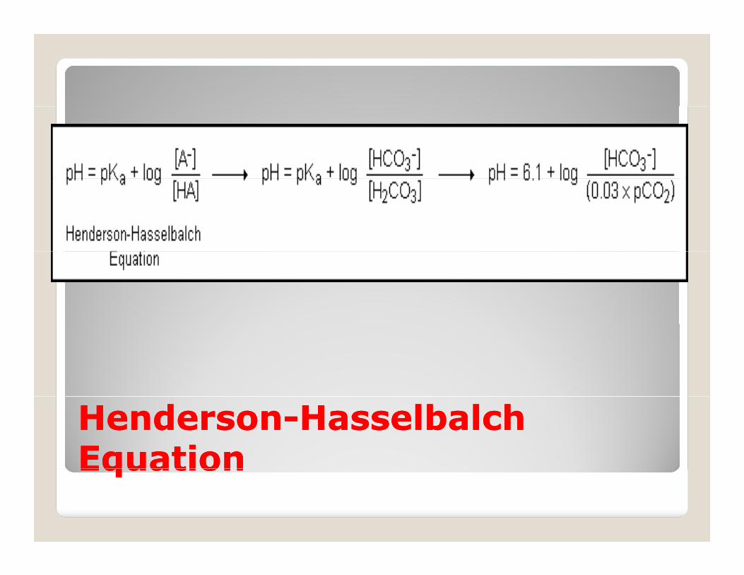
HendersonHenderson--HasselbalchHasselbalchEquationEquationEquationEquation


As dictated by the Henderson-Hasselbalch equation, disturbances in either the respiratory component disturbances in either the respiratory component (pCO2) or metabolic component (HCO3
-) can lead to alterations in pH.
Metabolic AcidosisMetabolic Acidosis(Too little HCO(Too little HCO --))
Metabolic AlkalosisMetabolic Alkalosis(Too much HCO(Too much HCO --))(Too little HCO(Too little HCO33 )) (Too much HCO(Too much HCO33 ))
Respiratory Acidosis Respiratory Acidosis Respiratory AlkalosisRespiratory Alkalosis(Too much CO(Too much CO22)) (Too little CO(Too little CO22))
Primary AcidPrimary Acid--Base DisordersBase DisordersPrimary AcidPrimary Acid Base DisordersBase Disorders

When a primary acid-base disorder exists, the body attempts to return the pH to normal via the “other half” of acid base metabolism.
Primary metabolic disorder Respiratory Primary metabolic disorder Respiratory compensation
Primary respiratory disorder Metabolic compensation
CompensationCompensationCompensationCompensation

Compensation (continued)Compensation (continued)p ( )p ( )
Primary DisorderPrimary Disorder Compensatory MechanismCompensatory Mechanism
Metabolic acidosisMetabolic acidosis Increased ventilationIncreased ventilation
Metabolic alkalosisMetabolic alkalosis Decreased ventilationDecreased ventilation
R i t id iR i t id i I d l b ti f HCOI d l b ti f HCORespiratory acidosisRespiratory acidosis Increased renal reabsorption of HCOIncreased renal reabsorption of HCO33--
in the proximal tubulein the proximal tubuleIncreased renal excretion of H in the Increased renal excretion of H in the
distal tubule distal tubule Respiratory alkalosisRespiratory alkalosis Decreased renal Decreased renal reabsorptionreabsorption of HCOof HCO33
--
in the proximal tubulein the proximal tubulein the proximal tubulein the proximal tubuleDecreased renal excretion of HDecreased renal excretion of H++ in the in the
distal tubule distal tubule

TypeType ofofDisturbanceDisturbance
PrimaryPrimary AlterationAlteration SecondarySecondaryResponseResponse
MechanismMechanism ofofSecondarySecondary
The Four Primary Acid-Base Disturbances
ResponseResponseMetabolicMetabolic acidosisacidosis Decrease in plasma
[HCO3-]
Decrease in Pa CO3 Hyperventilation
MetabolicMetabolic alkalosisalkalosis Increase in plasma[HCO3
-]Increase in PaCO3 Hypoventilation
[HCO3 ]
RespiratoryRespiratory acidosisacidosis Increase in PaCO3 Increase in plasma[HCO3
-]Acid titration of tissue buffers; transient increase in acid excretion and sustained enhancement of HCO3
-
reabsorption by kidneyreabsorption by kidney
RespiratoryRespiratory alkalosisalkalosis Decrease in Pa CO3 Decrease in plasma[HCO3
-]Alkaline titration of tissue buffers; transient
pp e ion of id e etion suppression of acid excretion and sustained reduction in bicarbonate reabsorption by kidney
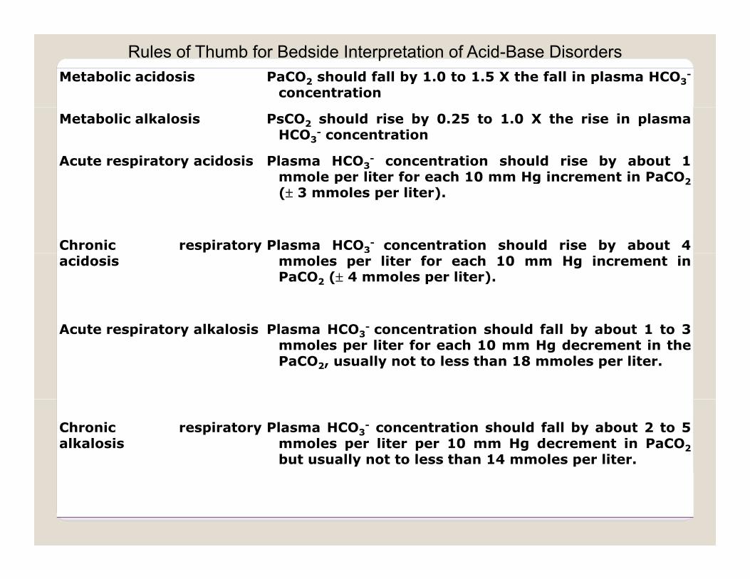
Metabolic acidosis PaCO2 should fall by 1.0 to 1.5 X the fall in plasma HCO3-
concentration
Rules of Thumb for Bedside Interpretation of Acid-Base Disorders
Metabolic alkalosis PsCO2 should rise by 0.25 to 1.0 X the rise in plasmaHCO3
- concentration
Acute respiratory acidosis Plasma HCO3- concentration should rise by about 1
mmole per liter for each 10 mm Hg increment in PaCO2mmole per liter for each 10 mm Hg increment in PaCO2( 3 mmoles per liter).
Chronic respiratory Plasma HCO3- concentration should rise by about 4
acidosis mmoles per liter for each 10 mm Hg increment inPaCO2 ( 4 mmoles per liter).
A t i t lk l i Pl HCO t ti h ld f ll b b t 1 t 3Acute respiratory alkalosis Plasma HCO3- concentration should fall by about 1 to 3
mmoles per liter for each 10 mm Hg decrement in thePaCO2, usually not to less than 18 mmoles per liter.
Chronic respiratoryalkalosis
Plasma HCO3- concentration should fall by about 2 to 5
mmoles per liter per 10 mm Hg decrement in PaCO2but usually not to less than 14 mmoles per liter.

REGULATION OF CO2 (Read also the separate article in the syllabus)
Plasma CO2 is determined by the rate of metabolic CO2 production2 2and by alveolar ventilation:
pCO2 = CO2 production x .84alveolar ventilation

H2 O H+ +OH−O O Only 1 in 14 million H2O molecules is
ionized to H+ and OH-
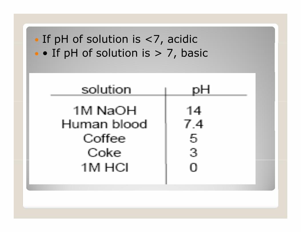
If pH of solution is <7, acidicp o so u o s , a d • If pH of solution is > 7, basic

Acids are compounds that donate a H+ tosolution
Bases are compounds that accept H+from solution

So what’s the big deal with H+?So what’s the big deal with H+?
• H+ is very reactive
• Almost all aspects of cell function can be influenced by H+
• Enzyme reactions are particularly sensitive to [H+]; there is an optimal pH above or below which the enzyme functions poorlyp y
• Normal extracell pH=7.4
• Acidosis pH<7.4 (death <6.8)
• Alkalosis pH>7 4 • Alkalosis pH>7.4

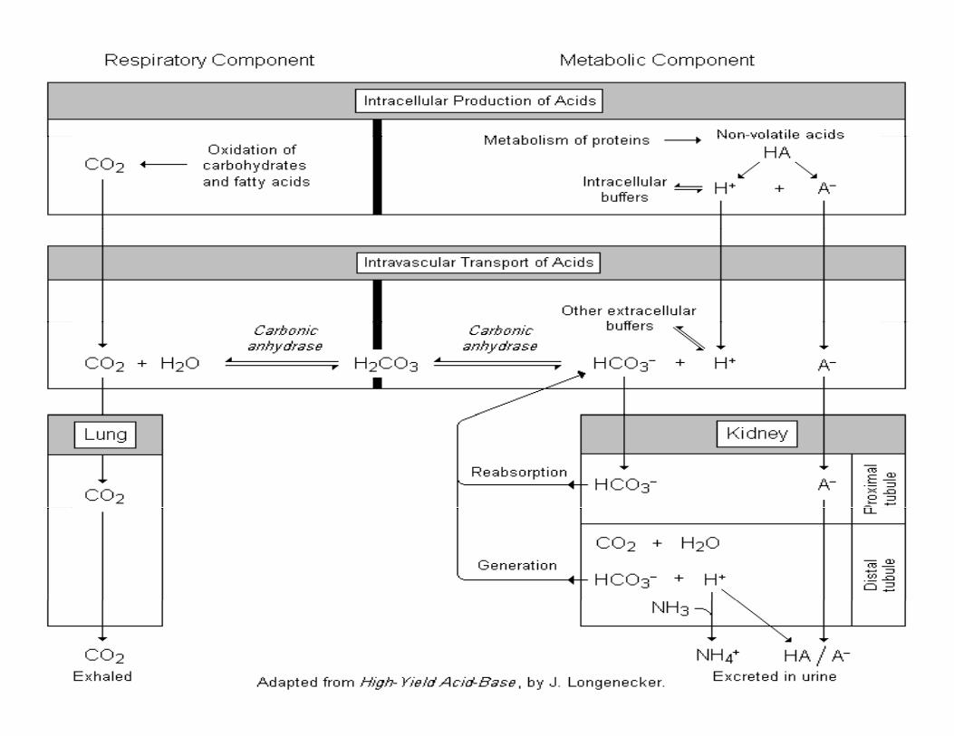
The body normally produces some acids:body o a y p odu s so a ds – Metabolism of proteins – Lactic acid from muscle
Disturbances of Acid-Base Balance1. RespiratoryRespiratory – changes in CO2
2. Metabolic 2. Metabolic – no change in CO2
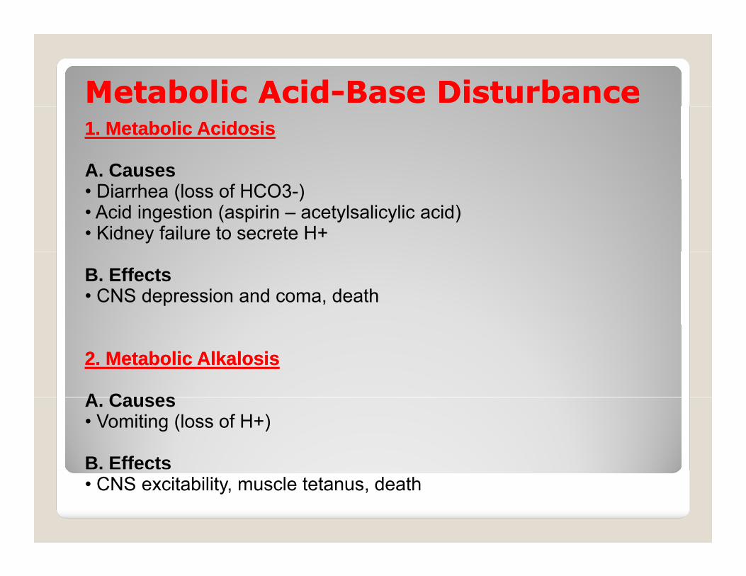
Metabolic AcidMetabolic Acid--Base DisturbanceBase Disturbance1. Metabolic Acidosis1. Metabolic Acidosis
A. Causes• Diarrhea (loss of HCO3-)• Acid ingestion (aspirin – acetylsalicylic acid)• Kidney failure to secrete H+
B. Effects• CNS depression and coma, death
2. Metabolic Alkalosis2. Metabolic Alkalosis
A CausesA. Causes• Vomiting (loss of H+)
B. Effects• CNS excitability, muscle tetanus, death

1. Fluid Buffering systemsu d u g sys s 2. Kidney 3. Respiratoryp y
AcidAcid--Base balanceBase balance
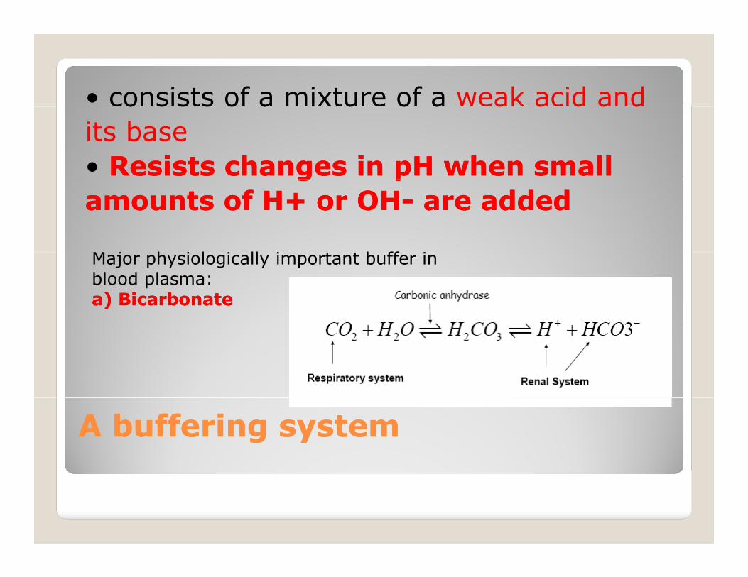
• consists of a mixture of a weak acid ando s s s o a u o a a a d a dits base• Resists changes in pH when smallResists changes in pH when smallg pg pamounts of H+ or OHamounts of H+ or OH-- are addedare added
M j h i l i ll i b ff iMajor physiologically important buffer inblood plasma:a) Bicarbonatea) Bicarbonate
A buffering systemA buffering system

General strategy
1. Balance the H+ intake and productionwith H+ excretion
2. Recover HCO3 to preserve bufferingcapability
Renal regulation of H+ and HCO3Renal regulation of H+ and HCO3
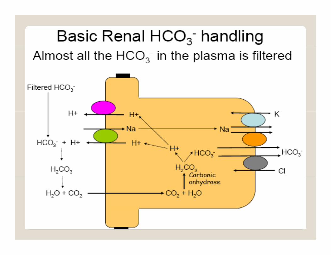

1. CO2 and H2O form H2CO3, which splits into H+ and HCO3
2. HCO3 moves to the interstitial fluid and blood
3. H+ is secreted into tubule, where it reacts with filtered HCO3 to CO2 d H2Oregenerate CO2 and H2O
4. For every HCO3- filtered, an HCO3 is formed within the tubular cell & transported to filtered, an HCO3 is formed within the tubular cell & transported to the interstitial fluid and blood
• “HCO3 reabsorption”
• A second important buffer in the tubular fluid is the phosphate system• Works in the tubular fluid to buffer H+ and allows for production of
HCO3new HCO3
A third important buffer in the tubular fluid is the ammonia system• Also works in the tubular fluid to buffer H+ and allows for • Also, works in the tubular fluid to buffer H+ and allows for production of new HCO3


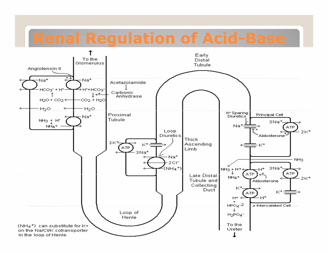
Renal Regulation of AcidRenal Regulation of Acid--BaseBase
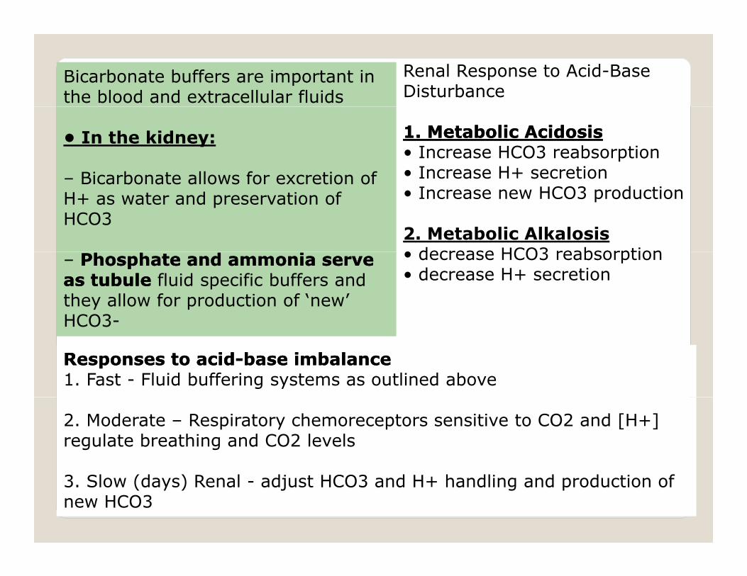
Bicarbonate buffers are important in the blood and extracellular fluids
Renal Response to Acid-Base Disturbance
• In the kidney:
– Bicarbonate allows for excretion of
11. Metabolic Acidosis. Metabolic Acidosis• Increase HCO3 reabsorption• Increase H+ secretion– Bicarbonate allows for excretion of
H+ as water and preservation of HCO3
• Increase new HCO3 production
2. Metabolic Alkalosis• decrease HCO3 reabsorption– Phosphate and ammonia serve Phosphate and ammonia serve
as tubule as tubule fluid specific buffers and they allow for production of ‘new’ HCO3-
• decrease HCO3 reabsorption• decrease H+ secretion
HCO3
Responses to acidResponses to acid--base imbalancebase imbalance1. Fast - Fluid buffering systems as outlined above
2. Moderate – Respiratory chemoreceptors sensitive to CO2 and [H+] regulate breathing and CO2 levels
3. Slow (days) Renal - adjust HCO3 and H+ handling and production of new HCO3

1 Check the pH1. Check the pH
If the pH < 7.35, acidemia (and at least 1 acidosis) is p , ( )present.
If th H > 7 45 lk l i ( d t l t 1 lk l i ) If the pH > 7.45, alkalemia (and at least 1 alkalosis) is present.
Practical ApproachPractical ApproachPractical ApproachPractical Approach

2 Check the pCO22. Check the pCO2
pH < 7.35 and pCO2 < 40 metabolic acidosisp p 2pH < 7.35 and pCO2 > 40 respiratory acidosis
pH > 7.45 and pCO2 < 40 respiratory alkalosispH > 7.45 and pCO2 > 40 metabolic acidosis
Practical ApproachPractical ApproachPractical ApproachPractical Approach

Practical ApproachPractical ApproachPractical ApproachPractical ApproachMost prominent disorderMost prominent disorder Compensation formulaCompensation formula
Metabolic acidosisMetabolic acidosis pCOpCO22 ≈ 1.5 [HCO≈ 1.5 [HCO33--] + 8 ] + 8
Metabolic alkalosisMetabolic alkalosis pCOpCO22 ≈ 0.9 [HCO≈ 0.9 [HCO33--] + 16 ] + 16
Respiratory acidosisRespiratory acidosis For every 10 ∆ in pCOFor every 10 ∆ in pCO22, pH decreases by:, pH decreases by:0.08 (in acute resp. acidoses)0.08 (in acute resp. acidoses)0 03 (in chronic resp acidoses)0 03 (in chronic resp acidoses)0.03 (in chronic resp. acidoses) 0.03 (in chronic resp. acidoses)
Respiratory alkalosisRespiratory alkalosis For every 10 ∆ in pCOFor every 10 ∆ in pCO22, pH increases by:, pH increases by:0.08 (in acute resp. alkaloses)0.08 (in acute resp. alkaloses)0.03 (in chronic resp. alkaloses) 0.03 (in chronic resp. alkaloses)

A i "G "Anion "Gap"1.An "artefact" of how we measure blood electrolytes 2.Determined by:y
Normal = 10 3 If the anion gap is normal with acidosis then Cl- has3.If the anion gap is normal with acidosis then Cl has increased to match HCO3- decline 4.If the anion gap is increased some other anion is involved

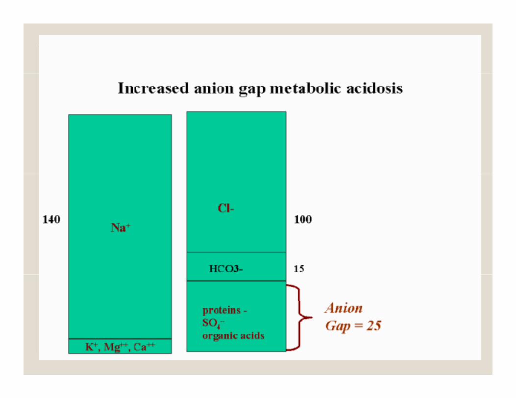


Calculate the anion gap
Anion gap = [Na+] – ( [Cl-] + [HCO3-] )
If the anion gap is elevated, an elevated gap t b li id i i lik l tmetabolic acidosis is likely present.
Calc late HCO3 deficitCalculate HCO3- deficitHCO3- deficit = (kg body weight)x (o.4) x (desired [HCO3-) – measured (HCO3-))
Practical ApproachPractical Approach
(desired [HCO3 ) – measured (HCO3 ))
Practical ApproachPractical Approach

The concentrations are expressed in units of milliequivalents/liter (mEq/L) or in millimoles/litre (mmol/L).
With potassiumWith potassium
It is calculated by subtracting the serum concentrations of chloride and bicarbonate (anions) from the concentrations of sodium sodium and potassium (and potassium (cationscations):):
([ ] [ ]) ([([ ] [ ]) ([ClCl ] [ CO3 ])] [ CO3 ]) = ([Na+] + [K+]) − ([= ([Na+] + [K+]) − ([ClCl−] + [HCO3−])−] + [HCO3−])
Without potassium (Daily practice)Without potassium (Daily practice)
However, the potassium is frequently ignored because potassium concentrations, being very low, usually have little effect on the calculated gap. This leaves the following equation:
= [Na+] − ([= [Na+] − ([ClCl--] + [HCO3−])] + [HCO3−])
Anion gapAnion gap
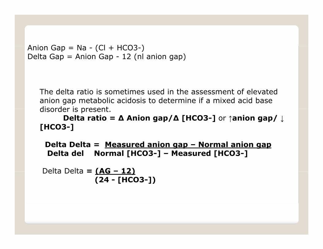
A i G N (Cl + HCO3 ) Anion Gap = Na - (Cl + HCO3-) Delta Gap = Anion Gap - 12 (nl anion gap)
The delta ratio is sometimes used in the assessment of elevated anion gap metabolic acidosis to determine if a mixed acid base disorder is present disorder is present.
Delta ratio = ∆ Anion gap/∆ [HCO3-] or ↑anion gap/ ↓[HCO3-]
Delta Delta = Measured anion gap – Normal anion gap Delta del Normal [HCO3-] – Measured [HCO3-]
Delta Delta = (AG – 12)Delta Delta = (AG 12)(24 - [HCO3-])

Delta ratio Assessment Guidelines
< 0.4 Hyperchloremic normal anion gap acidosis
< 1 High AG & normal AG acidosis
1 to 2 Pure Anion Gap Acidosis Lactic acidosis: average value 1.6DKA more likely to have a ratio closer to 1 due yto urine ketone loss
High AG acidosis and a concurrent metabolic > 2
galkalosisor a pre-existing compensated respiratory acidosis

High anion gap metabolic acidosis
. A high anion gap indicates acidosis. e.g. In uncontrolled diabetes, there is an increase in ketoacids (i.e. an increase in unmeasured anions) and a resulting increase in the anion gap. In these conditions, bicarbonate concentrations decrease, in response to the need to buffer the increased presence of acids (as a result of the underlying condition). The bicarbonate is consumed by the unmeasured anion (via its action as a buffer) resulting in a high unmeasured anion (via its action as a buffer) resulting in a high anion gap.
Lactic acidosis Ketoacidosis
MethanolPropylene Glycol
Diabetic ketoacidosis Alcohol abuseToxins: Toxins:
py yPhenforminAspirinCyanide, coupled with elevated venous oxygenation
Ethylene glycol Lactic acid Uremia
venous oxygenationIronIsoniazid
High anion gapHigh anion gapRenal failure,Renal failure,

In patients with a normal anion gap the drop in HCO3− is compensated for almost completely by an increase in Cl− and hence is also known as hyperchloremic acidosis.
The HCO3− lost is replaced by a chloride anion, and thus there is a normal anion gap.Gastrointestinal loss of HCO3 (i e diarrhea) (note: vomiting causes Gastrointestinal loss of HCO3− (i.e., diarrhea) (note: vomiting causes hypochloraemic alkalosis)
Renal loss of HCO3− (i.e. proximal renal tubular acidosis(RTA) also known as type 2 RTA)known as type 2 RTA)
Renal dysfunction (i.e. distal renal tubular acidosis also known as type 1 RTA)
Ingestions Ammonium chloride and Acetazolamide, ifosfamide.Hyperalimentation fluids (i.e. total parenteral nutrition)
Some cases of ketoacidosis, particularly during rehydration with Na+ t i i IV l ticontaining IV solutions.
Alcohol (such as ethanol) can cause a high anion gap acidosis in some patients, but a mixed picture in others due to concurrent metabolic alkalosis
Normal anion gapNormal anion gap
alkalosis. Mineralocorticoid deficiency (Addison's disease)

A low anion gap is frequently caused by hypoalbuminemiayp
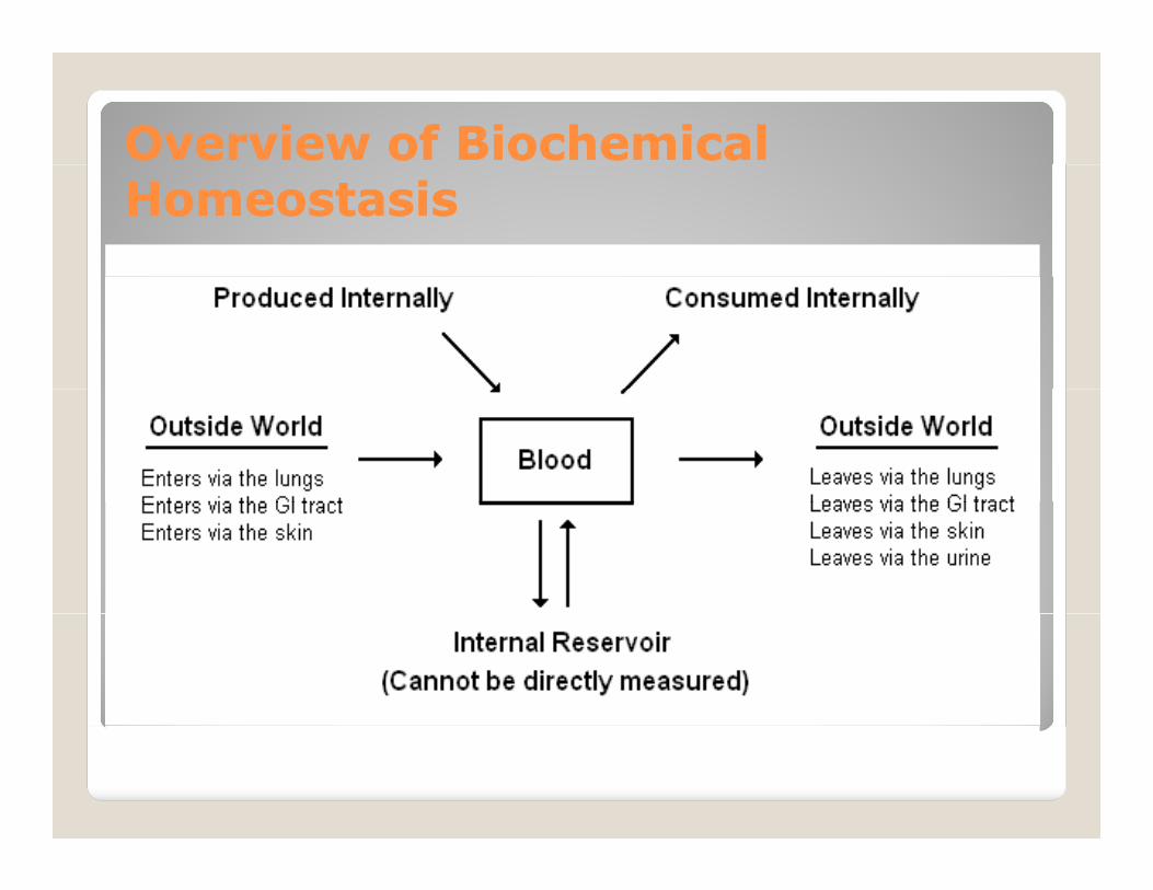
Overview of Biochemical Overview of Biochemical HomeostasisHomeostasis

Differential Differential DiagonsisDiagonsis for Acidfor Acid--Base DisordersBase Disorders