acid sphingomyelinase gene deficiency -
TRANSCRIPT

The American Journal of Pathology, Vol. 179, No. 5, November 2011
Copyright © 2011 American Society for Investigative Pathology.
Published by Elsevier Inc. All rights reserved.
DOI: 10.1016/j.ajpath.2011.07.019
Cardiovascular, Pulmonary, and Renal Pathology
Acid Sphingomyelinase Gene DeficiencyAmeliorates the Hyperhomocysteinemia-Induced
Glomerular Injury in MiceKrishna M. Boini,* Min Xia,* Caixia Li,*Chun Zhang,* Lori P. Payne,* Justine M. Abais,*Justin L. Poklis,* Philip B. Hylemon,† andPin-Lan Li*From the Departments of Pharmacology and Toxicology,* and
Microbiology and Immunology,† Medical College of Virginia
Campus, Virginia Commonwealth University, Richmond,
Virginia
Hyperhomocysteinemia (hHcys) enhances ceramideproduction, leading to the activation of NADPH oxi-dase and consequent glomerular oxidative stress andsclerosis. The present study was performed to de-termine whether acid sphingomyelinase (Asm), aceramide-producing enzyme, is implicated in thedevelopment of hHcys-induced glomerular oxida-tive stress and injury. Uninephrectomized Asm-knockout (Asm�/�) and wild-type (Asm�/�) mice,with or without Asm short hairpin RNA (shRNA)transfection, were fed a folate-free (FF) diet for 8weeks, which significantly elevated the plasma Hcyslevel compared with mice fed normal chow. By usingin vivo molecular imaging, we found that transfectedshRNAs were expressed in the renal cortex startingon day 3 and continued for 24 days. The FF diet sig-nificantly increased renal ceramide production, AsmmRNA and activity, urinary total protein and albuminexcretion, glomerular damage index, and NADPH-de-pendent superoxide production in the renal cortexfrom Asm�/� mice compared with that from Asm�/�
or Asm shRNA-transfected wild-type mice. Immuno-fluorescence analysis showed that the FF diet de-creased the expression of podocin but increaseddesmin and ceramide levels in glomeruli fromAsm�/� mice but not in those from Asm�/� and AsmshRNA-transfected wild-type mice. In conclusion, ourobservations reveal that Asm plays a pivotal role inmediating podocyte injury and glomerular sclerosis as-sociated with NADPH oxidase–associated local oxidativestress during hHcys. (Am J Pathol 2011, 179:2210–2219;
DOI: 10.1016/j.ajpath.2011.07.019)
2210
Hyperhomocysteinemia (hHcys) is an important patho-genic factor in the progression of end-stage renal dis-ease and in the development of cardiovascular compli-cations related to end-stage renal disease.1,2 Hcysinduces extracellular matrix accumulation and inhibits itsdegradation in glomeruli, ultimately leading to glomeru-losclerosis and loss of renal function.2–4 In addition, ourrecent studies5 revealed that hHcys initiates glomerulardamage by inducing podocyte injury. Furthermore, sev-eral studies2,6–10 have also demonstrated that local oxi-dative stress mediated by NADPH oxidase (Nox) is im-portantly involved in the progression of glomerular injuryassociated with hHcys. However, how the local oxidativestress is activated and thereby results in glomerular injuryduring hHcys has not yet been fully elucidated.
Previous studies11–19 have reported that sphingolipids(mainly ceramide) participate in signal transduction, cellmembrane formation, and plasma lipoprotein metabo-lism, all of which have an impact on the development ofatherosclerosis and other sclerotic diseases, such asinsulin resistance, obesity, Alzheimer’s disease, and cys-tic fibrosis. Ceramide production is mainly mediated viathe hydrolysis of membrane sphingomyelin by varioussphingomyelinases, such as acid sphingomyelinase(Asm) or neutral sphingomyelinase, or by de novo syn-thesis via serine palmitoyltransferase and ceramide syn-thase.20 Ceramide is subsequently metabolized into sph-ingosine by ceramidases, and sphingosine can befurther converted to sphingosine-1-phosphate via sphin-gosine kinase,20 in response to a variety of stimuli, in-cluding pro-inflammatory cytokines, oxidative stress, andincreased levels of free fatty acids. In addition, ceramideis considered a critical signaling molecule mediating the
Supported by grants from the NIH (DK54927, HL091464, HL57244, andHL075316).
Accepted for publication July 29, 2011.
Supplemental material for this article can be found at http://ajp.amjpathol.org or at doi: 10.1016/j.ajpath.2011.07.019.
Address reprint requests to Pin-Lan Li, M.D., Ph.D., Department ofPharmacology and Toxicology, Medical College of Virginia Campus, Vir-ginia Commonwealth University, 410 N. 12th St., Richmond, VA 23298.
E-mail: [email protected].
Asm in hHcys-Induced Glomerular Injury 2211AJP November 2011, Vol. 179, No. 5
activation of NADPH oxidase in different cells and tis-sues.4 Enhanced plasma Hcys concentrations increasethe ceramide production and NADPH activity in the kid-ney of hyperhomocysteinemic rats. Inhibition of ceramideproduction improved glomerular injury in those hyperho-mocysteinemic rats.4 However, it remains unknownwhether alterations of Asm gene expression and regula-tion are implicated in the development of hHcys-inducedglomerular oxidative stress and injury, ultimately resultingin glomerulosclerosis.
An inherited deficiency of Asm activity results in the typeA and B forms of Niemann-Pick disease, and Asm-knockoutmice are resistant to radiation21 and other forms of stress-induced apoptosis.22 Asm inhibition has rendered cells andanimals resistant to the apoptotic effects of diverse stimuli,including Fas-CD95,23 ischemia,24 radiation,25 chemo-therapy,26 and tumor necrosis factor-�.27 Asm knockoutor Asm inhibition had protective action during lung in-flammation and fibrosis,28 cystic fibrosis,29,30 obesity andassociated glomerular injury,31 liver fibrogenesis,32 andrenal fibrosis.33
The present study hypothesized that Asm gene defi-ciency protects glomeruli from hHcys-induced glomeru-lar oxidative stress and injury and thereby amelioratesglomerulosclerosis under such pathological conditions.To test this hypothesis, we first performed a series ofexperiments using Asm�/� and their wild-type (WT) litter-mates fed the normal chow or folate-free (FF) diet todetermine whether lack of the Asm gene alters renalceramide production, glomerular local oxidative stress,and podocyte injury in mice during hHcys. Then, welocally silenced the renal Asm gene using short hairpinRNA (shRNA) and observed the effects of renal Asmdeficiency on hHcys-induced glomerular oxidative stressand corresponding injury. Our results demonstrate thatan Asm gene defect or deficiency in the kidney improvesthe hHcys-induced local oxidative stress and glomerularinjury, ultimately preventing glomerulosclerosis.
Materials and Methods
Animals
Eight-week-old male C57BL/6J WT Asm�/� mice andtheir WT littermates were used in the present study. Tospeed up the damaging effects of hHcys on glomeruli,all mice were uninephrectomized, as previously de-scribed.4,5 This model has induced glomerular damageunrelated to the uninephrectomy and arterial blood pres-sure but specific to hHcys.5 After a 1-week recoveryperiod from uninephrectomy, mice were fed either a nor-mal chow or an FF diet (Dyets Inc., Bethlehem, PA) for 8weeks to induce hHcys. In another series of C57BL/6Jmice, Asm shRNA or a scrambled shRNA (Sigma, StLouis, MO) plasmid with a luciferase expression vectorwas cotransfected into the kidneys via intrarenal arteryinjection using the ultrasonographic microbubble system,as previously described.2 After delivery of plasmids intothe kidney, these uninephrectomized mice were main-
tained on a normal or an FF diet for 8 weeks. All protocolswere approved by the Institutional Animal Care and UseCommittee of Virginia Commonwealth University (Rich-mond, VA).
HPLC Analysis
Plasma and renal tissue Hcys levels were measured bythe high-performance liquid chromatography (HPLC)method, as previously described.5,34 A 100-�L plasmasample or standard solution mixed with 10 �L of internalstandard, thioglycolic acid (2.0 mmol/L) solution, wastreated with 10 �L of 10% tri-n-butylphosphine solution indimethylformamide at 4°C for 30 minutes. Then, 80 �L ofice-cold 10% trichloroacetic acid in 1 mmol/L EDTA wasadded and centrifuged to remove proteins in the sample.The supernatant, 100 �L, was transferred into the mixtureof 20 �L of 1.55 mol/L sodium hydroxide, 250 �L of 0.125mol/L borate buffer (pH 9.5), and 100 �L of 1.0 mg/mL4-fluoro-7-aminosulfonylbenzofurazan solution. The re-sulting mixture was incubated at 60°C for 30 minutes toaccomplish derivatization of thiols. HPLC was performedwith an HP 1100 series instrument (Agilent Technologies,Waldbronn, Germany) equipped with a binary pump, avacuum degasser, a thermostated column compartment,and an autosampler (Agilent Technologies). Separationwas performed at an ambient temperature on an analyt-ical column, Supelco LC-18-DB (Supelco, Bellefonte, PA;150 � 4.6 mm i.d., 5 �m particle size) with a SupelcosilLC-18 guard column (Supelco; 20 � 4.6 mm i.d., 5 �mparticle size). Fluorescence intensities were measuredwith an excitation wavelength of 385 nm and an emissionwavelength of 515 nm by a Hewlett-Packard model1046A fluorescence detector (Agilent Technologies). Thepeak area of the chromatographs was quantified with aHewlett-Packard 3392 integrator (Agilent Technologies).The analytical column was eluted with 0.1 mol/L potas-sium dihydrogen phosphate buffer (pH 2.1) containing6% acetonitrile (v/v) as the mobile phase, with a flow rateof 2.0 mL/minute.
Morphological Examination Findings
The fixed kidneys were paraffin embedded, and sectionswere prepared and stained with PAS stain. The glomer-ular damage index (GDI) was calculated from 0 to 4 onthe basis of the degree of glomerulosclerosis and mes-angial matrix expansion, as previously described.35 Ingeneral, we counted 50 glomeruli in total in each kidneyslice under a microscope, when each glomerulus wasgraded with level 0 to 4 damage, as follows: 0 representsno lesion; 1�, sclerosis of �25% of the glomerulus; 2�,sclerosis of 25% to 50% of the glomerulus; 3�, sclerosisof �50% to 75% of the glomerulus; and 4�, sclerosis of�75% of the glomerulus. A whole kidney average scle-rosis index was obtained by averaging scores fromcounted glomeruli.5 This observation was examined bytwo independent investigators (K.M.B. and M.X.) who
were blinded to the treatment of the experimental groups.
2212 Boini et alAJP November 2011, Vol. 179, No. 5
Asm Activity in Mice Lacking Asm
The activity of Asm was determined as previously de-scribed.36,37 Briefly, N-methyl-[14C]-sphingomyelin wasincubated with renal cortex tissue homogenates, and themetabolite of sphingomyelin, [14C]-choline phosphate,was quantified. An aliquot of homogenates (20 �g) wasmixed with 0.02 �Ci of N-methyl-[14C]-sphingomyelin in100-�L acidic reaction buffer containing 100 mmol/L so-dium acetate and 0.1% Triton X-100, pH 5.0, and incu-bated at 37°C for 15 minutes. The reaction was termi-nated by adding 1.5 mL of chloroform-methanol (2:1) and0.2 mL of double-distilled water. The samples were thenvortex mixed and centrifuged at 1000 � g for 5 minutes toseparate into two phases. A portion of the upper aqueousphase containing [14C]-choline phosphate was trans-ferred to scintillation vials and counted in a Beckmanliquid scintillation counter. The choline phosphate forma-tion rate (nmol/min/mg protein) was calculated to repre-sent the enzyme activity.
LC–Electrospray Ionization Tandem MS forQuantitation of Ceramide
The separation, identification, and quantitation of cer-amide in plasma were performed by LC–mass spectrom-etry (MS). The HPLC instrument was equipped with abinary pump, a vacuum degasser, a thermostated col-umn compartment, and an autosampler (Waters, Milford,MA). The HPLC separations were performed at 70°C onan RP C18 Nucleosil AB column (5 �m, 70 � 2-mm i.d.)from Macherey Nagel (Duren, Germany). The mobilephase was a gradient mixture formed as previously de-scribed.38 The plasma lipids were extracted according toprevious studies. To avoid any loss of lipids, the wholeprocedure was performed in siliconized glassware. MSdetection was performed using a Quattro II quadrupoleMS instrument (Micromass, Altrincham, UK) operatingunder MassLynx 3.5 and configured with a Z-spray elec-trospray ionization source. Source conditions were pre-viously described.4,38
Cell Culture
The conditionally immortalized mouse podocyte cell line,provided by Dr. P. E. Klotman (Division of Nephrology,Department of Medicine, Mount Sinai School of Medicine,New York, NY), was cultured on type I collagen–coatedflasks or plates in RPMI 1640 medium supplemented withrecombinant mouse interferon-� at 33°C. After differenti-ation at 37°C for 10 to 14 days without interferon-�, podo-cytes were used for the proposed experiments.
Confocal Microscopic Detection of MR and ItsColocalization with Asm in Podocytes
Podocytes were seeded on poly-L-lysine–coated cham-bers and treated with L-Hcys (40 �mol/L) or vehicle for 30minutes. In an additional group of cells, amitriptyline (20
�m; Sigma) was added to pretreat cells for 30 minutesbefore the addition of L-Hcys. The detection of membraneraft (MR) clusters and their colocalization were performedas previously described. Briefly, podocytes were washedwith cold PBS, fixed for 15 minutes in 4% paraformalde-hyde, and then blocked with 1% bovine serum albumin inPBS for 30 minutes. For detection of colocalization of MRand Asm, podocytes were first incubated with Alexa488-labeled cholera toxin B at 1 �g/mL (Molecular Probes,Eugene, OR), as previously described.39 Then, the podo-cytes were incubated overnight with goat anti-Asm (1:200; Santa Cruz Biotechnology, Santa Cruz, CA), fol-lowed by incubation with 5 �g/mL Texas Red–conjugated anti-goat IgG for an additional 1 hour at roomtemperature. After mounting, the slides were observedusing a confocal laser-scanning microscope (FluoviewFV1000; Olympus, Tokyo, Japan).39
Urinary Total Protein and Albumin ExcretionMeasurements
The 24-hour urine samples were collected using meta-bolic cages and subjected to total protein and albuminexcretion measurements.31 The total protein content inthe urine was detected by the Bradford method using aUV spectrophotometer. Urine albumin was detected us-ing a commercially available albumin enzyme-linked im-munosorbent assay kit (Bethyl Laboratories, Montgom-ery, TX). The urinary creatinine concentrations weremeasured by using a QuantiChrom creatinine assay kit(BioAssay System, Hayward, CA).40
Delivery of Asm shRNA into the Kidneys by anUltrasonograpic Microbubble Technique
After a 1-week recovery period from uninephrectomy,Asm shRNA or a scrambled shRNA plasmid with a lucif-erase expression vector was cotransfected into the kid-neys via intrarenal artery injection using the ultrasono-graphic microbubble system. A full description of theprocedures for the ultrasonographic microbubble genetransfer technique can be found in our previous studies.2
To daily monitor the efficiency of gene expressionthrough somatic plasmid transfection, mice were anes-thetized with isoflurane and an aqueous solution of lucife-rin (150 mg/kg) was injected i.p. 5 minutes before imag-ing. The anesthetized mice were imaged using theIVIS200 in vivo molecular imaging system (Xenogen, Hop-kinton, MA). Photons emitted from luciferase-expressingcells within the animal body and transmitted through tissuelayers were quantified over a defined period, ranging up to 5minutes, using the software program Living Image (Xenogen).The inhibitory efficiency of gene expression by Asm shRNAwas further confirmed by detection of the Asm mRNA level inmouse renal cortex using real-time RT-PCR.
ESR Analysis of O2· – Production
For detection of Nox-dependent superoxide (O2· –)
production, proteins from mouse renal cortex were ex-
tracted using sucrose buffer and resuspended with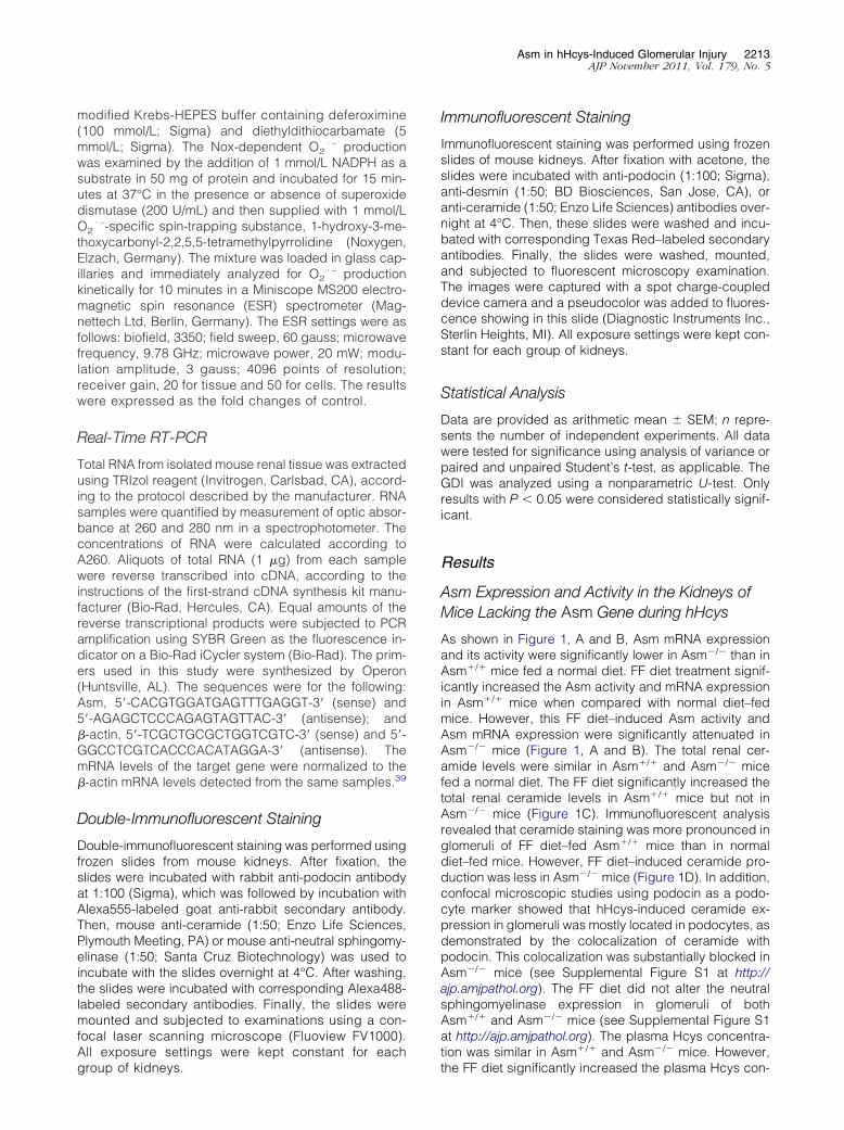
Asm in hHcys-Induced Glomerular Injury 2213AJP November 2011, Vol. 179, No. 5
modified Krebs-HEPES buffer containing deferoximine(100 mmol/L; Sigma) and diethyldithiocarbamate (5mmol/L; Sigma). The Nox-dependent O2
· – productionwas examined by the addition of 1 mmol/L NADPH as asubstrate in 50 mg of protein and incubated for 15 min-utes at 37°C in the presence or absence of superoxidedismutase (200 U/mL) and then supplied with 1 mmol/LO2
· –-specific spin-trapping substance, 1-hydroxy-3-me-thoxycarbonyl-2,2,5,5-tetramethylpyrrolidine (Noxygen,Elzach, Germany). The mixture was loaded in glass cap-illaries and immediately analyzed for O2
· – productionkinetically for 10 minutes in a Miniscope MS200 electro-magnetic spin resonance (ESR) spectrometer (Mag-nettech Ltd, Berlin, Germany). The ESR settings were asfollows: biofield, 3350; field sweep, 60 gauss; microwavefrequency, 9.78 GHz; microwave power, 20 mW; modu-lation amplitude, 3 gauss; 4096 points of resolution;receiver gain, 20 for tissue and 50 for cells. The resultswere expressed as the fold changes of control.
Real-Time RT-PCR
Total RNA from isolated mouse renal tissue was extractedusing TRIzol reagent (Invitrogen, Carlsbad, CA), accord-ing to the protocol described by the manufacturer. RNAsamples were quantified by measurement of optic absor-bance at 260 and 280 nm in a spectrophotometer. Theconcentrations of RNA were calculated according toA260. Aliquots of total RNA (1 �g) from each samplewere reverse transcribed into cDNA, according to theinstructions of the first-strand cDNA synthesis kit manu-facturer (Bio-Rad, Hercules, CA). Equal amounts of thereverse transcriptional products were subjected to PCRamplification using SYBR Green as the fluorescence in-dicator on a Bio-Rad iCycler system (Bio-Rad). The prim-ers used in this study were synthesized by Operon(Huntsville, AL). The sequences were for the following:Asm, 5=-CACGTGGATGAGTTTGAGGT-3= (sense) and5=-AGAGCTCCCAGAGTAGTTAC-3= (antisense); and�-actin, 5=-TCGCTGCGCTGGTCGTC-3= (sense) and 5=-GGCCTCGTCACCCACATAGGA-3= (antisense). ThemRNA levels of the target gene were normalized to the�-actin mRNA levels detected from the same samples.39
Double-Immunofluorescent Staining
Double-immunofluorescent staining was performed usingfrozen slides from mouse kidneys. After fixation, theslides were incubated with rabbit anti-podocin antibodyat 1:100 (Sigma), which was followed by incubation withAlexa555-labeled goat anti-rabbit secondary antibody.Then, mouse anti-ceramide (1:50; Enzo Life Sciences,Plymouth Meeting, PA) or mouse anti-neutral sphingomy-elinase (1:50; Santa Cruz Biotechnology) was used toincubate with the slides overnight at 4°C. After washing,the slides were incubated with corresponding Alexa488-labeled secondary antibodies. Finally, the slides weremounted and subjected to examinations using a con-focal laser scanning microscope (Fluoview FV1000).All exposure settings were kept constant for each
group of kidneys.Immunofluorescent Staining
Immunofluorescent staining was performed using frozenslides of mouse kidneys. After fixation with acetone, theslides were incubated with anti-podocin (1:100; Sigma),anti-desmin (1:50; BD Biosciences, San Jose, CA), oranti-ceramide (1:50; Enzo Life Sciences) antibodies over-night at 4°C. Then, these slides were washed and incu-bated with corresponding Texas Red–labeled secondaryantibodies. Finally, the slides were washed, mounted,and subjected to fluorescent microscopy examination.The images were captured with a spot charge-coupleddevice camera and a pseudocolor was added to fluores-cence showing in this slide (Diagnostic Instruments Inc.,Sterlin Heights, MI). All exposure settings were kept con-stant for each group of kidneys.
Statistical Analysis
Data are provided as arithmetic mean � SEM; n repre-sents the number of independent experiments. All datawere tested for significance using analysis of variance orpaired and unpaired Student’s t-test, as applicable. TheGDI was analyzed using a nonparametric U-test. Onlyresults with P � 0.05 were considered statistically signif-icant.
Results
Asm Expression and Activity in the Kidneys ofMice Lacking the Asm Gene during hHcys
As shown in Figure 1, A and B, Asm mRNA expressionand its activity were significantly lower in Asm�/� than inAsm�/� mice fed a normal diet. FF diet treatment signif-icantly increased the Asm activity and mRNA expressionin Asm�/� mice when compared with normal diet–fedmice. However, this FF diet–induced Asm activity andAsm mRNA expression were significantly attenuated inAsm�/� mice (Figure 1, A and B). The total renal cer-amide levels were similar in Asm�/� and Asm�/� micefed a normal diet. The FF diet significantly increased thetotal renal ceramide levels in Asm�/� mice but not inAsm�/� mice (Figure 1C). Immunofluorescent analysisrevealed that ceramide staining was more pronounced inglomeruli of FF diet–fed Asm�/� mice than in normaldiet–fed mice. However, FF diet–induced ceramide pro-duction was less in Asm�/� mice (Figure 1D). In addition,confocal microscopic studies using podocin as a podo-cyte marker showed that hHcys-induced ceramide ex-pression in glomeruli was mostly located in podocytes, asdemonstrated by the colocalization of ceramide withpodocin. This colocalization was substantially blocked inAsm�/� mice (see Supplemental Figure S1 at http://ajp.amjpathol.org). The FF diet did not alter the neutralsphingomyelinase expression in glomeruli of bothAsm�/� and Asm�/� mice (see Supplemental Figure S1at http://ajp.amjpathol.org). The plasma Hcys concentra-tion was similar in Asm�/� and Asm�/� mice. However,
the FF diet significantly increased the plasma Hcys con-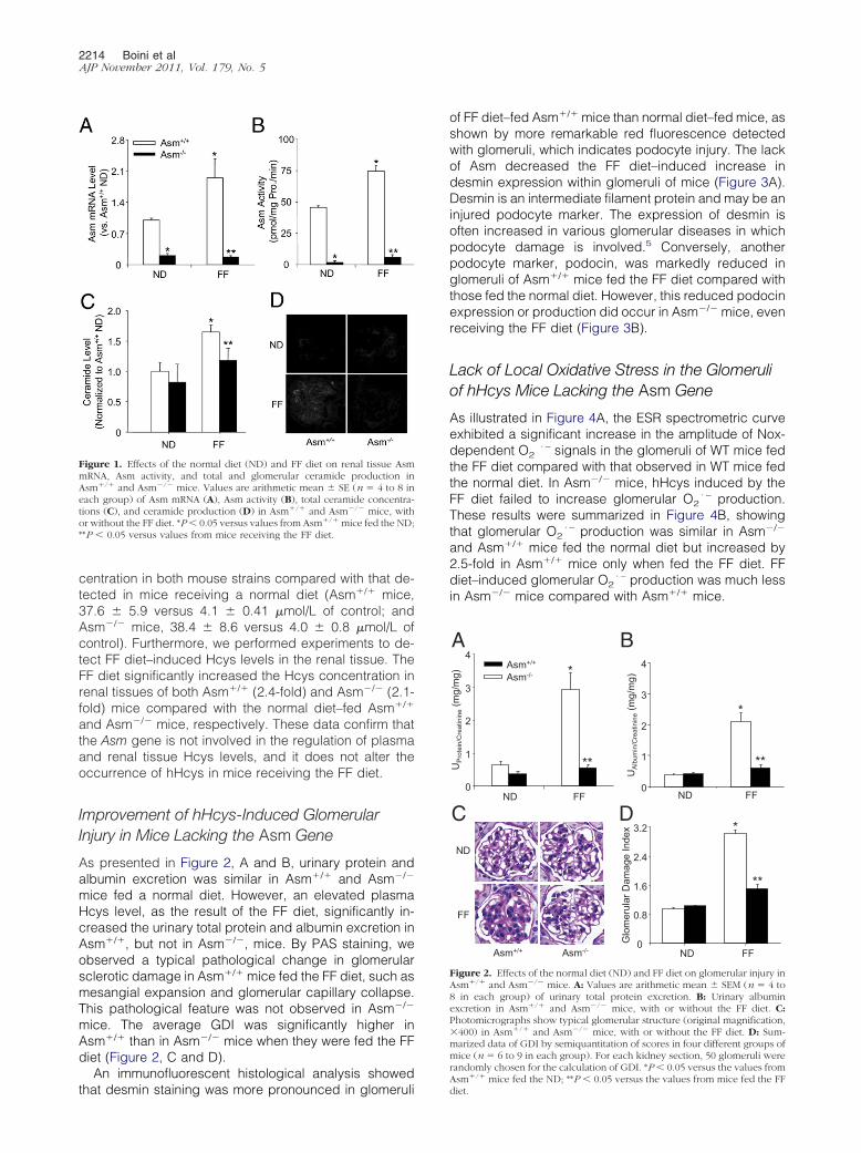
2214 Boini et alAJP November 2011, Vol. 179, No. 5
centration in both mouse strains compared with that de-tected in mice receiving a normal diet (Asm�/� mice,37.6 � 5.9 versus 4.1 � 0.41 �mol/L of control; andAsm�/� mice, 38.4 � 8.6 versus 4.0 � 0.8 �mol/L ofcontrol). Furthermore, we performed experiments to de-tect FF diet–induced Hcys levels in the renal tissue. TheFF diet significantly increased the Hcys concentration inrenal tissues of both Asm�/� (2.4-fold) and Asm�/� (2.1-fold) mice compared with the normal diet–fed Asm�/�
and Asm�/� mice, respectively. These data confirm thatthe Asm gene is not involved in the regulation of plasmaand renal tissue Hcys levels, and it does not alter theoccurrence of hHcys in mice receiving the FF diet.
Improvement of hHcys-Induced GlomerularInjury in Mice Lacking the Asm Gene
As presented in Figure 2, A and B, urinary protein andalbumin excretion was similar in Asm�/� and Asm�/�
mice fed a normal diet. However, an elevated plasmaHcys level, as the result of the FF diet, significantly in-creased the urinary total protein and albumin excretion inAsm�/�, but not in Asm�/�, mice. By PAS staining, weobserved a typical pathological change in glomerularsclerotic damage in Asm�/� mice fed the FF diet, such asmesangial expansion and glomerular capillary collapse.This pathological feature was not observed in Asm�/�
mice. The average GDI was significantly higher inAsm�/� than in Asm�/� mice when they were fed the FFdiet (Figure 2, C and D).
An immunofluorescent histological analysis showed
Figure 1. Effects of the normal diet (ND) and FF diet on renal tissue AsmmRNA, Asm activity, and total and glomerular ceramide production inAsm�/� and Asm�/� mice. Values are arithmetic mean � SE (n � 4 to 8 ineach group) of Asm mRNA (A), Asm activity (B), total ceramide concentra-tions (C), and ceramide production (D) in Asm�/� and Asm�/� mice, withor without the FF diet. *P � 0.05 versus values from Asm�/� mice fed the ND;**P � 0.05 versus values from mice receiving the FF diet.
that desmin staining was more pronounced in glomeruli
of FF diet–fed Asm�/� mice than normal diet–fed mice, asshown by more remarkable red fluorescence detectedwith glomeruli, which indicates podocyte injury. The lackof Asm decreased the FF diet–induced increase indesmin expression within glomeruli of mice (Figure 3A).Desmin is an intermediate filament protein and may be aninjured podocyte marker. The expression of desmin isoften increased in various glomerular diseases in whichpodocyte damage is involved.5 Conversely, anotherpodocyte marker, podocin, was markedly reduced inglomeruli of Asm�/� mice fed the FF diet compared withthose fed the normal diet. However, this reduced podocinexpression or production did occur in Asm�/� mice, evenreceiving the FF diet (Figure 3B).
Lack of Local Oxidative Stress in the Glomeruliof hHcys Mice Lacking the Asm Gene
As illustrated in Figure 4A, the ESR spectrometric curveexhibited a significant increase in the amplitude of Nox-dependent O2
· – signals in the glomeruli of WT mice fedthe FF diet compared with that observed in WT mice fedthe normal diet. In Asm�/� mice, hHcys induced by theFF diet failed to increase glomerular O2
· – production.These results were summarized in Figure 4B, showingthat glomerular O2
· – production was similar in Asm�/�
and Asm�/� mice fed the normal diet but increased by2.5-fold in Asm�/� mice only when fed the FF diet. FFdiet–induced glomerular O2
· – production was much lessin Asm�/� mice compared with Asm�/� mice.
D
FF
ND
Asm+/+ Asm-/-
C
B
0
0.8
1.6
2.4
3.2
ND FF
Glo
mer
ular
Dam
age
Inde
x *
**
0
1
2
3
4
ND FF
UP
rote
in/C
reat
inin
e(m
g/m
g) *
**
AAsm+/+
Asm-/-
0
1
2
3
4
ND FF
UA
lbum
in/C
reat
inin
e(m
g/m
g)
**
*
Figure 2. Effects of the normal diet (ND) and FF diet on glomerular injury inAsm�/� and Asm�/� mice. A: Values are arithmetic mean � SEM (n � 4 to8 in each group) of urinary total protein excretion. B: Urinary albuminexcretion in Asm�/� and Asm�/� mice, with or without the FF diet. C:Photomicrographs show typical glomerular structure (original magnification,�400) in Asm�/� and Asm�/� mice, with or without the FF diet. D: Sum-marized data of GDI by semiquantitation of scores in four different groups ofmice (n � 6 to 9 in each group). For each kidney section, 50 glomeruli wererandomly chosen for the calculation of GDI. *P � 0.05 versus the values from
�/�
Asm mice fed the ND; **P � 0.05 versus the values from mice fed the FFdiet.
Asm in hHcys-Induced Glomerular Injury 2215AJP November 2011, Vol. 179, No. 5
Decreased Asm Expression and Activity in theKidney with Local Gene Silencing
To further determine the implication of Asm in glomerularinjury during hHcys, an shRNA strategy was used tolocally silence this gene in the kidney and then observethe changes in glomerular function and pathological fea-tures during hHcys. As illustrated by images obtained byan in vivo molecular imaging system, luciferase gene ex-pression cotransfected with Asm shRNA could be detectedeven on the third day after the kidney was transfected byultrasonographic microbubble plasmid introduction. Inthe hemidissected kidney, all of the cortical regions ex-
A
Asm+/+ Asm-/-
ND
FF
B
Asm+/+ Asm-/-
ND
FF
Figure 3. Immunofluorescence staining of desmin and podocin in glomerulifrom Asm�/� and Asm�/� mice, with or without the FF diet. A: Typicalimages of desmin staining in glomeruli from Asm�/� and Asm�/� mice, withor without the FF diet (n � 6 in each group). B: Typical images of podocin
�/� �/�
staining in glomeruli from Asm and Asm mice, with or without the FFdiet (n � 6 in each group).hibited efficient gene transfection, as shown in green andred fluorescence (Figure 5, A and B). This is consistentwith previous studies2,40 showing that ultrasonographicmicrobubble gene introduction is an efficient techniquefor delivery of the gene into the glomerular cells, vascularendothelial cells, and fibroblasts.
AAsm+/+
Asm-/-
Asm+/+
Asm-/-
ND
FF
0
0.6
1.2
1.8
2.4
3.0
ND FF
O2.-
Pro
duct
ion
(Nor
mal
ized
to C
ontro
l)
*Asm-/-Asm+/+
B
**
Figure 4. Effects of the normal diet (ND) and FF diet on glomerular O2· –
production in Asm�/� and Asm�/� mice. A: Representative ESR spectratraces for O2
· – production in Asm�/� and Asm�/� mice. B: Values arearithmetic mean � SEM (n � 5 in each group) of O2
· – production in Asm�/�
and Asm�/� mice fed with the ND or FF diet. *P � 0.05 versus values fromAsm�/� mice fed the ND; **P � 0.05 versus values from mice fed the FF diet.
A in vivo
3D 6D
12D 24D
Ctrl Luci
ND
Control shRNA
FF
FE
B
C
0
0.5
1.0
1.5
2.0
ND FF
Asm
mR
NA
Lev
el(v
s C
trl)
*
*
**
Asm
Act
ivity
(pm
ol/m
g P
rot./
min
)
0
20
40
60
80
ND FF
DshRNAControl
***
ND FF
Cer
amid
e Le
vel
(Nor
mal
ized
to C
trl)
0
0.5
1.0
1.5
2.0*
**
Figure 5. Renal Asm gene silencing efficiency in C57BL/6J mice, with orwithout the FF diet. A: Daily imaging confirmation of gene transfection in thekidney by an in vivo molecular imaging system. D, days. B: Localization oftransfected gene expression in the hemidissected kidney on day 12 after genedelivery. Ctrl, control; Luci, luciferase. Values are arithmetic mean � SEM(n � 4 to 8 in each group) of Asm mRNA expression (C), Asm activity (D),total ceramide concentrations (E), and ceramide staining (F) in ND- or FFdiet–fed C57BL/6J mice, with or without Asm shRNA transfection. *P � 0.05
versus values from control mice fed the normal diet (ND); **P � 0.05 versusvalues from mice fed the FF diet.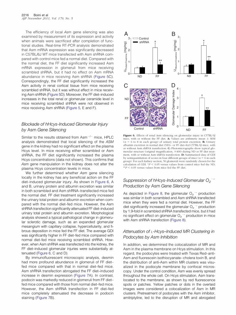
2216 Boini et alAJP November 2011, Vol. 179, No. 5
The efficiency of local Asm gene silencing was alsoexamined by measurement of its expression and activitywhen animals were sacrificed after completion of func-tional studies. Real-time RT-PCR analysis demonstratedthat Asm mRNA expression was significantly decreasedin C57BL/6J WT mice transfected with Asm shRNA com-pared with control mice fed a normal diet. Compared withthe normal diet, the FF diet significantly increased AsmmRNA expression in glomeruli from mice receivingscrambled shRNA, but it had no effect on Asm mRNAabundance in mice receiving Asm shRNA (Figure 5C).Correspondingly, the FF diet significantly increased theAsm activity in renal cortical tissue from mice receivingscrambled shRNA, but it was without effect in mice receiv-ing Asm shRNA (Figure 5D). Moreover, the FF diet–inducedincreases in the total renal or glomerular ceramide level inmice receiving scrambled shRNA were not observed inmice receiving Asm shRNA (Figure 5, E and F).
Blockade of hHcys-Induced Glomerular Injuryby Asm Gene Silencing
Similar to the results obtained from Asm�/� mice, HPLCanalysis demonstrated that local silencing of the ASMgene in the kidney had no significant effect on the plasmaHcys level. In mice receiving either scrambled or AsmshRNA, the FF diet significantly increased the plasmaHcys concentrations (data not shown). This confirms thatAsm gene manipulation in the kidney does not alter theplasma Hcys concentration levels in mice.
We further determined whether Asm gene silencinglocally in the kidney has any beneficial action on the FFdiet–induced glomerular injury. As shown in Figure 6, Aand B, urinary protein and albumin excretion was similarin both scrambled and Asm shRNA–transfected mice fedthe normal diet. FF diet treatment significantly increasedthe urinary total protein and albumin excretion when com-pared with the normal diet–fed mice. However, the AsmshRNA transfection significantly attenuated FF diet–inducedurinary total protein and albumin excretion. Morphologicalanalysis showed a typical pathological change in glomeru-lar sclerotic damage, such as an expanded glomerularmesangium with capillary collapse, hypercellularity, and fi-brous deposition in mice fed the FF diet. The average GDIwas significantly higher in FF diet–fed mice compared withnormal diet–fed mice receiving scrambled shRNA. How-ever, when Asm shRNA was transfected into the kidney, theFF diet–induced glomerular injuries were substantially at-tenuated (Figure 6, C and D).
By immunofluorescent microscopic analysis, desminhad more profound abundance in glomeruli of FF diet–fed mice compared with that in normal diet–fed mice.Asm shRNA transfection abrogated the FF diet–inducedincrease in desmin expression (Figure 7A). In contrast,podocin was markedly reduced in glomeruli from FF diet–fed mice compared with those from normal diet–fed mice.However, the Asm shRNA transfection in FF diet–fedmice completely attenuated the decrease in podocin
staining (Figure 7B).Suppression of hHcys-Induced Glomerular O2· –
Production by Asm Gene Silencing
As depicted in Figure 8, the glomerular O2· – production
was similar in both scrambled and Asm shRNA transfectedmice when they were fed a normal diet. However, the FFdiet significantly increased the glomerular O2
· – productionby 1.9-fold in scrambled shRNA transfected mice, but it hadno significant effect on glomerular O2
· – production in micewith Asm shRNA transfection (Figure 8).
Attenuation of L-Hcys–Induced MR Clustering inPodocytes by Asm Inhibition
In addition, we determined the colocalization of MR andAsm in the plasma membrane on Hcys stimulation. In thisregard, the podocytes were treated with Texas Red–anti-Asm and fluorescein isothiocyanate–cholera toxin B, andthe distribution of anti-Asm within MR clusters was visu-alized in the podocyte membrane by confocal micros-copy. Under the control condition, Asm was evenly spreadthroughout the whole cell. On Hcys stimulation, Asm trans-located to the membrane, as shown by red fluorescencespots or patches. Yellow patches or dots in the overlaidimages were considered a colocalization of Asm in MRclusters. Pretreatment of podocytes with the Asm inhibitor,
ND
FF
Control shRNA
C D
BControlshRNA
A
ND FF0
1
2
3
4
Glo
mer
ular
Dam
age
Inde
x
*
**
0
1
2
3
ND FF
UP
rote
in/C
reat
inin
e(m
g/m
g)
**
*
**
0
1
2
3
ND FF
UA
lbum
in/C
reat
inin
e(m
g/m
g) *
Figure 6. Effects of renal Asm silencing on glomerular injury in C57BL/6Jmice, with or without the FF diet. A: Values are arithmetic mean � SEM(n � 6 to 8 in each group) of urinary total protein excretion. B: Urinaryalbumin excretion in normal diet (ND)– or FF diet–fed C57BL/6J mice, withor without Asm shRNA transfection. C: Photomicrographs show typical glo-merular structure (original magnification, �400) during ND or FF diet treat-ment, with or without Asm shRNA transfection. D: Summarized data of GDIby semiquantitation of scores in four different groups of mice (n � 6 in eachgroup). For each kidney section, 50 glomeruli were randomly chosen for thecalculation of GDI. *P � 0.05 versus values from control mice fed the ND;**P � 0.05 versus values from mice fed the FF diet.
amitriptyline, led to the disruption of MR and abrogated

Asm in hHcys-Induced Glomerular Injury 2217AJP November 2011, Vol. 179, No. 5
patching and clustering of both fluorescein isothiocyanate–cholera toxin B and Asm after Hcys stimulation (see Sup-plemental Figure S2 at http://ajp.amjpathol.org).
Discussion
The goal of the present study is to determine whetherAsm, a ceramide-producing enzyme, is implicated in thedevelopment of hHcys-induced glomerular oxidativestress and injury. We found that FF diet treatment en-hanced the Asm activity, Asm mRNA expression, andceramide production, which was attributed to Nox-de-pendent O2
· – production and local oxidative stress inglomeruli and ultimately led to glomerulosclerosis. Our
ND
FF
Control shRNA
A
Control shRNA
B
ND
FF
Figure 7. Effects of renal Asm gene silencing on the expression of desminand podocin in glomeruli in C57BL/6J mice, with or without the FF diet. A:Typical images of desmin staining in glomeruli from C57BL/6J mice fed thenormal diet (ND) or FF diet, with or without Asm shRNA transfection (n � 6in each group). B: Typical images of podocin staining in glomeruli fromC57BL/6J mice fed the ND or FF diet, with or without Asm shRNA transfection(n � 6 in each group).
results demonstrate, for the first time to our knowledge,
that hHcys-induced glomerular oxidative stress and in-jury were attenuated by Asm gene deficiency in mice.
To examine the role of Asm in mediating hHcys-in-duced glomerular injury or sclerosis, we first inducedhHcys in Asm�/� mice and their WT littermates by feed-ing them an FF diet for 8 weeks. FF diet treatment signif-icantly increased plasma and renal tissue Hcys levels inboth Asm�/� and Asm�/� mice, indicating successfulestablishment of the hHcys and also suggesting that Asmitself is not involved in the global metabolism of Hcys. Theincreased plasma and renal tissue Hcys concentrationresulted in remarkable glomerular damage or sclerosis inAsm�/�, but not in Asm�/�, mice, suggesting that thelack of the Asm gene leads to blockade of pathogenicpathways mediating hHcys-induced glomerular injury.
Earlier studies reported that ceramide has been impli-cated in the regulation of kidney function. Ceramide as apotent regulator of cell proliferation, activation, and apop-tosis plays an important role in lipoprotein aggregationand may promote foam cell formation in atherosclero-sis.41 This lipid mediator has mediated the detrimental orpathogenic actions of many different injury factors in dif-ferent cells and tissues.12,41,42 In this regard, there arereports43 that serine palmitoyl–CoA transferase inhibitionby myriocin leads to a reduction of plasma sphingomy-elin, ceramide production, and atherosclerosis in apoli-poprotein E–knockout mice. We also demonstrated bypharmacological interventions that ceramide contributesto the development of chronic glomerular injury associ-ated with hHcys and obesity.4,31 The present study fur-ther demonstrated that Asm gene deficiency attenuatedhHcys-increased ceramide production in the kidney andthereby blocked glomerular injury associated with hHcys.Profiling analysis of renal ceramide demonstrated that,although C24 ceramide constituted the major ceramidecomponent that practically determines total ceramidelevels, the most dramatic changes in response to FFdiet–induced hHcys were observed from less-dominantceramide isoforms in mice. Correspondingly, local glo-merular ceramide was more abundant in Asm�/� than inAsm�/� mice when they both were fed the FF diet toinduce hHcys. Furthermore, Asm activity and Asm mRNAexpression in renal tissues were significantly increased in
Figure 8. Effect of renal Asm gene silencing on glomerular O2.� production
in C57BL/6J mice, with or without the FF diet. Values are arithmetic mean �SEM (n � 5 to 6 each group) of O2
.� production in C57BL/6J mice fed thenormal diet (ND) or FF diet with or without Asm shRNA transfection. *P �
0.05 versus values from control mice fed the ND; **P � 0.05 versus valuesfrom mice fed the FF diet.
2218 Boini et alAJP November 2011, Vol. 179, No. 5
FF diet–fed Asm�/� mice but not in Asm�/� mice. Inaddition, our studies demonstrated that Hcys stimulationenhanced the colocalization of MR and Asm in theplasma membrane and confirmed the translocation ofAsm into the cell membrane. These results together sug-gest that FF diet–induced hHcys increases the renal andglomerular ceramide levels, mainly because of activationof Asm and an increase in its expression.
Furthermore, we demonstrated that decreased cer-amide production via Asm has a protective role in theglomerular injury associated with hHcys. In accordancewith lowered ceramide production in Asm�/� mice fedthe FF diet, urinary albumin, protein excretion, and glo-merular podocyte injury and sclerosis were also signifi-cantly decreased compared with Asm�/� mice fed the FFdiet, suggesting that ceramide-associated renal injuryduring hHcys is alleviated in these Asm gene knockoutmice. Therefore, this sphingomyelinase could be a targetof a therapeutic strategy for hHcys-induced glomerularinjury or sclerosis.
To further explore the mechanism of glomerular injuryduring hHcys, we observed changes in podocyte func-tion in Asm�/� and Asm�/� mice. It has been well doc-umented that podocyte loss and dysfunction occur withthe onset and magnitude of glomerulosclerosis. Becausepodocytes serve as the final barrier against urinary pro-tein loss in the normal glomeruli, any change in podocytestructure and function may be intimately associated withproteinuria and consequent glomerular sclerosis.44 Thepresent study showed that podocin protein was markedlydecreased in FF diet–fed Asm�/� mice but not in micelacking Asm. In addition, we found that desmin, an inter-mediate filament protein and a specific and sensitivepodocyte injury marker, was increased in the glomeruliwhen Asm�/� mice received the FF diet. This increaseddesmin expression in the glomeruli was attenuated in FFdiet–fed Asm�/� mice. These results further support theview that hHcys-induced glomerular injury is associatedwith increased ceramide production via Asm.
Reportedly, oxidative stress has been implicated inthe development of glomerular injury and end-stagerenal disease. In our previous studies,4 Nox-derivedO2
· – production was an important mechanism mediat-ing hHcys-induced glomerular injury or damage. NA-DPH-dependent O2
· – production is an early event forhomocysteine-induced glomerular cell damage and glo-merular sclerosis.4,36 It is possible that hHcys-inducedNox activation is mediated by enhanced Asm activity.The present study hypothesized that Asm may be asso-ciated with local oxidative stress in the glomeruli of micewith hHcys. Indeed, electron-spin resonance analysisshowed that FF diet treatment significantly increasedNox-dependent O2
· – production in Asm�/�, but not inAsm�/�, mice. These results confirm the imperative roleof Asm in mediating O2
· – production through the activa-tion of Nox in the glomeruli during hHcys.
To further address the role of the Asm gene in medi-ating hHcys-induced podocyte and glomerular injury, alocal gene-silencing strategy was used in the presentstudy, in which ultrasonographic microbubble–mediated
plasmid delivery was used to introduce Asm shRNA intothe kidney. This method was highly efficient in deliveringplasmids into renal cells in vivo, which led to gene trans-fection and expression in most renal cells (90%), as con-firmed by earlier reports.2,45–48 By using an in vivo mo-lecular imaging system to daily monitor the efficiency ofAsm gene transfection in the kidney in living animals, weshowed that the transgene or shRNA expression vector(with the luciferase gene as an indicator) could be de-tected even 3 days after gene transfection and lasted for4 weeks by observation. This in vivo transgene monitoringimportantly guided our functional studies to define therole of the Asm gene in mediating glomerular damageassociated with hHcys. After completion of functionalprotocols, Asm mRNA expression, Asm activity, and re-nal and glomerular ceramide production were analyzedto confirm the efficient silencing of the Asm gene inshRNA-transfected kidneys. In such local Asm gene–silenced kidney, we found that the glomerular ceramidelevel was significantly decreased and that O2
· – produc-tion was markedly reduced. Silencing the Asm gene inthe kidney ameliorates proteinuria/albuminuria and podo-cyte injury, whereby glomerular sclerotic changes weresubstantially suppressed. These results from mice withlocal renal Asm gene silencing further support the previ-ous conclusion, drawn from studies using Asm-knockoutmice that Asm is importantly implicated in the develop-ment of podocytes and glomerular injury. The reductionof ceramide production or the inhibition of Nox is preven-tive to hHcys-induced glomerular injury. Further studieswith a careful experimental design for therapeutic actionvia this signaling system are needed to confirm its ther-apeutic potential. In conclusion, the present study dem-onstrated that Asm gene deficiency attenuates the hH-cys-induced glomerular oxidative stress and injury. Theamelioration of glomerular injury by Asm deficiency dur-ing hHcys implicates the pivotal role of Asm in hHcys-induced glomerulosclerosis.
References
1. Dennis VW, Robinson K: Homocysteinemia and vascular disease inend-stage renal disease. Kidney Int Suppl 1996, 57:S11–S17
2. Yi F, Xia M, Li N, Zhang C, Tang L, Li PL: Contribution of guaninenucleotide exchange factor Vav2 to hyperhomocysteinemic glomer-ulosclerosis in rats. Hypertension 2009, 53:90–96
3. Ingram AJ, Krepinsky JC, James L, Austin RC, Tang D, Salapatek AM,Thai K, Scholey JW: Activation of mesangial cell MAPK in response tohomocysteine. Kidney Int 2004, 66:733–745
4. Yi F, Zhang AY, Li N, Muh RW, Fillet M, Renert AF, Li PL: Inhibition ofceramide-redox signaling pathway blocks glomerular injury in hyper-homocysteinemic rats. Kidney Int 2006, 70:88–96
5. Zhang C, Hu JJ, Xia M, Boini KM, Brimson CA, Laperle LA, Li PL:Protection of podocytes from hyperhomocysteinemia-induced injuryby deletion of the gp91phox gene. Free Radic Biol Med 2010, 48:1109–1117
6. Tyagi N, Moshal KS, Sen U, Vacek TP, Kumar M, Hughes WM Jr,Kundu S, Tyagi SC: H2S protects against methionine-induced oxida-tive stress in brain endothelial cells. Antioxid Redox Signal 2009,11:25–33
7. Hwang SY, Siow YL, Au-Yeung KK, House J, OK: Folic acid supplemen-tation inhibits NADPH oxidase-mediated superoxide anion production inthe kidney. Am J Physiol Renal Physiol 2011, 300:F189–F198
8. Yi F, Li PL: Mechanisms of homocysteine-induced glomerular injuryand sclerosis. Am J Nephrol 2008, 28:254–264
Asm in hHcys-Induced Glomerular Injury 2219AJP November 2011, Vol. 179, No. 5
9. Li PL, Yi F, Li N: Hyperhomocysteinemia: association with renal trans-sulfuration and redox signaling in rats. Clin Chem Lab Med 2007,45:1688–1693
10. Papatheodorou L, Weiss N: Vascular oxidant stress and inflammationin hyperhomocysteinemia. Antioxid Redox Signal 2007, 9:1941–1958
11. Kaushal GP, Singh AB, Shah SV: Identification of gene family ofcaspases in rat kidney and altered expression in ischemia-reperfu-sion injury. Am J Physiol 1998, 274:F587–F595
12. Ueda N, Kaushal GP, Shah SV: Apoptotic mechanisms in acute renalfailure. Am J Med 2000, 108:403–415
13. Yin T, Sandhu G, Wolfgang CD, Burrier A, Webb RL, Rigel DF, Hai T,Whelan J: Tissue-specific pattern of stress kinase activation in isch-emic/reperfused heart and kidney. J Biol Chem 1997, 272:19943–19950
14. Zhang CE, Wei W, Liu YH, Peng JH, Tian Q, Liu GP, Zhang Y, WangJZ: Hyperhomocysteinemia increases beta-amyloid by enhancingexpression of gamma-secretase and phosphorylation of amyloid pre-cursor protein in rat brain. Am J Pathol 2009, 174:1481–1491
15. Schmitz-Peiffer C: Targeting ceramide synthesis to reverse insulinresistance. Diabetes 2010, 59:2351–2353
16. Ussher JR, Koves TR, Cadete VJ, Zhang L, Jaswal JS, Swyrd SJ,Lopaschuk DG, Proctor SD, Keung W, Muoio DM, Lopaschuk GD:Inhibition of de novo ceramide synthesis reverses diet-induced insu-lin resistance and enhances whole-body oxygen consumption. Dia-betes 2010, 59:2453–2464
17. Mielke MM, Haughey NJ, Ratnam Bandaru VV, Schech S, Carrick R,Carlson MC, Mori S, Miller MI, Ceritoglu C, Brown T, Albert M, Lyket-sos CG: Plasma ceramides are altered in mild cognitive impairmentand predict cognitive decline and hippocampal volume loss. Alzhei-mers Dement 2010, 6:378–385
18. Holland WL, Brozinick JT, Wang LP, Hawkins ED, Sargent KM, Liu Y,Narra K, Hoehn KL, Knotts TA, Siesky A, Nelson DH, KarathanasisSK, Fontenot GK, Birnbaum MJ, Summers SA: Inhibition of ceramidesynthesis ameliorates glucocorticoid-, saturated-fat-, and obesity-induced insulin resistance. Cell Metab 2007, 5:167–179
19. Summers SA: Ceramides in insulin resistance and lipotoxicity. ProgLipid Res 2006, 45:42–72
20. Futerman AH, Hannun YA: The complex life of simple sphingolipids.EMBO Rep 2004, 5:777–782
21. Garcia-Barros M, Paris F, Cordon-Cardo C, Lyden D, Rafii S, Haimo-vitz-Friedman A, Fuks Z, Kolesnick R: Tumor response to radiother-apy regulated by endothelial cell apoptosis. Science 2003, 300:1155–1159
22. Smith EL, Schuchman EH: The unexpected role of acid sphingomy-elinase in cell death and the pathophysiology of common diseases.FASEB J 2008, 22:3419–3431
23. Grassme H, Cremesti A, Kolesnick R, Gulbins E: Ceramide-mediatedclustering is required for CD95-DISC formation. Oncogene 2003,22:5457–5470
24. Yu ZF, Nikolova-Karakashian M, Zhou D, Cheng G, Schuchman EH,Mattson MP: Pivotal role for acidic sphingomyelinase in cerebralischemia-induced ceramide and cytokine production, and neuronalapoptosis. J Mol Neurosci 2000, 15:85–97
25. Paris F, Fuks Z, Kang A, Capodieci P, Juan G, Ehleiter D, Haimovitz-Friedman A, Cordon-Cardo C, Kolesnick R: Endothelial apoptosis asthe primary lesion initiating intestinal radiation damage in mice. Sci-ence 2001, 293:293–297
26. Dimanche-Boitrel MT, Meurette O, Rebillard A, Lacour S: Role of earlyplasma membrane events in chemotherapy-induced cell death. DrugResist Updat 2005, 8:5–14
27. Garcia-Ruiz C, Colell A, Mari M, Morales A, Calvo M, Enrich C,Fernandez-Checa JC: Defective TNF-alpha-mediated hepatocellularapoptosis and liver damage in acidic sphingomyelinase knockoutmice. J Clin Invest 2003, 111:197–208
28. Dhami R, He X, Schuchman EH: Acid sphingomyelinase deficiencyattenuates bleomycin-induced lung inflammation and fibrosis in mice.Cell Physiol Biochem 2010, 26:749–760
29. Becker KA, Riethmuller J, Luth A, Doring G, Kleuser B, Gulbins E:Acid sphingomyelinase inhibitors normalize pulmonary ceramide and
inflammation in cystic fibrosis. Am J Respir Cell Mol Biol 2010,42:716–724
30. Teichgraber V, Ulrich M, Endlich N, Riethmuller J, Wilker B, Oliveira-Munding CC, van Heeckeren AM, Barr ML, von Kurthy G, SchmidKW, Weller M, Tummler B, Lang F, Grassme H, Doring G, Gulbins E:Ceramide accumulation mediates inflammation, cell death and infec-tion susceptibility in cystic fibrosis. Nat Med 2008, 14:382–391
31. Boini KM, Zhang C, Xia M, Poklis JL, Li PL: Role of sphingolipidmediator ceramide in obesity and renal injury in mice fed a high-fatdiet. J Pharmacol Exp Ther 2010, 334:839–846
32. Moles A, Tarrats N, Morales A, Dominguez M, Bataller R, Caballeria J,Garcia-Ruiz C, Fernandez-Checa JC, Mari M: Acidic sphingomyeli-nase controls hepatic stellate cell activation and in vivo liver fibro-genesis. Am J Pathol 2010, 177:1214–1224
33. Achar E, Maciel TT, Collares CF, Teixeira VP, Schor N: Amitriptylineattenuates interstitial inflammation and ameliorates the progression ofrenal fibrosis. Kidney Int 2009, 75:596–604
34. Chen YF, Li PL, Zou AP: Effect of hyperhomocysteinemia on plasmaor tissue adenosine levels and renal function. Circulation 2002, 106:1275–1281
35. Raij L, Azar S, Keane W: Mesangial immune injury, hypertension, andprogressive glomerular damage in Dahl rats. Kidney Int 1984, 26:137–143
36. Yi F, Zhang AY, Janscha JL, Li PL, Zou AP: Homocysteine activatesNADH/NADPH oxidase through ceramide-stimulated Rac GTPaseactivity in rat mesangial cells. Kidney Int 2004, 66:1977–1987
37. Boini KM, Zhang C, Xia M, Han WQ, Brimson C, Poklis JL, Li PL:Visfatin-induced lipid raft redox signaling platforms and dysfunctionin glomerular endothelial cells. Biochim Biophys Acta 2010, 1801:1294–1304
38. Fillet M, Van Heugen JC, Servais AC, De Graeve J, Crommen J:Separation, identification and quantitation of ceramides in humancancer cells by liquid chromatography-electrospray ionization tan-dem mass spectrometry. J Chromatogr A 2002, 949:225–233
39. Zhang C, Hu JJ, Xia M, Boini KM, Brimson C, Li PL: Redox signalingvia lipid raft clustering in homocysteine-induced injury of podocytes.Biochim Biophys Acta 2010, 1803:482–491
40. van der Wouden EA, Sandovici M, Henning RH, de Zeeuw D, Deel-man LE: Approaches and methods in gene therapy for kidney dis-ease. J Pharmacol Toxicol Methods 2004, 50:13–24
41. Williams KJ, Tabas I: The response-to-retention hypothesis of earlyatherogenesis. Arterioscler Thromb Vasc Biol 1995, 15:551–561
42. Coroneos E, Martinez M, McKenna S, Kester M: Differential regulationof sphingomyelinase and ceramidase activities by growth factors andcytokines: implications for cellular proliferation and differentiation.J Biol Chem 1995, 270:23305–23309
43. Hojjati MR, Li Z, Zhou H, Tang S, Huan C, Ooi E, Lu S, Jiang XC: Effectof myriocin on plasma sphingolipid metabolism and atherosclerosisin apoE-deficient mice. J Biol Chem 2005, 280:10284–10289
44. Li N, Chen L, Yi F, Xia M, Li PL: Salt-sensitive hypertension inducedby decoy of transcription factor hypoxia-inducible factor-1alpha in therenal medulla. Circ Res 2008, 102:1101–1108
45. Hou CC, Wang W, Huang XR, Fu P, Chen TH, Sheikh-Hamad D, LanHY: Ultrasound-microbubble-mediated gene transfer of inducibleSmad7 blocks transforming growth factor-beta signaling and fibrosisin rat remnant kidney. Am J Pathol 2005, 166:761–771
46. Koike H, Tomita N, Azuma H, Taniyama Y, Yamasaki K, Kunugiza Y,Tachibana K, Ogihara T, Morishita R: An efficient gene transfermethod mediated by ultrasound and microbubbles into the kidney.J Gene Med 2005, 7:108–116
47. Lan HY, Mu W, Tomita N, Huang XR, Li JH, Zhu HJ, Morishita R,Johnson RJ: Inhibition of renal fibrosis by gene transfer of inducibleSmad7 using ultrasound-microbubble system in rat UUO model.J Am Soc Nephrol 2003, 14:1535–1548
48. Sheyn D, Kimelman-Bleich N, Pelled G, Zilberman Y, Gazit D, Gazit Z:
Ultrasound-based nonviral gene delivery induces bone formation invivo. Gene Ther 2008, 15:257–266