acknowledgements - university of manchester
TRANSCRIPT


1
Acknowledgements
Authors
Louisa Burton, Project Manager, Greater Manchester & Cheshire Cardiac and Stroke Network
Sarah Tyson, Professor of Rehabilitation, University of Manchester
Alison McGovern, Quality Improvement Manager, Greater Manchester & Cheshire Cardiac and Stroke Network
Sudi Sharifi, Senior Lecturer, University of Salford
With considerable thanks to the Greater
Manchester Stroke Rehabilitation teams who
participated in the G-MASTER project:
Bolton NHS Foundation Trust
Central Manchester & Manchester Children’s
Hospital NHS Trust
Pennine Acute Hospital Trust, Fairfield General
Hospital
Pennine Acute Hospital Trust, Royal Oldham
Hospital
Pennine Acute Hospital Trust, North Manchester
General Hospital
Salford Royal NHS Foundation Trust
Stockport NHS Foundation Trust
Tameside Hospital Foundation Trust
Trafford Healthcare NHS Trust
University Hospital of South Manchester NHS
Foundation Trust
Wrightington, Wigan & Leigh NHS Foundation
Trust

2
Contents
Introduction ...................................................................................................................................................................... 3
Swallow Screening ............................................................................................................................................................ 5
Greater Manchester Stroke Water Swallow Screening Tool: Instruction Manual ....................................................... 6
Greater Manchester Stroke Water Swallow Screening Tool ........................................................................................ 9
Nutritional Screening ...................................................................................................................................................... 11
Malnutrition Universal Screening Tool (BAPEN) ......................................................................................................... 12
Communication Screening .............................................................................................................................................. 18
National Institutes of Health Stroke Scale (NIHSS) Communication sections ............................................................ 19
Mood Screening .............................................................................................................................................................. 24
Greater Manchester Algorithm for Selecting Appropriate Mood Screening Measures for Patients with Stroke ...... 27
The Stroke Aphasic Depression Questionnaire (SADQ–H10)...................................................................................... 28
Depression Intensity Scale Circles (DISCs) .................................................................................................................. 29
Depression Intensity Scale Circles (DISCs) – Instructions for administration ............................................................. 29
GAD-7 .......................................................................................................................................................................... 32
Cognitive Screening ......................................................................................................................................................... 33
Montreal Cognitive Assessment (MoCA) .................................................................................................................... 34
Montreal Cognitive Assessment (MoCA): Administration and Scoring Instructions .................................................. 35
Measuring functional outcomes ..................................................................................................................................... 39
Independence in Activities of Daily Living – Barthel Index ......................................................................................... 39
The Barthel Index ............................................................................................................................................................ 40
Barthel Index Scoring Criteria ..................................................................................................................................... 41
Monitoring communication and swallowing abilities over time – Therapy Outcome Measures .................................. 42
Therapy Outcome Measures (Dysphasia/Aphasia) ..................................................................................................... 42
Therapy Outcome Measures (Dysphagia) ................................................................................................................... 44
Therapy Outcome Measures (Dysarthria) .................................................................................................................. 45
How to run an effective Stroke Multi-Disciplinary Team meeting ................................................................................. 46
Sample MDT Meeting Agenda ........................................................................................................................................ 49
Initial MDT form .......................................................................................................................................................... 51
Weekly MDT form ....................................................................................................................................................... 52
References ...................................................................................................................................................................... 54

3
Introduction
Background
Assessment is a crucial part of stroke
rehabilitation, providing a means of identifying
and measuring patients’ needs.
Although national guidance recommends the
use of standardised assessment tools and
outcome measures, clinicians have reported
pragmatic difficulties in selecting the best
tools to use in their practice. Research
evidence has demonstrated that using
standardised measures can be beneficial to
rehabilitation team communication, allowing
information about patients’ problems and
functional abilities to be shared in an efficient
way (Tyson et al., 2010). Measures can also
be used to monitor patients’ progress, to
inform clinical decision-making and to
demonstrate service effectiveness.
What did we aim to achieve?
The G-MASTER project aimed to:
Provide meaningful ways to measure
patient progress through rehabilitation
Provide clinicians with information
about valid and reliable tools that are
feasible to use in the clinical setting
Use of standardised measurements tools in
practice can:
Promote use of a common language to
enhance team communication
Facilitate transfer of information
between services, allowing them to
provide the best care for the patient
Help to ensure that multidisciplinary
discussions are patient-centred
Use of same tools across services can
prevent repetition of similar
assessments for patients
How did we decide what to measure?
Practicing stroke rehabilitation professionals
were asked about the domains of functioning,
which:
Are necessary to measure during
rehabilitation
All members of the rehabilitation team
need to know about to treat their
patient effectively
Focus groups of patients were also asked
about their most important problems after
they had a stroke and the ways these were
measured. Finally national guidelines were
explored to identify the areas requiring
measurement using standardised tools. “I think we all struggled to find
standardised assessments that we can use and it was really nice to have
something that’s been assessed as being a suitable tool” OT
4
What process did we use to identify tools?
The literature was searched to identify
appropriate validated tools to measure each
domain and the psychometric properties of
tools were assessed. In addition, tools were
assessed for clinical feasibility, including:
Time taken to administer and score the
tool
Need for specialist training for staff
Cost for purchase of the tool, specialist
equipment or record forms
Portability
In collaboration with practicing clinical staff, the
most robust tools were selected, and are
presented here with their instruction manuals
and guidelines for use. Where suitable tools
were not available, new measures were
developed and tested through collaborations of
clinical specialists.
How can the toolkit be used in practice?
The aim of the toolkit is for information to be
shared among the multidisciplinary team,
therefore the tools should be scored (if
appropriate; or the scores discussed) within
MDT meetings. As a result, a structured
approach to running MDT meetings
incorporating use of the tools into standardised
documentation has been developed and is
available at the end of this document.
For further information…
For further information on how the tools
were selected and other tools which were
considered, please contact:
Louisa Burton, G-MASTER Project Manager

5
Swallow Screening
What do the guidelines say?
Difficulties in swallowing are common after
stroke, affecting around 40% of patients. These
difficulties are associated with an increased risk of
aspiration and pneumonia and increased mortality
rates. Clinical guidelines therefore recommend
that every patient who has had a stroke should
have their ability to swallow screened by an
appropriately trained person using a recognised,
standard screening assessment before being given
oral food, fluids or oral medication (RCP). Patients
with swallowing difficulties should be referred to
and assessed by a Speech and Language Therapist
(RCP).
Could a validated tool be identified from the
existing research literature?
In 2011, a systematic literature search was
performed to identify whether a swallow
screening tool had been developed that met the
following criteria:
Psychometric criteria: sensitivity >80% and
specificity >60%
Clinical utility criteria: freely available,
manageable training requirements,
portable, administered in <10 minutes
Identified tools were also assessed to
ascertain whether the necessary detail to
enable use of the tool in practice was
provided
No tool could be identified that met the criteria
outlined above. However, much work and
training has been undertaken within local stroke
services to develop swallow screening tools and
protocols. The development of the Greater
Manchester Water Swallow Screening Tool aimed
to utilise the expertise of Speech & Language
Therapists
Therapists and dysphagia-trained nurses across
Greater Manchester to develop a series of
standardised core indicators to be included in all
swallow screening tools across the conurbation.
How was the screening tool developed?
A content analysis of seven screening tools
developed by services across Greater
Manchester was performed. Commonalities
between the screens were identified in three
categories: pre-screening checks (to determine
whether a swallow screen could be safely
completed), administration of the screening
instrument (in all cases this incorporated a water
swallow test) and clinical indicators of
disordered swallowing.
Speech and Language Therapists from across the
conurbation then took part in discussions to
agree common clinical indicators to be included
in all swallow screens. The aim was to provide a
core set of indicators to be included in all
screens, which teams could add to, to include
any other indicators or steps they felt were
appropriate and necessary according to local
policy. As a result of these discussions, a draft
screening tool was formed and this was sent to
the group for feedback and comments. A
second series of discussions was then held to
discuss the feedback, make resulting changes to
the tool and agree a final screening tool.
As dysphagia screening is completed in practice
by nursing staff, an instruction manual was
developed in collaboration with nurses working
in stroke services, to ensure the tool was easy to
use. Members of the Network’s Specialist
Nurses group were invited to comment on the
tool and revisions were made in line with their
feedback. Further validation work is now
needed to ensure sensitivity and specificity.

6
Greater Manchester Stroke Water Swallow Screening Tool: Instruction
Manual
General guidelines
This screen is intended to be carried out ONLY by professionals with water swallow screening
training
DO NOT administer the water swallow test unless you have first worked through the pre-
screening checks. If you are unsure at any point, discontinue the screen and refer to Speech
and Language Therapy.
Discontinuing – if you discontinue the screen at any point:
Keep the patient nil by mouth
Ensure that it is clear at what point the screen was discontinued, and sign and date the sheet.
File the screening form in the patient’s medical records and make an entry into the notes
stating that the screen was attempted and the reasons why it was discontinued.
Refer to Speech and Language Therapy if indicated and consider nutrition and hydration in line
with local protocol
Ensure that medication is administered through an alternative route (refer to pharmacy
guidelines on liquid/dispersible medications
Procedure
1.) Record the patient’s name, date of birth and NHS/hospital number on the form.
2.) Record the date and time you are carrying out the screen.
3.) Monitor vital signs, including BP, pulse and sats.
4.) Observe whether the patient can remain awake and alert for long enough to complete the
swallow screen – indicate on the form whether this is the case. As a guide, the screen may take
up to 15 minutes to complete. If the patient is drowsy and therefore inappropriate for
assessment, monitor alertness and repeat the screen as appropriate.
5.) Check that the patient can remain in an upright position, with supports if necessary, for long
enough to complete the swallow screen. If supports are necessary, ensure they are in place
before continuing. If the patient is unable to remain upright for long enough, mark the form
accordingly, discontinue the screen and refer to Speech and Language Therapy.

7
6.) Check whether there is oxygen in situ. If only nasal specs are in situ, mark ‘no’ on the form and
continue with the screen. If an oxygen mask is being used, check sats and consider whether
nasal specs can be used as an alternative. If not, mark ‘yes’ on the form and discontinue the
screening tool. Refer to Speech and Language Therapy if indicated in local protocol.
7.) Determine whether the patient has a chest infection, e.g. is temperature elevated? Ask the
medical team if you are unsure and indicate on the screening form. If the patient has a chest
infection, discontinue the swallow screening tool, manage medically and consider nutrition and
hydration in line with local protocol (consult nutrition nurse/dietitian as necessary). Repeat the
screen when the patient is free of infection.
8.) Ask the patient’s permission to check whether their mouth is clean. If necessary, e.g. if there
are excess secretions, thrush present or the patient’s mouth is dry, administer oral hygiene
according to local protocol.
9.) Find out whether the patient was on thickened fluids or modified diet prior to admission. This
may involve asking the patient directly, asking carers or family members or looking at the
patient’s medical records. If the patient was previously on thickened fluids or modified diet,
indicate the grade/consistency on the form, discontinue the screen and refer to Speech and
Language Therapy.
10.) Gain patient consent to swallow screen, e.g. say to the patient “swallowing can sometimes be
affected when someone has had a stroke, please could I check your swallow?” If the patient
asks for more information, use knowledge from your training to explain how and why
swallowing may be affected as a result of stroke.
11.) If the patient refuses to be screened, indicate this on the form, discontinue the screen and
refer to Speech and Language Therapy. Ensure that refusal is documented in the medical
notes. If the patient is unable to give verbal consent, e.g. due to aphasia or confusion, consider
whether screening is in the patient’s best interests and continue to water swallow test if
appropriate. If the patient is not cooperative with screening, discontinue, document as
appropriate and refer to Speech and Language Therapy.
12.) Proceed to water swallow test (overleaf). Ensure that you have a teaspoon, a glass and a jug of
drinking water ready.
13.) Give the patient 1 teaspoon of water and observe for two minutes for signs of difficulties.
Indicate on the form whether the following signs are present: coughing or choking upon
swallow or up to two minutes after swallow, no swallow triggered, wet or gurgly voice quality,
change in breathing pattern (may be either fast or laboured) or loss of water from mouth.
Other signs to look out for are drooling, poor lip closure, oral residue and an abnormal number
8
of swallows (too many or too few). Record these signs under any other difficulties if observed
and note them on the bottom of the sheet. If you observe difficulties at any time, discontinue
the screen and refer to Speech and Language Therapy. Consider nutrition and hydration in line
with local protocol (consult nutrition nurse or dietitian as appropriate).
14.) Give the patient a second teaspoon of water and observe for two minutes, discontinuing
immediately if any signs are present.
15.) Give the patient a third teaspoon of water and observe for two minutes, discontinuing
immediately if any signs are present.
16.) If you do not observe difficulties, give the patient a sip of water from a glass and observe for
two minutes for difficulties. DO NOT allow the patient to take the glass themselves at this
stage so you can control the amount of water given.
17.) Give the patient a second sip of water and observe for two minutes, discontinuing immediately
if any signs are present.
18.) Give the patient a third sip of water and observe for two minutes, discontinuing immediately if
any signs are present.
19.) If you do not observe any difficulties, give the patient 50ml of water in a glass. If appropriate,
give the cup to the patient and tell them to drink slowly. Monitor whether any of the signs of
swallowing difficulty are present, as well as monitoring the patient’s ability to self-feed and
noting signs of ataxia or coordination difficulties. If any signs of swallowing difficulty are
present, discontinue immediately, document and refer to Speech and Language Therapy. If the
patient has difficulties in self-feeding, ensure that this is documented in the notes and consider
local protocol.
20.) If none of the signs are present, the patient has passed the water swallow screen and can be
monitored on normal fluids. Mark the form as appropriate and write your name and signature
on the bottom of the form and your designation.
The Greater Manchester Stroke Water Swallow Screening Tool is a test of whether a patient
can swallow water only. Therefore if a patient passes the screen, this does not necessarily
mean they can be given solid foods – further assessment may be required in line with local
protocol.

9
Greater Manchester Stroke Water Swallow Screening Tool
TO BE CARRIED OUT BY DYSPHAGIA TRAINED NURSES ONLY
Patient’s name: _________________________ DOB: _______________ NHS No. _____________________
Time and date screening carried out: ___________________________________________
Before performing a water swallow test, work through the pre-screening checks below and delete as appropriate to indicate where the screening was terminated
Can the patient remain awake and maintain alertness for long enough to complete the swallow screen? Y / N Can the patient remain in an upright position (with supports if necessary) for long enough to complete the swallow screen? Y / N
If no to either question
Discontinue swallow screening tool and keep patient NBM. If patient is too drowsy and inappropriate for assessment, monitor alertness and repeat screen when patient can maintain alertness for long enough to complete the swallow screen. If discontinuing for other reason, refer to Speech & Language Therapy
Is there oxygen in situ (except nasal specs)? Y / N
Yes
Yes
Is the patient’s mouth clean? Y / N No
Administer oral hygiene before continuing
Was the patient on modified diet or thickened fluids prior to admission? Y / N If yes, please indicate consistency: _________________________________________________
Has the patient consented to swallow screen? Y / N Consent gained verbally Seen in best interests and consent assumed
by patient cooperation in swallow screen
If patient refuses assessment
Discontinue swallow screening tool and document. Keep patient NBM and refer to Speech & Language Therapy
Proceed to Water Swallow Test (overleaf)
Does the patient have a chest infection? Y / N
Yes
Discontinue swallow screening tool and keep patient NBM. Manage medically and consider nutrition and hydration in line with local protocol. Repeat screen when patient is free of infection
No
No
Yes
No Yes
Yes

10
Greater Manchester Stroke Water Swallow Screening Tool
TO BE CARRIED OUT BY DYSPHAGIA TRAINED NURSES ONLY
Ensure pre-screening checks overleaf have been carried out before administering the Water Swallow Test. If the patient displays any of the listed signs at any point during the Water Swallow Test,
discontinue immediately and indicate which signs were observed below Any other reason you feel the swallow is unsafe: _______________________________________________________________________________________________ _______________________________________________________________________________________________ Name and signature of person administering screen: ____________________________________________________
Give patient 50ml of water in a beaker or glass Are any of the following signs present at any time? □ Cough or choking on swallow or up to 2 minutes after swallow □ No swallow is triggered □ Wet or gurgly voice □ Change in breathing □ Loss of water from mouth □ Any other reason you feel the swallow is unsafe (document below)
Yes
Monitor on normal fluids.
Discontinue swallow screen. Keep patient NBM and refer to Speech & Language Therapy. Consider nutrition and hydration in line with local protocol.
No
No
No
Give patient 3 sips of water from a beaker or glass Are any of the following signs present at any time? □ Cough or choking on swallow or up to 2 minutes after swallow □ No swallow is triggered □ Wet or gurgly voice □ Change in breathing □ Loss of water from mouth □ Any other reason you feel the swallow is unsafe (document below)
Give patient 3 x 5 teaspoons water Are any of the following signs present at any time? □ Cough or choking on swallow or up to 2 minutes after swallow □ No swallow is triggered □ Wet or gurgly voice □ Change in breathing □ Loss of water from mouth □ Any other reason you feel the swallow is unsafe (document below)
Yes
Yes
Discontinue swallow screen. Keep patient NBM and refer to Speech & Language Therapy. Consider nutrition and hydration in line with local protocol.
Discontinue swallow screen. Keep patient NBM and refer to Speech & Language Therapy. Consider nutrition and hydration in line with local protocol.

11
Nutritional Screening
What do the guidelines say?
National guidelines state that all patients should be
screened for malnutrition and the risk of malnutrition
when first assessed (RCP). This should be completed by
a trained person using a validated procedure, for
example, clinical judgement or the Malnutrition
Universal Screening Tool (MUST; RCP). Screening should
be repeated weekly for all hospital in-patients (NICE).
Detailed nutritional assessment should be undertaken by
an appropriately trained health care professional if a
person with acute stroke is unable to take adequate
nutrition and fluids orally (RCP).
How was a tool selected?
As the guidelines recommend a tool, which is well-
validated and used across most of the Trusts involved in
the project, this tool was selected as part of the G-
MASTER toolkit.

12
Malnutrition Universal Screening Tool (BAPEN)

13

14

15

16

17

18
Communication Screening
What do the guidelines say?
Every patient who has had a stroke should be
assessed for communication disability
(RCSLT). The RCP Clinical Guideline for Stroke
(3rd Ed.) recommends that patients entering
rehabilitation with damage to the left
cerebral hemisphere should be screened for
aphasia using a formal measure, e.g. the
Frenchay Aphasia Screening Test or the
Sheffield Aphasia Screening Test.
Could a tool be identified from the existing
research literature?
A recent review of aphasia screening tools
that have been validated for use with stroke
patients was used as a starting point to
identify potential tools (Salter, Jutai et al.,
2006). Further tools were then identified
from the research literature through
systematic searches. These tools were then
considered in terms of the feasibility and
practicality of adopting them into clinical
practice.
When this information had been gathered,
the most feasible tools were considered in
terms of their psychometric properties, with
sensitivity >80% and specificity >60% needed
before a tool could be recommended.
These results were presented to a consensus
group of speech and language therapists
working in stroke services across the
conurbation and it was agreed that all stroke
patients should be screened for
communication problems.
The group agreed that it was not practical
for a Speech and Language Therapist to
assess all patients, therefore it was
suggested that a brief communication
screen be carried out as part of the initial
assessment process. The criteria for
selecting a tool set by the consensus group
were:
Easy to administer
Can be administered by any
member of the MDT
Takes less than 5 minutes to
administer
Following review of the available tools, the
group undertook a pilot of a translation of
the Language Screening Test (Flamand-
Roze et al., 2011). Work is currently on-
going to develop an English-language
version of this screen, which will be
available in 2013.
It was also suggested that where the NIHSS
screen was being used, the communication
sections could be utilised as a
communication screen. This represents a
practical alternative, although further
work is necessary to determine sensitivity
and specificity of use of this tool as a
communication screen. An online training
programme for this tool is freely available,
and can be accessed at:
http://nihss-
english.trainingcampus.net/uas/modules
/trees/windex.aspx

19
National Institutes of Health Stroke Scale (NIHSS) Communication sections (recommended for use as part of the NIHSS)
Record performance in each category after each subscale exam. Do not go back and change scores. Follow directions provided
for each exam technique. Scores should reflect what the patient does, not what the clinician thinks the patient can do. The
clinician should record answers while administering the exam and work quickly. Except where indicated, the patient should not
be coached (i.e., repeated requests to patient to make a special effort).
9. Best Language: A great deal of information about
comprehension will be obtained during the preceding sections
of the examination. For this scale item, the patient is asked to
describe what is happening in the attached picture, to name
the items on the attached naming sheet and to read from the
attached list of sentences. Comprehension is judged from
responses here, as well as to all of the commands in the
preceding general neurological exam. If visual loss interferes
with the tests, ask the patient to identify objects placed in the
hand, repeat, and produce speech. The intubated patient
should be asked to write. The patient in a coma (item 1a=3)
will automatically score 3 on this item. The examiner must
choose a score for the patient with stupor or limited
cooperation, but a score of 3 should be used only if the patient
is mute and follows no one-step commands.
0 = No aphasia; normal.
1 = Mild-to-moderate aphasia; some obvious loss of fluency or
facility of comprehension, without significant limitation on
ideas expressed or form of expression. Reduction of speech
and/or comprehension, however, makes conversation about
provided materials difficult or impossible. For example, in
conversation about provided materials, examiner can identify
picture or naming card content from patient’s response.
2 = Severe aphasia; all communication is through fragmentary
expression; great need for inference, questioning and guessing
by the listener. Range of information that can be exchanged is
limited; listener carries burden of communication. Examiner
cannot identify materials provided from patient response.
3 = Mute, global aphasia; no usable speech or auditory comprehension.
10. Dysarthria: If patient is thought to be normal, an adequate
sample of speech must be obtained by asking patient to read
or repeat words from the attached list. If the patient has
severe aphasia, the clarity of articulation of spontaneous
speech can be rated. Only if the patient is intubated or has
other physical barriers to producing speech, the examiner
should record the score as untestable (UN), and clearly write
an explanation for this choice. Do not tell the patient why he
or she is being tested.
0 = Normal.
1 = Mild-to-moderate dysarthria; patient slurs at least some
words and, at worst, can be understood with some difficulty.
2 = Severe dysarthria; patient’s speech is so slurred as to be
unintelligible in the absence of or out of proportion to any
dysphasia, or is mute/anarthric.
UN = Intubated or other physical barrier, explain: _______________________________________
1c. LOC commands: The patient is asked to open and close
the eyes and then to grip and release the non-paretic hand.
Substitute another one step command if the hands cannot be
used. Credit is given if an unequivocal attempt is made but
not completed due to weakness. If the patient does not
respond to command, the task should be demonstrated to him
or her (pantomime), and the result scored (i.e., follows none,
one or two commands). Patients with trauma, amputation, or
other physical impediments should be given suitable one-step
commands. Only the first attempt is scored.
0 = Performs both tasks correctly.
1 = Performs one task correctly.
2 = Performs neither task correctly.
Reproduced with kind permission from the National Institutes of Health

20

21

22

23

24
Mood Screening
What do the guidelines say?
Government guidelines recommend that every
patient entering stroke rehabilitation services
should be screened for depression using a
validated screening test (RCP, NICE, NAO). This
should be completed within six weeks of
diagnosis (NICE), with specified measures for
individuals with cognitive and communication
problems. It is best practice that patients are
asked to self-report on their mood and this may
be done by simplifying questionnaires to a
yes/no format or using pictorial measures where
communication difficulties complicate
assessment, although pictorial measures or
observational criteria alone should not be relied
upon as the only means of diagnosis (RCP).
How were the tools selected?
A systematic review of the literature was
completed to identify tools validated for use
with stroke patients. The psychometric
properties of these tools were extracted and
tool were selected if they had sensitivity >0.8
and specificity >0.6. The selected tools were
then scored on feasibility to use in clinical
practice, including cost to purchase the measure
and additional record forms, time and training
to administer. Considerations were also made
for patients with communication and cognitive
problems, which may complicate mood
screening, leading to the development of the
Greater Manchester Mood Tool Selection
Algorithm, which was developed to help
clinicians select appropriate tools for each
patient.
How are the tools scored?
The algorithm should be used to select the
most appropriate mood screening tools from
those available. Scores on any of the mood
screens should be discussed within the MDT to
decide if any action is needed in accordance
with the local mood pathway.
SADQ-H10
The Stroke Aphasic Depression Questionnaire
(SADQ-H10; Lincoln et al., 2000) can be
completed by any healthcare professional, but
it may be best scored as a team within a
multidisciplinary team meeting. The tool asks
the rater to score the patient on ten
behaviours, based on how many days in the
past week the patient has exhibited that
behaviour (every day, on 4-6 days, on 1-4
days, not at all). The scale should be
completed weekly and scored out of 30 with a
score of 6 or more points for two consecutive
weeks indicating mood problems.
GAD-7
The GAD-7 (Spitzer et al., 1999) is marked on
the same scale as the PHQ-9 and contains 7
further items. It should also be used as the
basis for an interview. Points are added to
give a score out of 21, with a score of 10 or
more indicating possible anxiety.

25
Turner-Stokes, L., Kalmus, M., Hirani, D. & Clegg, F. (2005). The Depression Intensity Scale Circles (DISCs): a first evaluation of a simple assessment tool for
depression in the context of brain injury. J NeurolNeurosurg Psychiatry, 76, 1273-1278
PHQ-9
The PHQ-9 (Spitzer et al., 1999) involves asking
the patient to rate how often they have
experienced nine symptoms over the last two
weeks. The PHQ-9 is best used as the basis for
an interview with the patient to introduce the
topic of mood problems after stroke; this
allows further discussion of symptoms and
clarification to be gained as to whether
symptoms are reflective of low mood or if this
is not the case (e.g. if the patient is
experiencing difficulties in sleeping due to the
noise in hospital). Items are scored based on
the patient’s responses as “not at all” (score
0), “several days” (score 1), “more than half
the days” (score 2) and “nearly every day”
(score 3). The points are then added together
to give a score out of 27 by any healthcare
professional, with a score of 11 or more points
indicating mood problems. As a rough guide,
scores of 0-4 points indicate few or no
symptoms, 5-14 indicate mild to moderate
symptoms and scores of 15 or above indicate
severe symptoms (House & Knapp, 2011).
DISCs
The DISCs (Turner-Stokes et al., 2005) can be
administered by any healthcare professional.
It consists of 6 circles to be shown to the
patient representing different severities of
depressed mood (see below left). There are
standardised instructions to be read to the
patient and to check understanding of the
scale. The patient is asked “Which of these
circles shows how depressed you feel today?”
The bottom circle (no depression) is scored as
0, with the highest score being 5 (most severe
depression). If the person scores 2 or more,
this may indicate mood problems.
DISCs
The DISCs (Turner-Stokes et al., 2005) can be
administered by any healthcare professional. It
consists of 6 circles to be shown to the patient
representing different severities of depressed
mood (see below left). There are standardised
instructions to be read to the patient and to
check understanding of the scale. The patient is
asked “Which of these circles shows how
depressed you feel today?” The bottom circle
(no depression) is scored as 0, with the highest
score being 5 (most severe depression). If the
person scores 2 or more, this may indicate
mood problems.
If the person
points to here
(2) or higher,
low mood may
be indicated
“I think if you’re looking at somebody’s
mood, that isn’t something that I think
any of us would want to just make an
assumption about, or treat somebody or
not treat somebody on gut feeling, so to
have a bit of an objective marker helps the
team focus a little better.”
Physiotherapist

26
What factors should be considered when
using the tools in practice?
The NICE Quality Standard for stroke
recommends that screening for mood
problems should occur within 6 weeks of
diagnosis, however there is no consensus on
the best time to screen for mood problems
post-stroke. Although research has
demonstrated that early identification and
treatment of mood problems can improve
outcomes, it is accepted that mood problems
may increase as the patient’s insight increases
during the recovery process.
Although there is no consensus on the best
time to screen, it has been recommended that
first assessment may best be timed between 3
and 6 weeks post-stroke (House & Knapp,
2011) and this may be completed by the
community stroke or early supported
discharge team if available. However, it is
important to ensure that responsibility is
allocated for completion of the assessment, to
ensure that patients experiencing difficulties
are not missed.
Where English is not the person’s first
language, a decision should be made as to
whether difficulties with English language may
interfere with the person’s ability to reliably
comprehend and respond to the mood
screening questions.
The person administering the tool should decide
as to whether it would be more appropriate to
use a tool that is less dependent on language
ability, however all attempts should be made to
encourage self-report either through PHQ-9 or
DISCs, and the observational tool (SADQ-H10)
should be used only as a last resort.
The recommended tools can be completed by
any member of the healthcare team, however it
is important to ensure that the member of staff
administering the measure is comfortable with
talking with the patient about distress and can
give a clear explanation to the patient about the
reasons for the assessment. Following
administration of the standardised tool, the
member of staff should discuss with the patient
their view of their current mood state and check
for distress not asked for within the tool (House
& Knapp, 2011). It is recommended that scores
are discussed within the multi-disciplinary team
and recommendations for treatment made as a
result of these discussions, rather than on the
basis of a score or individual opinion. The score
on any tool should be used to inform treatment,
however should not be considered as the sole
basis for diagnosis of mood problems.
The mood screening tools should be repeated as
and when appropriate to monitor changes. If
used, the SADQ-H10 should be scored in two
consecutive weeks.

27
Greater Manchester Algorithm for Selecting Appropriate Mood Screening
Measures for Patients with Stroke
Assessment Date:
Use algorithm to select appropriate mood screening tool
i.e. 24/03/11
Mood measure used
DISCs
Score/response 5
Classification of score
High
Ensure record of assessment is documented in the medical notes and discussed within MDT
Is the person confused? (i.e. reduced
alertness/awareness, delirium/delirious) Yes No
Complete SADQ-
H10 Does the person have a language problem? Yes
SLT input required – ensure referral
has been made No
Does the person have a
visual impairment?
Yes No
Does the person have a visual impairment?
Yes No
Complete
SADQ-H-10
Complete
DISCs
Complete PHQ-9 and GAD-7
(if visual impairment present,
read aloud questions)

28
The Stroke Aphasic Depression Questionnaire (SADQ–H10)
Please indicate on how many days of the last seven the person has shown the following behaviours
1. Did he/she have weeping spells this week? Every day On 4-6 days On 1-4 days Not at all 3 2 1 0 2. Did he/she have restless disturbed sleep this week? Every day On 4-6 days On 1-4 days Not at all 3 2 1 0 3. Did he/she avoid eye contact when you spoke to him/her? Every day On 4-6 days On 1-4 days Not at all 3 2 1 0 4. Did he/she burst into tears this week? Every day On 4-6 days On 1-4 days Not at all 3 2 1 0 5. Did he/she complain of aches and pains this week? Every day On 4-6 days On 1-4 days Not at all 3 2 1 0 6. Did he/she get angry this week? Every day On 4-6 days On 1-4 days Not at all 3 2 1 0 7. Did he/she refuse to participate in social activities this week? Every day On 4-6 days On 1-4 days Not at all 3 2 1 0 8. Was he/she restless and fidgety this week? Every day On 4-6 days On 1-4 days Not at all 3 2 1 0 9. Did he/she sit without doing anything this week? Every day On 4-6 days On 1-4 days Not at all 3 2 1 0 10. Did he/she keep him/herself occupied during the day? Every day On 4-6 days On 1-4 days Not at all 0 1 2 3
Lincoln, N. B., Sutcliffe, L. M., & Unsworth, G. (2000). Validation of the Stroke Aphasic Depression Questionnaire (SADQ) for use with patients in hospital. Clinical Neuropsychological Assessment, 1, 88–96.
http://www.nottingham.ac.uk/iwho/research/publishedassessments.aspx

29
Depression Intensity Scale Circles (DISCs)
Turner-Stokes, L., Kalmus, M., Hirani, D. & Clegg, F. (2005). The Depression Intensity Scale Circles (DISCs): a
first evaluation of a simple assessment tool for depression in the context of brain injury. J Neurol Neurosurg
Psychiatry, 76, 1273-1278

30
Depression Intensity Scale Circles (DISCs) – Instructions for
administration
The DISCs is displayed on a laminated card.
Each circle is 2cm in diameter.
The scale measures 15 cms from the centre of the bottom circle to the centre of the top circle.
A pictorial version also available
Instructions for administration:
Say to the patient:
This is a scale to measure depression
Please point to each of the circles in turn to make sure you can see them all.
[Continue only if satisfactorily accomplished]
The grey circles show how depressed you feel.
[Indicate the clear circle at the bottom]
The bottom circle shows no depression.
[Indicate the fully shaded circle at the top]
The top circle shows depression as bad as it can be.
[Point at each circle in ascending order]
As you go from the bottom circle to the top, you can see that depression is becoming more and
more severe.
Which of these circles shows how depressed you feel today?
To the administrator:
In your opinion was the person able to understand this scale?
Yes No
Comment:
Turner-Stokes, L., Kalmus, M., Hirani, D. & Clegg, F. (2005). The Depression Intensity Scale Circles (DISCs): a
first evaluation of a simple assessment tool for depression in the context of brain injury. J Neurol Neurosurg
Psychiatry, 76, 1273-1278

31
Patient Health Questionnaire (PHQ-9)
Over the last 2 weeks, how often
have you been bothered by any of the following problems?
Not at all
Several days
More than
half the days
Nearly every day
1
Little interest or pleasure in doing things
0 1 2 3
2
Feeling down, depressed, or hopeless
0 1 2 3
3 Trouble falling or staying asleep, or sleeping too much
0 1 2 3
4
Feeling tired or having little energy
0 1 2 3
5
Poor appetite or overeating
0 1 2 3
6 Feeling bad about yourself – or that you are a failure or have let yourself or your family down
0 1 2 3
7 Trouble concentrating on things, such as reading the newspaper or watching television
0 1 2 3
8 Moving or speaking so slowly that other people could have noticed. Or the opposite – being so fidgety or restless that you have been moving around a lot more than usual
0 1 2 3
9 Thoughts that you would be better off dead, or of hurting yourself in some way
0 1 2 3
Total scores
Add columns _______ +________+________
Total score ___________
PHQ-9 is adapted from PRIME MD TODAY, developed by Drs Robert L. Spitzer, Janet B. W. Williams, Kurt Kroenke, and colleagues, with an educational grant from Pfizer Inc. For research information, contact Dr Spitzer at [email protected]. Use of the PHQ-9 may only be made in accordance with the Terms of Use available at http://www.pfizer.com. Copyright © 1999 Pfizer Inc. All rights reserved. PRIME MD TODAY is a trademark of Pfizer Inc.

32
GAD-7
Over the last 2 weeks, how often have you been bothered by any of the following problems?
Not at all Several
days
More than half the days
Nearly every day
1
Feeling nervous, anxious or on edge
0 1 2 3
2
Not being able to stop or control worrying
0 1 2 3
3
Worrying too much about different things
0 1 2 3
4
Trouble relaxing
0 1 2 3
5
Being so restless that it is hard to sit still
0 1 2 3
6
Becoming easily annoyed or irritable
0 1 2 3
7
Feeling afraid as if something awful might happen
0 1 2 3
Add columns ________ + ________ + ________
Total score ___________
Developed by Drs. Robert L. Spitzer, Janet B.W. Williams, Kurt Kroenke and colleagues, with an educational grant from Pfizer Inc.
No permission required to reproduce, translate, display or distribute.
33
Cognitive Screening
What do the guidelines say?
Government guidelines recommend that every
patient entering stroke rehabilitation services
should be screened for cognitive problems using
a validated standardised measure (RCP, NICE,
NAO). This should be done within six weeks of
diagnosis (NICE).
How was the tool selected?
A systematic review of the literature was
completed to identify tools validated for use with
stroke patients. The psychometric properties of
these tools were extracted and tool were
selected if they had sensitivity >0.8 and
specificity >0.6. The selected tools were then
scored on feasibility to use in clinical practice,
including cost to purchase the measure and
additional record forms, time and training to
administer.
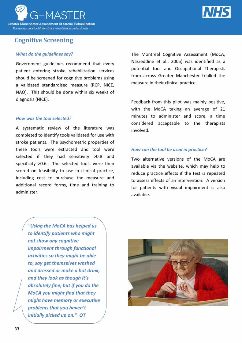
The Montreal Cognitive Assessment (MoCA;
Nasreddine et al., 2005) was identified as a
potential tool and Occupational Therapists
from across Greater Manchester trialled the
measure in their clinical practice.
Feedback from this pilot was mainly positive,
with the MoCA taking an average of 21
minutes to administer and score, a time
considered acceptable to the therapists
involved.
How can the tool be used in practice?
Two alternative versions of the MoCA are
available via the website, which may help to
reduce practice effects if the test is repeated
to assess effects of an intervention. A version
for patients with visual impairment is also
available.
“Using the MoCA has helped us
to identify patients who might
not show any cognitive
impairment through functional
activities so they might be able
to, say get themselves washed
and dressed or make a hot drink,
and they look as though it’s
absolutely fine, but if you do the
MoCA you might find that they
might have memory or executive
problems that you haven’t
initially picked up on.” OT

34
Montreal Cognitive Assessment (MoCA)

35
Montreal Cognitive Assessment (MoCA): Administration and Scoring
Instructions
The Montreal Cognitive Assessment (MoCA) was designed as a rapid screening instrument for mild cognitive
dysfunction. It assesses different cognitive domains: attention and concentration, executive functions, memory,
language, visuoconstructional skills, conceptual thinking, calculations, and orientation. Time to administer the MoCA
is approximately 10 minutes. The total possible score is 30 points; a score of 26 or above is considered normal.
1. Alternating Trail Making:
Administration: The examiner instructs the subject: “Please draw a line, going from a number to a letter in
ascending order. Begin here [point to (1)] and draw a line from 1 then to A then to 2 and so on. End here
[point to (E)].”
Scoring: Allocate one point if the subject successfully draws the following pattern: 1 – A – 2 – B – 3 – C – 4 –
D – 5 – E, without drawing any lines that cross. Any error that is not immediately self-corrected earns a
score of 0.
2. Visuoconstructional Skills (Cube):
Administration: The examiner gives the following instructions, pointing to the cube: “Copy this drawing as
accurately as you can, in the space below.”
Scoring: One point is allocated for a correctly executed drawing.
Drawing must be three-dimensional
All lines are drawn
No line is added
Lines are relatively parallel and their length is similar (rectangular prisms are accepted)
A point is not assigned if any of the above-criteria are not met.
3. Visuoconstructional Skills (Clock):
Administration: Indicate the right third of the space and give the following instructions: “Draw a clock. Put
in all the numbers and set the time to 10 past 11.”
Scoring: One point is allocated for each of the following three criteria:
Contour (1 pt.): the clock face must be a circle with only minor distortion acceptable (e.g., slight
imperfection on closing the circle);
Numbers (1 pt.): all clock numbers must be present with no additional numbers; numbers must be in
the correct order and placed in the approximate quadrants on the clock face; Roman numerals are
acceptable; numbers can be placed outside the circle contour;
Hands (1 pt.); there must be two hands jointly indicating the correct time; the hour hand must be
clearly shorter than the minute hand; hands must be centred within the clock face with their
junction close to the clock centre.
A point is not assigned for a given element if any of the above-criteria are not met.
MoCA Version August 18, 2010
© Z. Nasreddine MD www.mocatest.org

36
4. Naming:
Administration: Beginning on the left, point to each figure and say: “Tell me the name of this animal.”
Scoring: One point each is given for the following responses: (a) lion (2) rhinoceros or rhino (3) camel or
dromedary.
5. Memory
Administration: The examiner reads a list of 5 words at a rate of one per second, giving the following
instructions: “This is a memory test. I am going to read a list of words that you will have to remember now
and later on. Listen carefully. When I am through, tell me as many words as you can remember. It doesn’t
matter in what order you say them.” Mark a check in the allocated space for each word the subject
produces on this first trial. When the subject indicates that (s)he has finished (has recalled all words), or can
recall no more words, read the list a second time with the following instructions: “I am going to read the
same list for a second time. Try to remember and tell me as many words as you can, including words you
said the first time.” Put a check in the allocated space for each word the subject recalls after the second
trial.
At the end of the second trial, inform the subject that (s)he will be asked to recall these words again by
saying, “I will ask you to recall those words again at the end of the test.”
6. Attention:
Forward Digit Span: Administration: Give the following instruction: “I am going to say some numbers and
when I am through, repeat them to me exactly as I said them.” Read the five number sequence at a rate of
one digit per second.
Backward Digit Span: Administration: Give the following instruction: “Now I am going to say some more
numbers, but when I am through you must repeat them to me in the backwards order.” Read the three
number sequence at a rate of one digit per second.
Scoring: Allocate one point for each sequence correctly repeated, (N.B.: the correct response for the
backwards trial is 2-4-7).
Vigilance: Administration: The examiner reads the list of letters at a rate of one per second, after giving the
following instruction: “I am going to read a sequence of letters. Every time I say the letter A, tap your hand
once. If I say a different letter, do not tap your hand.”
Scoring: Give one point if there is zero to one errors (an error is a tap on a wrong letter or a failure to tap on
letter A).
MoCA Version August 18, 2010
© Z. Nasreddine MD www.mocatest.org

37
Serial 7s: Adminisration: The examiner gives the following instruction: “Now, I will ask you to count by
subtracting seven from 100, and then, keep subtracting seven from your answer until I tell you to stop.” Give
this instruction twice if necessary.
Scoring: This item is scored out of 3 points. Give no (0) points for no correct subtractions, 1 point for one
correct subtraction, 2 points for two-to-three correct subtractions, and 3 points if the participant
successfully makes four or five correct subtractions. Count each correct subtraction of 7 beginning at 100.
Each subtraction is evaluated independently; that is, if the participant responds with an incorrect number
but continues to correctly subtract 7 from it, give a point for each correct subtraction. For example, a
participant may respond “92 – 85 – 78 – 71 – 64” where the “92” is incorrect, but all subsequent numbers
are subtracted correctly. This is one error and the item would be given a score of 3.
7. Sentence repetition:
Administration: The examiner gives the gollowing instructions: “I am going to read you a sentence. Repeat it
after me, exactly as I say it [pause]: I only know that John is the one to help today.” Following the response,
say: “Now I am going to read you another sentence. Repeat it after me, exactly as I say it [pause]: The cat
always hid under the couch when dogs were in the room.”
Scoring: Allocate 1 point for each sentence correctly repeated. Repetition must be exact. Be alert for errors
that are omissions (e.g., omitting “only,” “always”) and substitutions/additions (e.g., John is the one who
helped today”; substituting “hides” for “hid,” altering plurals, etc.).
8. Verbal fluency:
Administration: The examiner gives the following instruction: “Tell me as many words as you can think of
that begin with a certain letter of the alphabet that I will tell you in a moment. You can say any kind of word
you want, except for proper nouns (like Bob or Boston), numbers, or words that begin with the same sound
but have a different suffix, for example, love, lover, loving. I will tell you to stop after one minute. Are you
ready? [Pause] Now, tell me as many words as you can think of that begin with the letter F. [time for 60 sec].
Stop.”
Scoring: Allocate one point if the subject generates 11 words or more in 60 sec. Record the subject’s
response in the bottom or side margins.
9. Abstraction:
Administration: The examiner asks the subject to explain what each pair of words has in common, starting
with the example: “Tell me how an orange and a banana are alike.” If the subject answers in a concrete
manner, then say only one additional time: “Tell me another way in which those items are alike.” If the
subject does not give the appropriate response (fruit), say, “Yes, and they are also both fruit.” Do not give
any additional instructions or clarification. After the practice trial, say: “Now, tell me how a train and a
bicycle are alike.” Following the response, administer the second trial, saying: “Now tell me how a ruler and
a watch are alike.” Do not give any additional instructions or prompts.
MoCA Version August 18, 2010
© Z. Nasreddine MD www.mocatest.org

38
Scoring: Only the last two item pairs are scored. Give 1 point to each item pair correctly answered. The
following responses are acceptable:
Train-bicycle = means of transportation, means of travelling, you take trips in both: Ruler-watch = measuring instruments, used to measure. The following responses are not acceptable: Train-bicycle = they have wheels; Ruler-watch = they have numbers.
10. Delayed recall:
Administration: The examiner gives the following instruction: “I read some words to you earlier, which I asked you to remember. Tell me as many of those words as you can remember.” Make a check mark ( √ ) for each of the words correctly recalled spontaneously without any cues, in the allocated space.
Scoring: Allocate 1 point for each word recalled freely without any cues.
11. Orientation:
Administration: The examiner gives the following instructions: “Tell me the date today.” If the subject does not give a complete answer, then prompt accordingly by saying: “Tell me the [year, month, exact date, and day of the week].” Then say: “Now, tell me the name of this place, and which city it is in.”
Scoring: Give one point for each item correctly answered. The subject must tell the exact date and the exact place (name of hospital, clinic, office). No points are allocated if subject makes an error of one day for the day and date.
TOTAL SCORE: Sum all subscores listed on the right-hand side. Add one point for an individual who has 12 years or fewer of formal education, for a possible maximum of 30 points. A final total score of 26 and above is considered normal.
MoCA Version August 18, 2010
© Z. Nasreddine MD www.mocatest.org
Optional:
Following the delayed free recall trial, prompt the subject with the semantic category cue provided below
for any word not recalled. Make a check mark ( √ ) in the allocated space if the subject remembered the
word with the help of a category or multiple-choice cue. Prompt all non-recalled words in this manner. If
the subject does not recall the word after the category cue, give him/her a multiple choice trial, using the
following example instruction, “Which of the following words do you think it was, NOSE, FACE, or HAND?”
Use the following category and/or multiple-choice cues for each word, when appropriate:
FACE: category cue: part of the body multiple choice: nose, face, hand
VELVET: category cue: type of fabric multiple choice: denim, cotton, velvet
CHURCH: category cue: type of building multiple choice: church, school, hospital
DAISY: category cue: type of flower multiple choice: rose, daisy, tulip
RED: category cue: a colour multiple choice: red, blue, green
Scoring: No points are allocated for words recalled with a cue. A cue is used for clinical information
purposes only and can give the test interpreter additional information about the type of memory disorder.
For memory deficits due to retrieval failures, performance can be improved with a cue. For memory
deficits due to encoding failures, performance does not improve with a cue.

39
Measuring functional outcomes
Independence in Activities of Daily Living – Barthel Index
What do the guidelines say?
National guidelines recommend that patients
who have had a stroke should be formally
assessed for their safety and independence in
activities of daily living (ADL), using a
standardised tool, preferably the Barthel
Activities of Daily Living Index (RCP).
How can the tool be used in practice?
The Barthel Index is the most widely known and
used test of independence in ADL (Wade &
Collin 1988; Collin et al 1988) and the preferred
measure of ADL according to RCP. The Barthel
Index should be completed in relation to what
the patient does day-to-day on the ward, not
what they can achieve in therapy sessions. The
main aim is to establish the degree of
independence from help, physical or verbal
however minor or for whatever reason. If a
patient’s performance of an activity is variable,
they should be scored at the lower level, in
order that subsequent discharge planning
reflects this variability. The use of aids to be
independent is allowed. Supervision for any
reason means the patient is not independent.
Observation is useful but direct testing is
unnecessary. Usually performance over the last
1-2 days is considered. The middle categories
imply that the patient supplies at least 50% of
the effort. There is a maximum score of 20.
It is recommended that the Barthel Index is scored
within 72 hours of admission to stroke
rehabilitation, as well as weekly as part of the
multidisciplinary team meeting to monitor patient
progress, and upon discharge, with scores
transferred to the community team involved in
patient follow-up. A premorbid score can be
obtained by asking the patient and/or their carers
and family what they were able to do before the
stroke.
“I think the Barthel certainly gives you
guidance really, because you can see
maybe they’ve been 7 for the past three
weeks, so even though in therapy they
might be improving in certain things,
their overall dependency isn’t
improving. Or it is improving, and so
you have bench-marking from the weeks
before.” Ward Manager

40
The Barthel Index
Name: ______________________________________ NHS No: _______________________________ D.O.B: _______________________________________
Date
Bowels
0 = incontinent (or needs to be given enemas): 1 = occasional accident (once a week): 2 = continent
0 1 2
0 1 2
0 1 2
0 1 2
0 1 2
Bladder
0 = incontinent or catheterised and unable to manage alone: 1 = occasional accident (maximum once per 24 hours): 2 = continent
0 1 2
0 1 2
0 1 2
0 1 2
0 1 2
Grooming
0 = needs help with personal care: 1 = independent (face, hair, teeth, shaving – implements provided)
0 1
0 1
0 1
0 1
0 1
Toilet Use
0 = dependent: 1 = needs some help but can do something alone: 2 = independent (on and off, dressing, wiping)
0 1 2
0 1 2
0 1 2
0 1 2
0 1 2
Feeding
0 = unable: 1 = needs help cutting, spreading butter etc: 2 = independent
0 1 2
0 1 2
0 1 2
0 1 2
0 1 2
Transfers (bed to chair and back)
0 = unable, no sitting balance: 1 = major help (1 or 2 people, physical), can sit: 2 = minor help (verbal or physical): 3 = independent
0 1 2 3
0 1 2 3
0 1 2 3
0 1 2 3
0 1 2 3
Mobility
0 = immobile: 1 = wheelchair independent, including corners: 2 = Walks with the help of 1 person (verbal or physical): 3 = independent (but may use aid; for example stick)
0 1 2 3
0 1 2 3
0 1 2 3
0 1 2 3
0 1 2 3
Dressing
0 = dependent: 1 = needs help but can do about half unaided: 2 = independent (including buttons/zips/laces, etc.)
0 1 2
0 1 2
0 1 2
0 1 2
0 1 2
Stairs
0 = unable: 1 = needs help (verbal, physical or carrying aid): 2 = independent
0 1 2
0 1 2
0 1 2
0 1 2
0 1 2
Bathing
0 = dependent: 1 = independent 0
1
0
1
0
1
0
1
0
1
Total Score (out of 20)

41
Barthel Index Scoring Criteria
Scoring Criteria Bowels (in the last week) 0 = incontinent or needs enema 1 = occasional accident (once per week) 2 = continent Bladder (in the last week) 0 = incontinent, or catheterised or unable to manage alone 1 = occasional accident (within last 24hrs) 2 = continent, including complete self-management of catheterisation Grooming (within last 24hrs) This refers to personal hygiene: cleaning teeth, fitting false teeth, combing hair, shaving, washing face. Implements can be supplied by a helper. 0 = Needs help with personal care 1 = Independent Toilet Use includes reaching the toilet/commode, undressing, cleaning self, dressing and leaving 0 = dependent 1 = Needs some help but can do some alone 2 = Independent Feeding involves eating any normal food (not restricted to soft food). Food can be cooked and served by others but not cut up 0 = Unable 1 = Needs help cutting, spreading, etc, but can feed him/herself 2 = Independent Transfer (bed to chair and back) 0 = Unable, no sitting balance, two people to help 1 = Major help – can sit but needs physical assistance of 1strong/skilled helper, 2 `normal' people 2 = Minor help – can be assisted easily by one person (verbally or physically) 3 = Independent, may use an aid.
Mobility refers to indoor mobility around the house or ward. An aid may be used. If using a wheelchair, corners and doors must be negoiated unaided. 0 = Immobile 1 = Wheelchair independent, including steering and corners/ doors 2 = Walks with help of 1 (verbal or physical) 3 = Independent although may use an aid Dressing 0 = Dependent 1 = Help with buttons, zips etc. but can put on some clothes unaided 2 = Independent - including buttons, zips, laces etc. Can select and put all clothes on/off although they may be adapted. Stairs To be independent the patient must carry any walking aids. 0 = Unable 1 = Needs help (verbal, physical, with aid) 2 = Independent Bath/Shower 0 = Dependent 1 = Independent – including getting in and out and washing self. If using a shower, the patient must be unsupervised and unaided.

42
Monitoring communication and swallowing abilities over time –
Therapy Outcome Measures
Whilst the Barthel Index provides a good
measure of independence in activities of daily
living after stroke, it does not take into account
difficulties in communication, which are
common post-stroke. The Therapy Outcome
Measures (TOMs; Enderby et al., 2006) for
communication difficulties allow for
measurement of impairments and functional
activities and can be scored on a weekly basis for
appropriate patients to track progress. There
are also measures for dysphagia, which help to
monitor progress in swallowing.
Measures of participation and well-
being/distress are also available (see Enderby, P.,
John, A. & Petheram, B. (2006). Therapy
Outcome Measures for Rehabilitation
Professionals (2nd Ed). Wiley: UK).
“It can be a benefit as it’s an actual
figure. Rather than saying they’ve
improved, I think to discuss it as a
figure helps to do it within the MDT,
so for a speech therapist to discuss
the level of aphasia between other
speech therapists, it might be quite
obvious how someone has improved,
but actually to another MDT member
that might not be so clear. Whereas
if you discuss it in TOMs and they go
up or they go down, I think it helps to
make it a bit clearer how that
patient is progressing.” Speech &
Language Therapist
To use the TOMs, identify the descriptor which
best fits the patient. It is not necessary for the
patient to have each feature mentioned. 0.5 can
be used to indicate if the patient is slightly better
or worse than an indicator.

43
Therapy Outcome Measures (Dysphasia/Aphasia)
Impairment
0 Aphasia affecting all modalities: Auditory and reading comprehension inconsistent even at one
keyword. No meaningful expression
1 Severe dysphasia/aphasia: Auditory and/or reading comprehension is consistent at one keyword level.
Occasionally understand and expresses limited amount
2 Severe/Moderate dysphasia/aphasia: Auditory and/or reading comprehension consistent at a minimum
of two or three keyword level. Some limited verbal and/or written expression used appropriately and
purposefully
3 Moderate dysphasia/aphasia: Constant auditory and/or reading comprehension for simple sentences or
structures. Inconsistent with complex commands and structures. Consistently reduced verbal and/or
written language structure and vocabulary. May have a specific more severe difficulty in one modality.
4 Mild dysphasia/aphasia: Occasional difficulties present in auditory and/or reading comprehension and
in verbal and/or written expression
5 No dysphasia/aphasia
Activity
0 Unable to communicate in any way. No effective communication. No interaction.
1 Occasionally able to make basic needs known with familiar persons or trained listeners in familiar
contexts. Minimal communication with maximal assistance
2 Limited functional communication. Consistently able to make basic needs/conversation understood but
is heavily dependent on cues and context. Communicates better with trained listener or family members
or in familiar settings. Frequent repetition required. Maintains meaningful interaction related to here and
now
3 Consistently able to make needs known but can sometimes convey more information than this. Some
inconsistency in unfamiliar settings. Is less dependent for intelligibility on cues and context. Occasional
repetition required. Communicates beyond here/now with familiar persons; needs cues and prompting
4 Can be understood most of the time by any listener despite communication irregularities. Holds
conversation; requires occasional prompts particularly with a wider range of people
5 Communicates effectively in all situations
From Enderby, P., John, A. & Petheram, B. (2006). Therapy Outcome Measures for Rehabilitation
Professionals (2nd Ed). Wiley: UK

44
Therapy Outcome Measures (Dysphagia)
Impairment
0 Aphagia: Not safe to swallow due to cognitive status/no bolus control/aspiration/absence of
oral/pharyngeal swallow. Clinical signs of aspiration. No cough reflex. May need regular suction.
1 Severe dysphagia: Weak oral movements/no bolus control/inadequate/inconsistent swallow reflex.
High risk of aspiration.
2 Severe/Moderate dysphagia: Cough/swallow reflexes evident but abnormal or delayed. Uncoordinated
oral movements. Risk of aspiration.
3 Moderate dysphagia: Swallow and cough reflex present. May have poor oral control. At risk of
occasional aspiration.
4 Mild oral/pharyngeal dysphagia: Incoordination but no clinical evidence of aspiration
5 No dysphagia/aphagia
Activity
0 Non-oral feeding to meet all hydration and nutritional needs. Unsafe to take practice amounts of
modified consistencies and unable to use compensatory strategies. Unable to manage secretions.
1 Non-oral feeding to meet most hydration and nutritional needs. Variable ability to take practice amounts
of modified consistencies using compensatory strategies. Some management of secretions. Needs
experienced supervision.
2 Non-oral feeding/supplements needed to meet hydration and nutritional needs. Consistently able to
take practice amount of modified consistencies using compensatory strategies. Needs experienced
supervision.
3 Consistently able to take modified consistencies using compensatory strategies. Needs some
supervision, may require feeding supplements, may eat extremely slowly.
4 Although eating and drinking is abnormal, it is good enough to meet nutritional requirements. No
supervision required. No alternative or supplement feeding. May avoid certain foods, drinks, or eating
situations.
5 Functionally eating and drinking a normal diet.
From Enderby, P., John, A. & Petheram, B. (2006). Therapy Outcome Measures for Rehabilitation
Professionals (2nd Ed). Wiley: UK

45
Therapy Outcome Measures (Dysarthria)
Impairment
0 Severe dysarthria: severe persistent articulatory/prosodic impairment. Inability to produce any
distinguishable speech sounds. No oral motor control. No respiratory support for speech.
1 Severe/moderate dysarthria: with consistent articulatory/prosodic impairment. Mostly open vowel
sounds with some consonant approximations/severe festination of speech. Extremely effortful or slow
speech; only 1 or 2 words per breath. Severely limited motor control.
2 Moderate dysarthria: with frequent episodes of articulatory/prosodic impairment. Most consonants
attempted but poorly represented acoustically/moderate festination. Very slow speech; manages up to 4
words per breath. Moderate limitation oral motor control.
3 Moderate/mild dysarthria: consistent omission/articulation of consonants. Variability of speed. Mild
limitation of oral motor control or prosodic impairment.
4 Mild dysarthria: slight or occasional omission/mispronunciation of consonants. Slight or occasional
difficulty with oral motor control/prosody or respiratory support.
5 No impairment.
Activity
0 Unable to communicate in any way. No effective communication. No interaction.
1 Occasionally able to make basic needs known with familiar persons or trained listeners in familiar
contexts. Minimal communication with maximal assistance.
2 Limited functional communication. Consistently able to make basic needs/conversation understood but
is heavily dependent on cues and context. Communicates better with trained listener or family members
or in familiar settings. Frequent repetition required. Maintained meaningful interaction related to here
and now.
3 Consistently able to make needs known but can sometimes convey more information than this. Some
inconsistency in unfamiliar settings. Is less dependent for intelligibility on cues and context. Occasional
repetition required. Communicates beyond here/now with familiar persons, needs some cues and
prompting.
4 Can be understood most of the time by any listener despite communication irregularities. Holds
conversation; requires special consideration, for example, patience, time, attention, especially with a wider
range of people.
5 Communicates effectively in all situations.
From Enderby, P., John, A. & Petheram, B. (2006). Therapy Outcome Measures for Rehabilitation
Professionals (2nd Ed). Wiley: UK

46
How to run an effective Stroke Multi-Disciplinary Team meeting
Guidelines
The RCP Clinical Guideline for Stroke (3rd Ed.)
states that a stroke service should have a co-
ordinated multi-disciplinary team that meets at
least once a week for the interchange of
information about patients. The following
guidance for running an MDT meeting has been
developed through the observation of meetings
across Greater Manchester and through gaining
feedback from staff about what works well in
these meetings. National guidance documents
and recommendations from the research
literature have also been incorporated.
Aims and purpose of the meeting
The multidisciplinary team should meet weekly to:
Develop a shared understanding of the patient’s problems
Plan and review multi-disciplinary patient goals
Monitor the progress of the patient throughout the rehabilitation process
Make quick and effective joint decisions about patient care and discharge plans
The meeting should also give the team the
opportunity to:
Facilitate greater understanding of a patient’s problems through discussion with other professionals with specialist expertise
Facilitate 24 hour rehabilitation and maintain consistency of care among the team, e.g. for therapists to hand over to nursing staff information on strategies and therapeutic activity to carry on outside of therapy time
Identify and set actions for team members to complete during the coming week
Ensure identified actions have been completed, e.g. referrals, assessments, arranging meetings and medical investigations
Goal-setting
Multidisciplinary goals should be planned and
reviewed in conjunction with the patient and
their family, and this may take place in informal
goal-setting or family meetings outside of the
main MDT meeting. Goals should take into
account the patient’s needs and aspirations,
and progress towards both short- and long-
term goals should be reviewed within the
formal MDT meeting.
Venue
The MDT meeting should be held in a room,
which is large enough for the team to
comfortably congregate in, with space for a
notes trolley if this is required. All members of
the team should have a seat and be able to see
and hear each other. The room should be in a
suitably quiet and cool environment and all
attempts should be made to protect patient
confidentiality, e.g. closing windows and doors
to prevent others listening to the discussion.
Interruptions to MDT meetings are common
and it should be ensured that other staff on the
unit are aware that the meeting should be
interrupted only in the event of an emergency.
It may be helpful to put a sign on the meeting
room door. It can also be useful to hold the
MDT meeting in a room off the main ward
where possible to prevent interruptions.

47
Attendance
MDT meetings should be regarded as a vital
aspect of patient care and prioritised as such by
all members of the team involved in patient
care. The start and finish times of the meeting
should be well-publicised among the team and
every effort should be made by members of the
team to keep to these times, to avoid wasting
the clinical time of other team members. If a
member of the team does not attend on time,
the meeting should go ahead without them. It
may help to schedule the meeting at the
beginning of the day or immediately after
lunch, so that team members are not in the
middle of other clinical tasks.
The MDT meeting should be attended by all
those involved in patients’ care. This should
include:
Doctor
Nurse
Occupational Therapist
Physiotherapist
Speech and Language Therapist
Social Worker
Anyone else who may be involved in patient care or discharge planning, e.g. clinical psychologist or counsellor, dietician, case manager, discharge facilitator, transfer of care nurse, Stroke Association Information, Advice and Support Service
Attendance should be monitored by recording
the disciplines present at each meeting within
the MDT documentation. If members of the
same discipline fail to attend three consecutive
MDT meetings, management should be alerted
by the chair and steps should be taken to
resolve any issues that are preventing
attendance.
Team members attending the MDT meeting
should ideally have knowledge of the patients
to be discussed, however where this is not
possible for all patients (due to sharing of
workload), detailed notes from a colleague
should be taken to the meeting. It is not
suggested that all members of every discipline
attend the whole MDT meeting and one
clinician may attend to represent all members
of their discipline. However, if one member of
a discipline is attending the MDT meeting on
behalf of their team, they should ensure that
they receive detailed notes from other
members of their team to feed back to the
MDT and they will also be responsible for
feeding back information about decisions
made at the MDT meeting to those members
of their team unable to attend.
If a member of the team is available only for
part of the meeting, it may be advisable to
alter the order of patients to be discussed, e.g.
should a speech therapist be available only at
the beginning of the meeting, patients with
speech problems should be discussed first.
“The MDT is more fluid and I think
that it’s a really good way to discuss
those patients and everybody gets a
platform as well, so I think it’s really
good that it’s not ‘what’s the speech
therapist got to say?’ it’s ‘what’s the
patient’s difficulties and what can
we input from each profession.’ I
think that works really well with it.”
Speech & Language Therapist

48
Alternatively, it may be useful to circulate an
agenda prior to the meeting confirming the times
at which each patient will be discussed to allow
professionals to attend only for the patients
whose care they are involved in. This may be
done through the allocation of a 5 minute slot per
patient and it is the responsibility of the chair to
ensure the discussions run to time.
Preparation
Team members should be aware of the
information they are likely to need during the
MDT meeting and should prepare this
information beforehand to allow quick and
effective feedback to the team. This is
particularly important for actions set from the
previous week and allocated to specific people, all
attempts should be made to ascertain whether
these actions have been completed before the
meeting. This may be in the form of a detailed
handover sheet or notebook. Should members of
the team be unable to attend the meeting, they
should provide a detailed handover and notes to
a colleague to share with the team.
Preparation should include:
Knowledge of the patient’s social and family circumstances
Communication from the patient/their family
Knowledge of the nursing and therapy needs of the patients
Progress made since the last meeting, to include completion of allocated actions
Results from patient assessments to review with the team where appropriate, e.g. mood scores
Leadership and chairing the MDT meeting
Every MDT meeting should have a chair and a
vice-chair, who should be prepared to take over
when the chair is absent, due to illness or annual
leave. These roles could be filled by any member
of the multi-disciplinary team and developing the
skills necessary to perform this role could form
part of a member of staff’s CPD. The chair
should facilitate discussion between all members
of the multi-disciplinary team and ensure that
the discussion is not dominated by a single
clinician or profession. Should conflict arise, the
chair should negotiate agreement and encourage
a climate of constructive criticism and open
discussion.
The chairperson is responsible for keeping the
discussion focussed on the agenda and ensuring
the meeting runs to time, and to facilitate quick
and effective decision-making about a patient’s
care or discharge. If a decision is clinically
complex and requires further discussion, the
chair should refer this point to another meeting
to be discussed in a patient case conference or
family meeting. This should be documented in
the action plan for the week.
Documentation
Notes of the discussion should be made using
standardised MDT documentation. This
documentation should include all aspects
mentioned above (an example of MDT
documentation can be on page 6). Action plans
allocated to specific members of the team should
be documented and these should be reviewed at
the following meeting. Responsibility for making
notes in patient files should be shared amongst
the members of the team. A member of each
discipline may also wish to record separate notes
to allow them to quickly feed back information to
members of their team who are unable to attend
the meeting.
“I think we all come to the meeting much
better prepared and we’re not scrabbling
through the notes trying to find the
information, but we come knowing their
scores and progress.” Physiotherapist

49
Structure
The MDT meeting should follow a pre-defined
structure to ensure that no aspect of patient care is
missed (see sample agenda).
The chair should introduce each patient with a brief
summary to include:
Name and age of patient
Diagnosis/summary of acute care and impairments upon initial assessment
Date of admission to service
Previous medical history
Social/family situation
Long-term MDT goals
The meeting should then follow a pre-defined
agenda to encompass:
Review of action points from the previous meeting
Scoring and review of scores on standardised measures and assessments
Discussion of progress towards goals and plans for management
Planning towards discharge (expected date of discharge and destination to be discussed and recorded following initial assessment)
Identification of actions to be completed with responsibility allocated to a member of the team and a date to complete by
Team Climate
All members of the team should be encouraged to
contribute to the discussions and openly seek
clarification if they feel something is unclear. There
should be a feeling of participative safety and trust
amongst the team and members should feel able to
express themselves freely. Members of the team
should have an understanding of the objectives the
team is aiming to achieve and team members
should help and support each other towards
achieving these objectives. There should be a pro-
discussion environment, allowing members of the
team to constructively challenge each other where
necessary.
Use of standardised measures
Standardised measures should be completed
during the multi-disciplinary team meeting to
document the patient’s progress through the
rehabilitation process, in order to focus the
discussions on the patient’s problems. All
team members should have an understanding
of the scores of the core measures being used
and contribute to scoring the measures where
appropriate. Progress should be assessed
using these standardised measures and they
should be used to inform clinical decision-
making, e.g. whether a patient is continuing to
make progress within rehabilitation.
After the meeting
Following the MDT meeting, members of the
stroke MDT should feed back actions and
decisions from the meeting to members of
their disciplinary team unable to attend the
meeting. Decisions taken at the meeting
should inform the plan of care and therapeutic
activity over the following week and team
members should ensure that they have
completed all actions they have been allocated
at the meeting prior to the date set for
completion.

50
Sample MDT Meeting Agenda
Brief introduction
State patient’s name, diagnosis, brief summary of acute care, admission date/days since stroke, previous level of functioning and social situation, long-term MDT goals
Review of previous actions
Score the toolkit – compare with previous scores to monitor progress o Score Barthel Index (even if you feel it will not have changed) – break down into sections and
score as a team. Record item scores o Has mood been scored? When was the screen completed (set date to complete screen if not
currently applicable), what was the score and interpretation, plans to review? o Has cognition been scored? When was the MoCA completed (set date to complete screen if not
currently applicable), what was the score and interpretation, plans to review? o What is the MUST score for this week? o Has a language screen been completed? o Score TOMs (aphasia/dysphagia/dysarthria)
Progress towards goals and plans for management What are the patient’s goals and how do these relate to assessment scores? Give update on progress towards goals and plans to manage lack of goal attainment if appropriate
Discharge planning State provisional place and estimated date of discharge and note any issues arising
Action Plans What actions are needed to support goal achievement and achieve discharge plans? Who should complete each action and by when?

51
Initial MDT form
Name:
DOB: NHS No:
Date of admission: Admission Barthel Index score:
Diagnosis/Summary of acute care (including date of thrombolysis if applicable):
NIHSS score:
Impairments upon initial assessment (tick all that apply) □ Motor □ Language/Communication □ Balance □ Sensory □ Visual/Perceptual □ Cognitive □ Continence □ Mood □ Swallowing
Previous medical history:
Social/family situation:
Long-term MDT goals:
Social Worker name and contact telephone number:

52
Weekly MDT form
Name:
DOB: NHS No:
Disciplines present: Doctor □ Nurse □ Occupational Therapist □ Physiotherapist □
Speech therapist □ Dietician □ Social Worker □ Other □ ____________________
Review of actions from previous meeting (note any actions not completed and reasons why – add to action plan overleaf if required)
Scoring of toolkit (score toolkit and compare with previous scores)
Barthel Index score: /20 Cognition – MoCA score: /30
Mood screen completed:
Date of completion: Score/Interpretation:
TOMs (aphasia) scores: Impairment /5, Activity /5 MUST score: /6
TOMs (dysphagia) scores: Impairment /5, Activity /5 Communication score:
Progress towards goals and plans for management (relate assessment scores to goals, record progress towards goals, plan to manage lack of goal attainment if appropriate)
Discharge planning (record provisional place and expected date of discharge below, and note any issues arising)
Provisional discharge destination:
Expected date of discharge:

53
Action plan (actions to be completed in the coming week, e.g. referrals to be made, assessments to be completed) Action Who completes? By when?

54
References
Collin C, Wade DT, Davies S, Horne V (1988). The Barthel ADL Index: A reliability study. Disability & Rehabilitation,
10 (2), 61-63.
Department of Health. National stroke strategy. London: Her Majesty's Stationery Office; 2007.
Elia M (2003). Screening for malnutrition: A multidisciplinary responsibility. Development and use of the
Malnutrition Universal Screening Tool (‘MUST’) for adults. Redditch: BAPEN.
Enderby P, John A & Petheram B. (2006). Therapy Outcome Measures for Rehabilitation Professionals (2nd Ed).
Wiley: UK
Flamand-Roze C, Falissard B, Roze E, Maintigneux L, Beziz J, Chacon A, Join-Lambert C, Adams D & Denier C (2011).
Validation of a new language screening tool for patients with acute stroke: The Language Screening Test (LAST).
Stroke, 42, 1224-1229
House, A. & Knapp, P. (2011). Psychological care after stroke. National Stroke Improvement Programme website.
Intercollegiate Stroke Working Party. National Clinical Guideline for stroke, 3rd Ed. London: Royal College of
Physicians; 2008.
Lincoln N, Sutcliffe L & Unsworth G. (2000) Validation of Stroke Aphasic Depression Questionnaire (SADQ) for use
with patients in hospital. Clinical Neuropsychological Assessment, 1, 88-96.
Nasreddine ZS, Phillips NA, Bédirian V, Charbonneau S, Whitehead V, Collin I, Cummings JL, Chertkow H (2005). The
Montreal Cognitive Assessment (MoCA©): A Brief Screening Tool For Mild Cognitive Impairment. J Am Geriatr Soc,
53, 695–699
National Audit Office. Progress in improving stroke care. London: The Stationery Office; 2010.
National Collaborating Centre for Chronic Conditions (2008). Stroke: national clinical guideline for diagnosis and
initial management of acute stroke and transient ischaemic attack (TIA). London: Royal College of Physicians.
National Institute for Health and Clinical Excellence (2010). Stroke Quality Standard (QS2). London: NICE.
Spitzer RL, Kroenke K, Williams, JBW, Löwe B (2006). A brief measure for assessing generalised anxiety disorder
(GAD-7). Arch Intern Med, 166 (10), 1092-1097.
Spitzer R, Kroenke K, Williams J. Validation and utility of a self-report version of PRIME-MD: The PHQ primary care
study (1999). JAMA, 282, 1737-1744.
Turner-Stokes L, Kalmus M, Hirani D, F C. The depression intesity scale circles (DISCs): a first evaluation of a simple
assessment tool for depression in the context of brain injury (2005). J Neurol Neurosurg Psychiatry, 76, 1273-1278.
Tyson S, Greenhalgh J, Long A, Flynn R. The use of measurement tools in clinical practice: an observational study of
neurorehabilitation (2010). Clinical Rehabilitation, 24, 74-81.
Wade DT & Collin C (1988). The Barthel ADL Index: A standard measure of physical disability? Disability &
Rehabilitation, 10 (2), 64-67.

55