acs and hyperlipidemia. which of the following statements is correct regarding the addition of...
TRANSCRIPT

ACS and Hyperlipidemia
Jonathon Firnhaber, MD, FAAFP Assistant Professor
The Brody School of Medicine at East Carolina University
Greenville, North Carolina

It is the policy of the AAFP that all individuals in a position to control content disclose any relationships with commercial interests upon nomination/invitation of participation. Disclosure documents are reviewed for potential conflicts of interest. If conflicts are identified, they are resolved prior to confirmation of participation. Only participants who have no conflict of interest or who agree to an identified resolution process prior to their participation were involved in this CME activity
Disclosure Statement
Dr. Firnhaber has nothing to disclose.

Learning Objectives
1. Identify the primary target for cholesterol lowering therapy based on the Adult Treatment Panel III report.
2. Categorize risk stratification for cholesterol therapy based upon LDL-C risk factors and the Framingham Data.
3. Recognize the importance of hyperlipidemia therapy in the metabolic syndrome.
4. Recognize the hyperlipidemia therapy of coronary artery equivalent patients.
5. Relate the importance and role of intensive LDL cholesterol lowering therapy.

Acute Coronary Syndrome
• Acute coronary syndrome – Unstable angina (UA) – Non-ST-elevation myocardial infarction
(NSTEMI) – ST-elevation myocardial infarction (STEMI)

1. A 52-year-old man presents to the ED with a 45-minute history of squeezing substernal chest pressure radiating to his left arm.
An ECG from a routine physical two months ago was normal.

Question 1 ECG

1. This patient’s history and ECG are most consistent with which one of the following?
A. Esophageal spasm B. Acute ST segment elevation MI (STEMI) C. Acute non-ST segment elevation MI (NSTEMI) D. Pericarditis
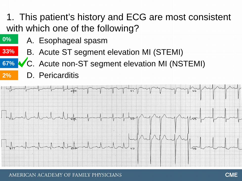
1. This patient’s history and ECG are most consistent with which one of the following?
A. Esophageal spasm B. Acute ST segment elevation MI (STEMI) C. Acute non-ST segment elevation MI (NSTEMI) D. Pericarditis 2%
0%
33%
67%

ECG Findings: ST Segment Depression, Inferior and Lateral Precordial Leads

Acute Coronary Syndrome
• UA and NSTEMI are closely related, with similar presentations, but with differing severity: – Both cause ST-segment depression or prominent T-
wave inversion – Ischemia severe enough to cause myocardial injury
with release of biomarkers (troponin I, troponin T, CK-MB) = NSTEMI
• STEMI is distinguished from UA/NSTEMI by presence of ST-segment elevation on ECG (followed by Q wave) – Warrants immediate reperfusion therapy

Acute Coronary Syndrome
Etiology: reduced myocardial perfusion • Reduced O2 supply (more common) • Increased O2 demand

Acute Coronary Syndrome
• Most commonly due to occlusive thrombus developing on top of a disrupted atherosclerotic plaque – Nearly 50% at sites with < 50% luminal narrowing
• Other etiologies: – Spasm at site of atherosclerotic plaque – Normal coronary arteries with spasm
• Prinzmetal’s angina; transient ST elevation associated
– Arterial inflammation (Kawasaki disease) – Cocaine-induced (treat with NTG and CCB)

2. This patient’s initial troponin is normal. Which of the following statements about cardiac biomarkers is correct?
A. A single negative troponin excludes MI B. Troponin is a better marker of reinfarction than
is CK-MB C. Myoglobin can be detected as early as 2 hours
after the onset of myocardial necrosis D. CK-MB and myoglobin rise and fall more
slowly than troponin

2. This patient’s initial troponin is normal. Which of the following statements about cardiac biomarkers is correct?
A. A single negative troponin excludes MI B. Troponin is a better marker of reinfarction than
is CK-MB C. Myoglobin can be detected as early as 2 hours
after the onset of myocardial necrosis D. CK-MB and myoglobin rise and fall more
slowly than troponin 41%
1%
18%
41%

Troponin
• Troponin is the biomarker of choice in the evaluation of ACS. Troponin I and T are equivalent.
• Detected 3-6 hours after the onset of ischemic symptoms
• Normal serial troponin levels exclude myocardial infarction, but do not exclude unstable angina.
• Troponin remains elevated for 7-14 days post-MI

Myoglobin
• Earliest marker of MI – Released from damaged muscle more rapidly than
CK-MB or troponin – Can be detected as early as 2 hours after cardiac
necrosis – Peaks at about 8 hours; remains elevated < 24 hours
• Sensitive but not specific – Cardiac origin must be confirmed with a more cardiac-
specific enzyme – False positives due to skeletal muscle injury

CK-MB
• Remains elevated for 36-48 hours following MI
• Early peak (12-18 hours) suggests reperfusion • CK-MB and myoglobin rise and fall more rapidly
than troponin – Better for diagnosing reinfarction

3. This patient continues to have pain despite treatment with IV NTG, morphine, ß-blocker, aspirin, and heparin. A repeat ECG shows persistent ST segment depression. What is the next most appropriate intervention?
A. Emergency PCI (percutaneous coronary intervention)
B. Fibrinolysis C. Emergency CABG D. Glycoprotein IIb/IIIa agent

3. This patient continues to have pain despite treatment with IV NTG, morphine, ß-blocker, aspirin, and heparin. A repeat ECG shows persistent ST segment depression. What is the next most appropriate intervention?
A. Emergency PCI (percutaneous coronary intervention)
B. Fibrinolysis C. Emergency CABG D. Glycoprotein IIb/IIIa agent 9%
83%
5%
3%

2007 ACC/AHA UA/NSTEMI Guideline Revision
Recommends initial invasive strategy in patients with the following characteristics: • Recurrent angina or ischemia at rest or with low-level
activities despite intensive medical therapy • Elevated cardiac biomarkers (TnT or TnI) • New or presumably new ST-segment depression • Signs or symptoms of HF or new or worsening mitral
regurgitation • High-risk findings from noninvasive testing

2007 ACC/AHA UA/NSTEMI Guideline Revision
Recommends initial invasive strategy in patients with the following characteristics: • Hemodynamic instability • Sustained ventricular tachycardia • PCI within 6 months • Prior CABG • High-risk score (eg, TIMI, GRACE) • Reduced left ventricular function (LVEF less than 40%)

ACS Therapy: Fibrinolysis
Fibrinolytic therapy or primary percutaneous coronary intervention (PCI) should be strongly considered in all STEMI patients with a symptom onset within 12 hours

ACS Therapy: Glycoprotein IIb/IIIa Inhibition
Glycoprotein IIb/IIIa receptor blockers • Inhibit platelet aggregation • Tirofiban (Aggrastat) • Eptifibatide (Integrilin) • Abciximab (ReoPro)

ACS Therapy: PCI with Stent
• Bare metal – Bare metal acts as foreign body, increasing risk of
in-stent thrombosis—clopidogrel + ASA decreases risk; continue 1 month post-stent if no MI (or 12 months if post-MI)
– Endothelialization may progress to in-stent stenosis • Drug-eluting
– Delay endothelialization, maintaining bare metal longer; continue clopidogrel + ASA for 12 months
– Sirolimus (Cypher), tacrolimus (Mahoroba), paclitaxel (Taxus)
ACS Therapy: Emergency CABG
Considered only if coronary anatomy is not suitable for PCI

4. A 70-year-old woman with history of hypertension and type 2 diabetes presents to the ED with a 14-hour history of profound shortness of breath, but no chest pain. Her current medications include chlorthalidone, lisinopril, and insulin.

Question 4 ECG

4. This patient’s history and ECG are most consistent with which one of the following?
A. Unstable angina B. Acute anterior wall ST-segment elevation
myocardial infarction C. Pericarditis D. Acute anterior wall ischemia

4. This patient’s history and ECG are most consistent with which one of the following?
A. Unstable angina B. Acute anterior wall ST-segment elevation
myocardial infarction C. Pericarditis D. Acute anterior wall ischemia 3%
1%
94%
3%

ECG Findings Q waves and ST-segment elevation with T-wave
inversion, anterior precordial leads (V2 – V5)

ACS: STEMI
ST segments: • Elevation occurs immediately post plaque-rupture and is
consistent with myocardial injury. • Resolution of ST elevation suggests reperfusion. • Persistent ST elevation may be seen with aneurysm
formation. • ST depression indicates myocardial ischemia.
Q waves: • Develop approximately 12 hours post plaque-rupture,
and are indicative of (electrically) dead myocardium (MI). • Typically permanent.

ACS: STEMI
• Anterior/anteroseptal – LAD – Leads V1 – V4
• Lateral – Circumflex – Leads V5 – V6
• Inferior – RCA – Leads II, III, aVF

5. Which of the following medications improves survival post-MI?
A. Digoxin B. Dihydropyridine calcium-channel blocker C. Long-acting nitrate D. ACE-inhibitor

5. Which of the following medications improves survival post-MI?
A. Digoxin B. Dihydropyridine calcium-channel blocker C. Long-acting nitrate D. ACE-inhibitor 95%
1%
2%
4%

Post-MI Survival
• ACE-inhibitors, ß-blockers, statins, and ASA improve survival post MI.
• Nitrates, clopidogrel, calcium-channel blockers, and digoxin may improve symptoms, but do not affect survival.

“ATP IV” Basics
4 statin benefit groups 1. Individuals with clinical ASCVD 2. With primary elevations of LDL-C > 190 mg/dL 3. 40-75 yrs with diabetes and LDL-C 70-189 4. Without clinical ASCVD or diabetes, age 40-75,
LDL-C 70-189, and estimated 10-year ASCVD risk of 7.5% or higher

“ATP IV” Basics
Why not other approaches? • Treat to target
– Current clinical data do not indicate what target should be
– Unclear magnitude of benefit one target or lower – Potential adverse effects from multi-drug therapy
• Lowest is best – Doesn’t consider potential adverse effects from multi-
drug therapy vs. magnitude of ASCVD event reduction

“ATP IV” Basics
Why not other approaches? • Treat level of ASCVD risk
– Current recommendations consider both risk reduction benefits and adverse effects of statin therapy
• Lifetime risk – Lack of data with RCTs > 15 years follow up – Lack of safety data with statin use > 10 years – Lack of data in individuals < 40 years of age

6. Which of the following statements is correct regarding the addition of nonstatin therapy to existing statin therapy?
A. The addition of niacin for individuals with treated LDL of 40-80 provides substantial ASCVD risk reduction.
B. The addition of fenofibrate for individuals using maximum tolerated statin intensity provides substantial ASCVD risk reduction.
C. The addition of omega-3 fatty acid for individuals using maximum tolerated statin intensity provides substantial ASCVD risk reduction.
D. As of yet, there are no data to show that adding a nonstatin drug to high intensity statin therapy will provide incremental ASCVD risk reduction with an acceptable margin of safety.
92%
0%
1%
6%

“ATP IV” Basics
• Current RCT data shows event reduction with maximum tolerated statin intensity rather than with specific targets
• Specific LDL targets may result in: – Undertreatment of those with high risk but only
marginally elevated LDL-C. Diabetics often have lower LDL-C than non-diabetics; “goal-directed” therapy may encourage use of a lower statin dose than is supported by RCTs
– Overtreatment with nonstatin therapy—which shows no additional risk reduction—to achieve an arbitrary target

“ATP IV” Basics
In selected individuals who are not in one of the four statin benefit groups, additional factors may be considered, including: • LDL > 160 or other evidence of genetic hyperlipidemia • FH premature ASCVD (male < 55, female < 65) • hs-CRP > 2 mg/dL • CAC score > 300 Agatston units or > 75th percentile • Ankle-brachial index (ABI) < 0.9 • Elevated lifetime risk of ASCVD

7. For which of the following groups does the ACC/AHA Expert Panel specifically make no recommendation for or against statin therapy?
A. Patients with NYHA Class II-IV ischemic systolic heart failure
B. Patients on maintenance hemodialysis C. Patients with untreated LDL-C < 100 mg/dL D. A and B E. A, B and C
23%
4%
21%
16%
36%

“ATP IV” Basics
There are no good RCTs to support (or refute) the use of statins in: • Patients with NYHA Class II-IV ischemic systolic heart
failure • Patients on maintenance hemodialysis

“ATP IV” Basics: Statin Intensity vs. Statin Benefit Group
1. Individuals with clinical ASCVD – Age < 75: High-intensity – Age > 75: Moderate-intensity
2. With primary elevations of LDL-C > 190 mg/dL – High-intensity
3. 40-75 yrs with diabetes and LDL-C 70-189 – Estimated 10-y ASCVD risk > 7.5%: High-intensity – Estimated 10-y ASCVD risk < 7.5%: Moderate-intensity
4. Without clinical ASCVD or diabetes, age 40-75, LDL-C 70-189 and estimated 10-year ASCVD risk of 7.5% or higher – Moderate-to-high intensity

“ATP IV” Basics: Statin Intensity
High-intensity (> 50% LDL-C reduction)
• Atorvastatin 40-80 mg • Rosuvastatin 20 mg
Moderate-intensity (30% to < 50%
LDL-C reduction)
• Atorvastatin 10-20 mg • Rosuvastatin 5-10 mg • Simvastatin 20-40 mg • Pravastatin 40-80 mg • Lovastatin 40 mg • Fluvastatin 40 mg bid

“ATP IV” Basics: The Risk Calculator
http://my.americanheart.org/cvriskcalculator
The information required to estimate ASCVD risk includes: age, sex, race, total cholesterol, HDL cholesterol, systolic blood pressure, blood pressure lowering medication use, diabetes status, and smoking status.


References
1. ACC/AHA 2007 Guidelines for the Management of Patients With Unstable Angina/Non–ST-Elevation Myocardial Infarction: Executive Summary A Report of the American College of Cardiology/American Heart Association Task Force on Practice Guidelines (Writing Committee to Revise the 2002 Guidelines for the Management of Patients With Unstable Angina/Non–ST-Elevation Myocardial Infarction) Circulation 2007;116;803-877.
2. Braunwald E, Antman EM, Beasley JW, et al: ACC/AHA 2002 Guideline Update for the Management of Patients with Unstable Angina and Non-ST-Elevation Myocardial Infarction: A Report of the American College of Cardiology/American Heart Association Task Force on Practice Guidelines. Committee on the Management of Patients with Unstable Angina. American College of Cardiology/American Heart Association, 2002, pp 6-7.
References
3. Jeremias A, Gibson M. Narrative review: alternative causes for elevated cardiac troponin levels when acute coronary syndromes are excluded. Ann Intern Med. 2005; 142(9): 786-91.
4. Miller CD, Roe MT, Mulgard J, et al. Impact of acute beta-blocker therapy for patients with non-ST-segment elevation myocardial infarction. Am J Med. 2007;120:685-692.
5. 2013 ACC/AHA Guideline on the Assessment of Cardiovascular Risk: A Report of the American College of Cardiology/American Heart Association Task Force on Practice Guidelines. Published online before print November 12, 2013, doi: 10.1161/01.cir.0000437741.48606.98

References 6. Poole-Wilson PA, Swedberg K, Cleland JG, et al. Comparison of
carvedilol and metoprolol on clinical outcomes in patients with chronic heart failure in the Carvedilol Or Metoprolol European Trial (COMET): randomised controlled trial. Lancet. 2003;362:7-13.
7. Daniels LB, Maisel AS. Natriuretic peptides. J Am Coll Cardiol. 2007;50(25):2357-2368.
8. Wu AH, Jaffe AS, Apple FS et al. National Academy of Clinical Biochemistry laboratory medicine practice guidelines: use of cardiac troponin and B-type natriuretic peptide or N-terminal proB-type natriuretic peptide for etiologies other than acute coronary syndromes and heart failure. Clin Chem. 2007;1;53(12):2086-96.
9. Hunt SA, American College of Cardiology, et al. ACC/AHA 2005 guidelines update for the diagnosis and management of chronic heart failure in the adult. J Am Coll Cardiol. 2005; 46(6):e1-82.

Answers
1. C 2. C 3. A 4. B 5. D 6. D 7. D