acta biologica marisiensis vesicular arbuscular …
TRANSCRIPT
ABMJ 2019, 2(1): 5-11 DOI: 10.2478/abmj-2019-0001
5
VESICULAR ARBUSCULAR MYCORRHIZA INFLUENCES THE HISTO-ANATOMIC
CHARACTERISTICS OF VEGETATIVE ORGANS IN ARTEMISIA ANNUA
Erzsébet DOMOKOS1*, Lilla Laura CSŐSZ1, Béla DARKÓ1, László JAKAB-FARKAS2
1Department of Fundamental Pharmaceutical Sciences, Discipline of Pharmaceutical Botany, University of
Medicine, Pharmacy, Sciences and Technology of Târgu Mureș, Romania 2Department of Electrical Engineering, Sapientia Hungarian
University of Transylvania, Cluj-Napoca, Romania
*Correspondence:
Erzsébet DOMOKOS
Received: 11 May 2019; Accepted: 12 June 2019; Published: 30 June 2019
Abstract: Recent studies have shown that vesicular-arbuscular mycorrhizae stimulate plant growth in case of
Artemisia annua plants. According to these studies mycorrhization can enhance plant height and biomasses,
shoot branching and inter-nodal length, foliar glandular hair density, and nutrient status of shoots and leafs. Contradictory data were obtained in case of leaf chlorophyll content and photosynthetic rate. The effects of
vesicular-arbuscular mycorrhizae on roots, shoots and leafs anatomy of A. annua have not been studied yet.
The aim of this paper was to compare the microscopic characteristics of the vegetative organs from the Artemisia annua plants treated with vesicular-arbuscular mycorrhizae, with those from the control plants.
Rhizophagus irregularis influenced the development of vascular tissues in root and stem of Artemisia plants
by increasing their surface in the organs. Mycorrhization also reduced the percentage of lignification in the
cortex of the root, increased the percentage of palisade parenchyma in leaf and had a positive effect on foliar glandular hair density. Further investigations are necessary to find out the role of these histo-anatomic
alterations in the growth and development of Artemisia plants.
Keywords: Rhizophagus irregularis, anatomy, histology, root, stem, leaf, glandular hair.
1. Introduction
Recent studies have shown that vesicular-
arbuscular mycorrhizae stimulate plant growth
in case of Artemisia annua (Chaudhary et al.,
2007; Kapoor et al., 2007; Awasthi et al., 2011;
Huang et al., 2011; Tan et al., 2013; Fortin and
Melchert, 2015; Giri, 2017; Domokos et al.,
2018). According to these studies
mycorrhization can enhance plant height and
biomasses, shoot branching and inter-nodal
length, foliar glandular hair density, and
nutrient status of shoots and leafs.
Contradictory data were obtained in case
of leaf chlorophyll content and photosynthetic
rate (Kapoor et al., 2007; Huang et al., 2011;
Rapparini et al., 2008). The effects of
vesicular-arbuscular mycorrhizae on roots,
shoots and leafs anatomy of A. annua have not
been studied. The hypothesis of this work was
that vesicular arbuscular mycorrhiza stimulates
plant growth by changes in vegetative organ
anatomy. Therefore the objective of the study
was to compare the microscopic characteristics
Acta Biologica Marisiensis
Bereitgestellt von Universitatea de Medicina si Farmacie Targu Mures | Heruntergeladen 08.10.19 12:00 UTC

Erzsébet Domokos et al.
6
of the vegetative organs from the Artemisia
annua plants treated with vesicular-arbuscular
mycorrhizae, with those from the control
plants.
2. Materials and Methods
The plants (Artemisia annua Anamed A-3,
Winnenden, Germany) were cultivated in 2017
in Corunca (Mureș County, 46°31’18.18’’N
and 24°35’53.78’’E) as previously described in
Domokos et al. (2018). For comparison of
microscopic features of vegetative organs, 20
plants treated with Rhizophagus irregularis and
20 control plants were used. Observations were
made on plants harvested in July. Sections of
vegetative organs were done by hand
microtome and razor. For staining iodine green
and ruthenium red was utilized (Tanase et al.,
2017). Microscopic images were obtained by a
Motic B3 (Hong Kong) optical microscope
equipped with a Canon EOS 1100D (Taiwan)
camera. Leaf surface of 40 treated plants and
40 control plants was observed with a JEOL
JSM-5200 (Japan) scanning electron
microscope. Analysis of obtained microscopic
images was performed with ImageJ Image
Processing and Analysis in Java Version 1.51j8
(National Institute of Mental Health, Bethesda,
MD, United States). The data did not have a
normal distribution (Shapiro-Wilk test), thus
for data comparing the Wilcoxon signed rank
test (Past 2.17, Hammer et al., 2001) was used.
3. Results and discussions
The cross section of the A. annua root had
a circular outline (Fig.1.). The root presented a
secondary structure. From the external part of
the root to the internal part, the following
tissues could be distinguished: periderm (cork)
formed by the phellogen, cortex with a few
tangentially elongated secretory ducts (Fig. 2),
three or more sclerenchyma bundels, secondary
phloem (in form of a thin ring surrounding the
secondary xylem), secondary xylem which
occupies the largest area and fills the pith too.
This structure had a lower degree of lignified
cells than the plants collected later, in the
flowering period, described by Ivănescu et al.
(2015).
Fig. 1. Artemisia annua roots-general view (4x): A. Plant treated with Rhizophagus irregularis;
B. Control plant (Photos: Erzsébet Domokos)
A B
Bereitgestellt von Universitatea de Medicina si Farmacie Targu Mures | Heruntergeladen 08.10.19 12:00 UTC

ABMJ 2019, 2(1): 5-11
7
The cross section of stem (on middle part)
was quasi-circular with more than 10 horns
(Fig. 3). The stem presented a primary
structure. From the external part of the stem to
the internal part, the following tissues could be
distinguished: epidermis with almost square or
rectangular cells covered by cuticle,
collenchyma layers under the epidermis, cortex
without secretory cavities, collateral vascular
bundles arranged in circle, and pith
parenchyma (Fig. 4.). Glandular hairs,
medifixed hairs, and stomata from the
epidermis were less than on the leaf surface.
These findings were in accordance with
Ivănescu et al. (2015) and Tu (2017).
Fig. 2. Secretory duct in the cortex of Artemisia annua root-detail (40x)
(Photo: Erzsébet Domokos)
Fig. 3. Artemisia annua stems-general view (4x): A. Plant treated with Rhizophagus irregularis;
B-control plant (Photos: Lilla Laura Csősz)
Fig. 4. Artemisia annua stem-detail (4x): epidermis, collenchyma layers, thin cortex, collateral
vascular bundles, sclerenchyma cap covering the phloem, band of multi-layered fascicular and
interfascicular cambium, pith (Photo: Lilla Laura Csősz)
A B
Bereitgestellt von Universitatea de Medicina si Farmacie Targu Mures | Heruntergeladen 08.10.19 12:00 UTC
Erzsébet Domokos et al.
8
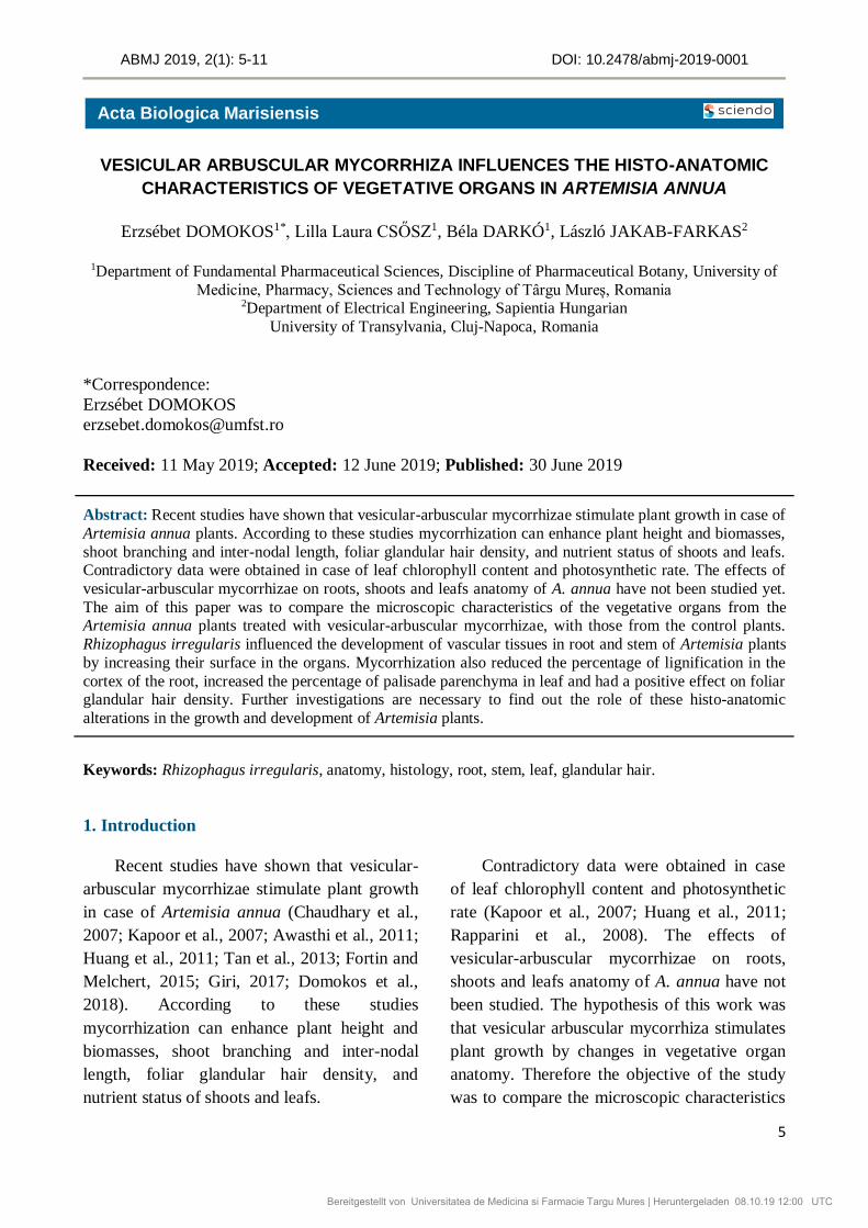
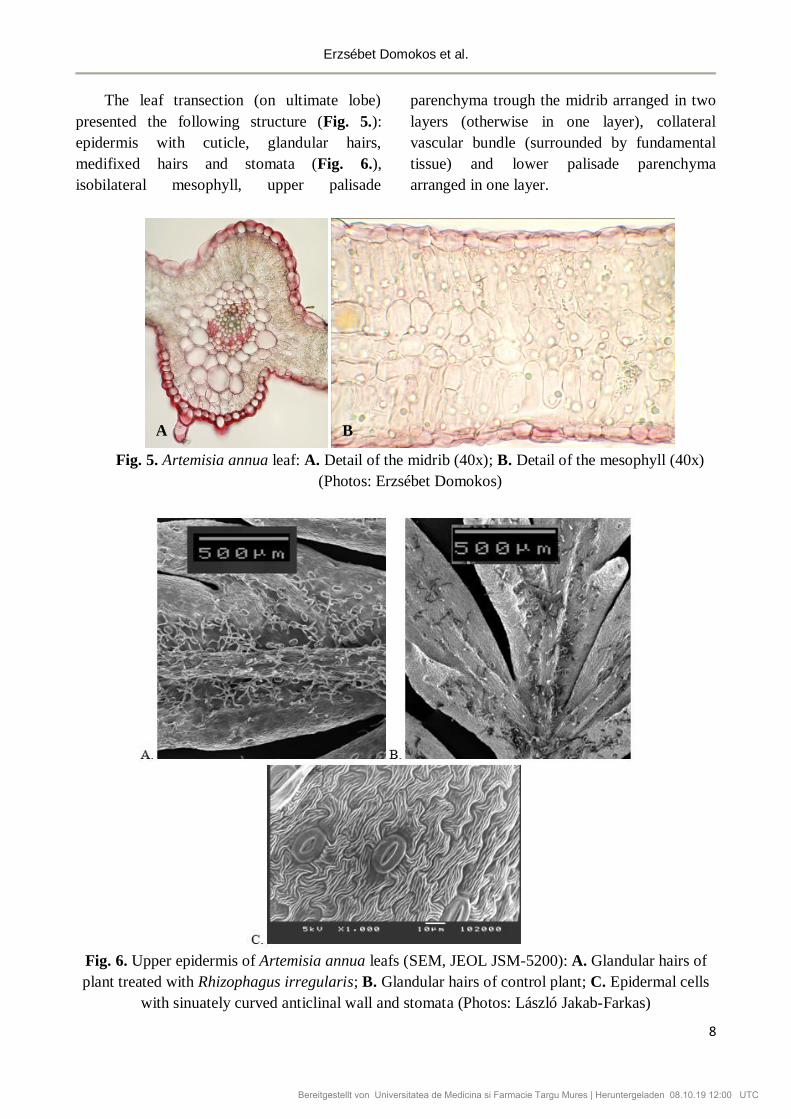
The leaf transection (on ultimate lobe)
presented the following structure (Fig. 5.):
epidermis with cuticle, glandular hairs,
medifixed hairs and stomata (Fig. 6.),
isobilateral mesophyll, upper palisade
parenchyma trough the midrib arranged in two
layers (otherwise in one layer), collateral
vascular bundle (surrounded by fundamental
tissue) and lower palisade parenchyma
arranged in one layer.
Fig. 5. Artemisia annua leaf: A. Detail of the midrib (40x); B. Detail of the mesophyll (40x)
(Photos: Erzsébet Domokos)
Fig. 6. Upper epidermis of Artemisia annua leafs (SEM, JEOL JSM-5200): A. Glandular hairs of
plant treated with Rhizophagus irregularis; B. Glandular hairs of control plant; C. Epidermal cells
with sinuately curved anticlinal wall and stomata (Photos: László Jakab-Farkas)
A B
Bereitgestellt von Universitatea de Medicina si Farmacie Targu Mures | Heruntergeladen 08.10.19 12:00 UTC
ABMJ 2019, 2(1): 5-11
9
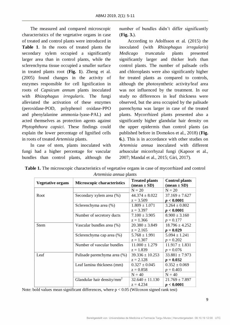
The measured and compared microscopic
characteristics of the vegetative organs in case
of treated and control plants were introduced in
Table 1. In the roots of treated plants the
secondary xylem occupied a significantly
larger area than in control plants, while the
sclerenchyma tissue occupied a smaller surface
in treated plants root (Fig. 1). Zheng et al.
(2005) found changes in the activity of
enzymes responsible for cell lignification in
roots of Capsicum annum plants inoculated
with Rhizophagus irregularis. The fungi
alleviated the activation of these enzymes
(peroxidase-POD, polyphenol oxidase-PPO
and phenylalanine ammonia-lyase-PAL) and
acted themselves as protection agents against
Phytophthora capsici. These findings could
explain the lower percentage of lignified cells
in roots of treated Artemisia plants.
In case of stem, plants inoculated with
fungi had a higher percentage for vascular
bundles than control plants, although the
number of bundles didn’t differ significantly
(Fig. 3.).
According to Adolfsson et al. (2015) the
inoculated (with Rhizophagus irregularis)
Medicago truncatula plants presented
significantly larger and thicker leafs than
control plants. The number of palisade cells
and chloroplasts were also significantly higher
for treated plants as compared to controls,
although the photosynthetic activity/leaf area
was not influenced by the treatment. In our
study no differences in leaf thickness were
observed, but the area occupied by the palisade
parenchyma was larger in case of the treated
plants. Mycorrhized plants presented also a
significantly higher glandular hair density on
the upper epidermis than control plants (as
published before in Domokos et al., 2018) (Fig.
6.). This is in accordance with other studies on
Artemisia annua inoculated with different
arbuscular micorrhyzal fungi (Kapoor et al.,
2007; Mandal et al., 2015; Giri, 2017).
Table 1. The microscopic characteristics of vegetative organs in case of mycorrhized and control
Artemisia annua plants
Vegetative organs Microscopic characteristics Treated plants
(mean ± SD)
Control plants
(mean ± SD)
N = 20 N = 20
Root Secondary xylem area (%) 44.374 ± 8.022 37.169 ± 7.627
z = 3.509 p < 0.0001
Sclerenchyma area (%) 1.809 ± 1.071 3.264 ± 0.802
z = 3.397 p < 0.0001
Number of secretory ducts 7.100 ± 3.905 8.900 ± 3.160 z = 1.366 p = 0.177
Stem Vascular bundles area (%) 20.380 ± 3.849 18.796 ± 4.252
z = 2.165 p = 0.029
Sclerenchyma cap area (%) 5.768 ± 1.991 5.094 ± 1.241 z = 1.307 p = 0.202
Number of vascular bundles 11.000 ± 1.279 11.917 ± 1.831
z = 1.839 p = 0.076
Leaf Palisade parenchyma area (%) 39.336 ± 10.253 33.881 ± 7.973 z = 2.128 p = 0.032
Leaf lamina thickness (mm) 0.327 ± 0.045 0.352 ± 0.069
z = 0.858 p = 0.403
N = 40 N = 40
Glandular hair density/mm2 32.640 ± 11.130 21.769 ± 7.897
z = 4.234 p < 0.0001
Note: bold values mean significant differences, where p < 0.05 (Wilcoxon signed rank test)
Bereitgestellt von Universitatea de Medicina si Farmacie Targu Mures | Heruntergeladen 08.10.19 12:00 UTC

Erzsébet Domokos et al.
10
Conclusions
Rhizophagus irregularis can influence the
development of vascular tissues in root and
stem of Artemisia plants by increasing their
surface in the organs. Mycorrhization also
reduces the percentage of lignification in the
cortex of the root, increases the percentage of
palisade parenchyma in leaf and has a positive
effect on foliar glandular hair density. Results
of this experiment (published earlier) showed
also that R. irregularis had a positive effect on
the biomasses of roots and herba. Further
investigations are necessary to find out the role
of these histo-anatomic alterations in the
growth and development of Artemisia plants.
Conflict of Interest
The authors declare that the research was
conducted in the absence of any commercial or
financial relationships that could be construed
as a potential conflict of interest.
References
1. Adolfsson L, Solymosi K, Andersson MX,
Keresztes Á, Uddling J, Schoefs B, et al.
(2015) Mycorrhiza Symbiosis Increases the
Surface for Sunlight Capture in Medicago
truncatula for Better Photosynthetic
Production. PLoS ONE 10(1): e0115314.
doi:10.1371/journal.pone.0115314
2. Chaudhary L V, Kapoor R, Bhatnagar K A
(2008) Effectiveness of two arbuscular
mycorrhizal fungi on concentrations of
essential oil and artemisinin in three
accessions of Artemisia annua L. Appl. Soil
Ecol. 40, 174–181. doi:
10.1016/j.apsoil.2008.04.003
3. Domokos E, Jakab-Farkas L, Darkó B,
Bíró-Janka B, Mara Gy, Albert Cs, Balog A
(2018) Increase in Artemisia annua plant
biomass content and guaiacol peroxidase
activity using the arbuscular mycorrhizal
fungus Rhizophagus irregularis. Front Plant
Sci. 9:478. doi: 10.3389/fpls.2018.00478
4. Fortin S, Melchert V (2015) Effect of
mycorrhizae on Artemisia annua.
Worcester Polytechnic Institute.
5. Giri B (2017) Mycorrhizal fungus
Rhizophagus fasciculatus promotes
artemisinin accumulation in Artemisia
annua. In Paper Presented at the Tropentag,
Future Agriculture: Socio-Ecological
Transitions and Bio-Cultural Shifts, Bonn.
6. Hammer Ø, Harper D A T, Ryan P D
(2001). PAST: Paleontological statistics
software package for education and data
analysis. Palaeontologia Electronica 4(1):
9pp. http://palaeo-electronica.org/2001_
1/past/issue1_01.htm
7. Huang J H, Tan J F, Jie H K, Zeng R S
(2011) Effects of inoculating arbuscular
mycorrhizal fungi on Artemisia annua
growth and its officinal components.
Yingyong Shengtai Xuebao, 22(6):1443–
1449.
8. Ivănescu B, Miron A, Lungu C (2015)
Histo-anatomy of vegetative organs of
some Artemisia species. The Medical-
Surgical Journal 119(3):917–924.
9. Kapoor R, Chaudhary V, Bhatnagar AK
(2007) Effects of arbuscular mycorrhiza
and phosphorus application on artemisinin
concentration in Artemisia annua L.
Mycorrhiza 17:581–587. doi:
10.1007/s00572-007-0135-4
10. Mandal S, Upadhyay S, Wajid S, Ram M,
Jain D C, Singh V P, Abdin M Z, Kapoor R
(2015) Arbuscular mycorrhiza increase
artemisinin accumulation in Artemisia
Bereitgestellt von Universitatea de Medicina si Farmacie Targu Mures | Heruntergeladen 08.10.19 12:00 UTC

ABMJ 2019, 2(1): 5-11
11
annua by higher expression of key
biosynthesis genes via enhanced jasmonic
acid levels. Mycorrhiza. 25(5):345-57. doi:
10.1007/s00572-014-0614-3
11. Rapparini F, Llusia J, Penuelas J (2008)
Effect of arbuscular mycorrhizal (AM)
colonization on terpene emission and
content of Artemisia annua L. Plant Biol
10:108–122. doi: 10.1055/s-2007-964963
12. Tan W D, Shen M J, Qiu H J, Zeng F L,
Huang J H, Huang R S, Luo W G, Liu Y X
(2013) Effects of different phosphorus
treatments on Arbuscular mycorrhizal
formation, growth and artemisinin content
of Artemisia annua. Journal of Southern
Agriculture 44(8):1303–1307.
13. Tanase C, Silvia O, Domokos E (2017)
Botanică farmaceutică-Îndrumător de
lucrări practice, Vol. 3. University Press,
Târgu Mureș
14. Tu Y (2017) From Artemisia annua L. to
artemisinins: the discovery and
developement of artemisinins and
antimalarial agents, Academic Press,
Cambridge
15. Zheng H Z, Cui C L, Zhang Y T, Wang D,
Jing Y, Kim K Y (2005) Active changes of
lignification-related enzymes in pepper
response to Glomus intraradices and/or
Phytophthora capsici. J Zhejiang Univ Sci
B. 6(8):778–86.
Bereitgestellt von Universitatea de Medicina si Farmacie Targu Mures | Heruntergeladen 08.10.19 12:00 UTC