actin cross-linking domain of aeromonas hydrophila repeat in toxin a (rtxa) induces host cell...
TRANSCRIPT

Gene 506 (2012) 369–376
Contents lists available at SciVerse ScienceDirect
Gene
j ourna l homepage: www.e lsev ie r .com/ locate /gene
Short Communication
Actin cross-linking domain of Aeromonas hydrophila repeat in toxin A (RtxA) induceshost cell rounding and apoptosis
Giovanni Suarez a,1, Bijay K. Khajanchi a,1, Johanna C. Sierra a,1, Tatiana E. Erova a,Jian Sha a, Ashok K. Chopra a,b,c,⁎a Department of Microbiology & Immunology, University of Texas Medical Branch, Galveston, TX 77555, USAb Institute of Human Infections & Immunity, University of Texas Medical Branch, Galveston, TX 77555, USAc Sealy Center for Vaccine Development, University of Texas Medical Branch, Galveston, TX 77555, USA
Abbreviations: Rtx, repeat in toxin; ACD, actin cross-green fluorescent protein; Act, Aeromonas cytotoxic enterottem; T2SS, type 2 secretion system; T3SS, type 3 secretionsystem; ADP, adenosine diphosphate; GAP, GTPase-actiglycine-repeat G1 protein family; VIP-2, vegetative insechemolysin-coregulated protein; RID, Rho-GTPase inactivacysteine protease domain; MARTX, multifunctional autopisopropyl-1-thiogalactopyranoside; rACD, recombinant ACSDS, sodium-dodecyl-sulfate; PAGE, polyacrylamide gelchemiluminescence; 7-AAD, 7-amino actinomycin D; EDacid; ABTS, 2,2″-Azino-bis[3-ethylbenzthiazoline-6-sulfonichloroacetic acid; HUS, hemolytic uremic syndrome.⁎ Corresponding author at: Department of Microbio
Galveston, TX 77555, USA. Tel.: +1 409 747 0578; fax:E-mail address: [email protected] (A.K. Chopra).
1 Contributed equally to the manuscript.
0378-1119/$ – see front matter © 2012 Elsevier B.V. Alldoi:10.1016/j.gene.2012.07.012
a b s t r a c t
a r t i c l e i n f oArticle history:Accepted 9 July 2012Available online 17 July 2012
Keywords:A. hydrophilaRtxActin cross-linking domain (ACD)HeLa Tet-Off systemCell roundingApoptosis
The repeat in toxin (Rtx) of an environmental isolate ATCC 7966 of Aeromonas hydrophila consists of six genes(rtxACHBDE) organized in an operon similar to the gene organization found for the Rtx of the Vibrio species.The first gene in this operon (rtxA) encodes an exotoxin in vibrios, while other genes code for proteins need-ed for proper activation of RtxA and in secretion of this toxin from Vibrio cholerae. However, the RtxA of ATCC7966, as well as from the clinical isolate SSU of A. hydrophila, was exclusively expressed and produced duringco-infection of this pathogen with the host, e.g., HeLa cells, indicating that rtxA gene expression required hostcell contact. Within the RtxA, an actin cross-linking domain (ACD) exists and to investigate the functionalityof this domain, several truncated versions of ACD were generated to discern its minimal biological active re-gion. Such genetically modified genes encoding ACD, which were truncated on either the NH2\ or theCOOH\ terminal, as well as on both ends, were expressed from a bidirectional promoter of thepBI-enhanced green fluorescent protein (EGFP) vector in a HeLa–Tet-Off cell system. We demonstratedthat only the full-length ACD of RtxA from A. hydrophila catalyzed the covalent cross-linking of the host cel-lular actin, whereas the ACD truncated on the NH2\, COOH\ or both ends did not exhibit such actincross-linking characteristics. Further, we showed that the full-length ACD of A. hydrophila RtxA disruptedthe actin cytoskeleton of HeLa cells, resulting in their rounding phenotype. Finally, our data providedevidence that the full-length ACD of RtxA induced host cell apoptosis. Our study is the first to report thatA. hydrophila possesses a functional RtxA having an ACD that contributes to the host cell apoptosis, andhence could represent a potential virulence factor of this emerging human pathogen.
© 2012 Elsevier B.V. All rights reserved.
1. Introduction
Aeromonas hydrophila is an emerging human pathogen, and its abil-ity to produce awide variety of virulence factors facilitates the organism
linking domain; EGFP, enhancedoxin; T1SS, type 1 secretion sys-system; T6SS, type 6 secretion
vating protein; VgrG1, valine–ticidal protein-2 domain; Hcp,tion domain; CPD, autocatalyticrocessing repeat in toxin; IPTG,D; MOI, multiplicity of infection;electrophoresis; ECL, enhancedTA, ethylenediaminotetraaceticc acid]; aa, amino acid; TCA, tri-
logy and Immunology, UTMB,+1 409 747 6869.
rights reserved.
in causing both intestinal and extra-intestinal infections (Chopra andHouston, 1999; Chopra et al., 1993, 1996;Merino et al., 1995, 1999). Im-portantly, Aeromonas species represented one of the most commonpathogens associated with skin and soft tissue infections among the2004 Tsunami survivors in Southern Thailand (Hiransuthikul et al.,2005). Also, an increased isolation rate of Aeromonas species wasreported in the floodwater samples following Hurricane Katrina inNew Orleans (Presley et al., 2006), suggesting that this microbe couldpose potential public health threats during natural disasters. The roleof A. hydrophila in causing necrotizing fasciitis was recently document-ed, which confirms some of the other earlier reports on the flesh-eatingnature of this pathogen (Abuhammour et al., 2006; Borger van der Burget al., 2006; Monaghan et al., 2008).
In our laboratory, we identified and characterized three entero-toxins from a diarrheal isolate SSU of A. hydrophila (Sha et al., 2002).Among these, the cytotoxic enterotoxin (Act) (Ferguson et al., 1997)was found to be the most potent toxin; it was secreted via the type 2secretion system (T2SS) and functioned as a hemolysin, a cytotoxin,or an enterotoxin, depending on the host target (Chopra and Houston,
370 G. Suarez et al. / Gene 506 (2012) 369–376
1999). Recently, we identified a new T3SS effector in A. hydrophila SSU,designated as AexU, which possessed adenosine diphosphate (ADP)-ribosyltransferase and GTPase-activating protein (GAP) activities thatled to host cell apoptosis (Sierra et al., 2007). In addition, we showedthat the T6SS effector protein, valine–glycine-repeat G1 (VgrG1) of A.hydrophila, also possessed actin ADP-ribosylating activity associatedwith its carboxyl-terminal vegetative insecticidal protein-2 (VIP-2) do-main that induced cell rounding followed by host cell apoptosis (Suarezet al., 2010a). On the other hand, the other T6SS effector protein,hemolysin-coregulated protein (Hcp), inhibited phagocytosis of A.hydrophila SSU by macrophages that allowed bacterial multiplicationand spread to different organs in a septicemicmousemodel of infection,leading to animal mortality (Suarez et al., 2010b).
Several studies reported that each Rtx (repeat in toxin) is a memberof a protein family that is produced by a wide range of Gram-negativebacteria (Kwak et al., 2011; Lee et al., 2008a, 2008b; Li et al., 2008)and functions as an important virulence factor. The Rtx of the Vibrio spe-cies is well characterized, and the rtx operon consists of six genes(rtxACHBDE) in which rtxA encodes an exotoxin, rtxC codes for anRtxA activator, rtxH encodes a conserved hypothetical protein andrtxBDE genes code for an ABC transporter (Li et al., 2008). Some impor-tant characteristics of this toxin include: i) it requires post-translationalmodification, i.e., acylation to become biologically active; ii) has aCOOH-terminal calcium-binding domain with tandem glycine/asparticacid-rich repeats; iii) it has a high molecular mass of usually 100to>400 kDa; and iv) it is delivered to the extracellular milieu throughthe T1SS (Boardman and Satchell, 2004; Li et al., 2008).
The Rtx was first detected in Vibrio cholerae (Lin et al., 1999), andlater, a similar toxin was identified in several other pathogens suchas Vibrio vulnificus (Lee et al., 2008a), Vibrio anguillarum (Li et al.,2008), Actinobacillus actinomycetemcomitans (Kraig et al., 1990),Actinobacillus pleuropneumoniae (Devenish et al., 1989), Morganellamorganii (Eberspacher et al., 1990), and Kingella kingae (Kehl-Fieand St Geme, 2007). Recent studies indicated that RtxA of V. choleraeharbored an actin cross-linking domain (ACD) that played a crucialrole in the pathogenesis of this organism (Cordero et al., 2006,2007; Sheahan et al., 2004). Other domains, such as a Rho-GTPase in-activation domain (RID), an autocatalytic cysteine protease domain(CPD), and an α/β-hydrolase domain, were also identified in theRtxA of several pathogens, including V. cholerae (Satchell, 2007).Later, RtxA of V. cholerae was re-designated as a multifunctionalautoprocessing repeat in toxin (MARTX) (Satchell, 2007) by virtueof its highly conserved structural and catalytic activity domains,which were assembled as mosaics. In addition, it was shown thatthe expression of the rtxA gene from V. vulnificus was induced afterhost cell contact (Kim et al., 2008), the RtxA enhanced the survivalof this pathogen during infection by evading phagocytosis (Lo etal., 2011), and, finally, the toxin assisted in the invasion process,i.e., translocation of bacteria from the intestinal tract to the blood-stream (Kim et al., 2008). An earlier study reported that the rtxAmu-tant of V. vulnificus was less virulent in a mouse model of infectionwhen compared to the parental strain, and in vitro data also showedthat the rtxAmutant had lower cytotoxicity when tested on differenthost cells and compared to the parental bacterium (Lee et al., 2008a).
In this study,we sought to characterize the biological functions of ACDlocated within the RtxA of the reference A. hydrophila strain ATCC 7966.Although RtxA is also found in the diarrheal isolate SSU of A. hydrophila,since the genome sequence of ATCC 7966 strain was available (Seshadriet al., 2006), emphasis was placed on characterizing ACD of RtxA fromthe latter strain. We showed that the ACD of RtxA from A. hydrophilainduced actin cross-linking of the host cells. In addition,wedemonstratedby using the pBI-EGFP vector that ACDwas associated with cell roundingand apoptosis when expressed and produced from the HeLa Tet-Off cells.Our study is the first to show that RtxA of A. hydrophila possesses a func-tionally active ACD, which could contribute to the pathogenesis duringthe infection process.
2. Materials and methods
2.1. Bacterial strains and vectors
A. hydrophila ATCC 7966 and SSU strains were grown in LuriaBertani (LB) medium at 27 °C or 37 °C with continuous shaking(180 rpm). The DNA fragment encoding the full-length ACD (Fig. 1)of RtxA from A. hydrophila ATCC 7966 was cloned into a pET-30a vec-tor (Novagen Madison, WI) for hyperexpression and purification pur-poses. The DNA fragment was cloned into the BglII and XhoI or SalIrestriction enzyme sites of the vector. The primers used to amplifythe DNA fragment are listed in Table 1. The recombinant proteincontained at the NH2-terminal end a histidine tag for nickel affinitychromatography. The recombinant plasmid was propagated andmaintained in Escherichia coli DH5α, and recombinant protein wasproduced in E. coli HMS174-DE3 cells (Novagen). Both the E. colistrains were grown in LB medium supplemented with 100 μg/mL ofkanamycin. The pBI-EGFP vector (Clontech, Mountain View, CA) wasused to express and produce recombinant proteins in the HeLaTet-Off cell system (Clontech).
To characterize the biological activity associated with ACD ofA. hydrophila RtxA, we generated several truncated versions ofthe DNA fragment encoding ACD, which were deleted either at the 5′-or the 3′-end region, as well as a fragment, which had deletions atboth the termini. In total, seven DNA fragments were generated whichwere truncated for the corresponding number of aa residues: 62(ΔN62), 121 (ΔN121), and 241(ΔN241) from the NH2-terminal end,61 (ΔC61), 120 (ΔC120), and 220 (ΔC220) from the COOH- terminalend, and finally one fragment that had a truncation of 62 aa residuesfrom the NH2\ and 61 aa residues from the COOH\ terminal end(ΔN62/ΔC61) (Fig. 1). The DNA fragments encoding the full-length,NH2-terminal or the COOH-terminal truncated version of ACD werecloned into the MluI and NheI restriction enzyme sites of the pBI-EGFPplasmid for ectopic expression and production of ACD of various lengthsfrom the HeLa–Tet-Off system (Fig. 1). This vector was propagated andmaintained in E. coli DH5α and grown in LB medium supplementedwith 100 μg/mL of ampicillin (Sigma-Aldrich, St Louis, MO).
2.2. Cell lines and transfections
HeLa Tet-Off cells were obtained from Clontech and grown inDulbecco's modified eagle medium (DMEM) with high glucose(Invitrogen-Gibco, Carlsbad, CA), supplemented with 100 μg/mL G-418(Cellgro, Herndon, VA) and 10% fetal bovine serum (FBS) (Tet system ap-proved; Clontech) (Sierra et al., 2010; Suarez et al., 2010a).
During transfection, HeLa Tet-Off cells were electroporated withthe pBI-EGFP plasmid (5 μg/mL) containing full-length and truncatedfragments of ACD by using 4-mm cuvettes in a Gene Pulser Xcell asdescribed in our previous studies (Sierra et al., 2010; Suarez et al.,2010a). Normal HeLa cells were obtained from the American TypeCulture Collection (Manassas, VA) and grown according to standardtissue culture techniques (Sierra et al., 2010; Suarez et al., 2010a).
2.3. Recombinant protein production
E. coli HMS174-DE3 cells containing the pET-30a recombinantplasmid-encoding ACD of RtxA from A. hydrophila were grown in10 mL of LB medium supplemented with kanamycin (100 μg/mL)overnight. The bacterial culture was diluted, induced with 1 mM ofIPTG (Sigma), and the recombinant proteins purified by using theProBond purification system (Invitrogen) according to the proceduredescribed in our previous studies (Sierra et al., 2010; Suarez et al.,2010a). Protein concentration was measured by using a Bradfordassay (Bio-Rad).

1 460
N62
N121
N241
C61
C120
C220
Full
N62/ C61
1 46851928 2388
ACD
RtxA (AHA_1359)
Fig. 1. Schematic representation of the full-length and truncated versions of ACD of RtxA from A. hydrophila ATCC 7966. Total numbers of amino acid (aa) residues of the RtxA, aswell as the aa residues that constitute ACD are indicated. The figure is not drawn to scale.
371G. Suarez et al. / Gene 506 (2012) 369–376
2.4. Antibody production
Female Swiss Webster mice (n=5; Taconic Farms, Germantown,NY) were immunized via the intraperitoneal route with 10 μg of pu-rified recombinant protein (ACD of RtxA) mixed with completeFreund's adjuvant (Sigma) and boosted with the rACD antigen onday 15 by using incomplete Freund's adjuvant (Sigma). Sera wereobtained frommice after bleeding them at weeks 2 and 4 after immu-nization. The antibody specificity was determined through Westernblot analysis by using whole E. coli lysates expressing and producingthe recombinant protein, as well as the purified recombinant protein(rACD) as the source of antigen.
2.5. Western blot analysis
HeLa cells were infectedwith A. hydrophilaATCC 7966 and SSU strains(multiplicity of infection [MOI] of 5) for 2 h at different temperatures(27 °C and 37 °C). Then, HeLa cells were lysed in Tris-Glycine Buffer,subjected to SDS-PAGE, and proteins were transferred to hybond-ECL ni-trocellulose membranes (GE Healthcare, Piscataway, NJ). Western blotanalysis was performed by using anti-ACD antibodies as described earlier(Sierra et al., 2010; Suarez et al., 2010a). For determination of actincross-linking, HeLa Tet-off cells expressing and producing different
Table 1Various primers used to amplify full-length and truncated versions of the gene encodingACD of RtxA from A. hydrophila ATCC 7966.
Name Primers
Full-length 5′-TCATACGCGTATGGTGGATCTCACCGGTCTG-3′5′-GGTCGCTAGCTCACAGTCCGCTTGCATCCTG-3′
ΔN241 5′-TCATACGCGTATGACGGCCGATCTGGCCCGGG-3′5′-GGTCGCTAGCTCACAGTCCGCTTGCATCCTG-3′
ΔC220 5′-TCATACGCGTATGGTGGATCTCACCGGTCTG-3′5′-GGTCGCTAGCTCACTTGCCGTAGATGGAGG-3′
ΔN121 5′-TCATACGCGTATGTTCACCCAGCACATCAGC-3′5′-GGTCGCTAGCTCACAGTCCGCTTGCATCCTG-3′
ΔC120 5′-TCATACGCGTATGGTGGATCTCACCGGTCTG-3′5′-GGTCGCTAGCTCAGGTCGCATTGTTGATGCC-3′
ΔN62 5′-TCATACGCGTATGCTGACCAAGGACATG-3′5′-GGTCGCTAGCTCACAGTCCGCTTGCATCCTG-3′
ΔC61 5′-TCATACGCGTATGGTGGATCTCACCGGTCTG-3′5′-GGTCGCTAGCTCAGACCAGATTGTTCACCGCC-3′
ΔN62/ΔC61 5′-TCATACGCGTATGCTGACCAAGGACATG-3′5′-GGTCGCTAGCTCAGACCAGATTGTTCACCGCC-3′
Underlines indicate restriction enzyme sites.
fragments of ACD were lysed after 24 h of transfection, and the proteinswere electrophoresed on a SDS-6% PAGE and then transferred tohybond-ECL nitrocellulose membranes. Western blot analysis wasperformed by using an anti-actin antibody (Santa Cruz Biotechnology,Santa Cruz, CA).
2.6. Host cell morphology
The morphology of HeLa Tet-Off cells was examined after 24 h oftransfection with the pBI-EGFP vector containing genes encoding eitherthe native or the truncated versions of ACD by fluorescence microscopyand flow cytometry by using Alexa-fluor 568-conjugated phalloidin(Invitrogen) as described in our previous studies (Sierra et al., 2010;Suarez et al., 2010a). Briefly, the cellswere fixed in plates after electropo-ration with Cytofix/Cytoperm (Becton Dickinson, San Diego, CA) for20 min in the dark. The cells were scraped and stained with Alexa-fluor 568-conjugated phalloidin, and, after washing, they were acquiredin a FACScan flow cytometer (Becton Dickinson) and analyzed by usingBD FACSDiva software. For fluorescence microscopy, the cells wereplaced on glass slides and the images acquired on a fluorescence micro-scope (Olympus BX51/DPManager v.1.2.1.107/DPController v.1.2.1.108,Olympus Optical CO. LTD).
2.7. Host cell viability
Incorporation of 7-amino actinomycin D (7-AAD) (BectonDickinson)was used to determine the HeLa Tet-Off cell viability according toestablished procedures, as described earlier (Sierra et al., 2007). HeLaTet-Off cells expressing different ACD fragments were detached fromthe tissue culture plate with 0.25% trypsin-EDTA, washed and then incu-bated for 10 minwith 7-AAD (5 μL per tube). Immediately after staining,the cells were examined in a FACScan flow cytometer to determine thepercentage of EGFP and 7-AAD double-positive cells.
2.8. Host cell apoptosis
The extent of apoptosis of HeLa Tet-Off cells expressing differentACD fragments was determined through detection of cytoplasmic nu-cleosomes by using the cell death ELISA kit (Roche, Indianapolis, IN)and following the manufacturer's instructions (Sierra et al., 2010;Suarez et al., 2010a). Briefly, the HeLa Tet-Off cell lysates, anti-DNA per-oxidase, and anti-histone biotinylated antibodies were incubated instreptavidin-coated, 96-well plates for 2 h at room temperature. Subse-quently, the plates were washed three times and incubated with ABTS(2, 2″-Azino-bis[3-ethylbenzthiazoline-6-sulfonic acid]) as the substrate.The color reaction was measured in a microplate reader at a 405-nm
372 G. Suarez et al. / Gene 506 (2012) 369–376
wavelength. Since the efficiency of transfection was different for each ofthe recombinant ACD fragment encoding gene, the optical density valuesat 405 nm were normalized to the percentage of transfection for each ofthe recombinant fragment.
2.9. Statistical analysis
A two-way ANOVA and Bonferoni post-test were used for statisti-cal analysis of the data by using GraphPad Prism V 4.02 for Windows(Software MacKiev, San Diego, CA).
3. Results
3.1. Characterization of RtxA ACD from A. hydrophila ATCC 7966
By genome sequence analysis of ATCC 7966 (Seshadri et al., 2006),we found that as with V. cholerae (Lin et al., 1999; Roig et al., 2011),the rtx operon of A. hydrophila also contained six genes (rtxACHBDE).The RtxA, as well as its ACD from A. hydrophila (AHA_1359), exhibited52% and 65% identity, respectively, with the corresponding full-lengthRtxA and its ACD of V. cholerae (VC1451) (Supplemental Fig. S1). Thefull-length RtxA of the ATCC 7966 strain has 4685 aa residues, and theACD is encoded by aa residues 1928–2388 (Fig. 1).
3.2. Expression of the rtxA gene in A. hydrophila ATCC 7966 and SSUstrains requires host cell contact
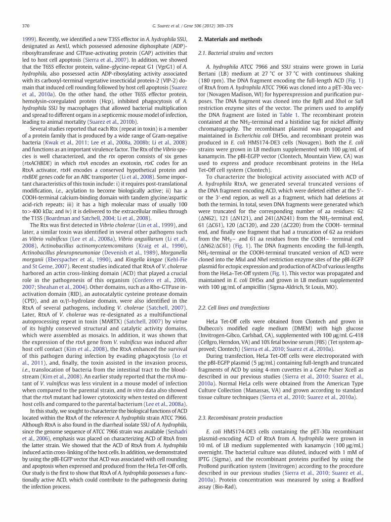
To investigate whether the RtxA of A. hydrophila is functional, weperformed Western blot analysis on samples by using anti-ACD anti-bodies after co-infection of HeLa cells with either the environmentalisolate ATCC 7966 or the diarrheal isolate SSU of A. hydrophila. These an-tibodies to ACD of RtxA were developed in the laboratory (Section 2.4)and could detect full-length RtxA. We used temperatures of 27 and37 °C (as they represent ambient vs. human body temperature) togrow the bacteria and to infect HeLa cells. As noted from Fig. 2, thefull-length RtxA (~520 kDa) was detected on Western blots in theco-culture samples of HeLa cells infected with either SSU (27 °C; lane
1 2 3 4 5 6
RtxA
225
102
130
MW
Fig. 2. Western blot analysis showing expression of the rtxA gene after co-infection ofHeLa cells with A. hydrophilaATCC and SSU strains. Lanes 1 and 2: expression and produc-tion of RtxA in the SSU strain after infection with HeLa cells at 27 °C (lane 1) and 37 °C(lane 2). Lanes 3 and 4: expression and production of RtxA in the ATCC 7966 strain afterinfection with HeLa cells at 27 °C (lane 3) and 37 °C (lane 4). Lanes 5 and 6: controluninfected HeLa cells grown at 27 °C and 37 °C, respectively. MW= markers (kDa).
1 and 37 °C; lane 2) or ATCC 7966 strain (27 °C; lane 3 and 37 °C;lane 4). Because of the crude nature of the hyper-immune sera toACD, these antibodies exhibited some non-specific reaction with theHeLa cell proteins (Fig. 2, lanes 1–6).
Importantly, RtxA was neither detected in the supernatant fractionsof the co-cultures nor in the culture filtrates (Supplemental Fig. S2) orcell lysates (Supplemental Fig. S3) of bacteria at either temperaturewhen grown in the absence of the host cells. A non-specific band of60–65 kDa was detected in the trichloroacetic acid (TCA) precipitatedbacterial culture filtrates in all the lanes, inclusive of uninfected HeLacells (Supplemental Fig. S2, lanes 1–5); however, no RtxA bandwas ob-served. The antibodies to ACD reacted with the correct size rACD (lane6), which served as a positive control.
Likewise, in the whole cell lysates of either SSU or ATCC 7966when grown at 27 °C, no RtxA band was detected on the Westernblot (Supplemental Fig. S3; lanes 1 and 2). Although a band of ap-proximately 60–65 kDa was observed in the cell lysates of thesetwo isolates (lanes 3 and 4) when grown at 37 °C, these sizes didnot match either with the molecular mass of full-length RtxA or itsACD domain, which should be approximately 520 kDa (Fig. 2) and50 kDa, respectively (Supplemental Fig. S3; lane 6). Therefore, theidentity of this band is currently unknown. Uninfected HeLa cell sam-ples (grown either at 27 °C or 37 °C) were used as controls, and noband corresponding to the size of RtxA was detected (SupplementalFig. S3; lane 5). We did notice some non-specific cross-reactingeukaryotic protein bands that interacted with antibodies to ACD ofA. hydrophila on the Western blots upon prolonged exposure. Thesedata demonstrated that RtxA of A. hydrophila was not secreted intothe medium and that the expression of the rtxA gene in A. hydrophilarequired host cell contact.
3.3. Full-length ACD of RtxA from A. hydrophila ATCC 7966 induces actincross-linking
To characterize the function of ACDwithin the RtxA of A. hydrophila,we first confirmed that all of the DNA fragments encoding truncatedversions of ACD were correctly produced from the HeLa Tet-Off cellsby Western blot analysis using anti-ACD antibodies of RtxA after 24 hof transfection (Fig. 3A). A representative blot, which showsfull-length ACD (lane 2), ΔN121 (lane 3), ΔN241 (lane 4), and ΔC220(lane 5), is depicted in Fig. 3A. Ectopic expression of the pBI-EGFPempty plasmid in HeLa–Tet-Off cells (lane 1), untransfected cells (lane7), and cells transfectedwith the VIP-2 domain of VgrG1 (lane 6) servedas non-actin cross-linking negative controls.Multiple bands observed inlanes 2 and 3 could represent either degradation products or differentlyspliced forms of ACD.
To further examine the functionality of ACD, we determined actincross-linking after expressing and producing these native and truncatedversions of ACD in the HeLa Tet-Off cells after 24 h of transfection byWestern blot analysis by using anti-actin antibodies. Interestingly, wefound that actin cross-linking was only detected (high molecularweight multiple bands) when the full-length gene encoding ACD wasexpressed from the HeLa Tet-Off system (Fig. 3B, lane 2) and not withthe NH2\ and/or \COOH terminal truncated proteins (Fig. 3B, lanes3–5). The band at 45 kDa in all the lanes represented monomericform of the cellular actin. These results indicated that full-length ACDof RtxA was required for the induction of actin cross-linking.
Since our truncations were based on the study on ACD of V. choleraeRtxA (Sheahan et al., 2004), which exhibits 65% sequence identity withthe ACD of A. hydrophilaATCC 7966 (Seshadri et al., 2006) (SupplementalFig. S1), we believe these ACD domains to have similar secondary struc-tures with α-helics and β strands. Consequently, the loss in the actincross-linking activity in the truncated versions of ACD of A. hydrophilawe observed might not be related to the differential folding of the mole-cule. However, in the future, we will develop site-directed mutations

373G. Suarez et al. / Gene 506 (2012) 369–376
within the ACD of A. hydrophila SSU and showwhich aa residues are keyplayers that contribute to this activity.
3.4. ACD of RtxA in A. hydrophila ATCC 7966 induces host cell roundingby disrupting actin cytoskeleton
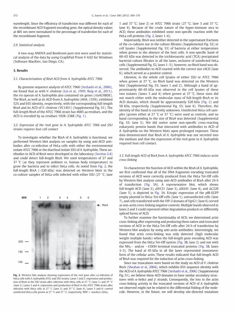
To examine the role of ACD of RtxA in inducing host cell toxicity, apBI-EGFP vector expressing DNA fragments encoding various truncatedversions of ACDwas electroporated into HeLa Tet-Off cells. After 24 h oftransfection, a rounded morphology was observed only in the HeLa–Tet-Off cells expressing and producing the full-length ACD (Fig. 4A,panel III). All of the HeLa Tet-Off cells successfully transfected with thepBI-EGFP plasmid were green due to the production of GFP. The actincytoskeleton of the cells was stained red by the Alexa 568 conjugatedphalloidin. As noted in panel III, the actin cytoskeleton was completelydisrupted in the HeLa–Tet-Off cells, which were green, indicating thatthe full-length ACD led to host cell killing and rounding due to actincross-linking (Fig. 3B). In contrast, none of the HeLa–Tet-Off cells ex-pressing and producing various truncated versions of ACD showed dis-ruption of the actin-cytoskeleton architecture, and a specific example ofHeLa–Tet-Off cells producing ACD ΔN62/ΔC61 is shown in panel II. Thedata presented in panel II is similar to the one shown in panel I in terms ofnormal morphology of the HeLa–Tet-Off cells that expressed the emptyvector. The VIP-2 domain of VgrG1 (a T6SS effector) of A. hydrophilawhen produced from the HeLa–Tet-Off cells exhibited typical roundingof the host cells, and served as a positive control. However, the VIP-2 do-main of VgrG1 induced cell-rounded morphology via actin-ADP-ribosylation (Fig. 4A, panel IV) (Suarez et al., 2010a) rather than actincross-linking, as observed with ACD of A. hydrophila. These differencesin activities are showed in Fig. 3B, lanes 2 and 6, as no actincross-linking was noted when the VIP-2 domain of VrgG1 encodinggene was expressed in the HeLa cells (lane 6) while ACD caused cellularactin cross-linking (lane 2).
AntiRtxA (ACD) Abs
A
B
1 2 3 4 5 6 7
52
38
31
24
1710
76102MW
Anti ActinAbs
Actin(monomers)
Actin Crosslinked
1 2 3 4 5 6 7
52
38
220
76
102
130
MW
Fig. 3. Expression and production of the full-length and truncated versions of ACD fromthe pBI-EGFP plasmid in HeLa Tet-Off cells. A) Western blot analysis of HeLa Tet-Off celllysates expressing and producing different fragments of ACD of RtxA from A. hydrophilaATCC 7966. Lanes 1: control, empty vector; 2: full length ACD; 3: NH2-terminal truncatedACD (ΔN121) 4: NH2-terminal truncated ACD (ΔN241); 5: COOH-terminal truncated ACD(ΔC220);6: VIP-2 domainof VgrG1; and 7: UntransfectedHeLa cells. B)Western blot anal-ysis showing actin cross-linking induced by different fragments of ACD. Lanes 1: control,vector only; 2: full-length ACD; 3: NH2-terminal truncatedACD (ΔN121); 4: NH2-terminaltruncated ACD (ΔN241); 5: COOH-terminal truncated ACD (ΔC220); 6: VIP-2 domain ofVgrG1; and 7: untransfected HeLa cells. Abs = antibodies. MW= markers (kDa).
In addition, we quantified by flow cytometry the disruption of theactin cytoskeleton of HeLa–Tet-Off cells expressing and producing vari-ous truncated versions of ACD by using Alexa 568-conjugated phalloidinfor staining of the actin cytoskeleton on GFP-positive cells. A much re-duced percentage of phalloidin-positive HeLa Tet-Off cells (40%) wasdetected when the full-length ACD was expressed and produced(Fig. 4B, panel III) when compared to HeLa–Tet-Off cells producing vari-ous truncated versions of ACD (more than 85%). We have representativedata with ACD ΔN62/ΔC61 in panel II in which 88% of the GFP-positivecells had intact actin filaments. This number of 88% phalloidin-positivecells was similar to that of phalloidin-positive HeLa–Tet-Off cells (89%)expressing an empty vector (Fig. 4B, panel I). These data lead us to con-clude that the full- length ACD of RtxA in A. hydrophila was necessaryto disrupt the actin cytoskeleton, resulting in host cell rounding.
3.5. ACD of RtxA in A. hydrophila ATCC 7966 induces apoptosis of HeLaTet-Off cells
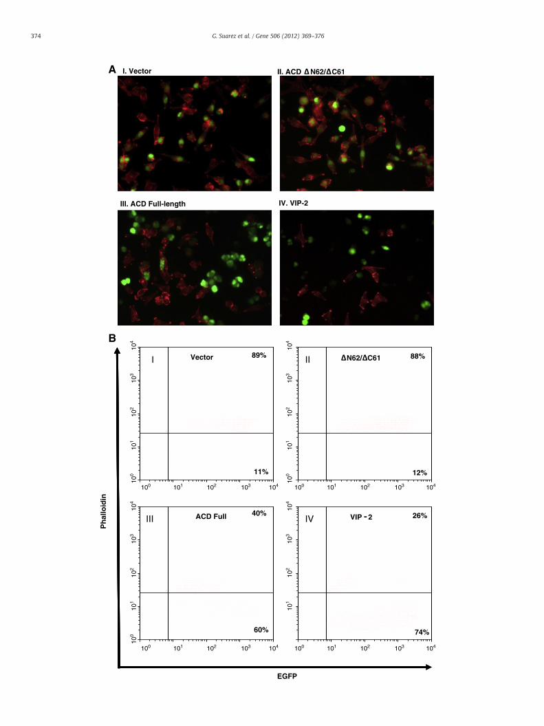
The viability of HeLa Tet-Off cells expressing and producing varioustruncated versions of ACD was measured by incorporating 7-amino ac-tinomycin D (AAD). The percentage of 7-AAD-positive cells was signif-icantly higher (Fig. 5) inHeLa Tet-Off cells expressing andproducing thefull-length ACD than that of HeLa cells expressing various truncatedversions of ACD (Fig. 5). Representative data with NH2\ terminal trun-cation of ACD are shown here; however, similar results were seen withthe COOH-terminal truncation of ACD orwhen ACDwas truncated fromboth the NH2\ and COOH\ termini (data not shown). HeLa–Tet-Offcells expressing and producing either the empty vector or the VIP-2 do-main of VgrG1 of A. hydrophila served as a negative and positive control,respectively.
We then measured the rate of apoptosis induced by various trun-cated versions of ACD in HeLa Tet-Off cells by determining the level ofcytoplasmic nucleosomes. A significant increase in the cytoplasmicnucleosomes (Fig. 6) was noted in HeLa Tet-Off cells expressing andproducing the full-length ACD, which was similar to HeLa cells pro-ducing the VIP-2 domain of VgrG1 (Fig. 6). The level of cytoplasmicnucleosomes in HeLa–Tet-Off cells producing various truncatedversions of ACD was similar (only 4 shown here) to HeLa cells ex-pressing the empty vector. These results indicated that ACD of RtxAof A. hydrophila was able to induce apoptosis in host cells.
4. Discussion
The ability of A. hydrophila to produce numerous virulence factorsunderlines its potential to cause a wide range of human diseases frommild diarrhea to severe forms of infections, such as necrotizing fasciitisand hemolytic uremic syndrome (HUS) (Abuhammour et al., 2006;Figueras et al., 2007; Janda and Abbott, 2010). Secreted/effector pro-teins that are delivered through different secretion systems have beenidentified in various species of Aeromonas that contributed to host cellcytotoxicity during infection (Ferguson et al., 1997; Sierra et al., 2007;Suarez et al., 2010a, 2010b). Earlier, RtxA was shown to be secretedthrough the T1SS of V. cholerae and contributed to pathogenic sequelaein the host (Boardman and Satchell, 2004; Cordero et al., 2006; Sheahanet al., 2004). In this study we demonstrated that RtxA of A. hydrophilaATCC 7966 possessed a functional ACD that induced host cell roundingand apoptosis.
A recent study on comparative sequence analysis of differentVibrio species (e.g., V. cholerae, V. vulnificus, and V. anguillarum)and that of A. hydrophila ATCC 7966 revealed a similar genomicorganization of Rtx operon (Roig et al., 2011). In addition, inthis comparative analysis it was noticed that ACD was only present inRtxA of V. vulnificus isolates belonging to biotype 2, while RtxA of all thetested strains of V. cholerae had ACD. Importantly, ACD was not found inRtxA of other tested Gram-negative organisms, including other Vibriospecies (Roig et al., 2011). From these data, we hypothesized that the
104
103
102
101
100
104
103
102
101
100
104
103
102
101
100
104
103
102
101
89% 88%Vector N62/ C61III
100 101 102 103 104 100 101 102 103 104
100 101 102 103 104 100 101 102 103 104
26%40%
11% 12%
ACD Full VIP 2
Ph
allo
idin
III IV
74%60%
EGFP
III. ACD Full-length IV. VIP-2
I. Vector II. ACD N62/ C61A
B
374 G. Suarez et al. / Gene 506 (2012) 369–376
7-AAD Incorporation (24h)
Vecto
rFull
N62N12
1N24
1VIP
-2
0
10
20
30
ACD
***
***%
Pos
itive
Cel
ls
Fig. 5. Viability of HeLa Tet-Off cells expressing genes encoding different fragments ofACD of A. hydrophila RtxA. The percentage of dead and/or dying cells was quantified byincorporation of 7-AAD and flow cytometry on HeLa Tet-Off cells expressing vectoralone (pBI-EGFP) or producing different fragments of ACD (as indicated) after 24 h oftransfection. Results from three different assays were plotted, and statistical signifi-cance is indicated by asterisks (pb0.001).
Apoptosis - Nucleosomes (24h)
Vecto
rFull
N62N12
1N24
1C22
0VIP
-2
0
1
2
3
4
5
ACD
******
Nor
mal
ized
OD
405
Fig. 6. Apoptosis of HeLa Tet-Off cells expressing genes encoding different fragments ofACD of A. hydrophila RtxA. Apoptosis rates were measured by quantification of cyto-plasmic nucleosomes in lysates of HeLa Tet-Off cells expressing vector alone(pBI-EGFP), or genes encoding various truncated versions (as indicated) of ACD fromA. hydrophila RtxA. Results from three different assays were plotted, and statistical sig-nificance is indicated by asterisks (pb0.001).
375G. Suarez et al. / Gene 506 (2012) 369–376
RtxA of A. hydrophilawould have similar biological functions to those ofV.cholerae and biotype 2 strains of V. vulnificus. We provided evidence thatexpression of the rtxA gene in A. hydrophila required host cell contact,whichwas in accordancewith a previous study performed onV. vulnificus(Kim et al., 2008). However, unlike V. cholerae RtxA (Fullner andMekalanos, 2000), we found that A. hydrophila RtxA was not secretedinto the medium, indicating that although the genomic organization ofRtx operons is similar in these pathogens, they possess subtle differencesas well.
We noted that the full-length ACD of RtxA in A. hydrophila possessedactin cross-linking activity that resulted in a cell-rounding phenotype ofHeLa Tet-Off cells. An earlier study has shown that in V. cholerae, the cellrounding was not completely eliminated when ACD was deleted fromRtxA. It was later discovered that cell rounding was also associated withanother domain of RtxA of Vibrio, referred to as a Rho-GTPase inactivationdomain (RID) (Sheahan and Satchell, 2007). Therefore, in V. cholarae, cellrounding was induced by two domains of RtxA (ACD and RID); however,sequence annotation of A. hydrophila ATCC 7966 RtxA revealed that it didnot contain an RID domain, possibly meaning that in A. hydrophila theACD domain might primarily be associated with the induction of ahost cell-rounding phenotype. Additionally, it is important to mentionthat A. hydrophila produces VgrG1 with ADP-ribosyltransferase activitythat induces host cell-rounding phenotype. This activity prevents theformation of F-actin by the ADP-ribosylation of G-actin leading toapoptosis (Suarez et al., 2010a).
In agreementwith the present study, itwas reported thatV. vulnificusRtxAplayed an important role in the apoptotic death of human epithelialcells, as it was noticed that the RtxA mutant induced significantly lowerlevels of apoptotic death when compared to the parental strain (Lee etal., 2008a). In addition, these investigators further demonstratedthat V. vulnificus RtxA induced apoptotic death through a
Fig. 4. Full-length ACD causes host cell rounding. A) Morphological changes in HeLa Tet-Offrom A. hydrophila ATCC 7966. Host cells were stained for actin-cytoskeleton by using Alexatein (EGFP) encoding gene was detected in cells successfully transfected with the pBI-EGFP esion of ACD (II), full-length ACD (III), and the VIP-2 domain of VgrG1 of A. hydrophilaactin-cytoskeleton as measured by fluorescent phalloidin staining of HeLa Tet-Off expressTet-Off cells stained with Alexa fluor 568-phalloidin and expressing different encoding fragmof positive cells from a representative experiment is shown in the plotted quadrants. Three
mitochondria-dependent pathway (Lee et al., 2008a). In futurestudies, we plan to explore in detail the mechanism(s) as tohow A. hydrophila RtxA of both ATCC 7966 and SSU induces ap-optosis in host cells.
Taken together, our data showed that A. hydrophila possesses afunctional RtxA that leads to host cell rounding and apoptotic death.It appears that A. hydrophila produces several toxins and/or effectorproteins (Act, AexU, VgrG1, RtxA) that have identical functions,such as apoptotic cell death. From these observations, it can be spec-ulated that redundancy in toxin functions of A. hydrophila may benecessary to cause a successful infection in the host. In the future,we plan to investigate the detailed mechanisms of regulation of ex-pression and production of these different toxins and /or effector pro-teins in A. hydrophila both in vitro and in vivo models of infection.Likewise, we will delineate the function of RtxA in bacterial virulencein vivo by generating a knockout mutant. In the long run, such studieswould be beneficial for discovering the appropriate target for the devel-opment of novel therapeutics. Together, the Rtx operon of A. hydrophilahas some unique characteristics as well as similarities with the Rtxoperon of Vibrio species. The actin cross-linking domain of A. hydrophilahas not been characterized previously, and our current study shed lighton how this domain leads to cytotoxic effects on the host cells.
Supplementary data to this article can be found online at http://dx.doi.org/10.1016/j.gene.2012.07.012.
Acknowledgments
This study was supported by the NIH/NIAID (AI41611) grant aswell as funding from the EPA. We thank Ms. Mardelle J. Susman foreditorial assistance.
f cells induced by the expression and production of different fragments of ACD of RtxAFluor 568-phalloidin (red), and the expression of the enhanced green fluorescent pro-mpty vector (I), with vector containing genes encoding the ΔN62/ΔC61 truncated ver-used as a positive control (IV). Original magnification is 40×. B) Quantification ofing and producing different versions of ACD. Flow cytometry dot plots showing HeLaents of ACD of RtxA. The analysis was performed on EGFP-positive cells. The percentageindependent experiments were performed and similar data were obtained.
376 G. Suarez et al. / Gene 506 (2012) 369–376
References
Abuhammour, W., Hasan, R.A., Rogers, D., 2006. Necrotizing fasciitis caused by Aeromonashydrophilia in an immunocompetent child. Pediatr. Emerg. Care 22, 48–51.
Boardman, B.K., Satchell, K.J., 2004. Vibrio cholerae strainswithmutations in an atypical type Isecretion system accumulate RTX toxin intracellularly. J. Bacteriol. 186, 8137–8143.
Borger van der Burg, B.L., Bronkhorst, M.W., Pahlplatz, P.V., 2006. Aeromonas hydrophilanecrotizing fasciitis. A case report. J. Bone Joint Surg. Am. 88, 1357–1360.
Chopra, A.K., Houston, C.W., 1999. Enterotoxins in Aeromonas-associated gastroenteri-tis. Microbes Infect. 1, 1129–1137.
Chopra, A.K.,Houston, C.W., Peterson, J.W., Jin, G.F., 1993. Cloning, expression, and sequenceanalysis of a cytolytic enterotoxin gene from Aeromonas hydrophila. Can. J. Microbiol.39, 513–523.
Chopra, A.K., Peterson, J.W., Xu, X.J., Coppenhaver, D.H., Houston, C.W., 1996. Molecularand biochemical characterization of a heat-labile cytotonic enterotoxin fromAeromonas hydrophila. Microb. Pathog. 21, 357–377.
Cordero, C.L., Kudryashov, D.S., Reisler, E., Satchell, K.J., 2006. The actin cross-linkingdomain of the Vibrio cholerae RTX toxin directly catalyzes the covalent cross-linking of actin. J. Biol. Chem. 281, 32366–32374.
Cordero, C.L., Sozhamannan, S., Satchell, K.J., 2007. RTX toxin actin cross-linking activity inclinical and environmental isolates ofVibrio cholerae. J. Clin.Microbiol. 45, 2289–2292.
Devenish, J., Rosendal, S., Johnson, R., Hubler, S., 1989. Immunoserological comparisonof 104-kilodalton proteins associated with hemolysis and cytolysis in Actinobacilluspleuropneumoniae, Actinobacillus suis, Pasteurella haemolytica, and Escherichia coli.Infect. Immun. 57, 3210–3213.
Eberspacher, B., Hugo, F., Pohl, M., Bhakdi, S., 1990. Functional similarity between thehaemolysins of Escherichia coli andMorganella morganii. J. Med. Microbiol. 33, 165–170.
Ferguson,M.R., et al., 1997.Hyperproduction, purification, andmechanismof actionof the cy-totoxic enterotoxin produced by Aeromonas hydrophila. Infect. Immun. 65, 4299–4308.
Figueras, M.J., Horneman, A.J., Martinez-Murcia, A., Guarro, J., 2007. Controversial data onthe association of Aeromonas with diarrhoea in a recent Hong Kong study. J. Med.Microbiol. 56, 996–998 (author reply 98).
Fullner, K.J., Mekalanos, J.J., 2000. In vivo covalent cross-linking of cellular actin by theVibrio cholerae RTX toxin. EMBO J. 19, 5315–5323.
Hiransuthikul, N., Tantisiriwat, W., Lertutsahakul, K., Vibhagool, A., Boonma, P., 2005.Skin and soft-tissue infections among tsunami survivors in southern Thailand.Clin. Infect. Dis. 41, e93–e96.
Janda, J.M., Abbott, S.L., 2010. The genus Aeromonas: taxonomy, pathogenicity, and in-fection. Clin. Microbiol. Rev. 23, 35–73.
Kehl-Fie, T.E., St Geme, J.W., 2007. Identification and characterization of an RTX toxin inthe emerging pathogen Kingella kingae. J. Bacteriol. 189, 430–436.
Kim, Y.R., et al., 2008. Vibrio vulnificus RTX toxin kills host cells only after contact of thebacteria with host cells. Cell. Microbiol. 10, 848–862.
Kraig, E., Dailey, T., Kolodrubetz, D., 1990. Nucleotide sequence of the leukotoxin genefrom Actinobacillus actinomycetemcomitans: homology to the alpha-hemolysin/leukotoxin gene family. Infect. Immun. 58, 920–929.
Kwak, J.S., Jeong, H.G., Satchell, K.J., 2011. Vibrio vulnificus rtxA1 gene recombinationgenerates toxin variants with altered potency during intestinal infection. Proc.Natl. Acad. Sci. U. S. A. 108, 1645–1650.
Lee, B.C., Choi, S.H., Kim, T.S., 2008a. Vibrio vulnificus RTX toxin plays an important rolein the apoptotic death of human intestinal epithelial cells exposed to Vibriovulnificus. Microbes Infect. 10, 1504–1513.
Lee, B.C., et al., 2008b. Vibrio vulnificus rtxE is important for virulence, and its expres-sion is induced by exposure to host cells. Infect. Immun. 76, 1509–1517.
Li, L., Rock, J.L., Nelson, D.R., 2008. Identification and characterization of a repeat-in-toxin gene cluster in Vibrio anguillarum. Infect. Immun. 76, 2620–2632.
Lin, W., et al., 1999. Identification of a Vibrio cholerae RTX toxin gene cluster that is tightlylinked to the cholera toxin prophage. Proc. Natl. Acad. Sci. U. S. A. 96, 1071–1076.
Lo, H.R., et al., 2011. RTX toxin enhances the survival of Vibrio vulnificus during infec-tion by protecting the organism from phagocytosis. J. Infect. Dis. 203, 1866–1874.
Merino, S., Rubires, X., Knochel, S., Tomas, J.M., 1995. Emerging pathogens: Aeromonasspp. Int. J. Food Microbiol. 28, 157–168.
Merino, S., Aguilar, A., Nogueras, M.M., Regue, M., Swift, S., Tomas, J.M., 1999. Cloning,sequencing, and role in virulence of two phospholipases (A1 and C) frommesophilic Aeromonas sp. serogroup O:34. Infect. Immun. 67, 4008–4013.
Monaghan, S.F., Anjaria, D., Mohr, A., Livingston, D.H., 2008. Necrotizing fasciitis andsepsis caused by Aeromonas hydrophila after crush injury of the lower extremity.Surg. Infect. (Larchmt.) 9, 459–467.
Presley, S.M., et al., 2006. Assessment of pathogens and toxicants in New Orleans, LAfollowing Hurricane Katrina. Environ. Sci. Technol. 40, 468–474.
Roig, F.J., Gonzalez-Candelas, F., Amaro, C., 2011. Domain organization and evolution ofmultifunctional autoprocessing repeats-in-toxin (MARTX) toxin in Vibrio vulnificus.Appl. Environ. Microbiol. 77, 657–668.
Satchell, K.J., 2007. MARTX, multifunctional autoprocessing repeats-in-toxin toxins. In-fect. Immun. 75, 5079–5084.
Seshadri, R., et al., 2006. Genome sequence of Aeromonas hydrophila ATCC 7966T: jackof all trades. J. Bacteriol. 188, 8272–8282.
Sha, J., Kozlova, E.V., Chopra, A.K., 2002. Role of various enterotoxins in Aeromonashydrophila-induced gastroenteritis: generation of enterotoxin gene-deficientmutants and evaluation of their enterotoxic activity. Infect. Immun. 70,1924–1935.
Sheahan, K.L., Satchell, K.J., 2007. Inactivation of small Rho GTPases by themultifunctional RTX toxin from Vibrio cholerae. Cell. Microbiol. 9, 1324–1335.
Sheahan, K.L., Cordero, C.L., Satchell, K.J., 2004. Identification of a domain within themultifunctional Vibrio cholerae RTX toxin that covalently cross-links actin. Proc.Natl. Acad. Sci. U. S. A. 101, 9798–9803.
Sierra, J.C., et al., 2007. Biological characterization of a new type III secretion sys-tem effector from a clinical isolate of Aeromonas hydrophila—part II. Microb.Pathog. 43, 147–160.
Sierra, J.C., Suarez, G., Sha, J., Baze, W.B., Foltz, S.M., Chopra, A.K., 2010. Unraveling themechanism of action of a new type III secretion system effector AexU fromAeromonas hydrophila. Microb. Pathog. 49, 122–134.
Suarez, G., Sierra, J.C., Erova, T.E., Sha, J., Horneman, A.J., Chopra, A.K., 2010a. A typeVI secretion system effector protein, VgrG1, from Aeromonas hydrophila thatinduces host cell toxicity by ADP ribosylation of actin. J. Bacteriol. 192,155–168.
Suarez, G., Sierra, J.C., Kirtley, M.L., Chopra, A.K., 2010b. Role of Hcp, a type 6 secretionsystem effector, of Aeromonas hydrophila in modulating activation of host immunecells. Microbiology 156, 3678–3688.