actin polymerization stimulated by contractile activation regulates force development in canine...
TRANSCRIPT

The objective of this study was to evaluate the role of actin
filament polymerization in the contraction of smooth muscle.
The activation of a number of non-muscle cells including
neutrophils, platelets and fibroblasts has been shown to
stimulate the rapid polymerization of actin from monomeric
globular (G) actin to filamentous (F) actin (Cano et al. 1991;
Symons & Mitchison, 1991; Hartwig, 1992; Theriot, 1994;
Iwig et al. 1995). In unstimulated smooth muscle tissues,
actin exists predominantly in filamentous form (Lehman et
al. 1996); however, there is little evidence as to whether
additional actin polymerization is stimulated in smooth
muscle in response to the contractile activation.
Previous work from our laboratory and others has
demonstrated that the physical length of smooth muscle at
the time of contractile activation has long-lasting effects on
its mechanical properties for the duration of the period of
contractile activation (Gunst, 1986; Harris & Warshaw, 1991;
Gunst et al. 1993; Meiss, 1993). Differences in mechanical
properties caused by activation of the muscle at different
lengths can be observed by subsequently measuring its
contractile properties at the same muscle length during the
same contraction. These persistent effects of length history
are not the result of mechanically induced alterations in
myosin light chain phosphorylation (Mehta et al. 1996). We
have hypothesized that differences in smooth muscle
contractility that are established by contracting muscles at
different muscle lengths may result from changes in the
organization of the actin filament network that enable the
cell to optimize the organization of its contractile apparatus
to its mechanical environment at the time of contractile
activation (Gunst et al. 1993, 1995; Pavalko et al. 1995;
Wang et al. 1996; Tang et al. 1999). Changes in the
organization of the actin cytoskeleton might occur via the
remodelling of actin filaments andÏor through changes in
the sites of actin filament attachment to the smooth muscle
membrane (Pavalko et al. 1995; Wang et al. 1996; Tang et
al. 1999). Thus the polymerization of even a small pool of
G_actin might hypothetically have significant physiological
effects on smooth muscle contractility.
In non-muscle cells, mechanical strain is sensed by trans-
membrane integrins and transduced by integrin-associated
cytoskeletal proteins into signalling pathways that regulate
Journal of Physiology (1999), 519.3, pp.829—840 829
Actin polymerization stimulated by contractile activation
regulates force development in canine tracheal smooth muscle
Dolly Mehta and Susan J. Gunst
Department of Physiology and Biophysics, Indiana University School of Medicine,
Indianapolis, IN 46202, USA
(Received 23 March 1999; accepted after revision 15 June 1999)
1. The role of actin polymerization in the regulation of smooth muscle contractility was
investigated in canine trachealis muscle strips. The effect of contractile activation on the
content of monomeric globular (G)-actin was estimated by the method of DNase I inhibition.
The G_actin content was 30% lower in extracts of muscle strips activated with 10¦Æ Ò
acetylcholine (ACh) than in extracts from unstimulated muscle strips. The decrease in
G_actin in response to contractile stimulation was prevented by latrunculin-A, an agent that
prevents actin polymerization by binding to G_actin monomers.
2. The inhibition of actin polymerization by latrunculin-A markedly depressed force
development in response to ACh but had no effect on ACh-induced myosin light chain (MLC)
phosphorylation. Latrunculin also suppressed the length sensitivity of force during ACh-
induced isometric contractions. The actin-capping agent cytochalasin_D also markedly
inhibited force and caused only a slight decrease in MLC phosphorylation. Cytochalasin_D
also inhibited force in á_toxin-permeabilized muscle strips that were activated either by Ca¥
or by ACh at constant pCa. No disorganization of smooth muscle cell ultrastructure was
detected by electron microscopy or by immunofluorescence microscopy of muscles treated
with either agent.
3. The results suggest that the polymerization of actin is stimulated by the contractile
activation of tracheal smooth muscle and that this actin polymerization contributes directly
to force development. In addition, actin filament remodelling contributes to the length
sensitivity of tracheal smooth muscle contractility.
9416

strain-sensitive cellular processes (Burridge & Chrzanowska-
Wodnicka, 1996; Shyy & Chien, 1997; Ingber, 1997). In a
number of cell types, cytoskeletal remodelling can be
stimulated by an integrin-associated mechano-transduction
pathway (Wang et al. 1993; Banes et al. 1995; Shyy &
Chien, 1997; Glogauer et al. 1998). Alterations in cell shape
or stiffness in response to mechanical stimuli may be
mediated by remodelling the length or attachment sites of
actin filaments (Hartwig, 1992; Theriot, 1994). Analogous
signalling pathways may mediate cytoskeletal remodelling
in response to mechanical stimuli in smooth muscle cells,
and this may contribute to length-sensitive changes in
contractility. A mechanosensitive signal transduction
pathway involving integrin-associated cytoskeletal proteins
is present in tracheal smooth muscle. The tyrosine
phosphorylation of the integrin-associated focal adhesion
proteins paxillin and focal adhesion kinase (FAK) is
regulated in activated tracheal smooth muscle in response to
changes in mechanical strain (Tang et al. 1999).
The objective of the present study was to evaluate the
effects of contractile activation on actin polymerization in
tracheal smooth muscle, and to determine the role of actin
polymerization in force development. We investigated
whether actin filament dynamics contribute to the changes
in smooth muscle contractility that occur in response to
changes in muscle length. Two approaches were used to
assess the role of actin polymerization in smooth muscle
contraction. First, the effects of inhibiting actin filament
polymerization on myosin light chain phosphorylation and
force development were evaluated using cytochalasin_D and
latrunculin-A. Cytochalasin_D and latrunculin-A act by
different mechanisms to inhibit actin filament polymerization
(Cooper, 1987; Coue et al. 1987). Second, changes in the
concentration of G_actin in response to smooth muscle
contraction were assessed using the DNase I inhibition assay
(Blikstad et al. 1978).
METHODS
Tissue preparation and measurement of contractile force
Mongrel dogs (20—25 kg) were anaesthetized with pentobarbital
sodium (150 mg kg¢ i.v.) and killed by rapid exsanguination, as
approved by the Indiana University Animal Care and Use
Committee. A 10—15 cm segment of extrathoracic trachea was
immediately removed and immersed in physiological saline solution
(PSS) at 22°C of the following composition (mÒ): 110 NaCl, 3·4
KCl, 2·4 CaClµ, 0·8 MgSOÚ, 25·8 NaHCO×, 1·2 KHµPOÚ and 5·6
glucose. The solution was aerated with 95% Oµ—5% COµ to
maintain a pH of 7·4. Rectangular strips of trachealis muscle
12—15 mm long and 2—3 mm wide were dissected from the trachea
after removal of the epithelium and connective tissue layer. Muscle
strips were equilibrated for approximately 90 min after being
mounted in a tissue bath and attached to a force transducer (Grass)
at a resting tension of 2 g. The optimal length for maximal active
force (Lo) was determined by increasing muscle length progressively
during successive stimulations with 10¦Ç Ò acetylcholine (ACh)
until the force of active contraction reached a maximum.
After the determination of Lo, muscle strips were maintained in
PSS containing the vehicle (0·1% DMSO), incubated for 1 h with
cytochalasin_D (0·5, 1·0 or 10 ìÒ ), or incubated for 45 min with
latrunculin-A (0·1, 0·5 or 1 ìÒ) dissolved in 0·1% DMSO. The
strips were then stimulated with 10¦Æ Ò ACh for 5 min after which
they were quickly frozen with liquid Nµ-cooled tongs for the
measurement of MLC phosphorylation. Up to 14 muscle strips from
a single trachea were studied concurrently. Duplicate muscle strips
were used for each measurement.
The effects of cytochalasin_D and latrunculin-A on the length
dependence of force and MLC phosphorylation were assessed at
muscle lengths between Lo and 0·6Lo as follows. Muscle strips were
contracted isometrically at the predetermined lengths repeatedly
using 10¦Ç Ò ACh until they developed constant force at that
length. They were then incubated with 1 ìÒ cytochalasin_D or
0·5 ìÒ latrunculin-A for 45 min. Strips were then stimulated with
10¦Ç Ò ACh for 5 min and contractile force was determined. In
some experiments, strips were then frozen for the measurement of
MLC phosphorylation.
Measurement of myosin light chain phosphorylation
Frozen muscle strips were immersed in acetone containing 10%
(wÏv) trichloroacetic acid and 10 mÒ dithiothreitol (DTT) (acetone—
TCA—DTT) cooled to −80°C with crushed dry ice. Strips were
thawed in acetone—TCA—DTT at room temperature and then
washed with acetone—DTT. Myosin light chains were extracted for
60 min in 8 Ò urea, 20 mÒ Tris, 22 mÒ glycine and 10 mÒ DTT.
Proteins were separated by glycerol—urea polyacrylamide gel electro-
phoresis and blotted onto nitrocellulose. MLCs were specifically
labelled with polyclonal rabbit anti-myosin light chain 20 antibody.
The primary antibody was detected with125
I-labelled recombinant
Protein A (New England Nuclear). Unphosphorylated and
phosphorylated bands of myosin light chains were localized on
nitrocellulose membranes by autoradiography. Bands were cut out
and counted in a gamma counter. Background counts were
subtracted and fractional phosphorylation was calculated as the
ratio of phosphorylated myosin light chains to total myosin light
chains.
Permeabilization of muscle strips
A modification of the method of Kitazawa et al (1989) was used to
permeabilize the muscle strips. Muscle strips (0·1—0·2 mm wide and
7—10 mm long) were incubated at 22°C in a relaxing solution
composed of (mÒ): 8·5 NaµATP, 4 K-EGTA, 1 DTT, 10 sodium
creatinine phosphate, 20 imidazole, 8·9 magnesium acetate, 100·5
potassium acetate and 1 mg ml¢ creatine phosphokinase (Sigma
cat. no. C 3755; 310 U mg¢). After 20 min, the strips were then
incubated in the same solution with the addition of á_toxin
(350 u ml¢) (Calbiochem), plus 0·1 mg ml¢ phosphocreatine kinase
and 1 ìÒ leupeptin (a protease inhibitor) for another 20—25 min.
Intracellular Ca¥ stores were depleted by incubating the strips in
10 ìÒ calcium ionophore A23187 in relaxing solution. An
algorithm of Fabiato & Fabiato (1979) was used to calculate the
composition of relaxing or contracting solutions containing free
Ca¥ from pCa 9 to pCa 5. For the measurement of isometric force,
permeabilized muscle strips were mounted in tissue baths and
attached to Gould GM-2 force transducers. In each experiment,
permeabilization of the strips was verified by contracting the
muscles with contracting solution at pCa 5.
Measurement of monomeric actin (G_actin) in tracheal
muscle strips
The content of G_actin in smooth muscle strips was estimated by
measuring the inhibition of DNase I activity by G_actin (Blikstad et
D. Mehta and S. J. Gunst J. Physiol. 519.3830

al. 1978). Muscle strips were subjected to treatment with ACh,
latrunculin, or cytochalasin_D and then frozen and pulverized
under liquid Nµ. The pulverized powder was transferred to dry ice-
cooled centrifuge tubes for the extraction of protein. After the
addition of 800 ìl of extraction buffer the sample was quickly
vortexed. The extraction buffer (pH 6·9) contained 60 mÒ Pipes,
25 mÒ Hepes, 10 mÒ EGTA, 2 MgClµ, 0·5% Triton X-100, 0·1 mÒ
DTT, 0·5 mÒ phenylmethylsulfonylfluoride (PMSF) and protease
inhibitors (0·01 mg ml¢ each of chymotrypsin, leupeptin, aprotinin
and pepstatin). The sample was then kept at 4°C for 5 min after
which it was centrifuged at 14 000 g for 8 min. The supernatant was
then transferred to another tube for the measurement of G_actin
content.
The DNase I inhibition assay was performed at 25°C. The same
time course for protein extraction and G_actin determination was
maintained for each sample. One millilitre of DNA solution (100 ìg
of calf thymus DNA dissolved in 0·1 Ò Tris-HCl (pH 7·4), 4 mÒ
MgSOÚ and 1·8 mÒ CaClµ) was added to 10 ìl of DNase I solution
(1 ìg of enzyme in 0·05 Ò Tris-HCl (pH 7·4), 0·01 mÒ PMSF, and
0·1 mÒ CaClµ). The production of DNA oligonucleotides due to
hydrolysis of DNA was then monitored by recording the hyper-
chromicity at 260 nm as a function of time using a Beckman UV
spectrophotometer. The concentration of G_actin in different
volumes (5—30 ìl) of muscle extract was determined using multiple
aliquots from each muscle extract sample. Extract samples were
mixed with DNase I solution for 10 s before the addition of DNA
solution and the reaction rate was followed for up to 3 min. Muscle
extract volumes were chosen to allow 30—70% inhibition of DNase
activity. DNase activity was also measured in the presence of the
same volume of sample buffer without the addition of muscle extract.
The concentration of G_actin in the muscle extract that caused 50%
inhibition of DNase activity was estimated from a standard curve
that was determined using known amounts of purified actin. The
concentration of protein in each sample of muscle extract was
estimated by using a standard micro protein assay (Pierce). G_actin
content of the muscle extract was then normalized as a fraction of
soluble protein. The accuracy of the assay in smooth muscle extracts
was confirmed by adding known amounts of purified G_actin to
samples of muscle extracts and verifying that this resulted in
predicted increases in the inhibition of DNase I activity.
In separate experiments, the proportion of G_actin to total actin in
tracheal muscle strips was estimated by quantifying the actin
content contained in the supernatant of muscle extracts relative to
the actin contained in total muscle homogenates, assuming that
most of the actin in the supernatant was in the form of G_actin.
Proteins in the supernatant and in total muscle homogenates were
separated by 15% sodium dodecyl sulfate—polyacrylamide gel
electrophoresis, transferred to nitrocellulose, and blocked with a
5% solution of non-fat dry milk. The membranes were probed with
mouse monoclonal G_actin antibody (Sigma) and then with horse-
radish peroxidase and anti-mouse immunoglobulin (Amersham) for
visualization by chemiluminescence. Actin in the supernatant and
total muscle homogenate was quantified by densitometry and
calculated as a ratio to determine the proportion of G_actin to total
actin.
Electron microscopy of tracheal smooth muscle tissues
Muscle strips were fixed at room temperature in 2% glutaraldehyde
in 0·075 Ò cacodylate containing 1·2 mÒ calcium and 4·5% sucrose
at pH 7·4. The tissues were postfixed in 2% osmium in 0·1 Ò
cacodylate buffer for 2 h, rinsed with buffer and in block stained
with saturated aqueous uranyl acetate for 90 min. They were then
dehydrated in graded alcohols and embedded in Spurr’s resin.
Longitudinal sections 60—70 nm thick were then cut with a
Sorwall MT5000 ultramicrotome with the muscle tissue oriented
longitudinally parallel to the edge of the Dupont diamond micro-
tome knife. The tissue sections were picked up on uncoated grids and
viewed at a magnification of ²10000 under the electron microscope.
Fluorescence imaging of smooth muscle cells
Tracheal muscle strips (1 mm wide, 10—20 mm long) were
transferred to 5 ml of dissociation solution (composition, mÒ: 125
NaCl, 4·7 KCl, 0·25 CaClµ, 1·0 MgClµ, 10 Hepes, 0·25 EDTA, 10
ª_glucose, 10 taurine, pH 7), with the addition of collagenase
(400 U ml¢, type I), papain (30 U ml¢, type IV) bovine serum
albumin (1 mg ml¢) and DTT (1 mÒ). All enzymes were from
Sigma. The strips were then maintained in a 37°C shaking water
bath at 80 oscillations min . After 40 min, the strips were washed
several times with Hepes-buffered saline solution (composition, mÒ:
130 NaCl, 5 KCl, 1·0 CaClµ, 1·0 MgClµ, 20 Hepes, 10 ª_glucose,
pH 7·4) and gently triturated to liberate individual smooth muscle
cells (Halayko et al. 1996). Dissociated cells were allowed to settle
onto coverslips for 40 min.
Dissociated smooth muscle cells adhering to coverslips were
incubated in Hepes-buffered saline containing 0·1% DMSO, 10 ìÒ
cytochalasin_D, or 1 ìÒ latrunculin for 45 min after which they
were fixed with 3·7% (vÏv) paraformaldehyde in phosphate-
buffered saline (composition, mÒ: 137 NaCl, 4·3 NaµHPOÚ, 1·4
KHµPOÚ, 2·7 KCl, pH 7·4) for 10 min. The cells were washed three
times in Tris-buffered saline (TBS) containing 50 mÒ Tris, 100 mÒ
NaCl, 0·1% NaN× and then permeabilized for 2 min with 0·2%
Triton X_100 dissolved in TBS. Cells were rinsed again three times
in TBS and labelled with rhodamine-phalloidin (1:400, Molecular
Probes) at room temperature. After 25 min the cells were washed
again to remove excess label and coverslips were mounted onto
slides using 10—15 ìl mounting medium containing antifade agents
(gelvatol with glycerol, n-propyl gallate and sodium azide). Cells
were then viewed using a BioRad 1024 MRC laser confocal
microscope and viewed with a ²100 oil immersion objective
(numerical aperture 1·4).
Data analysis
Comparisons among different groups were performed by one-way
analysis of variance or Kruskal—Wallis one-way analysis of
variance. Differences between the two groups were analysed by
Student’s t test or Dunn’s method. Statistical analysis was performed
using SigmaStat software. Values of n represent the number of
experiments used to obtain each value. P < 0·05 was considered to
be significant.
RESULTS
Effect of latrunculin-A and cytochalasin_D on active
force and MLC phosphorylation during isometric
contraction of tracheal muscle strips with
acetylcholine
Force and MLC phosphorylation in response to 10¦Æ Ò ACh
were assessed in muscle strips treated for 45 min with 0,
0·1, 0·5 or 1 ìÒ of latrunculin-A, an agent that binds to
monomeric actin and prevents its assembly into actin
filaments (Fig. 1A and B). Force was significantly reduced in
all latrunculin-A treated muscle strips as compared to that
in untreated muscle strips. The reduction in force increased
Smooth muscle contraction stimulates actin polymerizationJ. Physiol. 519.3 831
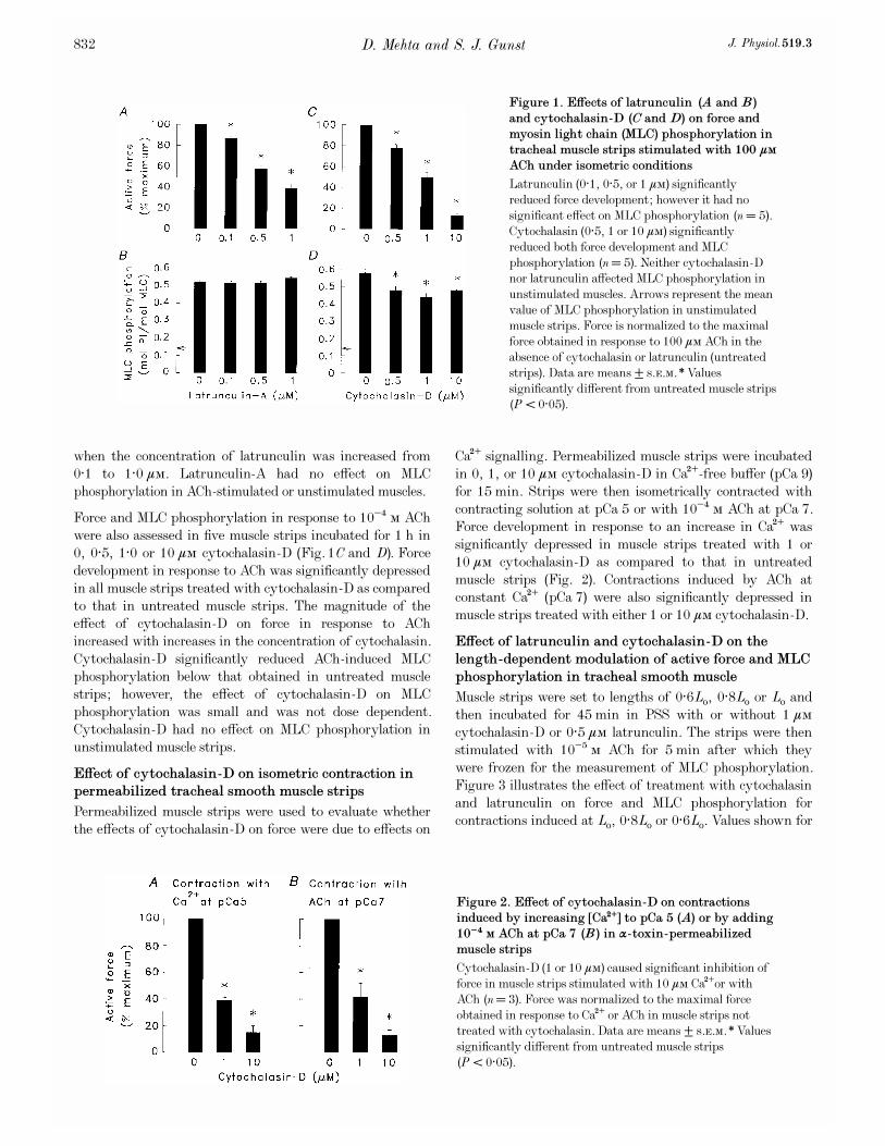
when the concentration of latrunculin was increased from
0·1 to 1·0 ìÒ. Latrunculin-A had no effect on MLC
phosphorylation in ACh-stimulated or unstimulated muscles.
Force and MLC phosphorylation in response to 10¦Æ Ò ACh
were also assessed in five muscle strips incubated for 1 h in
0, 0·5, 1·0 or 10 ìÒ cytochalasin_D (Fig. 1C and D). Force
development in response to ACh was significantly depressed
in all muscle strips treated with cytochalasin_D as compared
to that in untreated muscle strips. The magnitude of the
effect of cytochalasin_D on force in response to ACh
increased with increases in the concentration of cytochalasin.
Cytochalasin_D significantly reduced ACh-induced MLC
phosphorylation below that obtained in untreated muscle
strips; however, the effect of cytochalasin_D on MLC
phosphorylation was small and was not dose dependent.
Cytochalasin_D had no effect on MLC phosphorylation in
unstimulated muscle strips.
Effect of cytochalasin_D on isometric contraction in
permeabilized tracheal smooth muscle strips
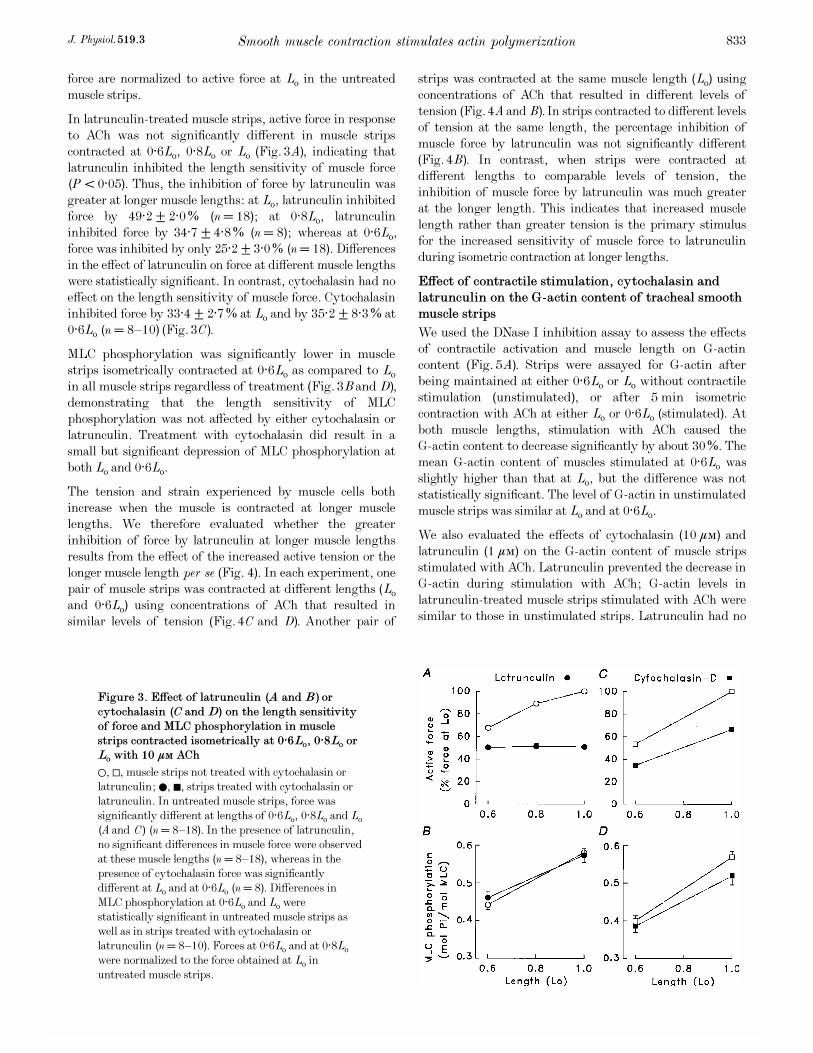
Permeabilized muscle strips were used to evaluate whether
the effects of cytochalasin_D on force were due to effects on
Ca¥ signalling. Permeabilized muscle strips were incubated
in 0, 1, or 10 ìÒ cytochalasin_D in Ca¥-free buffer (pCa 9)
for 15 min. Strips were then isometrically contracted with
contracting solution at pCa 5 or with 10¦Æ Ò ACh at pCa 7.
Force development in response to an increase in Ca¥ was
significantly depressed in muscle strips treated with 1 or
10 ìÒ cytochalasin_D as compared to that in untreated
muscle strips (Fig. 2). Contractions induced by ACh at
constant Ca¥ (pCa 7) were also significantly depressed in
muscle strips treated with either 1 or 10 ìÒ cytochalasin_D.
Effect of latrunculin and cytochalasin_D on the
length-dependent modulation of active force and MLC
phosphorylation in tracheal smooth muscle
Muscle strips were set to lengths of 0·6Lo, 0·8Lo or Lo and
then incubated for 45 min in PSS with or without 1 ìÒ
cytochalasin_D or 0·5 ìÒ latrunculin. The strips were then
stimulated with 10¦Ç Ò ACh for 5 min after which they
were frozen for the measurement of MLC phosphorylation.
Figure 3 illustrates the effect of treatment with cytochalasin
and latrunculin on force and MLC phosphorylation for
contractions induced at Lo, 0·8Lo or 0·6Lo. Values shown for
D. Mehta and S. J. Gunst J. Physiol. 519.3832
Figure 1. Effects of latrunculin (A and B)
and cytochalasin_D (C and D) on force and
myosin light chain (MLC) phosphorylation in
tracheal muscle strips stimulated with 100 ìÒ
ACh under isometric conditions
Latrunculin (0·1, 0·5, or 1 ìÒ) significantly
reduced force development; however it had no
significant effect on MLC phosphorylation (n = 5).
Cytochalasin (0·5, 1 or 10 ìÒ) significantly
reduced both force development and MLC
phosphorylation (n = 5). Neither cytochalasin_D
nor latrunculin affected MLC phosphorylation in
unstimulated muscles. Arrows represent the mean
value of MLC phosphorylation in unstimulated
muscle strips. Force is normalized to the maximal
force obtained in response to 100 ìÒ ACh in the
absence of cytochalasin or latrunculin (untreated
strips). Data are means ± s.e.m. *Values
significantly different from untreated muscle strips
(P < 0·05).
Figure 2. Effect of cytochalasin_D on contractions
induced by increasing [Ca¥] to pCa 5 (A) or by adding
10¦Æ Ò ACh at pCa 7 (B) in á_toxin-permeabilized
muscle strips
Cytochalasin_D (1 or 10 ìÒ) caused significant inhibition of
force in muscle strips stimulated with 10 ìÒ Ca¥or with
ACh (n = 3). Force was normalized to the maximal force
obtained in response to Ca¥ or ACh in muscle strips not
treated with cytochalasin. Data are means ± s.e.m. *Values
significantly different from untreated muscle strips
(P < 0·05).
force are normalized to active force at Lo in the untreated
muscle strips.
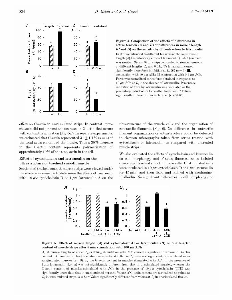
In latrunculin-treated muscle strips, active force in response
to ACh was not significantly different in muscle strips
contracted at 0·6Lo, 0·8Lo or Lo (Fig. 3A), indicating that
latrunculin inhibited the length sensitivity of muscle force
(P < 0·05). Thus, the inhibition of force by latrunculin was
greater at longer muscle lengths: at Lo, latrunculin inhibited
force by 49·2 ± 2·0% (n = 18); at 0·8Lo, latrunculin
inhibited force by 34·7 ± 4·8% (n = 8); whereas at 0·6Lo,
force was inhibited by only 25·2 ± 3·0% (n = 18). Differences
in the effect of latrunculin on force at different muscle lengths
were statistically significant. In contrast, cytochalasin had no
effect on the length sensitivity of muscle force. Cytochalasin
inhibited force by 33·4 ± 2·7% at Lo and by 35·2 ± 8·3% at
0·6Lo (n = 8—10) (Fig. 3C).
MLC phosphorylation was significantly lower in muscle
strips isometrically contracted at 0·6Lo as compared to Lo
in all muscle strips regardless of treatment (Fig. 3B and D),
demonstrating that the length sensitivity of MLC
phosphorylation was not affected by either cytochalasin or
latrunculin. Treatment with cytochalasin did result in a
small but significant depression of MLC phosphorylation at
both Lo and 0·6Lo.
The tension and strain experienced by muscle cells both
increase when the muscle is contracted at longer muscle
lengths. We therefore evaluated whether the greater
inhibition of force by latrunculin at longer muscle lengths
results from the effect of the increased active tension or the
longer muscle length per se (Fig. 4). In each experiment, one
pair of muscle strips was contracted at different lengths (Lo
and 0·6Lo) using concentrations of ACh that resulted in
similar levels of tension (Fig. 4C and D). Another pair of
strips was contracted at the same muscle length (Lo) using
concentrations of ACh that resulted in different levels of
tension (Fig. 4A and B). In strips contracted to different levels
of tension at the same length, the percentage inhibition of
muscle force by latrunculin was not significantly different
(Fig. 4B). In contrast, when strips were contracted at
different lengths to comparable levels of tension, the
inhibition of muscle force by latrunculin was much greater
at the longer length. This indicates that increased muscle
length rather than greater tension is the primary stimulus
for the increased sensitivity of muscle force to latrunculin
during isometric contraction at longer lengths.
Effect of contractile stimulation, cytochalasin and
latrunculin on the G_actin content of tracheal smooth
muscle strips
We used the DNase I inhibition assay to assess the effects
of contractile activation and muscle length on G_actin
content (Fig. 5A). Strips were assayed for G_actin after
being maintained at either 0·6Lo or Lo without contractile
stimulation (unstimulated), or after 5 min isometric
contraction with ACh at either Lo or 0·6Lo (stimulated). At
both muscle lengths, stimulation with ACh caused the
G_actin content to decrease significantly by about 30%. The
mean G_actin content of muscles stimulated at 0·6Lo was
slightly higher than that at Lo, but the difference was not
statistically significant. The level of G_actin in unstimulated
muscle strips was similar at Lo and at 0·6Lo.
We also evaluated the effects of cytochalasin (10 ìÒ) and
latrunculin (1 ìÒ) on the G_actin content of muscle strips
stimulated with ACh. Latrunculin prevented the decrease in
G_actin during stimulation with ACh; G_actin levels in
latrunculin-treated muscle strips stimulated with ACh were
similar to those in unstimulated strips. Latrunculin had no
Smooth muscle contraction stimulates actin polymerizationJ. Physiol. 519.3 833
Figure 3. Effect of latrunculin (A and B) or
cytochalasin (C and D) on the length sensitivity
of force and MLC phosphorylation in muscle
strips contracted isometrically at 0·6Lo, 0·8Lo or
Lo with 10 ìÒ ACh
1, ±, muscle strips not treated with cytochalasin or
latrunculin; 0, þ, strips treated with cytochalasin or
latrunculin. In untreated muscle strips, force was
significantly different at lengths of 0·6Lo, 0·8Lo and Lo
(A and C) (n = 8—18). In the presence of latrunculin,
no significant differences in muscle force were observed
at these muscle lengths (n = 8—18), whereas in the
presence of cytochalasin force was significantly
different at Lo and at 0·6Lo (n = 8). Differences in
MLC phosphorylation at 0·6Lo and Lo were
statistically significant in untreated muscle strips as
well as in strips treated with cytochalasin or
latrunculin (n = 8—10). Forces at 0·6Lo and at 0·8Lo
were normalized to the force obtained at Lo in
untreated muscle strips.
effect on G_actin in unstimulated strips. In contrast, cyto-
chalasin did not prevent the decrease in G_actin that occurs
with contractile activation (Fig. 5B). In separate experiments,
we estimated that G_actin represented 31 ± 1·1% (n = 4) of
the total actin content of the muscle. Thus a 30% decrease
in the G_actin content represents polymerization of
approximately 10% of the total actin in the cell.
Effect of cytochalasin and latrunculin on the
ultrastructure of tracheal smooth muscle
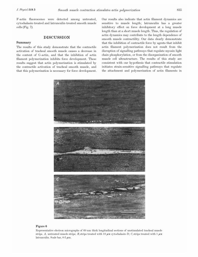
Sections of tracheal smooth muscle strips were viewed under
the electron microscope to determine the effects of treatment
with 10 ìÒ cytochalasin_D or 1 ìÒ latrunculin-A on the
ultrastructure of the muscle cells and the organization of
contractile filaments (Fig. 6). No differences in contractile
filament organization or ultrastructure could be detected
in electron micrographs taken from strips treated with
cytochalasin or latrunculin as compared with untreated
muscle strips.
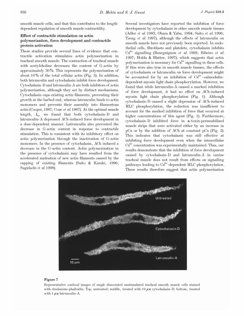
We also evaluated the effects of cytochalasin and latrunculin
on cell morphology and F-actin fluorescence in isolated
dissociated tracheal smooth muscle cells. Unstimulated cells
were incubated in 10 ìÒ cytochalasin_D or 1 ìÒ latrunculin
for 45 min, and then fixed and stained with rhodamine-
phalloidin. No significant differences in cell morphology or
D. Mehta and S. J. Gunst J. Physiol. 519.3834
Figure 5. Effect of muscle length (A) and cytochalasin_D or latrunculin (B) on the G_actin
content of muscle strips after 5 min stimulation with 100 ìÒ ACh
A, at muscle lengths of either Lo or 0·6Lo, stimulation with ACh caused a significant decrease in G_actin
content. Differences in G_actin content in muscles at 0·6Lo or Lo were not significant in stimulated or in
unstimulated muscles (n = 8). B, the G_actin content in muscles stimulated with ACh in the presence of
1 ìÒ latrunculin (Lat-A) was not significantly different from that in unstimulated muscles, whereas the
G_actin content of muscles stimulated with ACh in the presence of 10 ìÒ cytochalasin (CCD) was
significantly lower than that in unstimulated muscles. Values of G_actin content are normalized to values at
Lo in unstimulated strips (n = 8). *Values significantly different from values at Lo in unstimulated tissues.
Figure 4. Comparison of the effects of differences in
active tension (A and B) or differences in muscle length
(C and D) on the sensitivity of contraction to latrunculin
In strips contracted to different tensions at the same muscle
length (A), the inhibitory effect of latrunculin (Lat_A) on force
was similar (B) (n = 6). In strips contracted to similar tensions
at different lengths, Lo and 0·6Lo (C), latrunculin caused
significantly more force inhibition at Lo (D) (n = 6). 4,
contraction with 10 ìÒ ACh;$, contraction with 0·1 ìÒ ACh.
Force was normalized to the force obtained in response to
10 ìÒ ACh at Lo in the absence of latrunculin. Percentage
inhibition of force by latrunculin was calculated as the
percentage reduction in force after treatment. *Values
significantly different from each other (P < 0·05).
F-actin fluorescence were detected among untreated,
cytochalasin-treated and latrunculin-treated smooth muscle
cells (Fig. 7).
DISCUSSION
Summary
The results of this study demonstrate that the contractile
activation of tracheal smooth muscle causes a decrease in
the content of G_actin, and that the inhibition of actin
filament polymerization inhibits force development. These
results suggest that actin polymerization is stimulated by
the contractile activation of tracheal smooth muscle, and
that this polymerization is necessary for force development.
Our results also indicate that actin filament dynamics are
sensitive to muscle length; latrunculin has a greater
inhibitory effect on force development at a long muscle
length than at a short muscle length. Thus, the regulation of
actin dynamics may contribute to the length dependence of
smooth muscle contractility. Our data clearly demonstrate
that the inhibition of contractile force by agents that inhibit
actin filament polymerization does not result from the
disruption of signalling pathways that regulate myosin light
chain phosphorylation, or from the disorganization of smooth
muscle cell ultrastructure. The results of this study are
consistent with our hypothesis that contractile stimulation
initiates strain-sensitive signalling pathways that regulate
the attachment and polymerization of actin filaments in
Smooth muscle contraction stimulates actin polymerizationJ. Physiol. 519.3 835
Figure 6
Representative electron micrographs of 60 nm thick longitudinal sections of unstimulated tracheal muscle
strips. A, untreated muscle strips; B, strips treated with 10 ìÒ cytochalasin_D; C, strips treated with 1 ìÒ
latrunculin. Scale bar, 0·5 ìm.
smooth muscle cells, and that this contributes to the length-
dependent regulation of smooth muscle contractility.
Effect of contractile stimulation on actin
polymerization, force development and contractile
protein activation
These studies provide several lines of evidence that con-
tractile activation stimulates actin polymerization in
tracheal smooth muscle. The contraction of tracheal muscle
with acetylcholine decreases the content of G_actin by
approximately 30%. This represents the polymerization of
about 10% of the total cellular actin (Fig. 5). In addition,
both latrunculin and cytochalasin inhibit force development.
Cytochalasin_D and latrunculin-A are both inhibitors of actin
polymerization, although they act by distinct mechanisms.
Cytochalasin caps existing actin filaments, preventing their
growth at the barbed end, whereas latrunculin binds to actin
monomers and prevents their assembly into filamentous
actin (Cooper, 1987; Coue et al. 1987). At the optimal muscle
length, Lo, we found that both cytochalasin_D and
latrunculin-A depressed ACh-induced force development in
a dose-dependent manner. Latrunculin also prevented the
decrease in G_actin content in response to contractile
stimulation. This is consistent with its inhibitory effect on
actin polymerization through the inactivation of G_actin
monomers. In the presence of cytochalasin, ACh induced a
decrease in the G_actin content. Actin polymerization in
the presence of cytochalasin may have resulted from the
accelerated nucleation of new actin filaments caused by the
capping of existing filaments (Saito & Karaki, 1996;
Sugidachi et al. 1998).
Several investigators have reported the inhibition of force
development by cytochalasin in other smooth muscle tissues
(Adler et al. 1983; Obara & Yabu, 1994; Saito et al. 1996;
Tseng et al. 1997), although the effects of latrunculin on
smooth muscle have not previously been reported. In endo-
thelial cells, fibroblasts and platelets, cytochalasin inhibits
Ca¥ signalling (Bourguignon et al. 1993; Ribeiro et al.
1997; Holda & Blatter, 1997), which suggests that actin
polymerization is necessary for Ca¥ signalling in these cells.
If this were also true in smooth muscle tissues, the effects
of cytochalasin or latrunculin on force development might
be accounted for by an inhibition of Ca¥—calmodulin-
dependent myosin light chain phosphorylation. However, we
found that while latrunculin-A caused a marked inhibition
of force development, it had no effect on ACh-induced
myosin light chain phosphorylation (Fig. 1). Although
cytochalasin_D caused a slight depression of ACh-induced
MLC phosphorylation, the reduction was insufficient to
account for the marked inhibition of force that occurred at
higher concentrations of this agent (Fig. 1). Furthermore,
cytochalasin_D inhibited force in á_toxin-permeabilized
muscle strips that were activated either by an increase in
pCa or by the addition of ACh at constant pCa (Fig. 2).
This indicates that cytochalasin was still effective at
inhibiting force development even when the intracellular
Ca¥ concentration was experimentally maintained. Thus, our
results demonstrate that the inhibition of force development
caused by cytochalasin_D and latrunculin-A in canine
tracheal muscle does not result from effects on signalling
pathways leading to Ca¥-dependent MLC phosphorylation.
These results therefore suggest that actin polymerization
D. Mehta and S. J. Gunst J. Physiol. 519.3836
Figure 7
Representative confocal images of single dissociated unstimulated tracheal smooth muscle cells stained
with rhodamine-phalloidin. Top, untreated; middle, treated with 10 ìÒ cytochalasin_D; bottom, treated
with 1 ìÒ latrunculin-A.

plays a direct role in force development in smooth muscle
that is independent of signalling events regulating the
phosphorylation of myosin light chains.
The results of previous studies investigating the mechanism
for the effects of cytochalasin on smooth muscle contraction
are conflicting. Tseng et al. (1997) reported that in bovine
tracheal smooth muscle, the inhibition of force caused by
cytochalasin-B, a less specific agent than cytochalasin_D,
was associated with a decrease in myosin light chain
phosphorylation. They concluded that the inhibitory effect
of cytochalasin on force was due to the inhibition of Ca¥
signalling and MLC phosphorylation caused by disruption
of the actin cytoskeleton. In contrast, Saito et al. (1996)
found that cytochalasin_D inhibited contractions induced by
norepinephrine and K¤ in rat aorta smooth muscle without
affecting intracellular Ca¥ or MLC phosphorylation.
Consistent with this, Obara & Yabu (1994) found no effect of
cytochalasin-B on voltage-dependent Ca¥ currents and
membrane potentials in taenia coli smooth muscle. Our
present results demonstrating that smooth muscle contraction
is inhibited by latrunculin, which has no effect on myosin
light chain phosphorylation, provide unequivocal evidence
that the inhibition of actin polymerization per se inhibits
smooth muscle contraction.
Role of actin polymerization in smooth muscle
contraction
The activation of cells such as platelets, neutrophils and
fibroblasts results in the rapid polymerization of monomeric
globular (G) actin into filamentous (F) actin (Cano et al.
1991; Symons & Mitchison, 1991; Hartwig, 1992; Iwig et
al. 1995). This polymerization occurs primarily at the
‘barbed end’ of the actin filament, where the filament links
to transmembrane integrins (Cooper, 1987, 1991; Schafer
& Cooper, 1995). In these cells, actin polymerization is
regulated by the availability of free barbed ends of actin
filaments as well as by the pool of available actin monomers.
Most actin filament barbed ends are not freely accessible in
resting cells (Hartwig, 1992; Hug et al. 1995) as they are
blocked from elongation by capping proteins such as gelsolin
and CapZ (Barkalow et al. 1996). These proteins are also
present in differentiated smooth muscle (Pollard & Cooper,
1986; Schafer & Cooper, 1995). The activation of platelets
and other non-muscle cells causes the uncapping of actin
filaments, thereby increasing the number of free barbed
ends and enabling actin polymerization to proceed (Symons
& Mitchison, 1991; Hartwig, 1992, 1995; Nachmias et al.
1996). In platelets, the interaction of the endogenous
capping protein gelsolin with actin can be inhibited by
PIPµ, a by-product of phosphatidyl inositol breakdown that
is also produced in smooth muscle in response to stimulation
(Janmey, 1994). The capping of the barbed ends of actin
filaments may be coupled to detachment of the filament
from the membrane (Carlier, 1998).
Cytochalasin_D binds to the barbed (fast-growing) ends of
actin filaments and acts as a capping protein (Cooper, 1987).
As the binding of cytochalasin is not regulated by PIPµ,
actin filaments capped by cytochalasin do not become
uncapped during activation of the cell. Thus they do not
undergo polymerization at the barbed end, and they may
not attach to the integrin complex and participate in force
transmission during cell activation. Thus the effects of
cytochalasin on force in smooth muscle may be related to
its ability to block the attachment of actin filaments to the
integrin complex, as well as to its ability to prevent
filament polymerization at the barbed end. In contrast,
the latrunculin does not interfere with the uncapping or
attachment of filamentous actin to the integrin complex, but
prevents actin polymerization by complexing with free actin
monomers and thereby inactivating the pool of monomeric
actin (Coue et al. 1987).
Role of actin filament dynamics in the length
sensitivity of smooth muscle force
A striking finding of our study was that the inhibitory
effect of latrunculin on contractile force was length sensitive,
suggesting that mechanical strain may stimulate the rate of
actin polymerization. The inhibition of force caused by
latrunculin was greatest when the strips were activated at
the longest length, Lo, and smallest when the muscles were
activated at 0·6Lo. As a result, in the presence of latrunculin,
there were no significant differences in force development at
muscle lengths between 0·6Lo and Lo in response to
acetylcholine (Fig. 3). Myosin light chain phosphorylation is
length sensitive in smooth muscle, and the length sensitivity
of contractile activation has been proposed as a mechanism to
account for the length-dependent modulation of contractility
(Rembold & Murphy, 1990; Mehta et al. 1996). However,
latrunculin did not affect the length sensitivity of myosin
light chain phosphorylation; thus the differences in its effect
on contraction at different muscle lengths could not have
resulted from a suppression of the mechanosensitivity of
myosin activation. We observed a slight difference in the
mean levels of G_actin in muscles activated with ACh at Lo
and 0·6Lo using the DNase inhibition assay, but this
difference was not statistically significant (Fig. 5A). This
suggests that differences in the amount of actin polymerized
at different lengths may be small and difficult to discriminate
using this assay. Alternatively, mechanical strain may affect
the dynamics of actin polymerization rather than the total
amount of G_actin polymerized into filaments during
contractile activation.
We evaluated whether differences in the sensitivity of
contractions at Lo and 0·6Lo to inhibition by latrunculin
were related directly to the difference in muscle length or to
the differences in active tension at these muscle lengths. Our
results indicated that muscle length rather than tension is
the primary determinant of latrunculin sensitivity (Fig. 4).
These results are consistent with our recent report that the
muscle length rather than the tension is the stimulus for the
length-sensitive increases in the tyrosine phosphorylation
of paxillin and focal adhesion kinase during the contractile
Smooth muscle contraction stimulates actin polymerizationJ. Physiol. 519.3 837
activation of tracheal smooth muscle (Tang et al. 1999).
These proteins have been implicated in the signalling
pathway for cytoskeletal remodelling in non-muscle cells
(Burridge & Chrzanowska-Wodnicka, 1996; Shyy & Chien,
1997; Schmidt et al. 1998; Glogauer et al. 1998). Taken in
sum, our observations suggest a mechanosensitive process in
smooth muscle in which externally imposed strain activates
an integrin-mediated signalling pathway leading to actin
filament polymerization and cytoskeletal reorganization.
Actin polymerization in smooth muscle cells might involve
the lengthening of existing actin filaments andÏor the
nucleation of new actin filaments. This could enable the
smooth muscle cell to adapt the arrangement of its contractile
apparatus to externally imposed changes in the shape of the
cell. In tracheal smooth muscle, contractile activation at
different lengths results in persistent differences in stiffness
that suggest differences in cell structure (Gunst et al. 1995;
Gunst &Wu, 1996; Gunst, 1999).
In our study, the effects of cytochalasin_D contrasted with
those of latrunculin in that cytochalasin inhibited force
development proportionally during contraction at all muscle
lengths whereas latrunculin inhibited proportionally more
at long muscle lengths (Fig. 3). The absence of an effect of
cytochalasin on the length sensitivity of active force has
been previously reported for bovine tracheal muscle (Youn et
al. 1998). The difference in the inhibitory effect of latrunculin
and cytochalasin on the length sensitivity of active force can
be interpreted in terms of the mechanisms of action of these
agents. Latrunculin prevents the polymerization of new
actin filaments or the lengthening of existing filaments.
This should prevent the strain-sensitive remodelling of actin
filaments, although existing filaments could continue to
participate in force transmission. In contrast, cytochalasin
caps existing F-actin filaments and may therefore inhibit
their attachment and participation in force transmission
(Carlier, 1998). Only the actin filaments left uncapped by
cytochalasin_D could continue to participate in force trans-
mission. Therefore, in the presence of cytochalasin, force
development at all muscle lengths should be proportionally
inhibited.
Effect of cytochalasin and latrunculin on smooth
muscle cell structure
We used electron microscopy and immunofluorescence
microscopy to evaluate the effects of cytochalasin and
latrunculin on actin filament integrity and the ultra-
structure of tracheal smooth muscle. Electron micrographs
of tracheal muscle strips treated with cytochalasin or
latrunculin revealed no differences in contractile filament
organization or ultrastructure as compared with untreated
muscle strips (Fig. 6). We also detected no significant
differences in the morphology of untreated, cytochalasin-
treated or latrunculin-treated smooth muscle cells stained
for F-actin using rhodamine-phalloidin (Fig. 7). Thus the
effects of these agents on force are unlikely to result from
the disruption of actin filament integrity or disorganization
of the contractile apparatus.
Conclusion
In conclusion, our results suggest that the contractile
activation of tracheal smooth muscle stimulates actin
polymerization, and that this actin polymerization con-
tributes directly to force development. The polymerization
of actin in response to contractile stimuli may modulate
the organization or length of actin filaments as well as the
attachment of actin filaments to the cell membrane for the
transmission of force. The inhibition of force by inhibitors
of actin polymerization does not result from the disruption
of signalling pathways leading to myosin light chain
phosphorylation, or from the disorganization of cell
structure. Actin polymerization may also play an important
role in regulating the length sensitivity of force development.
Adler, K. B., Krill, J., Alberghini, T. V. & Evans, J. N. (1983).Effect of cytochalasin D on smooth muscle contraction. Cell Motility3, 545—551.
Banes, A. J., Tsuzaki, M., Yamamoto, J., Fischer, T., Brigman, B.,
Brown, T. & Miller, L. (1995). Mechanoreception at the cellularlevel: the detection, interpretation, and diversity of responses tomechanical signals. Biochemistry and Cell Biology 73, 349—365.
Barkalow, K., Witke, W., Kwiatkowski, D. J. & Hartwig, J. H.
(1996). Coordinated regulation of platelet actin filament barbedends by gelsolin and capping protein. Journal of Cell Biology 134,389—399.
Blikstad, I., Markey, F., Carlsson, L., Persson, T. & Lindberg,
U. (1978). Selective assay of monomeric and filamentous actin in cellextracts, using inhibition of deoxyribonuclease I. Cell 15, 935—943.
Bourguignon, L. Y., Iida, N. & Jin, H. (1993). The involvement ofthe cytoskeleton in regulating IP3 receptor-mediated internal Ca¥release in human blood platelets. Cell Biology International 17,751—758.
Burridge, K. & Chrzanowska-Wodnicka, M. (1996). Focaladhesions, contractility, and signaling. Annual Review of Cell and
Developmental Biology 12, 463—518.
Cano, M. L., Lauffenburger, D. A. & Zigmond, S. H. (1991).Kinetic analysis of F-actin depolymerization in polymorphonuclearleukocyte lysates indicates that chemoattractant stimulationincreases actin filament number without altering the filament lengthdistribution. Journal of Cell Biology 115, 677—687.
Carlier, M. F. (1998). Control of actin dynamics. Current Opinion in
Cell Biology 10, 45—51.
Cooper, J. A. (1987). Effects of cytochalasin and phalloidin on actin.Journal of Cell Biology 105, 1473—1478.
Cooper, J. A. (1991). The role of actin polymerization in cell motility.Annual Review of Physiology 53, 585—605.
Coue, M., Brenner, S. L., Spector, I. & Korn, E. D. (1987).Inhibition of actin polymerization by latrunculin A. FEBS Letters
213, 316—318.
Fabiato, A. & Fabiato, F. (1979). Calculator programs for computingthe composition of the solutions containing multiple metals andligands used for experiments in skinned muscle cells. Journal de
Physiologie 75, 463—505.
D. Mehta and S. J. Gunst J. Physiol. 519.3838

Glogauer, M., Arora, P., Chou, D., Janmey, P. A., Downey, G. P. &
McCulloch, C. A. (1998). The role of actin-binding protein 280 inintegrin-dependent mechanoprotection. Journal of Biological
Chemistry 273, 1689—1698.
Gunst, S. J. (1986). Effect of length history on contractile behavior ofcanine tracheal smooth muscle. American Journal of Physiology
250, C146—154.
Gunst, S. J. (1999). Applicability of sliding filamentÏcrossbridgeparadigm to smooth muscle. Reviews of Physiology Biochemistry
and Pharmacology 134, 7—61.
Gunst, S. J., Meiss, R. A., Wu, M. F. & Rowe, M. (1995).Mechanisms for the mechanical plasticity of tracheal smooth muscle.American Journal of Physiology 268, C1267—1276.
Gunst, S. J. & Wu, M. F. (1996). Canine tracheal smooth musclestiffness during stretch depends on muscle length at contractiononset. Biophysical Journal 70, A45.
Gunst, S. J., Wu, M. F. & Smith, D. D. (1993). Contraction historymodulates isotonic shortening velocity in smooth muscle. American
Journal of Physiology 265, C467—476.
Halayko, A. J., Salari, H., Ma, X. & Stephens, N. L. (1996).Markers of airway smooth muscle cell phenotype. American Journal
of Physiology 270, L1040—1051.
Harris, D. E. & Warshaw, D. M. (1991). Length vs. active forcerelationship in single isolated smooth muscle cells. American Journal
of Physiology 260, C1104—1112.
Hartwig, J. H. (1992). Mechanisms of actin rearrangementsmediating platelet activation. Journal of Cell Biology 118,1421—1442.
Hartwig, J. H., Bokoch, G. M., Carpenter, C. L., Janmey, P. A.,
Taylor, L. A., Toker, A. & Stossel, T. P. (1995). Thrombinreceptor ligation and activated Rac uncap actin filament barbedends through phosphoinositide synthesis in permeabilized humanplatelets. Cell 82, 643—653.
Holda, J. R. & Blatter, L. A. (1997). Capacitative calcium entry isinhibited in vascular endothelial cells by disruption of cytoskeletalmicrofilaments. FEBS Letters 403, 191—196.
Hug, C., Jay, P. Y., Reddy, I., McNally, J. G., Bridgman, P. C.,
Elson, E. L. & Cooper, J. A. (1995). Capping protein levelsinfluence actin assembly and cell motility in Dictyostelium. Cell 81,591—600.
Ingber, D. E. (1997). Tensegrity: the architectural basis of cellularmechanotransduction. Annual Review of Physiology 59, 575—599.
Iwig, M., Czeslick, E., Muller, A., Gruner, M., Spindler, M. &
Glaesser, D. (1995). Growth regulation by cell shape alteration andorganization of the cytoskeleton. European Journal of Cell Biology
67, 145—157.
Janmey, P. A. (1994). Phosphoinositides and calcium as regulators ofcellular actin assembly and disassembly. Annual Review of
Physiology 56, 169—191.
Kitazawa, T., Kobayashi, S., Horiuti, K., Somlyo, A. V. & Somlyo,
A. P. (1989). Receptor-coupled, permeabilized smooth muscle. Roleof the phosphatidylinositol cascade, G-proteins, and modulation ofthe contractile response to Ca¥. Journal of Biological Chemistry
264, 5339—5342.
Lehman, W., Vibert, P., Craig, R. & Barany, M. (1996). Actin andthe structure of smooth muscle thin filaments. In Biochemistry of
Smooth Muscle Contraction, ed. Barany, M., pp. 47—60. AcademicPress Inc., San Diego, CA, USA.
Mehta, D., Wu, M. F. & Gunst, S. J. (1996). Role of contractileprotein activation in the length-dependent modulation of trachealsmooth muscle force. American Journal of Physiology 270,C243—252.
Meiss, R. A. (1993). Persistent mechanical effects of decreasinglength during isometric contraction of ovarian ligament smoothmuscle. Journal of Muscle Research and Cell Motility 14, 205—218.
Nachmias, V. T., Golla, R., Casella, J. F. & Barron-Casella, E.
(1996). Cap Z, a calcium insensitive capping protein in resting andactivated platelets. FEBS Letters 378, 258—262.
Obara, K. & Yabu, H. (1994). Effect of cytochalasin B on intestinalsmooth muscle cells. European Journal of Pharmacology 255,139—147.
Pavalko, F. M., Adam, L. P., Wu, M. F., Walker, T. L. & Gunst,
S. J. (1995). Phosphorylation of dense-plaque proteins talin andpaxillin during tracheal smooth muscle contraction. American
Journal of Physiology 268, C563—571.
Pollard, T. D. & Cooper, J. A. (1986). Actin and actin-bindingproteins. A critical evaluation of mechanisms and functions. AnnualReview of Biochemistry 55, 987—1035.
Rembold, C. M. & Murphy, R. A. (1990). Muscle length, shortening,myoplasmic [Ca¥], and activation of arterial smooth muscle.Circulation Research 66, 1354—1361.
Ribeiro, C. M., Reece, J. & Putney, J. W. Jr (1997). Role of thecytoskeleton in calcium signaling in NIH 3T3 cells. An intactcytoskeleton is required for agonist-induced [Ca¥]é signaling, butnot for capacitative calcium entry. Journal of Biological Chemistry
272, 26555—26561.
Saito, S. Y., Hori, M., Ozaki, H. & Karaki, H. (1996). CytochalasinD inhibits smooth muscle contraction by directly inhibitingcontractile apparatus. Journal of Smooth Muscle Research 32,51—60.
Saito, S. & Karaki, H. (1996). A family of novel actin-inhibitingmarine toxins. Clinical and Experimental Pharmacology and
Physiology 23, 743—746.
Schafer, D. A. & Cooper, J. A. (1995). Control of actin assembly atfilament ends. Annual Review of Cell and Developmental Biology
11, 497—518.
Schmidt, C., Pommerenke, H., Durr, F., Nebe, B. & Rychly, J.
(1998). Mechanical stressing of integrin receptors induces enhancedtyrosine phosphorylation of cytoskeletally anchored proteins.Journal of Biological Chemistry 273, 5081—5085.
Shyy, J. Y. & Chien, S. (1997). Role of integrins in cellular responsesto mechanical stress and adhesion. Current Opinion in Cell Biology
9, 707—713.
Sugidachi, A., Ogawa, T., Asai, F., Saito, S., Ozaki, H., Fusetani,
N., Karaki, H & Koike, H. (1998). Inhibition of rat plateletaggregation by mycalolide-B, a novel inhibitor of actinpolymerization with a different mechanism of action fromcytochalasin_D. Thrombosis and Haemostasis 79, 614—619.
Symons, M. H. & Mitchison, T. J. (1991). Control of actinpolymerization in live and permeabilized fibroblasts. Journal of Cell
Biology 114, 503—513.
Tang, D. C., Mehta, D. & Gunst, S. J. (1999). Mechanosensitivetyrosine phosphorylation of paxillin and focal adhesion kinase intracheal smooth muscle. American Journal of Physiology 276,C250—258.
Theriot, J. A. (1994). Regulation of the actin cytoskeleton in livingcells. Seminars in Cell Biology 5, 193—199.
Tseng, S., Kim, R., Kim, T., Morgan, K. G. & Hai, C. M. (1997).F_actin disruption attenuates agonist-induced [Ca¥], myosinphosphorylation, and force in smooth muscle. American Journal of
Physiology 272, C1960—1967.
Wang, N., Butler, J. P. & Ingber, D. E. (1993).Mechanotransduction across the cell surface and through thecytoskeleton. Science 260, 1124—1127.
Smooth muscle contraction stimulates actin polymerizationJ. Physiol. 519.3 839
Wang, Z., Pavalko, F. M. & Gunst, S. J. (1996). Tyrosinephosphorylation of the dense plaque protein paxillin is regulatedduring smooth muscle contraction. American Journal of Physiology
271, C1594—1602.
Youn, T., Kim, S. A. & Hai, C. M. (1998). Length-dependentmodulation of smooth muscle activation: effects of agonist,cytochalasin, and temperature. American Journal of Physiology
274, C1601—1607.
Acknowledgements
We are grateful to Dr Simon Atkinson for his advice and assistance
with the DNase inhibition assay. We also thank Ming-Fang Wu for
his assistance with experiments and for his help in preparing the
figures. This work was supported by a US PHS grant HL29289, the
Midwest Affiliate of the American Heart Association, and the
Showalter Foundation.
Corresponding author
S. J. Gunst: Department of Physiology and Biophysics, Indiana
University School of Medicine, 635 Barnhill Drive, Indianapolis,
IN 46202-5126, USA.
Email: [email protected]
D. Mehta and S. J. Gunst J. Physiol. 519.3840