activated peripheral lymphocytes with increased expression ... · activated peripheral lymphocytes...
TRANSCRIPT
437437437437437Mem Inst Oswaldo Cruz, Rio de Janeiro, Vol. 101(4): 437-449, June 2006
Activated peripheral lymphocytes with increased expression ofcell adhesion molecules and cytotoxic markers are associated
with dengue fever diseaseElzinandes L Azeredo, Sonia MO Zagne**, Allan R Alvarenga, Rita MR Nogueira,
Claire F Kubelka, Luzia M de Oliveira-Pinto*/+
Laboratório de Imunologia Viral, Departamento de Virologia *Laboratório de Auto-imunidade e Imuno-regulação, Departamento deImunologia, Instituto Oswaldo Cruz-Fiocruz, Av. Brasil 4365, 21045-900 Rio de Janeiro, RJ, Brasil **Departamento de Clínica
Médica, Hospital Universitário Antônio Pedro, Universidade Federal Fluminense, Niterói, RJ, Brasil
The immune mechanisms involved in dengue fever and dengue hemorrhagic/dengue shock syndrome are not wellunderstood. The ex vivo activation status of immune cells during the dengue disease in patients was examined. CD4and CD8 T cells were reduced during the acute phase. Interestingly, CD8 T cells co-expressing activation markerHLA-DR, Q, P, and cytolytic granule protein-Tia-1 were significantly higher in dengue patients than in controls.Detection of adhesion molecules indicated that in dengue patients the majority of T cells (CD4 and CD8) express theactivation/memory phenotype, characterized as CD44HIGH and lack the expression of the naïve cell marker, CD62LLOW.Also, the levels of T cells co-expressing ICAM-1 (CD54), VLA-4, and LFA-1 (CD11a) were significantly increased.CD8 T lymphocytes expressed predominantly low levels of anti-apoptotic molecule Bcl-2 in the acute phase, possi-bly leading to the exhibition of a phenotype of activated/effector cells. Circulating levels of IL-18, TGF-β1 andsICAM-1 were significantly elevated in dengue patients. Early activation events occur during acute dengue infec-tion which might contribute to viral clearance. Differences in expression of adhesion molecules among CD4 andCD8 T cells might underlie the selective extravasation of these subsets from blood circulation into lymphoid organsand/or tissues. In addition, activated CD8 T cells would be more susceptible to apoptosis as shown by the alterationin Bcl-2 expression. Cytokines such as IL-18, TGF-β1, and sICAM-1 may be contributing by either stimulating orsuppressing the adaptative immune response, during dengue infection, thereby perhaps establishing a relationshipwith disease severity.
Key word: dengue - patients lymphocytes activation
Approximately 1 billion human infections with den-gue virus (DENV) are estimated to occur annually. Themajority of these cases develop mild self-limited denguefever (DF), whereas only a small proportion develop se-vere hemorrhagic manifestations, dengue hemorrhagicfever/dengue shock syndrome (DHF/DSS), at the end ofthe acute phase of illness. DHF is a severe febrile diseasecharacterized by abnormalities in homeostasis and in-creased vascular permeability, which may result in DSS asreviewed by Rothman (2004).
There is a correlation between viral load and diseaseseverity (Vaughn et al. 2000). In this way, immunity to agiven dengue virus serotype (DENV 1, DENV 2, DENV 3,and DENV 4) provides good protection against re-infec-tion by that same serotype, however subsequent infec-tion by other viral serotypes markedly increases the riskof DHF/DSS, contributing to the hypothesis by the Ab(antibody) mediated enhancement of infection (Thein etal. 1997). In conjunction to the role of Abs in immuno-pathogenesis of dengue infection, a mechanism of mo-
Financial support: CNPq, Faperj, IOC-Fiocruz+Corresponding author: [email protected] 8 March 2006Accepted 3 May 2006
lecular mimicry has recently been proposed, in which Absdirected against the DENV non-structural protein (NS1)cross-react with endothelial cells, resulting in endothelialcells activation, increased levels of cytokine, chemokine,and adhesion molecule expression and consequentlyPBMC adhesion to endothelial cells. This activation mayfavour the vasculopathy observed in DENV infection viathe transcription factor NF-κB (Lin et al. 2005).
Immune response to DENV infection may also under-lie the pathogenesis of disease. In this context, a shiftfrom a Th1-type cytokine response in DF to a Th2-typecytokine response in DHF has been correlated with in-creasing severity of the illness (Chaturvedi et al. 1999).Among cytokines, tumour necrosis factors-α (TNF-α),interleukin-6 (IL-6), IL-10 (Iyngkaran et al. 1995, Azeredoet al. 2001) and more recently, transforming growth factor-β1 (TGF- β1) (Agarwal et al. 1999) and macrophage migra-tion inhibitory factor (MIF) (Chen et al. 2006) are frequentlyassociated with severity and platelet decay (Green et al.1999b, Azeredo et al. 2001). Elevated levels of soluble TNF-α receptors, soluble CD8, and soluble IL-2 receptor (Greenet al. 1999b), and soluble VCAM-1 (vascular cell adhesionmolecule 1) (Murgue et al. 2001) have been reported inplasma or serum of DHF patients compared to the levelsfound in early course infection DF patients. However, arise in cytokines and soluble receptors is an indirect mea-surement of immune activation not revealing which im-mune cells are activated. Thus, the percentage CD8 T and

438438438438438 Activation of lymphocytes from dengue patients • Elzinandes L Azeredo et al.
NK cells expressing CD69 rose more in children who de-veloped DHF than in those with only DF, demonstratingthat cellular immune activation is present early in acutedengue and is related to disease severity (Green et al.1999a). More recently, our group demonstrated that pe-ripheral NK and NKT cells rates are increased in adultBrazilian mild dengue patients. The majority of these NKcells display early markers for activation (CD69, HLA-DR,and CD38), cell adhesion molecules (CD44, CD11a) be-sides the intracellular cytotoxic granule, Tia-1 (Azeredoet al. 2006). A study displayed that during acute infectionfew DENV-specific T cells of low affinity for the infectingvirus were recovered in Thai children, contributing tohigher viral loads and increased immunopathology(Mongkolsapaya et al. 2003). Impaired proliferation of Tcells to a wide variety of stimuli, specific or not, was sup-pressed during acute dengue infection in subjects witheither DHF (Mathew et al. 1999) or DF (Mathew et al.1999, Azeredo et al. 2001). Nevertheless, these investiga-tions have not fully elucidated the complex mechanismsof immunopathology during DENV infection.
Thus, in adult Brazilian dengue patients, we determinedthe ex vivo activation status of immune cells over theperiod of acute to convalescence infection. Most activa-tion markers studied were induced in CD8 lymphocytesduring acute phase, returning to normal expression onconvalescence, showing the prevalence of this cell re-sponse over CD4 cell during infection. Moreover, in anattempt to better understand how it correlates with dis-ease severity, we observed that some of these activationmarkers were more pronounced in CD8 cells among sever-ity of DF cases.
MATERIALS AND METHODS
Study population and blood samples - From 2001 to2002 the DENV 1 and 3 outbreak, heparinized peripheralblood samples were obtained from 60 dengue infectedpatients (35 females, 25 males, age range 15-73 years), andamong those a follow-up blood samples was obtainedfrom 18 dengue infected patients, attended at two healthcentres in Niterói, state of Rio de Janeiro (Posto de Saúdede Itaipu and Centro Previdenciário de Niterói). All pa-tients presented clinical diagnosis of dengue infectionaccording to the criteria of the World Health Organization(WHO 2002). Among the dengue patients, 16 cases werehospitalised due to severity but, upon subsequent inde-pendent review of the medical record, only two of themwere considered to meet all criteria for DHF. There wasnot one fatal case in this outbreak. Fifteen healthy indi-viduals with similar age range (9 females, 6 males, agerange 18-50 years), without any febrile or other illnessesin the previous three months, were included as controls.
A detailed physical examination was performed to de-tect hemorrhagic manifestations (positive tourniquet testfor capillary fragility, skin haemorrhages, epistaxis, gingi-val, gastrointestinal or urinary tract haemorrhage), signsof plasma leakage (pleural or pericardial effusion, ascites),signs of circulatory failure (cold extremities, cyanosis,hypotension, tachycardia), and hepatomegally. Informedconsent was obtained from all patients or their guardiansprior to blood collection.
The diagnosis of DENV infection was confirmed byanti-dengue enzyme-linked immunoassorbent assay(ELISA)-IgM, serotype specific reverse trancription-poly-merase chain reaction (RT-PCR), or by virus isolation.Dengue viral isolations were attempted from serumsamples of patients in the acute phase, using Aedesalbopictus C6/36 cell line. Isolates were demonstrated byimmunofluorescence assay. Alternatively, virus was anti-genically detected in monocytes by flow cytometry(Neves-Souza et al. 2005) in 23 out of 35 patients from 1-10days after disease outset but not after 11 days. Dengueimmune response was considered as primary or second-ary by IgG ELISA according to previously establishedcriteria.
For these studies, 76 samples were considered to bean acute sample up to study day 1 to 10 from the patientshad at least one of the following symptoms: fever, head-ache, rash, myalgia, retro-orbital pain, arthralgia, and oth-ers symptoms associated or not with haemorrhagic mani-festations, particularly, petechiae and tourniquet-positivetest. Convalescent samples were obtained from other 12patients 11 days or later after their onset disease. Thesubjects in convalescence phase were not febrile neitherhad others characteristics symptoms at that time.
As other investigators have previously reported (Har-ris et al. 2000, Phuong et al. 2004, Wilder-Smith & Schwartz2005) we also were unable to meet WHO criteria (WHO2002) for severity classification. Indeed in Nicaragua,Harris et al. (2000) defined a severe patient group withsigns of shock that do not fit DHF/DSS classification there-fore designating a new disease category: dengue withsigns associated with shock. More recently, Balmasedaet al. (2005) confirmed that strict application of the WHOcriteria fails to detect a significant number of patients withsevere manifestations of dengue, especially in adults inNicaraguan hospitals. We considered all patients withthrombocytopenia (< 100,000 counts/mm3) and hypoten-sion (postural hypotension with decrease in systolic arte-rial pressure in 20 mmHg in supine position or systolicarterial pressure < 90 mmHg) as severe cases. Based onclinical grounds, we established that among dengue pa-tients, 31 had platelet levels ≥100.000counts/mm3, with-out hypotension and were classified as mild dengue; 19other patients had thrombocytopenia and hypotension,successively receiving parenteral hydration for at least6 h and were classified as severe dengue (due to not meet-ing classical DHF/DSS classification). Ten other patientshad thrombocytopenia but had no other severe manifes-tation.
The procedures performed during this work were ap-proved by the Ethics Committee of the Oswaldo Cruz Foun-dation, Brazilian Health Ministry (recognized by the Bra-zilian National Ethics Committee) under number 111/00.
Purification and cryopreservation of human periph-eral blood mononuclear cells (PBMCs) - PBMCs fromhealthy individuals and dengue infected patients wereobtained from 30 ml of heparinized venous blood. Bloodsamples were diluted 1:1 with culture medium RPMI 1640(Sigma) and PBMC were separated by performing Hys-topaque (Sigma) (d = 1077g/ml) and centrifuged at 400 g
439439439439439Mem Inst Oswaldo Cruz, Rio de Janeiro, Vol. 101(4), June 2006
for 30 min. The PBMC layer was washed twice in RPMI1640 medium. The viability of PBMC was greater than95% after Trypan blue exclusion. Approximately 107
PBMCs were re-suspended in 1 ml of solution destinedfor freezing [90% inactivated FBS (Gibco Invitrogen Co.)plus 10% DMSO (Sigma)] and stored initially at –70o C for24 h before introduction into liquid nitrogen, and aliquotswere cryopreserved for later study.
Reagents and monoclonal antibodies - The mouseanti-human surface antigen mAbs used in this study in-cluded either FITC-, PE or Cycrome (Cy)-conjugated with:anti-CD8 mAb (IgG1, clone DK25), anti-HLA-DP, DQ, DRmAb (IgG1, clone CR3/43), anti-CD29 mAb (IgG2a, cloneK20), and anti-CD44 mAb (IgG1, clone DF1485) fromDAKO; anti-CD49d mAb (IgG1, clone 9F10), anti-CD11amAb (IgG1, clone HI111), and anti-CD3 mAb (IgG3, cloneSP34), from BD Biosciences Pharmingen as well as anti-CD54 mAb (IgG1, clone HA58) and anti-CD4 mAb (IgG2a,clone S3.5) from Caltag. Intracellular detection of Tia-1proteins was performed with unconjugated monoclonalanti-human Tia-1 (IgG1, clone 2G9A10F5) from Coulter(US) plus polyclonal rabbit anti-mouse Ig conjugated toFITC from Dako (cat. # F0313). For Bcl-2 proteins we usedPE-conjugated anti-Bcl-2 mAb (IgG1, clone 100) fromCaltag. Matching isotype control for each antibody wasincluded in all experiments.
Extra- and intracellular staining by flow cytometry -Cryopreserved PBMCs (107 cells) were thawed and dividedinto aliquots each containing 2 × 10⁵ cells in order to stainfor flow cytometry analysis. PBMCs were washed in PBS-BSA-NaN3 [PBS pH 7.2, supplemented with 1% (w/v) BSA(Sigma) and 0.1% NaN3 (Sigma)] and triple-stained for 30min at 4ºC with specific mAbs, described above in dilu-tions recommended by the manufacturer. Stained cells werethen washed in PBS-BSA-NaN3 and fixed in PBS-BSA-NaN3 containing 1% paraformaldehyde (PFA) (Sigma) for15 min at 4ºC. Fixed cells were then immediately applied toa FACSCalibur (BD Biosciences).
The intracellular expression of Tia-1 and Bcl-2 pro-teins was analyzed on thawed PBMC. Cells were quicklyfirst co-stained with PE-conjugated anti-CD4 and Cy-con-jugated anti-CD8 mAbs, washed twice with PBS-BSA-NaN3 and then fixed with PBS-BSA-NaN3 containing 1%PFA. These cells were washed twice in cold PBS prior topermeabilization by saponine 0.05% (w/v) (Sigma) for 5min at room temperature. PBMCs were stained withunconjugated anti-Tia-1 mAb in 0.05% saponine bufferfor 45 min at 4ºC. Cells were washed once in PBS thenstained with FITC-conjugated anti-mouse and finally fixedin 1% PFA. PBMCs, pre-stained with Cy-conjugated anti-CD8 and FITC-conjugated anti-CD4 were fixed, per-meabilized and then co-stained for 45 min with PE-conju-gated Bcl2 mAb as previously described. For each sample,10,000-20,000 events were acquired, and analyses wereperformed with the WinMDI 2.8 and FlowJo version 4.3software.
Cytokine detection assays - Plasma samples were ob-tained from 43 dengue patients and 15 controls stored inaliquots at –70°C until use. Levels of soluble factors were
determined by ELISA kits (R&D Systems) in compliancewith manufacturer’s directions. The limits of sensitivityfor TGF-β1, IL-18 and sICAM-1 were 25.6, 31.2 and 2.47pg/ml, respectively.
Statistical analysis - The non-parametric Mann-Whitney U was used to evaluate variations in the expres-sion of cell surface markers between patients and controldonors. P values lower than 0.05 were considered to beassociated with statistical significance. Correlation be-tween cell surface marker expression and cytokine pro-duction was estimated by Spearman regression analysis.The Prism 4 statistics program was used for all analyses(GraphPad, US).
RESULTS
T lymphocyte subsets and clinical characterizationof adult Brazilian patients with dengue disease - Leuko-penia was present in 64% and thrombocytopenia in 47%of investigated dengue patients. Most patients, comparedto controls, presented a significantly reduced mean per-centage of CD4 T lymphocytes during acute phase ofinfection, which did not return to normal in convalescencedays studied (sample up to study day 11 to 30). Therewere no significant differences in the mean percentage ofCD8 T cells between dengue patients during acute or con-valescence phases and control individuals. Strikingly, bothabsolute CD4 and CD8 T cell counts decreased in denguepatients when compared to controls (Table I). In addition,there was no significant difference in CD4/CD8 ratios (con-trols 1.7 ± 0.3, days 1-10 1.5 ± 0.75 and days >11 1.7 ± 0.8).
Among 44 patients with DENV-1, 10 were classified assecondary infection, and among 16 patients with DENV-3, 9 were considered secondary infection. We suspectthat the in general secondary infections may be more se-rious than primary and also that DENV-3 is more severethan DENV-1. However our statistical analysis did notconfirm this hypothesis. Either severe patient samplingwas not big enough to permit group subdivisions or theless frequent incidence of severe cases in primary infec-tion/DENV-1 would hide this effect.
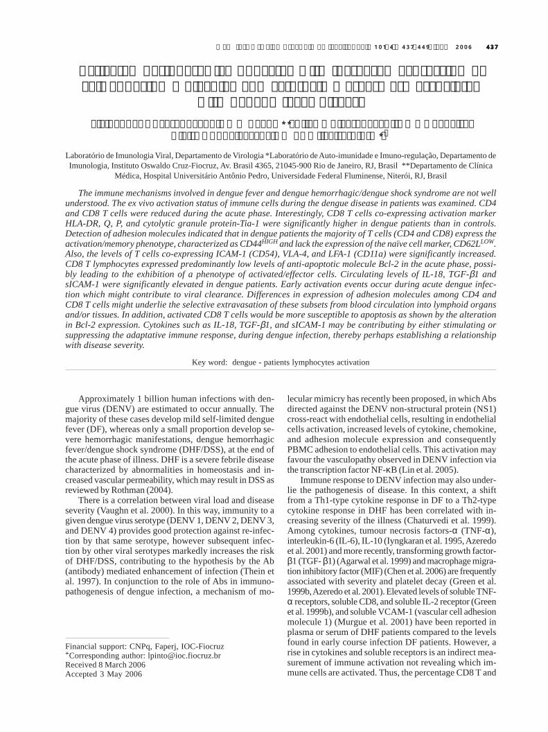
Predominance of activated and cytotoxic CD8 T lym-phocytes in dengue patients - We evaluated the degreeof activation and cytotoxic capability of T lymphocytesin the course of dengue disease. The mean percentage ofCD8 T lymphocytes expressing human leukocyte antigensgrouped as class II MHC genes (HLA- DP, DQ, DR) (Fig.1A, B) or intracellular cytotoxic granule (Tia-1) amongtotal CD8 T lymphocytes (Fig. 1C, D) was significantlyhigher in dengue patients during 1-10 days after diseaseonset as compared to that of healthy individuals (valuesfor HLA class II expression among CD8 T lymphocytes:5.8 ± 4.0% on controls vs 15.2 ± 10.4% at days 1-10 vs 7.5± 4.2% at > 11 days and for Tia-1 expression among CD8 Tlymphocytes: 6.6 ± 6.1% on controls vs 20.3 ±16.1% atdays 1-10 days vs 4.9 ± 5.1% at > 11 days). Besides eachillustration we presented the respective isotype controldemonstrating the labelling specificity. There was no sig-nificant increase of these activation molecules among CD4T lymphocytes (data not shown). In view of the key roleof CD8 T lymphocytes in the course of viral infections,
440440440440440 Activation of lymphocytes from dengue patients • Elzinandes L Azeredo et al.
TABLE IClinical features of dengue patients included in the study
Controls DENV 1-10 DENV >11
Number of samples 13 76 12
Laboratory findingsHematocrit (%) 38 ± 2a 42 ± 3.7 40.8 ± 2.8Leukocytes (counts/mm3) 5826 ± 1281 3913 ± 1680*b 8033 ± 4045Platelets (counts × 103/mm3) 271 ± 36 152 ± 59*** 245 ± 97
Immunological parametersCD4+CD3+ T lymphocytes (%) 46.2 ± 5.3 37.6 ± 10.2** 38.2 ± 5.5**CD4 T lymphocytes (counts/mm3) 513.1± 134.7 247 ± 185.8*** 318. 2 ± 176.5**CD8+CD3+ T lymphocytes (%) 27.9 ± 3.5 26.8 ± 8.8 24.9 ± 5.9CD8 T lymphocytes (counts/mm3) 304.8 ± 69.2 190.7 ± 180*** 203 ± 115
a: average ± standard deviation from patient and control determinations; b: statistical significance was assessed by the Mann-Whitney U test to evaluate differences in T lymphocytes between dengue patients and controls; P values: *p < 0.05, ** p < 0.01,***p < 0.0001.
Fig. 1: activation of CD8 T lymphocytes during dengue disease. Peripheral blood mononuclear cells from healthy subjects (n = 9) orsamples from dengue patients taken at different time after disease onset: at acute (1-10 days, n = 31) and convalescent phase (after 11 days,n = 8) were labelled as described in Materials and Methods and analyzed by flow cytometry within lymphocyte gate. Representative contourplots of CD8 T cells from a control donor, a dengue patient at day 5 and its respective isotype control are shown demonstrating thelabelling specificity. Numbers in each quadrant indicate the percentage of A: HLA-DR, DP, DQ+ or C: Tia-1+ cells within CD8 subsets.Mean percentages of B: HLA-DR, DP, DQ+ or D: Tia-1+ cells among CD8 T lymphocytes are shown for each patient and controls.Statistical significance was assessed by the Mann-Whitney U test and * represent P < 0.05.
441441441441441Mem Inst Oswaldo Cruz, Rio de Janeiro, Vol. 101(4), June 2006
the following analysis was designed to provide additionalknowledge of the role mediated by these cells duringDENV infection.
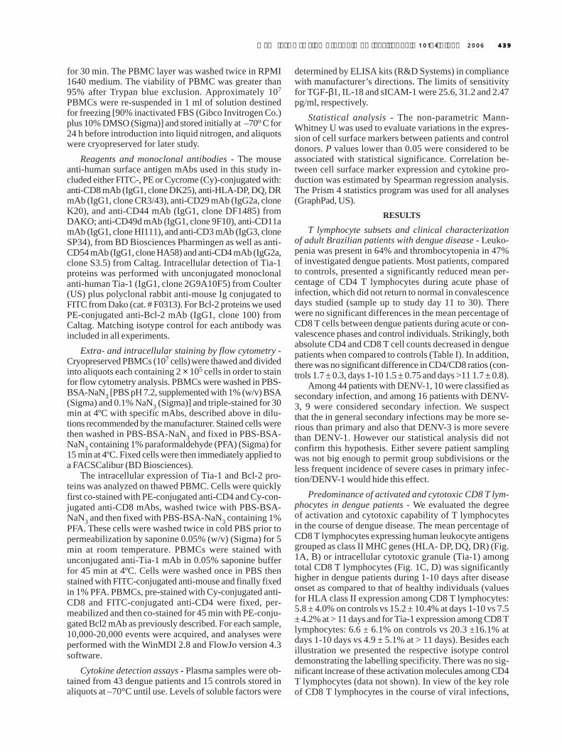
Adhesion molecules are up regulated mainly in den-gue patient CD8 T lymphocytes - We evaluated the ex-pression of molecules known to mediate adhesive inter-actions among lymphocytes, endothelial cells and matrix
proteins, including VLA-4 (CD49d/CD29), ICAM-1 (CD54),and LFA-1 (CD11a/CD18) (Fig. 2). Initially, we regardedthe co-expression of CD49d and CD29 (α4β1 integrin, VLA-4) on T lymphocytes from our study groups. There weresignificant increases in the mean percentage of CD4 andCD8 T lymphocytes expressing the VLA-4 and LFA-1 (val-ues for LFA-1 expression among CD4 T lymphocytes: 31,3
Fig. 2: CD8 T lymphocytes from dengue patients expressing adhesion molecules during acute phase of disease. Peripheral blood mono-nuclear cells from healthy subjects (n = 12) or for samples from dengue patients taken at different periods after disease onset: at acute (1-10 days, n = 26) and convalescent phase (after 11 days, n = 10) were labelled as described in Materials and Methods and analyzed by three-colour flow cytometry for particular adhesion molecule expression in CD4 and CD8 T cells. A: ex vivo co-expression of CD49d and CD29on CD8 T lymphocytes was illustrated from a representative healthy control and a dengue patient with acute infection. B: meanpercentages of CD49dCD29 (VLA-4) among CD4 and CD8 T lymphocytes; C: ICAM-1 or D: LFA-1 among CD8 T lymphocytes are showin dengue patients and controls. Statistical significance was assessed by the Mann-Whitney U test.Values for VLA-4 expression among CD4 T lymphocytes - Controls 41.3 ± 10%, dengue patients at 1-10 days 54 ± 11.2%, and patientsat > 11 days 45.6 ± 12.2% (controls vs acute dengue patients, p = 0.0136 in Mann-Whitney U test). Values for VLA-4 expression amongCD8 T lymphocytes: controls 42.7 ± 6.5, dengue patients at 1-10 days 61 ± 13.2% and patients at > 11 days 46.1 ± 12.1% (controls vsacute dengue patients, p = 0.0038 and acute vs convalescent dengue patients p = 0.0308).Values for ICAM-1 expression among CD8 T lymphocytes - Controls 18.8 ± 8.9, dengue patients at 1-10 days 29.8 ± 16.9%, and denguepatients at > 11 dpi 23.1± 13.2%.Values for LFA-1 expression among CD8 T lymphocytes - Controls 52.9 ± 17.2, dengue patients at 1-10 days 71.4 ± 16.3% and dengueconvalescent patients at > 11 days 61.7 ± 24.3% (controls vs acute dengue patients, p = 0.0045).
442442442442442 Activation of lymphocytes from dengue patients • Elzinandes L Azeredo et al.
± 4.7% on controls vs 43.4. ± 19.9% at days 1-10, p = 0.01)among T cells from dengue patients 1-10 days after dis-ease onset compared with controls (Fig. 2A, B, D). Al-though not significant, the mean percentage of CD8 Tcells expressing ICAM-1 among total CD8 T lymphocytes1-10 days after disease onset was higher than those ob-served in control individuals (controls 18 ± 8.9 vs denguepatients days 1-10 29.8 ± 16.9) (Fig. 2C). ICAM-1 in CD4 Tcells was not higher in dengue patients compared to thatof control individuals (controls 16.9 ± 11.1 vs dengue pa-tients days 1-10 19.8 ± 17.3).
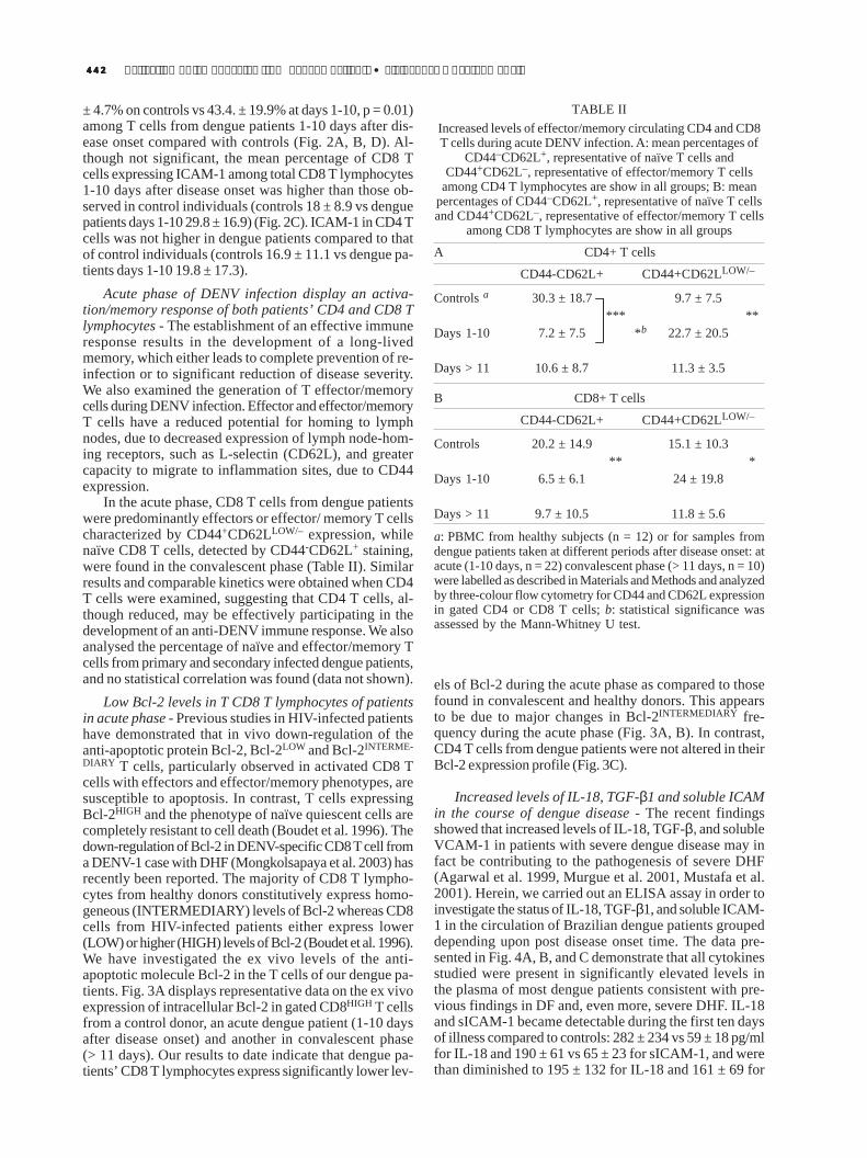
Acute phase of DENV infection display an activa-tion/memory response of both patients’ CD4 and CD8 Tlymphocytes - The establishment of an effective immuneresponse results in the development of a long-livedmemory, which either leads to complete prevention of re-infection or to significant reduction of disease severity.We also examined the generation of T effector/memorycells during DENV infection. Effector and effector/memoryT cells have a reduced potential for homing to lymphnodes, due to decreased expression of lymph node-hom-ing receptors, such as L-selectin (CD62L), and greatercapacity to migrate to inflammation sites, due to CD44expression.
In the acute phase, CD8 T cells from dengue patientswere predominantly effectors or effector/ memory T cellscharacterized by CD44+CD62LLOW/– expression, whilenaïve CD8 T cells, detected by CD44-CD62L+ staining,were found in the convalescent phase (Table II). Similarresults and comparable kinetics were obtained when CD4T cells were examined, suggesting that CD4 T cells, al-though reduced, may be effectively participating in thedevelopment of an anti-DENV immune response. We alsoanalysed the percentage of naïve and effector/memory Tcells from primary and secondary infected dengue patients,and no statistical correlation was found (data not shown).
Low Bcl-2 levels in T CD8 T lymphocytes of patientsin acute phase - Previous studies in HIV-infected patientshave demonstrated that in vivo down-regulation of theanti-apoptotic protein Bcl-2, Bcl-2LOW and Bcl-2INTERME-DIARY T cells, particularly observed in activated CD8 Tcells with effectors and effector/memory phenotypes, aresusceptible to apoptosis. In contrast, T cells expressingBcl-2HIGH and the phenotype of naïve quiescent cells arecompletely resistant to cell death (Boudet et al. 1996). Thedown-regulation of Bcl-2 in DENV-specific CD8 T cell froma DENV-1 case with DHF (Mongkolsapaya et al. 2003) hasrecently been reported. The majority of CD8 T lympho-cytes from healthy donors constitutively express homo-geneous (INTERMEDIARY) levels of Bcl-2 whereas CD8cells from HIV-infected patients either express lower(LOW) or higher (HIGH) levels of Bcl-2 (Boudet et al. 1996).We have investigated the ex vivo levels of the anti-apoptotic molecule Bcl-2 in the T cells of our dengue pa-tients. Fig. 3A displays representative data on the ex vivoexpression of intracellular Bcl-2 in gated CD8HIGH T cellsfrom a control donor, an acute dengue patient (1-10 daysafter disease onset) and another in convalescent phase(> 11 days). Our results to date indicate that dengue pa-tients’ CD8 T lymphocytes express significantly lower lev-
els of Bcl-2 during the acute phase as compared to thosefound in convalescent and healthy donors. This appearsto be due to major changes in Bcl-2INTERMEDIARY fre-quency during the acute phase (Fig. 3A, B). In contrast,CD4 T cells from dengue patients were not altered in theirBcl-2 expression profile (Fig. 3C).
Increased levels of IL-18, TGF-β1 and soluble ICAMin the course of dengue disease - The recent findingsshowed that increased levels of IL-18, TGF-β, and solubleVCAM-1 in patients with severe dengue disease may infact be contributing to the pathogenesis of severe DHF(Agarwal et al. 1999, Murgue et al. 2001, Mustafa et al.2001). Herein, we carried out an ELISA assay in order toinvestigate the status of IL-18, TGF-β1, and soluble ICAM-1 in the circulation of Brazilian dengue patients groupeddepending upon post disease onset time. The data pre-sented in Fig. 4A, B, and C demonstrate that all cytokinesstudied were present in significantly elevated levels inthe plasma of most dengue patients consistent with pre-vious findings in DF and, even more, severe DHF. IL-18and sICAM-1 became detectable during the first ten daysof illness compared to controls: 282 ± 234 vs 59 ± 18 pg/mlfor IL-18 and 190 ± 61 vs 65 ± 23 for sICAM-1, and werethan diminished to 195 ± 132 for IL-18 and 161 ± 69 for
TABLE IIIncreased levels of effector/memory circulating CD4 and CD8T cells during acute DENV infection. A: mean percentages of
CD44–CD62L+, representative of naïve T cells andCD44+CD62L–, representative of effector/memory T cells
among CD4 T lymphocytes are show in all groups; B: meanpercentages of CD44–CD62L+, representative of naïve T cellsand CD44+CD62L–, representative of effector/memory T cells
among CD8 T lymphocytes are show in all groups
A CD4+ T cells
CD44-CD62L+ CD44+CD62LLOW/–
Controls a 30.3 ± 18.7 9.7 ± 7.5*** **
Days 1-10 7.2 ± 7.5 *b 22.7 ± 20.5
Days > 11 10.6 ± 8.7 11.3 ± 3.5
B CD8+ T cells
CD44-CD62L+ CD44+CD62LLOW/–
Controls 20.2 ± 14.9 15.1 ± 10.3** *
Days 1-10 6.5 ± 6.1 24 ± 19.8
Days > 11 9.7 ± 10.5 11.8 ± 5.6
a: PBMC from healthy subjects (n = 12) or for samples fromdengue patients taken at different periods after disease onset: atacute (1-10 days, n = 22) convalescent phase (> 11 days, n = 10)were labelled as described in Materials and Methods and analyzedby three-colour flow cytometry for CD44 and CD62L expressionin gated CD4 or CD8 T cells; b: statistical significance wasassessed by the Mann-Whitney U test.

443443443443443Mem Inst Oswaldo Cruz, Rio de Janeiro, Vol. 101(4), June 2006
sICAM-1 on days > 11. The plasma levels of TGF-β1 in-creased in the acute phase and reached peak levels byday 11 onwards (controls 1888 ± 1044 pg/ml, 15107 ± 10187pg/ml on days 1-10 and 27486 ± 12700 pg/ml on days >11).
Correlation between IL-18 with soluble and surfaceICAM and percentage T cells expressing HLA-DR amongCD8 T lymphocytes - Interestingly, we observed a signifi-cant positive correlation between the level of IL-18 inplasma of the dengue patients and the CD8 T cells ex-pressing HLA class II. In addition, levels of IL-18 havebeen directly correlated with soluble ICAM-1 in plasma
and also with CD8 T cells expressing ICAM-1 (Table III).In fact, previous data demonstrated that IL-18 increasesthe expression of adhesion molecules ICAM-1 andVCAM-1 (Morel et al. 2001), which facilitate the emigra-tion of neutrophils and lymphocytes in containing a ni-dus of infection, thereby contributing to the primary pro-cess in inflammatory diseases.
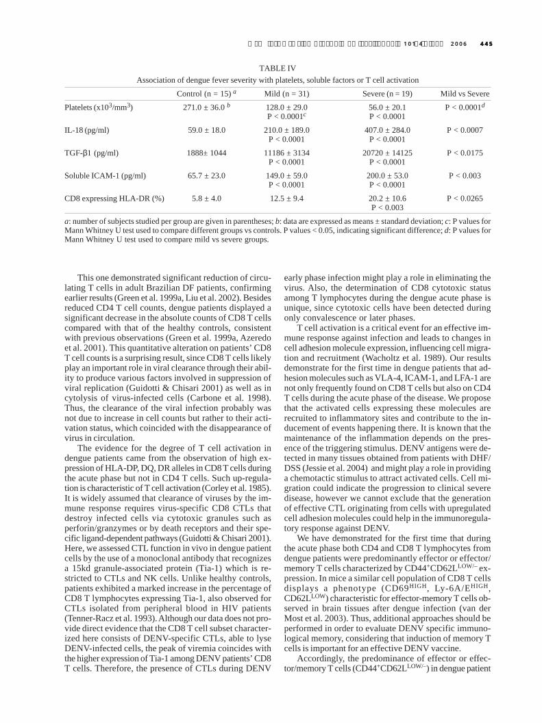
A significant increase of IL-18, TGF-b1, and sICAM-1 and activation cell markers on T lymphocytes in se-vere dengue compared to mild dengue patients - Basedon gravity classification, in severe dengue CD4+CD3+ andCD8+CD3+ relative rates and absolute cell counts are not
Fig. 3: down-regulation of Bcl-2 expression in CD8 T cells from dengue patients. A: ex vivo expression of Bcl-2 on CD8 T lymphocytesfrom a representative control and dengue patients during the acute and convalescent phases, respectively. Populations expressing low,intermediary, and high levels of Bcl-2 were determined based in previously report (Boudet et al. 1996). Bcl-2LOW, Bcl-2INTERMEDIARY
and Bcl-2HIGH percentages are indicated within CD8HIGH subset; B: data represent the CD8 T lymphocytes differential expressing levelsof Bcl-2 of PBMCs isolated from controls (n = 8) and from dengue patients taken at different periods after disease onset: at acute (1-10days n = 10) and convalescent phases (> 11 days n = 9); C: similar analysis was performed by CD4 T lymphocytes. Statistical significancewas assessed by the Mann-Whitney U test.Values for differential expression of Bcl-2 among CD8 T lymphocytes:Bcl-2LOW - Controls 14.0 ± 7.1%, dengue patients at acute phase 39.4 ± 18.8%, and at convalescent phase 24.9 ± 16.5% (controls vs acutedengue patients, p = 0.0199). Bcl-2NORMAL - Controls 76.0 ± 15.2%, dengue patients at acute phase 54.0 ± 17.9%, and at convalescentphase 66.5 ± 15.9 (controls vs acute patients, p = 0.0148). Bcl2HIGH - Controls 9.2 ± 10.1%, dengue patients at acute phase 6.5 ± 3.0%,and at convalescent phase 8.4 ± 2.4%.

444444444444444 Activation of lymphocytes from dengue patients • Elzinandes L Azeredo et al.
significantly affected as compared to mild dengue (forCD4 T cells, severe dengue 39 ± 13% and 290 ± 270 counts/mm3 vs mild dengue 36.9 ± 9.3% and 204 ± 125 counts/mm3 and for CD8 T cells, severe dengue 26.2 ± 9.4% and241.3 ± 286.8 counts/mm3 vs mild dengue 26.7 ± 8.3% and151.4 ± 107.4 counts/mm3). Absolute counts of controlswere higher for both CD4 and CD8 T cells than mild orsevere dengue.
In addition, we investigate whether soluble proteinsdetected in plasma by ELISA assay and expression of cellactivation and/or adhesion molecules on T lymphocytesdiffers between two dengue patients groups mild and se-vere in order to and predict gravity of dengue infectiondisease. Evaluating the whole dengue group we foundsignificantly increased levels of IL-18, TGF-β1, andsICAM-1, plasmatic in severe dengue as compared to milddengue (Table IV). Additionally, expression of HLA-DR,Tia-1 and ICAM-1 in CD8 T cells was higher in severedengue patients and even more in DHF individuals com-pared to mild dengue. Moreover, expression of LFA-1 andVLA-4 in CD4 T cells was higher in Severe Dengue pa-tients and even more in a DHF individual compared toMild dengue (Fig. 5). No differences were encountered
between the two groups of dengue in ICAM-1 and LFA-1-expressed by CD8 and CD4 T lymphocytes. Moreover,the majority of activation and cell adhesion moleculesevaluated were greatly increased in two only DHF patientevaluated.
DISCUSSION
During the last two decades, incidence of dengue inBrazil has been increasing gradually. In fact, extensiveepidemics of DENV 1 and DENV 2 emerged, and after theappearance of DENV 3, recognized in Rio de Janeiro dur-ing 2002, the disease became alarming (WHO 2002). It isimportant to mention that dengue incidence in Brazil oc-curs mainly in adults while in Asia it is predominantly apaediatric disease. The definition of dengue severity inLatin Americas has often been a matter of debate as se-vere cases did not satisfy WHO criteria for DHF/DSS.Mainly in Latin American adults, but also in patients fromAsian countries, this issue has become striking (Guzman& Kouri 2003, Phuong et al. 2004). Circulatory collapse isfrequently associated with dengue without trombocy-topenia or haemorrhagic manifestations. Concerning ourpatient cohort epidemics in Rio de Janeiro in 2002, mostsevere cases were associated with shock but without con-sistent haemorrhagic manifestations. Shock has been con-sidered as high risk even without trombocytopenia (Lumet al. 2002), as registered during several fatal cases in thissame Brazilian epidemics (unpublished observations).Harris et al. (2000) classified their patients in Nicaragua inaccordance with criteria that did not meet WHO defini-tions, considering that severe cases with signs of shockneed not include trombocytopenia or haemoconcen-tration. In this context, additional studies in well-charac-terized patient cohorts from different geographic regionsare essential to advance this research and guide new ap-proaches towards prevention and treatment of DHF(Rothman 2004).
Fig. 4: alterations in IL-18, TGF-β1 and sICAM-1 plasma levels during DENV infection. Significant increase in A: IL-18 levels in denguepatients are increased on acute phase (Mann-Whitney U test, p = 0.0001); B: high levels of sICAM-1 were detected during acute phase ofdisease (p = 0.0001). During convalescence phase of disease, sICAM-1 plasma levels were statistically significantly increased (p = 0.0003)when compared to controls; C: TGF-β1 plasma levels increased at acute phase, and reached peak levels by day 11 onwards. High levels ofTGF-β1 were detected during convalescence phase of disease (controls vs dengue patients p = 0.0001) and significantly higher whencompared with patients at acute phase (p = 0.0017).
TABLE IIIPositive correlation between IL-18 with percent of HLA-DP,
DQ, DR, and ICAM-1 among CD8 T lymphocytes andsoluble ICAM-1
Spearman rank correlation r P N
HLA class II among CD8 T cells 0.5140 0.0121 23sICAM-1 0.4993 0.0001 66ICAM-1 among CD8 T cells 0.7086 0.0001 25
N: number of samples studied per group.
445445445445445Mem Inst Oswaldo Cruz, Rio de Janeiro, Vol. 101(4), June 2006
TABLE IVAssociation of dengue fever severity with platelets, soluble factors or T cell activation
Control (n = 15) a Mild (n = 31) Severe (n = 19) Mild vs Severe
Platelets (x103/mm3) 271.0 ± 36.0 b 128.0 ± 29.0 56.0 ± 20.1 P < 0.0001d
P < 0.0001c P < 0.0001
IL-18 (pg/ml) 59.0 ± 18.0 210.0 ± 189.0 407.0 ± 284.0 P < 0.0007P < 0.0001 P < 0.0001
TGF-β1 (pg/ml) 1888± 1044 11186 ± 3134 20720 ± 14125 P < 0.0175P < 0.0001 P < 0.0001
Soluble ICAM-1 (pg/ml) 65.7 ± 23.0 149.0 ± 59.0 200.0 ± 53.0 P < 0.003P < 0.0001 P < 0.0001
CD8 expressing HLA-DR (%) 5.8 ± 4.0 12.5 ± 9.4 20.2 ± 10.6 P < 0.0265P < 0.003
a: number of subjects studied per group are given in parentheses; b: data are expressed as means ± standard deviation; c: P values forMann Whitney U test used to compare different groups vs controls. P values < 0.05, indicating significant difference; d: P values forMann Whitney U test used to compare mild vs severe groups.
This one demonstrated significant reduction of circu-lating T cells in adult Brazilian DF patients, confirmingearlier results (Green et al. 1999a, Liu et al. 2002). Besidesreduced CD4 T cell counts, dengue patients displayed asignificant decrease in the absolute counts of CD8 T cellscompared with that of the healthy controls, consistentwith previous observations (Green et al. 1999a, Azeredoet al. 2001). This quantitative alteration on patients’ CD8T cell counts is a surprising result, since CD8 T cells likelyplay an important role in viral clearance through their abil-ity to produce various factors involved in suppression ofviral replication (Guidotti & Chisari 2001) as well as incytolysis of virus-infected cells (Carbone et al. 1998).Thus, the clearance of the viral infection probably wasnot due to increase in cell counts but rather to their acti-vation status, which coincided with the disappearance ofvirus in circulation.
The evidence for the degree of T cell activation indengue patients came from the observation of high ex-pression of HLA-DP, DQ, DR alleles in CD8 T cells duringthe acute phase but not in CD4 T cells. Such up-regula-tion is characteristic of T cell activation (Corley et al. 1985).It is widely assumed that clearance of viruses by the im-mune response requires virus-specific CD8 CTLs thatdestroy infected cells via cytotoxic granules such asperforin/granzymes or by death receptors and their spe-cific ligand-dependent pathways (Guidotti & Chisari 2001).Here, we assessed CTL function in vivo in dengue patientcells by the use of a monoclonal antibody that recognizesa 15kd granule-associated protein (Tia-1) which is re-stricted to CTLs and NK cells. Unlike healthy controls,patients exhibited a marked increase in the percentage ofCD8 T lymphocytes expressing Tia-1, also observed forCTLs isolated from peripheral blood in HIV patients(Tenner-Racz et al. 1993). Although our data does not pro-vide direct evidence that the CD8 T cell subset character-ized here consists of DENV-specific CTLs, able to lyseDENV-infected cells, the peak of viremia coincides withthe higher expression of Tia-1 among DENV patients’ CD8T cells. Therefore, the presence of CTLs during DENV
early phase infection might play a role in eliminating thevirus. Also, the determination of CD8 cytotoxic statusamong T lymphocytes during the dengue acute phase isunique, since cytotoxic cells have been detected duringonly convalescence or later phases.
T cell activation is a critical event for an effective im-mune response against infection and leads to changes incell adhesion molecule expression, influencing cell migra-tion and recruitment (Wacholtz et al. 1989). Our resultsdemonstrate for the first time in dengue patients that ad-hesion molecules such as VLA-4, ICAM-1, and LFA-1 arenot only frequently found on CD8 T cells but also on CD4T cells during the acute phase of the disease. We proposethat the activated cells expressing these molecules arerecruited to inflammatory sites and contribute to the in-ducement of events happening there. It is known that themaintenance of the inflammation depends on the pres-ence of the triggering stimulus. DENV antigens were de-tected in many tissues obtained from patients with DHF/DSS (Jessie et al. 2004) and might play a role in providinga chemotactic stimulus to attract activated cells. Cell mi-gration could indicate the progression to clinical severedisease, however we cannot exclude that the generationof effective CTL originating from cells with upregulatedcell adhesion molecules could help in the immunoregula-tory response against DENV.
We have demonstrated for the first time that duringthe acute phase both CD4 and CD8 T lymphocytes fromdengue patients were predominantly effector or effector/memory T cells characterized by CD44+CD62LLOW/– ex-pression. In mice a similar cell population of CD8 T cellsdisplays a phenotype (CD69HIGH, Ly-6A/EHIGH,CD62LLOW) characteristic for effector-memory T cells ob-served in brain tissues after dengue infection (van derMost et al. 2003). Thus, additional approaches should beperformed in order to evaluate DENV specific immuno-logical memory, considering that induction of memory Tcells is important for an effective DENV vaccine.
Accordingly, the predominance of effector or effec-tor/memory T cells (CD44+CD62LLOW/–) in dengue patient

446446446446446 Activation of lymphocytes from dengue patients • Elzinandes L Azeredo et al.
Fig. 5: representative dot plots demonstrating A: CD8 cell frequencies expressing HLA-DP, DQ, DR, Tia-1, ICAM-1; B: CD4 cellfrequencies expressing LFA-1 and co-CD49d/CD29 for each individual divided among mild DF, severe DF and DHF.
T lymphocytes studied here was paralleled by significantincrease of Bcl-2LOW CD8 T lymphocytes during the acutephase. Mongkolsapaya et al. (2003) described low levelsof Bcl-2 expression and a TUNEL positive reaction (amarker for DNA fragmentation) in DENV-specific CD8 cellsduring the acute phase in Thai patients. These observa-tions led us to propose that in dengue patients, as in HIV+
patients, there is a relationship between T cell suscepti-bility to apoptosis and the activation state. In convales-cence, the percentage of effector/memory T cells is re-
established, and Bcl-2LOW CD8 T lymphocytes no longerpredominate when viral clearance is achieved (> 11 daysof infection). In consideration, the lack of physiologicalprotection by Bcl-2 may contribute in vivo with alterationsin T cell homeostasis during DENV infection and/or mayconstitute an example of primary control of immune re-sponse.
In a cohort of dengue patients studied, we observedthat in contrast to IL-18 and sICAM-1, which displayedhigh levels during the early acute phase, TGF-β1 was con-
447447447447447Mem Inst Oswaldo Cruz, Rio de Janeiro, Vol. 101(4), June 2006
siderably more concentrated during the late phases ofdisease, especially in the convalescence phase. It has beenestablished that soluble factors as chemokines andcytokines act through specific receptors on immune cellsto activate and mobilize the response to infection (Hartyet al. 2000, Guidotti & Chisari 2001). However, cytokinesexert a double-edge sword function: they are essential forthe appropriate function of the immune system but arealso potentially toxic mediators of immunopathology un-der conditions of excess or deregulated production (Slifka& Whitton 2000). In this context, IL-18 has been reportedto be an IFN-γ inducer possibly being produced after NKactivation and contributing to induce an efficient innateimmunity, therefore perhaps stimulating CTL activity inorder to achieve viral clearance (Fujioka et al. 1999). Onthe other hand, IL-18 was associated with pathogenesisin autoimmunity, arthritis, and sepsis, probably by an in-direct pathway by inducement of proinflammatorycytokine synthesis (IL-1 and TNF-α) and chemokines (IL-8 and MIP-1α) (Dinarello & Fantuzzi 2003).
Moreover, circulating forms of ICAM-1 were detectedin plasma of our dengue patients, as soluble VCAM-1was encountered in the plasma of children hospitalizedfor dengue in French Polynesia (Murgue et al. 2001). Cir-culating forms of adhesion molecules are generally thoughtto be released from the cell surface by secretion or pro-teolytic cleavage. They are naturally found in serum ofhealthy individuals but reach elevated levels in variousdiseases, such as infections by Plasmodium falciparum,Schistosoma mansoni, HCV and HIV (Jakobsen et al. 1994,Nordoy et al. 1996, Kaplanski et al. 1997).
TGF-β has been reported to inhibit many T cell func-tions, including proliferation and development of cyto-toxic cells. However, under certain circumstances, TGF-βhas also been proved to stimulate T cells, partly by pre-venting apoptosis and also inducing T cell proliferation(Lee & Rich 1993, Cerwenka et al. 1996). The up-regula-tion of TGF-β1, in late stages of DENV infection mightsuggest that TGF-β activity may suppress the inflamma-tory response after elimination of DENV and acting in ananti-inflammatory manner in the late steps, prevent pos-sible harm to host by prolonged inflammation.
In accordance with previous studies, when we classi-fied our group of dengue patients based on degree ofseverity, high levels of IL-18, soluble ICAM-1, and TGF-β1 were frequently correlated with severe dengue cases.Feasibly elevated levels of soluble ICAM-1 in the plasmaof dengue patients might mediate endothelial cell activa-tion and consequently, the plasma leakage phenomenonobserved during dengue disease, one of the major hall-marks distinguishing DHF from DF. In fact, it was demon-strated that culture supernatants from DENV infected pri-mary human monocytes could activate endothelial cellsby eliciting expression of VCAM-1 and ICAM-1 in thesecells (Anderson et al. 1997). Interestingly, the IL-18 plasmalevels were positively correlated with sICAM-1 levels to-gether with the expression of CD54 on T CD8 cells, asdescribed previously that IL-18 increases the expressionof ICAM-1 (Morel et al. 2001) and VCAM-1 (Vidal-Vana-clocha et al. 2000), enhancing the recruitment of lympho-cytes into inflammatory tissues. Furthermore, our study
revealed that CD8+ T cells expressing HLA-DR were sig-nificantly present in higher frequency in Severe Den-gue cases. Although CD8+ T-cell activation might be ex-pected to have an association with a better clinical out-come during viral infections, in long-term HIV-1 infectionhigh levels of cell surface expression of CD38 on CD8 T-cell are instead linked with faster disease progression, inpart independent of the predictive value of plasma viralburden and CD4+ T-cell number (Liu et al. 1998). Morerecently, a low CD8+HLA-DR+ cell percentage has beenconsidered a significant predictor of immunological long-term HIV disease nonprogressor (ILNTP) status (Paul etal. 2005).
Recently, the phenomenon of “original antigenic sin”,first described for B cell responses against influenza vi-rus subtypes (Fazekas de St & Webster 1966), was ob-served in dengue infection. Infection with a DENV sero-type generated by CD8 T cells with a higher affinity to asecond and presumably previously encountered DENVserotype, leads to the belief that cross-reactive memoryCD8 T cells had preferentially expanded over T cells morespecific to the serotype causing infection (Mongkolsapayaet al. 2003).
Under the study conditions, immune status activationwas found significantly higher in DF patients comparedwith healthy controls and may allow early assessment ofthe severity of DF. In fact, the pathophysiological eventsof dengue disease is frequently associated with high de-gree of T cell activation, but we cannot exclude a possibil-ity that these activated CD8 T cells identified in DF pa-tients act on infected cells and efficiently clear the virus.The reason why T-cell activation is associated with pooroutcome in DENV disease remains unknown. Further ad-ditional factors such as immunological exhaustion, hypo-responsiveness of T cells to their cognate antigens andalterations in the T-cell receptor repertoire must be estab-lished in order to evaluate how T-cell activation could beinfluencing the severity of dengue infection.
ACKNOWLEDGMENTS
To Dr Marize Miagostovich and Ms Eliana Saraiva for thehelp during laboratorial diagnosis and the technical supportfrom Ms Mariana Lopes and Ms Maryrose Lavatori. To DrJoseli Lannes-Vieira, Department of Immunology, Fiocruz, andDr Ricardo Galler for critically reviewing the manuscript.
REFERENCES
Agarwal R, Elbishbishi EA, Chaturvedi UC, Nagar R, MustafaAS 1999. Profile of transforming growth factor-beta 1 inpatients with dengue haemorrhagic fever. Int J Exp Pathol80: 143-149.
Anderson R, Wang S, Osiowy C, Issekutz AC 1997. Activa-tion of endothelial cells via antibody-enhanced dengue vi-rus infection of peripheral blood monocytes. J Virol 71:4226-4232.
Azeredo EL, De Oliveira-Pinto LM, Zagne SM, Cerqueira DI,Nogueira RM, Kubelka CF 2006. NK cells, displaying earlyactivation, cytotoxicity and adhesion molecules, are associ-ated with mild dengue disease. Clin Exp Immunol 14: 345-356.
Balmaseda A, Hammond SN, Perez MA, Cuadra R, Solano S,
448448448448448 Activation of lymphocytes from dengue patients • Elzinandes L Azeredo et al.
Rocha J, Idiaquez W, Harris E 2005. Assessment of theWorld Health Organization Scheme for Classification ofDengue Severity In Nicaragua. Am J Trop Med Hyg 73:1059-1062.
Boudet F, Lecoeur H, Gougeon ML 1996. Apoptosis associ-ated with ex vivo down-regulation of Bcl-2 and up-regula-tion of Fas in potential cytotoxic CD8+ T lymphocytesduring HIV infection. J Immunol 156: 2282-2293.
Carbone FR, Kurts C, Bennett SR, Miller JF, Heath WR 1998.Cross-presentation: a general mechanism for CTL immu-nity and tolerance. Immunol Today 19: 368-373.
Cerwenka A, Kovar H, Majdic O, Holter W 1996. Fas- andactivation-induced apoptosis are reduced in human T cellspreactivated in the presence of TGF-beta 1. J Immunol156: 459-464.
Chaturvedi UC, Elbishbishi EA, Agarwal R, Raghupathy R,Nagar R, Tandon R, Pacsa AS, Younis OI, Azizieh F 1999.Sequential production of cytokines by dengue virus-infectedhuman peripheral blood leukocyte cultures. J Med Virol59: 335-340.
Chen LC, Lei HY, Liu CC, Shiesh SC, Chen SH, Liu HS, Lin YS,Wang ST, Shyu H W, Yeh TM 2006. Correlation of serumlevels of macrophage migration inhibitory factor with dis-ease severity and clinical outcome in dengue patients. Am JTrop Med Hyg 74: 142-147.
Corley RB, LoCascio NJ, Ovnic M, Haughton G 1985. Twoseparate functions of class II (Ia) molecules: T-cell stimula-tion and B-cell excitation. Proc Natl Acad Sci USA 82: 516-520.
Dinarello CA, Fantuzzi G 2003. Interleukin-18 and host de-fense against infection. J Infect Dis 187 (Suppl. 2): S370-384.
Fazekas de St G, Webster RG 1966. Disquisitions of originalantigenic sin. I. Evidence in man. J Exp Med 124: 331-345.
Fujioka N, Akazawa R, Ohashi K, Fujii M, Ikeda M, KurimotoM 1999. Interleukin-18 protects mice against acute herpessimplex virus type 1 infection. J Virol 73: 2401-2409.
Green S, Pichyangkul S, Vaughn DW, Kalayanarooj S,Nimmannitya S, Nisalak A, Kurane I, Rothman AL, EnnisFA 1999a. Early CD69 expression on peripheral blood lym-phocytes from children with dengue hemorrhagic fever.J Infect Dis 180: 1429-35.
Green S, Vaughn DW, Kalayanarooj S, Nimmannitya S,Suntayakorn S, Nisalak A, Lew R, Innis BL, Kurane I,Rothman AL, Ennis FA 1999b. Early immune activation inacute dengue illness is related to development of plasmaleakage and disease severity. J Infect Dis 179: 755-762.
Guidotti LG, Chisari FV 2001. Noncytolytic control of viralinfections by the innate and adaptive immune response.Annu Rev Immunol 19: 65-91.
Guzman MG, Kouri G 2003. Dengue and dengue hemorrhagicfever in the Americas: lessons and challenges. J Clin Virol27: 1-13.
Harris E, Videa E, Perez L, Sandoval E, Tellez Y, Perez ML,Cuadra R, Rocha J, Idiaquez W, Alonso RE, Delgado MA,Campo LA, Acevedo F, Gonzalez A, Amador JJ, BalmasedaA 2000. Clinical, epidemiologic, and virologic features ofdengue in the 1998 epidemic in Nicaragua. Am J Trop MedHyg 63: 5-11.
Harty JT, Tvinnereim AR, White DW 2000. CD8+ T cell effec-tor mechanisms in resistance to infection. Annu Rev Immunol18: 275-308.
Iyngkaran N, Yadav M, Sinniah M 1995. Augmented inflam-matory cytokines in primary dengue infection progressingto shock. Singapore Med J 36: 218-221.
Jakobsen PH, Morris-Jones S, Ronn A, Hviid L, Theander TG,Elhassan IM, Bygbjerg IC, Greenwood BM 1994. Increasedplasma concentrations of sICAM-1, sVCAM-1 andsELAM-1 in patients with Plasmodium falciparum or P.vivax malaria and association with disease severity. Immu-nology 83: 665-669.
Jessie K, Fong MY, Devi S, Lam SK, Wong KT 2004. Localiza-tion of dengue virus in naturally infected human tissues, byimmunohistochemistry and in situ hybridization. J InfectDis 189: 1411-1418.
Kaplanski G, Farnarier C, Payan MJ, Bongrand P, Durand JM1997. Increased levels of soluble adhesion molecules in theserum of patients with hepatitis C. Correlation with cytokineconcentrations and liver inflammation and fibrosis. DigDis Sci 42: 2277-84.
Lee HM, Rich S 1993. Differential activation of CD8+ T cellsby transforming growth factor-beta 1. J Immunol 151: 668-677.
Lin CF, Chiu SC, Hsiao YL, Wan SW, Lei HY, Shiau AL, Liu HS,Yeh TM, Chen S H, Liu CC, Lin YS 2005. Expression ofcytokine, chemokine, and adhesion molecules during endot-helial cell activation induced by antibodies against denguevirus nonstructural protein 1. J Immunol 174: 395-403.
Liu CC, Huang KJ, Lin YS, Yeh TM, Liu HS, Lei HY 2002.Transient CD4/CD8 ratio inversion and aberrant immuneactivation during dengue virus infection. J Med Virol 68:241-252.
Liu Z, Cumberland WG, Hultin LE, Kaplan AH, Detels R, GiorgiJV 1998. CD8+ T-lymphocyte activation in HIV-1 diseasereflects an aspect of pathogenesis distinct from viral bur-den and immunodeficiency. J Acquir Immune Defic SyndrHum Retrovirol 18: 332-340.
Lum LC, Goh AY, Chan PW, El-Amin AL, Lam SK 2002. Riskfactors for hemorrhage in severe dengue infections. J Pediatr140: 629-631.
Mathew A, Kurane I, Green S, Vaughn DW, Kalayanarooj S,Suntayakorn S, Ennis F A, Rothman AL 1999. Impaired Tcell proliferation in acute dengue infection. J Immunol 162:5609-5615.
Mongkolsapaya J, Dejnirattisai W, Xu XN, Vasanawathana S,Tangthawornchaikul N, Chairunsri, A, Sawasdivorn, S,Duangchinda, T, Dong, T, Rowland-Jones, S, Yenchit-somanus PT, McMichael A, Malasit P, Screaton G 2003.Original antigenic sin and apoptosis in the pathogenesis ofdengue hemorrhagic fever. Nat Med 9: 921-927.
Morel JC, Park CC, Woods JM, Koch AE 2001. A novel rolefor interleukin-18 in adhesion molecule induction throughNF kappa B and phosphatidylinositol (PI) 3-kinase-de-pendent signal transduction pathways. J Biol Chem 276:37069-37075.
Murgue, B, Cassar, O, Deparis, X 2001. Plasma concentrationsof sVCAM-1 and severity of dengue infections. J Med Virol65: 97-104.
Mustafa AS, Elbishbishi EA, Agarwal R, Chaturvedi UC 2001.
449449449449449Mem Inst Oswaldo Cruz, Rio de Janeiro, Vol. 101(4), June 2006
Elevated levels of interleukin-13 and IL-18 in patients withdengue hemorrhagic fever. FEMS Immunol Med Microbiol30: 229-233.
Neves-Souza PC, Azeredo EL, Zagne SM, Valls-de-Souza R,Reis SR, Cerqueira DI, Nogueira RM, Kubelka CF 2005.Inducible nitric oxide synthase (iNOS) expression in mono-cytes during acute Dengue Fever in patients and during invitro infection. BMC Infect Dis 5: 64.
Nordoy I, Aukrust P, Muller F, Froland SS 1996. Abnormallevels of circulating adhesion molecules in HIV-1 infectionwith characteristic alterations in opportunistic infections.Clin Immunol Immunopathol 81: 16-21.
Paul ME, Mao C, Charurat M, Serchuck L, Foca M, Hayani K,Handelsman EL, Diaz C, McIntosh K, Shearer WT 2005.Predictors of immunologic long-term nonprogression in HIV-infected children: implications for initiating therapy. J Al-lergy Clin Immunol 115: 848-855.
Phuong CX, Nhan NT, Kneen R, Thuy PT, van Thien C, NgaNT, Thuy TT, Solomon T, Stepniewska K, Wills B 2004.Clinical diagnosis and assessment of severity of confirmeddengue infections in Vietnamese children: is the World HealthOrganization classification system helpful? Am J Trop MedHyg 70: 172-179.
Rothman AL 2004. Dengue: defining protective versus patho-logic immunity. J Clin Invest 113: 946-951.
Slifka MK, Whitton JL 2000. Clinical implications of dysregu-lated cytokine production. J Mol Med 78: 74-80.
Tenner-Racz K, Racz P, Thome C, Meye, CG, Anderson PJ,Schlossman SF, Letvin N L 1993. Cytotoxic effector cellgranules recognized by the monoclonal antibody TIA-1 arepresent in CD8+ lymphocytes in lymph nodes of human
immunodeficiency virus-1-infected patients. Am J Pathol142: 1750-1758.
Thein S, Aung MM, Shwe TN, Aye M, Zaw A, Aye K, AyeKM, Aaskov J 1997. Risk factors in dengue shock syn-drome. Am J Trop Med Hyg 56: 566-572.
van der Most RG, Murali-Krishna K, Ahmed R 2003. Pro-longed presence of effector-memory CD8 T cells in thecentral nervous system after dengue virus encephalitis.Int Immunol 15: 119-125.
Vaughn DW, Green S, Kalayanarooj S, Innis BL, NimmannityaS, Suntayakorn S, Endy TP, Raengsakulrach B, RothmanAL, Ennis FA, Nisalak A 2000. Dengue viremia titer, anti-body response pattern, and virus serotype correlate withdisease severity. J Infect Dis 181: 2-9.
Vidal-Vanaclocha F, Fantuzzi G, Mendoza L, Fuentes AM,Anasagasti MJ, Martin J, Carrascal T, Walsh P, ReznikovLL, Kim SH, Novick D, Rubinstein M, Dinarello CA 2000.IL-18 regulates IL-1beta-dependent hepatic melanoma me-tastasis via vascular cell adhesion molecule-1. Proc NatlAcad Sci USA 97: 734-739.
Wacholtz MC, Patel SS, Lipsky PE 1989. Leukocyte function-associated antigen 1 is an activation molecule for human Tcells. J Exp Med 170: 431-48.
Wilder-Smith A, Schwartz E 2005. Dengue in travelers. N EnglJ Med 353: 924-932.
WHO-World Health Organization 2002. Communicable Dis-ease Surveillance and Response (CSR) Disease OutbreaksReported 8 May Dengue/Dengue Haemorrhagic Fever inBrazil - Update 2 http://www.who.int/disease-outbreak-news/n2002/may/8may2002.html.
