activation and regulation of systemic inflammation in ards ... · inflammation in ards ... nf...
TRANSCRIPT

DOI 10.1378/chest.08-2408 2009;136;1631-1643; Prepublished online October 3, 2009;Chest
and Scott E. SinclairG. Umberto Meduri, Djillali Annane, George P. Chrousos, Paul E. Marik Prolonged Glucocorticoid TherapyInflammation in ARDS : Rationale for Activation and Regulation of Systemic
http://chestjournal.chestpubs.org/content/136/6/1631.full.html
services can be found online on the World Wide Web at: The online version of this article, along with updated information and
ISSN:0012-3692)http://chestjournal.chestpubs.org/site/misc/reprints.xhtml(
of the copyright holder.may be reproduced or distributed without the prior written permission Northbrook, IL 60062. All rights reserved. No part of this article or PDFby the American College of Chest Physicians, 3300 Dundee Road,
2009Physicians. It has been published monthly since 1935. Copyright is the official journal of the American College of ChestCHEST
© 2009 American College of Chest Physicians by David Wallace on June 21, 2010chestjournal.chestpubs.orgDownloaded from

Activation and Regulation of SystemicInflammation in ARDSRationale for Prolonged Glucocorticoid Therapy
G. Umberto Meduri, MD, FCCP; Djillali Annane, MD, PhD;George P. Chrousos, MD; Paul E. Marik, MD, FCCP;and Scott E. Sinclair, MD, FCCP
Experimental and clinical evidence has demonstrated a strong cause-and-effect relationshipbetween persistence vs reduction in systemic inflammation and progression (unresolving) vsresolution (resolving) of ARDS. In this review, the cellular mechanisms involved in activating andregulating inflammation are contrasted between patients with resolving and unresolving ARDS.At the cellular level, patients with unresolving ARDS have deficient glucocorticoid (GC)-mediated down-regulation of inflammatory cytokine and chemokine transcription despite ele-vated levels of circulating cortisol, a condition defined as systemic inflammation-associatedacquired GC resistance. These patients, contrary to those with resolving ARDS, have persistentelevation in levels of both systemic and BAL fluid inflammatory cytokines and chemokines,markers of alveolar-capillary membrane permeability and fibrogenesis. At the tissue level, thecontinued production of inflammatory mediators leads to tissue injury, intravascular andextravascular coagulation, and the proliferation of mesenchymal cells, all resulting in maladap-tive lung repair and progression of extrapulmonary organ dysfunction. In ARDS, down-regulation of systemic inflammation is essential to restoring homeostasis, decreasing morbidity,and improving survival. Prolonged low-to-moderate dose GC therapy promotes the down-regulation of inflammatory cytokine transcription at the cellular level. Eight controlled studieshave consistently reported a significant reduction in markers of systemic inflammation, pulmo-nary and extrapulmonary organ dysfunction scores, duration of mechanical ventilation, and ICUlength of stay. In the aggregate (n � 628), reduction in mortality was substantial for all patients(relative risk [RR], 0.75; 95% CI, 0.63 to 0.89; p < 0.001; I2, 43%) and for those treated before day14 (RR, 0.71; 95% CI, 0.59 to 0.85; p < 0.001; I2, 40%). (CHEST 2009; 136:1631–1643)
Abbreviations: ACM � alveolar-capillary membrane; ACTH � adrenocorticotropic hormone; ALI � acute lung injury;GC � glucocorticoid; GC-GR� � glucocorticoid-activated-glucocorticoid receptor � complex; GR� � glucocorticoid recep-tor �; HDR � host defense response; HPA � hypothalamic-pituitary-adrenal; IL � interleukin; LIS � lung injury score;MODS � multiple-organ dysfunction syndrome; NF � nuclear factor; PBL � peripheral blood leukocyte; PEEP �positive end-expiratory pressure; PGCT � prolonged glucocorticoid treatment; RR � relative risk; TNF � tumornecrosis factor
I n this review, we examine the cellular mechanismsinvolved in activating and regulating inflammation
to provide a pathophysiologic rationale for low-to-moderate dose prolonged glucocorticoid treatment(PGCT) in patients with acute lung injury (ALI) andARDS. Current understanding places dysregulatedsystemic inflammation, with its persistent elevationof circulating inflammatory cytokines and chemo-kines over time, as the central pathogenetic processfor the dysfunction and failure of vital organs, theleading cause of short-term and long-term morbidity
and mortality in patients with ARDS.1–10 Longitudi-nal measurements of inflammatory cytokine levelshave shown that systemic and pulmonary inflamma-tion persists for several weeks and extends wellbeyond the clinical resolution of respiratory failureand extubation.1,7,9,11–15 A strong cause-and-effectrelationship between persistence vs reduction insystemic inflammation and progression vs resolu-tion of ARDS was provided by comparing longitu-dinal intracellular and extracellular measurements ofinflammation in improvers vs nonimprovers before
CHEST Special Feature
www.chestjournal.org CHEST / 136 / 6 / DECEMBER, 2009 1631
© 2009 American College of Chest Physicians by David Wallace on June 21, 2010chestjournal.chestpubs.orgDownloaded from
and after blind randomization to PGCT vs plac-ebo.7,9,13–15 These findings demonstrate the follow-ing: (1) down-regulation of systemic inflammation isstrongly associated with restoration of homeostasis,reduction in morbidity, and improved survival; and(2) the glucocorticoid (GC)-activated GC receptor isa major regulator of inflammation the antiinflamma-tory activity of which can be significantly enhancedwith prolonged GC administration.
For the purpose of this review, we conducted anelectronic search through Medline for the period1966 to October 2008. The search was restricted tostudies in adults and used the search terms “ARDS,”“adult respiratory distress syndrome,” “acute respi-ratory distress syndrome,” “noncardiogenic pulmo-nary edema,” “respiratory insufficiency,” “respiratoryfailure,” “acute lung injury” AND “inflammation,”“cytokines,” “chemokines,” “nuclear factor-kB,” “glu-cocorticoid receptor,” “sepsis,” and “systemic inflam-matory response.” The search for controlled trialsinvestigating PGCT in ALI-ARDS was previouslydescribed.16,17
Systemic Inflammation and Tissue HostDefense Response
Systemic inflammation is a highly organized re-sponse to infectious and noninfectious threats tohomeostasis that includes the activation of at leastthe following five major programs: (1) tissue hostdefense response (HDR),18; (2) acute-phase reaction;(3) sickness syndrome (including sickness behavior)19;(4) pain program mediated by the afferent sensory andautonomic systems; and (5) the stress program medi-ated by the hypothalamic-pituitary-adrenal (HPA) axisand the locus ceruleus-norepinephrine/sympatheticnervous system.20 The main effectors of systemicinflammation are inflammatory cytokines, such astumor necrosis factor (TNF)-�, interleukin (IL)-1�,
and IL-6; chemokines and other mediators of inflam-mation; the acute-phase reactants, mostly of hepaticorigin, such as C-reactive protein (CRP), fibrinogen,and plasminogen activator inhibitor-1; and the effec-tors of the sensory afferent system, such as substanceP, and of the stress system, such as hypothalamiccorticotropin-releasing hormone and vasopressin,cortisol, the catecholamines norepinephrine and epi-nephrine, and peripheral neuronal corticotropin-releasing hormone (reviewed in Elenkov et al20).Excessive release of inflammatory mediators into thecirculation induces tissue changes in vital organsleading to multiple-organ dysfunction syndrome(MODS).21,22
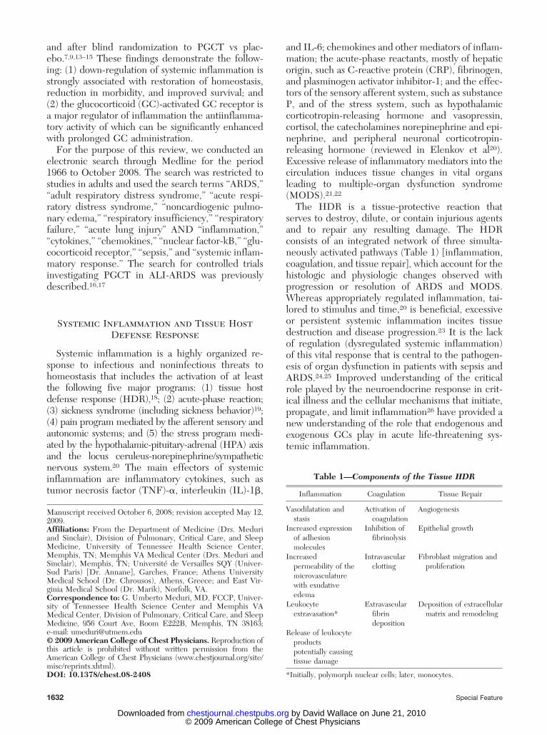
The HDR is a tissue-protective reaction thatserves to destroy, dilute, or contain injurious agentsand to repair any resulting damage. The HDRconsists of an integrated network of three simulta-neously activated pathways (Table 1) [inflammation,coagulation, and tissue repair], which account for thehistologic and physiologic changes observed withprogression or resolution of ARDS and MODS.Whereas appropriately regulated inflammation, tai-lored to stimulus and time,20 is beneficial, excessiveor persistent systemic inflammation incites tissuedestruction and disease progression.23 It is the lackof regulation (dysregulated systemic inflammation)of this vital response that is central to the pathogen-esis of organ dysfunction in patients with sepsis andARDS.24,25 Improved understanding of the criticalrole played by the neuroendocrine response in crit-ical illness and the cellular mechanisms that initiate,propagate, and limit inflammation26 have provided anew understanding of the role that endogenous andexogenous GCs play in acute life-threatening sys-temic inflammation.
Manuscript received October 6, 2008; revision accepted May 12,2009.Affiliations: From the Department of Medicine (Drs. Meduriand Sinclair), Division of Pulmonary, Critical Care, and SleepMedicine, University of Tennessee Health Science Center,Memphis, TN; Memphis VA Medical Center (Drs. Meduri andSinclair), Memphis, TN; Universite de Versailles SQY (Univer-Sud Paris) [Dr. Annane], Garches, France; Athens UniversityMedical School (Dr. Chrousos), Athens, Greece; and East Vir-ginia Medical School (Dr. Marik), Norfolk, VA.Correspondence to: G. Umberto Meduri, MD, FCCP, Univer-sity of Tennessee Health Science Center and Memphis VAMedical Center, Division of Pulmonary, Critical Care, and SleepMedicine, 956 Court Ave, Room E222B, Memphis, TN 38163;e-mail: [email protected]© 2009 American College of Chest Physicians. Reproduction ofthis article is prohibited without written permission from theAmerican College of Chest Physicians (www.chestjournal.org/site/misc/reprints.xhtml).DOI: 10.1378/chest.08-2408
Table 1—Components of the Tissue HDR
Inflammation Coagulation Tissue Repair
Vasodilatation andstasis
Activation ofcoagulation
Angiogenesis
Increased expressionof adhesionmolecules
Inhibition offibrinolysis
Epithelial growth
Increasedpermeability of themicrovasculaturewith exudativeedema
Intravascularclotting
Fibroblast migration andproliferation
Leukocyteextravasation*
Extravascularfibrindeposition
Deposition of extracellularmatrix and remodeling
Release of leukocyteproductspotentially causingtissue damage
*Initially, polymorph nuclear cells; later, monocytes.
1632 Special Feature
© 2009 American College of Chest Physicians by David Wallace on June 21, 2010chestjournal.chestpubs.orgDownloaded from

Progression of ARDS: Resolving vsUnresolving
ARDS is a disease of multifactorial etiology that ischaracterized by a specific morphologic lesiontermed diffuse alveolar damage.27 ARDS developsrapidly, in most patients within 12 to 48 h ofexposure to infectious or noninfectious insults thatcan affect the lung directly (via the alveolar compart-ment) or indirectly (via the vascular compartment).28
At presentation, (early) ARDS manifests with se-vere, diffuse, and spatially inhomogeneous HDRof the pulmonary lobules, leading to a breakdownin the barrier integrity and gas exchange functionof the lung. Every anatomical component of thepulmonary lobule (epithelium, endothelium, andinterstitium) is involved, including the respiratorybronchioles, alveolar ducts, and alveoli, as well asarteries and veins. Diffuse injury to the alveolar-capillary membrane (ACM) causes edema of theairspaces and interstitium with a protein-rich neu-trophilic exudate, resulting in severe gas exchangeand lung compliance abnormalities.29 Although theterm “syndrome” was applied in its original descrip-tion,30 ARDS meets all the constitutive elements of adisease process.31 Translational clinical research hasconstructed, through a holistic level of inquiry, apathophysiologic model of ARDS that fits pathogen-esis (biology) with morphologic (pathology) and clin-ical (physiology) findings observed during the longi-tudinal course of the disease.31
The lung injury score (LIS) quantifies the impairedrespiratory physiology in ARDS patients through theuse of a 4-point score that is based on the level ofpositive end-expiratory pressure (PEEP), the Pao2/Fio2 ratio, the quasistatic lung compliance, and thedegree of infiltration seen on a chest radiograph (1point per quadrant of chest radiograph involved).32
Based on simple physiologic criteria, the evolution ofARDS can be divided into resolving and unresolvingbased on achieving a 1-point reduction in LIS by day7 (Table 2). Even though, at the onset of ARDS, thetwo groups may appear similar, daily measurementof LIS, MODS score, and CRP levels allow earlyidentification of nonimprovers. Patients whose LISfails to improve in the first week of receiving me-chanical ventilation (ie, unresolving ARDS) havesignificantly higher levels of inflammatory cytokinesat the onset of the disease,1,7 and persistent elevationin circulating and BAL fluid levels of inflammatorycytokines1–8,10,11 and chemokines,9 markers of ACMpermeability11,33,34 and fibrogenesis.13 Systemic hy-percytokinemia produces fever (systemic inflamma-tory response syndrome) in the absence of infec-tions6,35,36 and creates an environment favoringbacterial growth and the development of nosocomial
infections (Fig 1).6,37 Systemic hypercytokinemia isalso involved in the pathogenesis of the morbiditythat is frequently encountered in patients with sepsisand ARDS, including hyperglycemia,38 short-termand long-term neurologic dysfunction (delirium,39
neuromuscular weakness,39 and posttraumatic stressdisorder40), and sudden cardiac events in thosepersons with underlying atherosclerosis (Fig 1).41
At the tissue level, persistent production of inflam-matory mediators sustains inflammation with result-ing tissue injury, intravascular and extravascular co-agulation (exudation) in previously spared lobules,and proliferation of mesenchymal cells (fibroprolif-eration) with deposition of extracellular matrix inpreviously affected lobules (intraalveolar, interstitial,and endovascular), resulting in maladaptive lungrepair evolving ultimately into fibrosis (Fig 2).18 Inunresolving ARDS, lobules with exudation can beseen adjacent to lobules with fibroproliferation42 andhave been described in detail.43 Persistent endothe-lial and epithelial injury leads to protracted vascularpermeability (capillary leak) in the lung and system-ically. Intravascular coagulation and fibroprolifera-
Table 2—Progression of ARDS
Variables Resolving Unresolving
Onset of ARDSSystemic inflammation* Moderate ExaggeratedHPA axis response† Adequate Inadequate
Over timeSystemic inflammation Regulated DysregulatedCellular activation/regulation
of inflammation‡GR�-driven NF-�B-driven
Inflammation, markers§ Decreasing Persistentelevation
ACM permeability, markers Decreasing Persistentelevation
Fibrogenesis, markers Decreasing IncreasingLung repair (histology) Adaptive MaladaptiveReduction in LIS � 1 point by
day 7� 1 point by
day 7ICU mortality Low High
*In one study, one receiver operating curve analysis revealed that atthe onset of ARDS, plasma levels of IL-1� (Endogen; Boston, MA),TNF-�, IL-6, (Genzyme; Cambridge, MA), and IL-8 (R & DSystems; Minneapolis, MN) � 400 pg/mL were prognostic of death.When IL-1� levels on day 1 of ARDS were categorized as either� 400 pg/mL or � 400 pg/mL, high values of IL-1� were prognosticof death (RR, 3.75; 95% CI, 1.08 to 13.07) and independent of thepresence of sepsis or shock, acute physiology and chronic healthevaluation (APACHE) II score, cause of ARDS, and MODS score.For IL-1 �, nonsurvivors exhibited consistently elevated values overtime, while values for survivors decreased markedly during the firstweek.
†Cortisol/ACTH ratio, 15.‡Inflammation: systemic (effects of longitudinal plasma samples onnormal PBLs) and pulmonary (tissue immunohistochemistry inunresolving ARDS).
§Plasma and BAL fluid.
www.chestjournal.org CHEST / 136 / 6 / DECEMBER, 2009 1633
© 2009 American College of Chest Physicians by David Wallace on June 21, 2010chestjournal.chestpubs.orgDownloaded from

tion decrease the available pulmonary vascularbed, while intraalveolar fibrin deposition promotescell-matrix organization by fibroproliferation.44
Figure 1 displays the pathophysiologic manifesta-tions of dysregulated systemic inflammation inARDS. Predictors of poor outcome in ARDS, asreported in the literature, are clinical expressionsof persistent and exaggerated (dysregulated) sys-temic inflammation.18
Cellular Regulation of Inflammation:Interaction Between Activated Nuclear
Factor-�B and GC Receptor �
The body needs mechanisms to keep acuteinflammation in check,25 and the GC-activated GCreceptor � (GR�) complex (GC-GR�) is the mostimportant physiologic inhibitor of inflammation,26
affecting thousands of genes involved in stress-related homeostasis with more transactivation thantransrepression.45,46 It is now appreciated that theubiquitously present cytoplasmic transcription fac-tors nuclear factor (NF)-�B, activated by inflamma-tory signals, and GR�, activated by endogenous orexogenous GCs, have diametrically opposed func-tions that counteract each other in regulating thetranscription of inflammatory genes.47,48 NF-�B isrecognized as the principal driver of the inflamma-
tory response and is responsible for the transcriptionof � 100 genes, including TNF-�, IL-1�, and IL-6.49
NF-�B activation is central to the pathogenesis ofsepsis, lung inflammation, and ALI.50,51 At the mo-lecular level, GCs also have very rapid (within min-utes) nongenomic effects via interaction with mem-brane sites or the release of chaperone proteins fromthe GC receptor. These effects include mainly amodulation of cellular responses with decreases incell adhesion and phosphotyrosine kinases, and anincrease in annexin 1 externalization.52
The adrenal gland does not store cortisol; in-creased secretion occurs from increased synthesisunder adrenocorticotropic hormone (ACTH) con-trol. During systemic inflammation, peripherallygenerated TNF-� and IL-1� stimulate the HPAaxis53,54 to limit the inflammatory response throughthe synthesis of cortisol.55 Cortisol, which is secretedinto the systemic circulation, readily penetrates cellmembranes and exerts its antiinflammatory effectsby activating cytoplasmic GR�. Once activated,NF-�B and GR� can mutually repress each otherthrough a protein-protein interaction that preventstheir binding to and proper interaction with pro-moter and/or enhancer DNA and the subsequentregulation of transcriptional activity. The activationof one transcription factor in excess of the binding(inhibitory) capacity of the other shifts cellular re-
Figure 1. Pathophysiologic manifestations of dysregulated systemic inflammation in ARDS. Dysregu-lated systemic inflammation leads to changes at the pulmonary and systemic levels.18 In the lungs,persistent elevation of inflammatory mediators sustains inflammation with resulting tissue injury, ACMpermeability, intravascular and extravascular coagulation in previously spared lobules, and proliferationof mesenchymal cells with deposition of extracellular matrix in previously affected lobules, resulting inmaladaptive lung repair. This manifests clinically with a failure to improve gas exchange and lungmechanics, and persistent BAL fluid neutrophilia. Systemic manifestations include (1) systemicinflammatory response syndrome in the absence of infection, (2) progression of MODS, (3) positivefluid balance, and (4) increased rate of nosocomial infections. Additional morbidity attributed toelevated cytokinemia includes hyperglycemia,38 short-term and long-term neurologic dysfunction(delirium,39 neuromuscular weakness,39 and posttraumatic stress disorder40), and sudden cardiac eventsin those with underlying atherosclerosis.41
1634 Special Feature
© 2009 American College of Chest Physicians by David Wallace on June 21, 2010chestjournal.chestpubs.orgDownloaded from

sponses toward increased (dysregulated) or de-creased (regulated) transcription of inflammatorymediators over time.15 In sepsis and ARDS, theeffect of endogenous cortisol on target tissue isblunted at least partly as a result of decreasedGR-mediated activity, allowing an uninhibited in-crease of NF-�B activation in immune cells overtime and, hence, leading to an impaired down-regulation of systemic inflammation.7,56,57
Interaction Between Activated NF-�B andGR� in ARDS
Using an ex vivo model of systemic inflamma-tion, a 2005 study7 investigated the intracellular
upstream and downstream events associated withDNA binding of NF-�B and GR� in naïve periph-eral blood leukocytes (PBLs) stimulated with longi-tudinal plasma specimens obtained from 28 ARDSpatients (with ARDS caused by sepsis in most pa-tients). Intracellular and extracellular laboratoryfindings were correlated with physiologic progres-sion (resolving vs unresolving) of ARDS in the firstweek of mechanical ventilation and after blind ran-domization to PGCT vs placebo on mean (� SD) day9 � 3 of ARDS (described in the next section).7,15
The exposure of naïve cells to longitudinal plasmasamples from the patients led to divergent directionsin NF-�B and GR� activation that reflected theseverity of systemic inflammation (defined by plasmaTNF-� and IL-1� levels). The activation of onetranscription factor in excess of the other shiftedcellular responses toward decreased (GR�-driven) orincreased (NF-�B-driven) transcription of inflamma-tory mediators over time.7
Plasma samples from patients with declining in-flammatory cytokine levels (regulated systemic in-flammation) over time elicited a progressive increasein all measured aspects of GC-GR�-mediated activ-ity (p � 0.0001), and a corresponding reduction inNF-�B nuclear binding (p � 0.0001) and transcrip-tion of TNF-� and IL-1�.7 In contrast, plasmasamples from patients with sustained elevations ininflammatory cytokine levels elicited only modestlongitudinal increases in GC-GR�-mediated activity(p � 0.04) and a progressive increase in NF-�Bnuclear binding over time (p � 0.0001) that wasmost striking in nonsurvivors (dysregulated, NF-�B-driven response).7 These findings demonstrate thatinsufficient GC-GR�-mediated activity is an impor-tant mechanism for the early loss of homeostaticautoregulation (ie, the down-regulation of NF-�Bactivation). The divergent directions in NF-�B andGR� activation (Fig 3, left) in patients with regulatedvs dysregulated systemic inflammation places insuf-ficient GC-GR�-mediated activity as an early crucialevent leading to unchecked NF-�B activation.7 De-ficient GR� activity in naïve cells exposed to plasmafrom patients with dysregulated inflammation wasobserved despite elevated levels of circulating corti-sol and ACTH, implicating inflammatory cytokine-driven excess NF-�B activation as an importantmechanism for target organ insensitivity (resistance)to cortisol.7 The concept of inflammation-associatedintracellular GC resistance in patients with sepsisand ALI is supported by in vitro and animal stud-ies.58,59 In vitro studies60–62 have shown that cyto-kines may induce, in a dose-dependent fashion,resistance to GCs by reducing GR� binding affinityto cortisol and/or DNA GC response elements.Because GC resistance is most frequently observed
Figure 2. Evolution of ARDS: adaptive vs maladaptive response.Top: progression of the HDR in patients with adaptive andmaladaptive lung repair. In the first group, the HDR is initiallyless severe and diminishes over time, allowing for the restorationof anatomy and function. In the second group, the HDR isinitially more severe and continues unrestrained over time,leading to continuing inflammatory insults and amplification ofintravascular and extravascular coagulation and fibroproliferation,resulting in maladaptive lung repair. Maladaptive lung repairmanifests clinically with persistent hypoxemia, failure to improvelung mechanics, and prolonged mechanical ventilation. Bottom:in patients with adaptive response, with progressive reduction inNF-�B-driven TNF-� and IL-1� levels over time, previouslyspared lobules are not subjected to new insults, while previouslyaffected lobules undergo an adaptive repair leading to therestoration of anatomy and function. In patients with maladaptiveresponse who have persistent elevation in NF-�B-driven TNF-�and IL-1� levels over time, previously spared lobules are sub-jected to new insults, while previously affected lobules undergo amaladaptive repair (unrestrained coagulation and fibroprolifera-tion), leading to fibrosis.
www.chestjournal.org CHEST / 136 / 6 / DECEMBER, 2009 1635
© 2009 American College of Chest Physicians by David Wallace on June 21, 2010chestjournal.chestpubs.orgDownloaded from

in patients with excessive inflammation, it remainsunclear whether it is a primary phenomenon and/orwhether the antiinflammatory capacity of GCs issimply overwhelmed by an excessive synthesis ofproinflammatory cytokines.63
The above findings are in agreement with twolongitudinal studies56,57 that investigated NF-�Bbinding activity directly in the peripheral bloodmononuclear cells of patients with sepsis or trauma(reviewed in Meduri and Yates64). In both stud-ies,56,57 nonsurvivors, contrary to survivors, had aprogressive increase in NF-�B activity over time.In one longitudinal study,57 nonsurvivors of septicshock had, by day 2 to 6, a 200% increase inNF-�B activity from day 1. Similarly, in the above-referenced study,7 NF-�B binding activity on day 3of ARDS clearly separated patients by outcome,providing an argument for the early initiation ofPGCT. The degree of NF-�B and GR� activationalso affects the histologic progression of ARDS. Inimmunohistochemical analysis of lung tissue, lobuleswith histologically severe vs mild fibroproliferationhad higher mean nuclear uptake of NF-�B (13 � 1.3
vs 7 � 2.9, respectively; p � 0.01) and a lower GR�/NF-�B ratio nuclear uptake (0.5 � 0.2 vs 1.5 � 0.2,respectively; p � 0.007).7 Thus, measurements incirculating and tissue cells have established thefollowing: (1) that an increase in NF-�B activationover time is a significant premortem pathogeneticcomponent of lethal sepsis and ARDS; and (2) thatan increase in GC-GR�-mediated activity is requiredfor NF-�B down-regulation.
Pathophysiology of ARDS and the Effectof GC Treatment
In a randomized trial,65 longitudinal measure-ments of biomarkers provided compelling evidencethat prolonged methylprednisolone treatment mod-ifies, at the cellular level, the core pathogeneticmechanism (systemic inflammation-acquired GC re-sistance) of ARDS, and positively affects the biology,histology, and physiology of the disease process.64
Normal blood leukocytes exposed to plasma samplescollected during GC vs placebo treatment exhibited
Figure 3. Longitudinal relation on natural logarithmic scales between mean levels of nuclear NF-�Band nuclear GR�: resolving vs unresolving ARDS (left) and after randomization to methylprednisolonevs placebo (right). The data are from Meduri and colleagues.7,15 Left: plasma samples from patientswith sustained elevation in cytokine levels over time (triangles) elicited only a modest longitudinalincrease in GC-GR�-mediated activity (p � 0.04) and a progressive significant (p � 0.0001) increase inNF-�B nuclear binding over time (dysregulated, NF-�B-driven response). In contrast, in patients withregulated inflammation (squares), an inverse relation was observed between these two transcriptionfactors, with the longitudinal direction of the interaction shifting to the left (decreased NF-�B) andupward (increased GC-GR�). The first interaction is defined as being driven by NF-�B (a progressiveincrease in NF-�B-DNA binding and transcription of TNF-� and IL-1�) and the second interaction asbeing driven by the GR� response (progressive increase in GR�-DNA binding, transcription of IL-10,and repression of TNF-� and IL-1�). Right: longitudinal relation on natural logarithmic scales betweenmean levels of nuclear NF-�B and nuclear GR� observed by exposing naïve PBL to plasma samplescollected at randomization (R), and after 3, 5, 7, and 10 days in the methylprednisolone (squares) andplacebo (triangles) groups. With methylprednisolone, contrary to placebo, the intracellular relationbetween the NF-�B and GR� signaling pathways changed from an initial NF-�B-driven andGR-resistant state to a GR�-driven and GR-sensitive one. It is important to compare the two figuresto appreciate how methylprednisolone supplementation restored the equilibrium between theactivation and suppression of inflammation that is distinctive of a regulated inflammatory response.
1636 Special Feature
© 2009 American College of Chest Physicians by David Wallace on June 21, 2010chestjournal.chestpubs.orgDownloaded from

rapid, progressive, significant increases in GC-GR�-mediated activities (GR� binding to NF-�B, GR�binding to GC response element on DNA, stimulationof inhibitory protein I�B�, and stimulation of IL-10transcription), and significant reductions in NF-�B–�b-DNA binding (Fig 3, right) and the transcriptionof TNF-� and IL-1�.15 A PGCT-induced increase inGC receptor nuclear translocation was also reported inpolymorphonuclear leukocytes of patients with sepsis.66
In ARDS patients, methylprednisolone treatment, con-trary to placebo, led to a rapid and sustained reductionin mean plasma and BAL fluid levels of TNF-�, IL-1�,IL-6, IL-8, soluble intercellular adhesion molecule-1,IL-1 receptor antagonist, soluble TNF receptor 1 and2, and procollagen amino terminal propeptide type Iand III, and increases in IL-10 and anti-inflammatory-to-pro-inflammatory cytokine ratios (IL-1 receptorantagonist/IL-1�, IL-10/TNF-�, and IL-10/IL-1�ratios).9,13–15 During PGCT, the reduction in in-flammation-coagulation-fibroproliferation at thetissue level (Fig 1) was associated with a parallelimprovement in the following: (1) pulmonary organdysfunction scores10,65,67–72 and extrapulmonary or-gan dysfunction scores10,65,68–70,72; and (2) indexes ofACM permeability.12,72 Importantly, the extent ofbiological improvement in markers of systemic andpulmonary inflammation demonstrated during pro-longed methylprednisolone administration is supe-rior (qualitatively and quantitatively) to any otherinvestigated intervention in ARDS patients.64 Exper-imental evidence supporting the use of PGCT inALI-ARDS patients has been reviewed.73
PGCT in ALI-ARDS: Review of theLiterature
Eight controlled studies (five randomized10,65,70–72
and three concurrent case-controlled67–69) have evalu-
ated the effectiveness of PGCT in patients with earlyALI-ARDS (n � 314)10,70,71 and late ARDS(n � 314)65,67–69,72 and were the subject of tworecent metaanalyses.16,17 Table 3 shows dosages anddurations of treatment, while Table 4 shows mortal-ity and important patient-centered outcome vari-ables. These trials consistently reported that treat-ment-induced reduction in markers of systemicinflammation10,65,67–72 was associated with significantimprovement in Pao2/Fio2 ratios,10,65,67–72 a signifi-cant reduction in multiple organ dysfunctionscore,10,65,68–70,72 duration of mechanical ventila-tion,10,65,70–72 and ICU length of stay (allp � 0.05).10,65,70,72 These findings10,65,67–72 provideadditional support for a causal relationship be-tween reductions in systemic inflammation andresolution of ARDS that is further reinforced byexperimental and clinical data showing that re-bound inflammation following the early removalof GC treatment leads to the recrudescence ofARDS that improves with the reinstitution oftreatment.13,67,74 –78
Four of the five randomized trials10,65,70,72 providedKaplan-Meier curves for the continuation of me-chanical ventilation; each showed a twofold orgreater rate of extubation in the first 5 to 7 days oftreatment. In the ARDS Network trial,72 thetreated group had, before the discontinuation oftreatment, a noteworthy reduction of 9.5 daysin the mean (� SD) duration of mechanical ven-tilation (14.1 � 1.7 days vs 23.6 � 2.9 days, respec-tively; p � 0.006) and more patients discharged fromthe hospital to home after initial weaning from mechan-ical ventilation (62% vs 49%, respectively; p � 0.006).As shown in Figure 4, an analysis of randomized trialsshowed a sizable increase in the number of mechan-ical ventilation-free days (weighted mean difference,6.58 days; 95% CI, 2.93 days to 10.23 days;
Table 3—Prolonged GC Treatment in ALI-ARDS Patients
Study/Year Type of Study Patients, No. Initial Daily Dose* Duration, d Taper (Duration, d)
Early ALI-ARDS 3/3 RCTs 314 40–80 mg 7–21 1/3 YesConfalonieri et al70/2005 RCT 46 48 mg 7 NoAnnane et al71/2006 RCT 177 40 mg 7 NoMeduri et al10/2007 RCT 91 1 mg/kg Up to 21† Yes (7)
Late ARDS 2/5 RCTs 314 100–250 mg 7–27 5/5 YesMeduri et al65/1998 RCT 24 2 mg/kg Up to 21† Yes (11)Keel et al67/1998 Case control 31 100–250 mg 8 Yes (NA)Varpula et al68/2000 Case control 31 120 mg Up to 27 Yes (NA)Huh et al69/2002 Case control 48 2 mg/kg Up to 21† Yes (11)Steinberg et al72/2006 RCT 180 2 mg/kg Up to 21† Yes (3–4)
NA � not available; RCT � randomized controlled trial.*Methylprednisolone equivalent.†Treatment was continued at full dose for 14 days followed by a lower dosage for 7 days before final tapering. In two trials,10,15 if the patient wasextubated before day 14, the treatment protocol was advanced to day 15. In one trial,23 treatment was rapidly tapered (0.5 to 1.5 days) 48 h afterextubation.
www.chestjournal.org CHEST / 136 / 6 / DECEMBER, 2009 1637
© 2009 American College of Chest Physicians by David Wallace on June 21, 2010chestjournal.chestpubs.orgDownloaded from

p � 0.001) and ICU-free days to day 28 (weightedmean difference, 7.02 days; 95% CI, 3.20 days to10.85 days; p � 0.001) that was threefold greaterthan the one reported with low-tidal volume venti-lation (12 � 11 days vs 10 � 11 days, respectively;p � 0.007)79 or conservative strategy of fluid man-agement (14.6 � 0.5 days vs 12.1 � 0.5 days, respec-tively; p � 0.001).80 The reductions in duration ofmechanical ventilation and ICU length of stay areassociated with a substantial reduction in health-careexpenditures.81 Controlled trials10,70,82,83 have alsoprospectively evaluated the impact of the early initi-ation of GC treatment on preventing progression ofthe temporal continuum of systemic inflammation in
patients with, or at risk for, ARDS. A prospectivecontrolled study (n � 72) found that the intraopera-tive IV administration of 250 mg of methylpred-nisolone just before pulmonary artery ligation duringpneumonectomy reduces the incidence of postsurgi-cal ARDS (0% vs 13.5%, respectively; p � 0.05) andduration of hospital stay (6.1 days vs 11.9 days,respectively; p � 0.02).82 Early treatment with hy-drocortisone in patients with severe community-acquired pneumonia prevented progression to septicshock (0% vs 43%, respectively; p � 0.001) andARDS (0% vs 17%, respectively; p � 0.11)70; inpatients with early ARDS, prolonged methylpred-nisolone treatment prevented progression to respi-
Figure 4. Effects of PGCT on mechanical ventilation-free days (top) and ICU-free days (bottom) today 28.
Table 4—Prolonged GC Treatment in ALI-ARDS Patients
Study/Year
Hospital Mortality* forTreatment Initiated, %
Reduction inInflammation
Improvement inPao2/Fio2 Ratio
Reduction inMV
DurationReduction in
ICU StayRate
of Infection†At Any TimeBeforeDay 14
Early ALI-ARDS (� 3 d) 40 vs 60 40 vs 60 3 of 3 3 of 3 3 of 3 2 of 2 0.30 vs 0.39Confalonieri et al70/2005‡ 0.0 vs 30 0.0 vs 30 Yes Yes Yes Yes 0 vs 0.17Annane et al71/2006 64 vs 73 64 vs 73 Yes Yes Yes NR 0.14 vs 0.13Meduri et al10/2007‡ 24 vs 43 24 vs 43 Yes Yes Yes Yes 0.63 vs 1.43
Late ARDS (� 5 d) 28 vs 43 26 vs 45 5 of 5 5 of 5 2 of 3 2 of 3 0.43 vs 0.51Meduri et al65/1998 12 vs 62 13 vs 57 Yes Yes Yes Yes 1.50 vs 1.25Keel et al67/1998 38 vs 67 NA Yes Yes NR NR 0 vs NRVarpula et al68/2000 19 vs 20 (30 d) 19 vs 20 (30 d) Yes Yes No No 0.56 vs 0.33Huh et al69/2002 43 vs 74 43 vs 74 Yes Yes NR NR NRSteinberg et al72/2006 29 vs 29 (60 d) 27 vs 36 (60 d) Yes Yes Yes Yes 0.31 vs 0.47
Early and late ARDS 35 vs 51 35 vs 54 8 of 8 8 of 8 5 of 6 4 of 5 0.36 vs 0.44
See Table 3 for expansion of abbreviations.*Mortality is reported as hospital mortality unless specified otherwise in parenthesis.†Values are given as No. of infections divided by No. of patients.‡In two positive trials,10,70 improvement in lung function (Pao2/Fio2 ratio or LIS) was the primary outcome variable.
1638 Special Feature
© 2009 American College of Chest Physicians by David Wallace on June 21, 2010chestjournal.chestpubs.orgDownloaded from

ratory failure requiring mechanical ventilation (42%vs 100%, respectively; p � 0.02)83 or progressionto unresolving ARDS (8% vs 36%, respectively;p � 0.002).10
Treatment decisions involve a tradeoff betweenbenefits and risks, as well as costs.84 Side effectsattributed to steroid treatment, such as an increasedrisk of infection and neuromuscular dysfunction,have partly tempered enthusiasm for their broaderuse in patients with sepsis and ARDS.85 In morerecent years, however, substantial evidence has ac-cumulated6,37,39 showing that systemic inflammationis also implicated in the pathogenesis of these com-plications (Fig 2), suggesting that treatment-induceddown-regulation of systemic inflammation could the-oretically prevent, or partly offset, their developmentand/or progression. As shown in Table 4, GC treat-ment was not associated with an increased rate ofnosocomial infection. Contrary to older studies86,87
investigating a time-limited (24 to 48 h), massive,daily dose of GCs (methylprednisolone, up to 120mg/kg/d), the newer trials17 have not reported anincreased rate of nosocomial infections. Moreover,new cumulative evidence37,88 indicates that, in pa-tients with ARDS and severe sepsis, the down-regulation of life-threatening systemic inflammationwith prolonged low-to-moderate-dose GC treatmentimproves innate immunity89,90 and provides an envi-ronment less favorable to the intracellular and extra-cellular growth of bacteria.91,92
In the reviewed studies,17 the incidence ofneuromuscular weakness was similar in the corti-costeroid-treated group and the control group(17% vs 18%, respectively). In agreement, tworecent publications93,94 found no association be-tween PGCT and electrophysiologically or clinicallyproven neuromuscular dysfunction. Given that neu-romuscular dysfunction is an independent predictorof prolonged weaning95 and ARDS randomized trialshave consistently reported a sizable and significantreduction in the duration of mechanical ventila-
tion,10,65,70–72 clinically relevant neuromuscular dys-function caused by GC or GC-induced hyperglyce-mia is unlikely. The aggregate of these consistentlyreproducible findings shows that desirable effects(Table 4) clearly outweigh undesirable effects andprovide a strong (grade 1B) level of evidence that thesustained antiinflammatory effect achieved duringPGCT accelerates the resolution of ARDS, leadingto earlier removal of the patient from mechanicalventilation. Importantly, the low cost of off-patentmethylprednisolone (in the United States, the cost isapproximately $240 for 28 days of IV therapy10)makes this treatment globally and equitably avail-able. All but three controlled studies67,68,72 showed areduction in ICU or hospital mortality, and, in oneretrospective subgroup analysis,71 mortality benefitswere limited to those with relative adrenal insuffi-ciency. The ARDS Network trial72 reported thattreated patients had a lower mortality rate (27% vs36%, respectively; p � 0.14) when randomized be-fore day 14 of ARDS and an increased mortality ratewhen randomized after day 14 of ARDS (8% vs 35%,respectively; p � 0.01). The latter subgroup(n � 48), however, had large differences in baselinecharacteristics, and the mortality difference lost sig-nificance (p � 0.57) when the analysis was adjustedfor these imbalances.96
As a result of the marked differences in studydesign and patient characteristics, the limited size ofthe studies (� 200 patients), the cumulative mortal-ity summary of these studies should be interpretedwith some caution. Nevertheless, in the aggregate(n � 628), absolute and relative reductions in mor-tality rate were substantial for all patients (16% and31%, respectively) and for those treated before day14 (19% and 35%, respectively). As shown in Figure5, GC treatment was associated with a markedreduction in the risk of death for all patients (relativerisk [RR], 0.75; 95% CI, 0.63 to 0.89; p � 0.001; I2,43%) and for those treated before day 14 (RR, 0.71;95% CI, 0.59 to 0.85; p � 0.001; I2, 40%). However,
Figure 5. Effects of PGCT on survival of ARDS patients.
www.chestjournal.org CHEST / 136 / 6 / DECEMBER, 2009 1639
© 2009 American College of Chest Physicians by David Wallace on June 21, 2010chestjournal.chestpubs.orgDownloaded from

there was a moderate degree of heterogeneity acrossthe studies, namely, different timing for initiation,different doses, different duration of treatment, anddifferent study design. Subgroup and metaregressionanalyses, however, showed that heterogeneity hadminimal effect on treatment efficacy.17 For thisreason, a recent consensus statement52 recom-mended the early initiation of PGCT in patients withsevere ARDS (Pao2/Fio2 ratio � 200 with PEEP of10 cm H2O) and before day 14 for patients withunresolving ARDS (Table 5), grading the evidencefor a survival benefit as weak (grade 2b).
Recommendations for Treatment andFuture Research
The results of one randomized trial10 in patientswith early severe ARDS have indicated that methyl-prednisolone, 1 mg/kg/d, given as an infusion andtapered over 4 weeks is associated with a favorablerisk-benefit profile when secondary preventive mea-sures are implemented. For patients with unresolvingARDS, beneficial effects were shown for treatment(methylprednisolone, 2 mg/kg/d) initiated before day
14 of ARDS and continued for at least 2 weeksfollowing extubation.65,72 If treatment is initiated afterday 14, there is no evidence of either benefit orharm.16,96 The treatment response should be moni-tored with daily measurement of LIS, MODS score,and CRP level.10,70
We believe that secondary prevention is importantto minimize serious complications associated with, ormasked by, PGCT. GC treatment should be admin-istered as a continuous infusion (while the patient isin the ICU) to minimize glycemic variations.97,98 Thefollowing two medications should be avoided at allcosts: neuromuscular blocking agents to minimizethe risk of neuromuscular weakness99; and etomi-date, which causes the suppression of cortisol syn-thesis.100 GC treatment blunts the febrile response;therefore, infection surveillance is essential to iden-tify early and to treat nosocomial infections. Second-ary prevention includes surveillance bronchoscopicor nonbronchoscopic BAL fluid sampling at 5- to7-day intervals in intubated patients (without contra-indication) and a systematic diagnostic protocol101 ifthe following conditions develop: (1) a change intemperature (fever or hypothermia); (2) an increasein immature neutrophil count; (3) an unexplainedincrease in minute ventilation (� 30%); (4) an unex-plained increase in MODS score; (5) worseningmetabolic acidosis; or (6) an unexplained increase inCRP level. Underscoring its clinical relevance, in arandomized trial,10 infection surveillance identified56% of nosocomial infections in patients withoutfever. Finally, a slow GC dosage reduction (9 to 12days) after a complete course allows the recovery ofGC receptor numbers and the HPA axis, therebyreducing the risk of rebound inflammation. Labora-tory evidence of physiologic deterioration (ie, wors-ening Pao2/Fio2 ratio) associated with rebound in-flammation (increased serum CRP concentration)after the completion of PGCT may require thereinstitution of treatment.
To conclude, we have provided considerable evi-dence for a cause-and-effect relationship betweenpersistence vs reduction in systemic inflammationand progression vs resolution of ARDS. In ARDSpatients, GC receptor-mediated down-regulation ofsystemic inflammation is essential to restore ho-meostasis, decrease morbidity, and improve survival,and can be significantly enhanced with prolongedlow-to-moderate-dose GC treatment. The findingsof controlled trials provide strong evidence (grade1B) for improvement in patient-centered outcome(sizable reduction in duration of mechanical ventila-tion and ICU length of stay) and weak evidence(grade 2B) for a survival benefit. The findingsreported10 with low-dose methylprednisolone(1 mg/kg/d) in patients with early severe ARDS should
Table 5—Methylprednisolone Treatment of EarlySevere ARDS and Late Unresolving ARDS
Time Administration Form Dosage
Early severe ARDSLoading Bolus over 30 min 1 mg/kgDays 1 to 14*†‡ Infusion at 10 mL/h 1 mg/kg/dDays 15 to 21*‡ Infusion at 10 mL/h 0.5 mg/kg/dDays 22 to 25*‡ Infusion at 10 mL/h 0.25 mg/kg/dDays 26 to 28*‡ Infusion at 10 mL/h 0.125 mg/kg/dUnresolving ARDSLoading Bolus over 30 min 2 mg/kgDays 1 to 14*†‡ Infusion at 10 mL/h 2 mg/kg/dDays 15 to 21*‡ Infusion at 10 mL/h 1 mg/kg/dDays 22 to 25*‡ Infusion at 10 mL/h 0.5 mg/kg/dDays 26 to 28*‡ Infusion at 10 mL/h 0.25 mg/kg/dDays 29 to 28*‡ Bolus over 30 min 0.125 mg/kg/d
The dosage is adjusted to body weight and rounded up to the nearest10 mg (ie, 77 mg rounded up to 80 mg). The infusion is obtained byadding the daily dosage to 240 mL of normal saline solution. Earlysevere ARDS � Pao2/Fio2 ratio � 200 with PEEP of 10 cm H2O;Unresolving ARDS � � 1-point reduction in LIS by day 7 of ARDS.*Five days after the patient is able to ingest medications, methyl-prednisolone is administered per os in one single daily equivalentdose. Enteral absorption of methylprednisolone is compromised fordays after extubation. Prednisone (available in 1-mg, 5-mg, 10-mg,and 20-mg strengths) can be used in place of methylprednisolone.
†If between days 1 and 14 the patient is extubated, the patient isadvanced to day 15 of drug therapy and tapered according toschedule.
‡When patients leave the ICU, if they are still not tolerating enteralintake for at least 5 days, they should be given the dosage specified,but divided into two doses and given every 12 h IV push until theycan tolerate the ingestion of medications by mouth.
1640 Special Feature
© 2009 American College of Chest Physicians by David Wallace on June 21, 2010chestjournal.chestpubs.orgDownloaded from
be replicated in a larger trial of patients with ALI-ARDS. The new trial should have mortality as theprimary end point, avoid internal crossover, and incor-porate secondary prevention measures. Similarly, thefindings of a preliminary trial70 investigating PGCTin severe CAP (the leading cause of ARDS) shouldbe replicated in a large multicenter study. In the bestinterest of the public, we strongly urge governmentalsupport for the conduct of these multicenter trials.
Acknowledgments
Financial/nonfinancial disclosures: The authors have re-ported to the ACCP that no significant conflicts of interest existwith any companies/organizations whose products or servicesmay be discussed in this article.Other contributions: We are grateful to Dr. David Armbrusterfor critical review of the manuscript.
References1 Meduri GU, Headley S, Kohler G, et al. Persistent elevation
of inflammatory cytokines predicts a poor outcome inARDS. Plasma IL-1 � and IL-6 levels are consistent andefficient predictors of outcome over time. Chest 1995;107:1062–1073
2 Roumen RM, Hendriks T, van der Ven-Jongekrijg J, et al.Cytokine patterns in patients after major vascular surgery,hemorrhagic shock, and severe blunt trauma. Relation withsubsequent adult respiratory distress syndrome and multipleorgan failure. Ann Surg 1993; 218:769–776
3 Romaschin AD, DeMajo WC, Winton T, et al. Systemicphospholipase A2 and cachectin levels in adult respiratorydistress syndrome and multiple-organ failure. Clin Biochem1992; 25:55–60
4 Groeneveld AB, Raijmakers PG, Hack CE, et al. Interleukin8-related neutrophil elastase and the severity of the adultrespiratory distress syndrome. Cytokine 1995; 7:746–752
5 Baughman RP, Gunther KL, Rashkin MC, et al. Changes inthe inflammatory response of the lung during acute respi-ratory distress syndrome: prognostic indicators. Am J RespirCrit Care Med 1996; 154:76–81
6 Headley AS, Tolley E, Meduri GU. Infections and theinflammatory response in acute respiratory distress syn-drome. Chest 1997; 111:1306–1321
7 Meduri GU, Muthiah MP, Carratu P, et al. Nuclear factor-�B- and glucocorticoid receptor �-mediated mechanismsin the regulation of systemic and pulmonary inflammationduring sepsis and acute respiratory distress syndrome:evidence for inflammation-induced target tissue resis-tance to glucocorticoids. Neuroimmunomodulation 2005;12:321–338
8 Parsons PE, Eisner MD, Thompson BT, et al. Lower tidalvolume ventilation and plasma cytokine markers of inflam-mation in patients with acute lung injury. Crit Care Med2005; 33:1–6
9 Sinclair S, Bijoy J, Golden E, et al. Interleukin-8 and solubleintercellular adhesion molecule-1 during acute respiratorydistress syndrome and in response to prolonged methylpred-nisolone treatment. Minerva Pneumol 2006; 45:93–104
10 Meduri GU, Golden E, Freire AX, et al. Methylpred-nisolone infusion in early severe ARDS: results of a random-ized controlled trial. Chest 2007; 131:954–963
11 Meduri GU, Kohler G, Headley S, et al. Inflammatorycytokines in the BAL of patients with ARDS: persistentelevation over time predicts poor outcome. Chest 1995;108:1303–1314
12 Meduri GU, Headley S, Tolley E, et al. Plasma and BALcytokine response to corticosteroid rescue treatment in lateARDS. Chest 1995; 108:1315–1325
13 Meduri GU, Tolley EA, Chinn A, et al. Procollagen types Iand III aminoterminal propeptide levels during acute respi-ratory distress syndrome and in response to methylpred-nisolone treatment. Am J Respir Crit Care Med 1998;158:1432–1441
14 Headley AS, Meduri GU, Tolley E, et al. Infections, SIRS,and CARS during ARDS and in response to prolongedglucocorticoid treatment [abstract]. Am J Respir Crit CareMed 2000; 161:A378
15 Meduri GU, Tolley EA, Chrousos GP, et al. Prolongedmethylprednisolone treatment suppresses systemic inflam-mation in patients with unresolving acute respiratory distresssyndrome: evidence for inadequate endogenous glucocorti-coid secretion and inflammation-induced immune cell resis-tance to glucocorticoids. Am J Respir Crit Care Med 2002;165:983–991
16 Meduri GU, Marik PE, Chrousos GP, et al. Steroid treatmentin ARDS: a critical appraisal of the ARDS network trial and therecent literature. Intensive Care Med 2008; 34:61–69
17 Tang B, Craig J, Eslick G, et al. Use of corticosteroids inacute lung injury and acute respiratory distress syndrome: asystematic review and meta-analysis. Crit Care Med; 2009;37:1594–1603
18 Meduri GU. The role of the host defence response in theprogression and outcome of ARDS: pathophysiological cor-relations and response to glucocorticoid treatment. EurRespir J 1996; 9:2650–2670
19 Dantzer R, Kelley KW. Twenty years of research on cyto-kine-induced sickness behavior. Brain Behav Immun 2007;21:153–160
20 Elenkov IJ, Iezzoni DG, Daly A, et al. Cytokine dysregula-tion, inflammation and well-being. Neuroimmunomodula-tion 2005; 12:255–269
21 Imai Y, Parodo J, Kajikawa O, et al. Injurious mechanicalventilation and end-organ epithelial cell apoptosis and organdysfunction in an experimental model of acute respiratorydistress syndrome. JAMA 2003; 289:2104–2112
22 Ranieri VM, Giunta F, Suter PM, et al. Mechanical venti-lation as a mediator of multisystem organ failure in acuterespiratory distress syndrome. JAMA 2000; 284:43–44
23 Suffredini AF, Fantuzzi G, Badolato R, et al. New insightsinto the biology of the acute phase response. J Clin Immunol1999; 19:203–214
24 Englert JA, Fink MP. The multiple organ dysfunctionsyndrome and late-phase mortality in sepsis. Curr Infect DisRep 2005; 7:335–341
25 Mizgerd JP. Acute lower respiratory tract infection. N EnglJ Med 2008; 358:716–727
26 Rhen T, Cidlowski JA. Antiinflammatory action of glucocor-ticoids: new mechanisms for old drugs. N Engl J Med 2005;353:1711–1723
27 Katzenstein AL, Bloor CM, Leibow AA. Diffuse alveolardamage: the role of oxygen, shock, and related factors; areview. Am J Pathol 1976; 85:209–228
28 Hudson LD, Milberg JA, Anardi D, et al. Clinical risks fordevelopment of the acute respiratory distress syndrome.Am J Respir Crit Care Med 1995; 151:293–301
29 Steinberg KP, Hudson LD. Acute lung injury and acuterespiratory distress syndrome: the clinical syndrome. ClinChest Med 2000; 21:401–417, vii
www.chestjournal.org CHEST / 136 / 6 / DECEMBER, 2009 1641
© 2009 American College of Chest Physicians by David Wallace on June 21, 2010chestjournal.chestpubs.orgDownloaded from
30 Ashbaugh DG, Bigelow DB, Petty TL, et al. Acute respira-tory distress in adults. Lancet 1967; 2:319–323
31 Cotran RS, Kumar V, Robbins SL. Cellular injury andcellular death. In: Cotran RS, Kumar V, Robbins SL, eds.Pathologic basis of disease. Philadelphia, PA: WB Saunders,1994; 1–34
32 Murray JF, Matthay MA, Luce JM, et al. An expandeddefinition of the adult respiratory distress syndrome. AmRev Respir Dis 1988; 138:720–723
33 Steinberg KP, Milberg JA, Martin TR, et al. Evolution ofbronchoalveolar cell populations in the adult respiratorydistress syndrome. Am J Respir Crit Care Med 1994;150:113–122
34 Gunther K, Baughman RP, Rashkin M, et al. Bronchoalveo-lar lavage results in patients with sepsis-induced adultrespiratory distress syndrome: evaluation of mortality andinflammatory response [abstract]. Am Rev Respir Dis 1993;147:A346
35 Andrews CP, Coalson JJ, Smith JD, et al. Diagnosis ofnosocomial bacterial pneumonia in acute, diffuse lung in-jury. Chest 1981; 80:254–258
36 Meduri GU, Belenchia JM, Estes RJ, et al. Fibroprolifera-tive phase of ARDS: clinical findings and effects of cortico-steroids. Chest 1991; 100:943–952
37 Meduri GU. Clinical review: a paradigm shift; the bidirec-tional effect of inflammation on bacterial growth—clinicalimplications for patients with acute respiratory distresssyndrome. Crit Care 2002; 6:24–29
38 Raghavan M, Marik PE. Stress hyperglycemia and adrenalinsufficiency in the critically ill. Semin Respir Crit Care Med2006; 27:274–285
39 Pustavoitau A, Stevens RD. Mechanisms of neurologicfailure in critical illness. Crit Care Clin 2008; 24:1–24, vii
40 von Kanel R, Hepp U, Kraemer B, et al. Evidence forlow-grade systemic proinflammatory activity in patients withposttraumatic stress disorder. J Psychiatr Res 2007; 41:744–752
41 El Gamal A, Aleech Y, Umberger R, et al. Sudden cardiacarrest is a leading cause of death in patients with ASCVDadmitted to the ICU with acute systemic inflammation.Chest 2008; 134:e587
42 Bachofen A, Weibel ER. Alterations of the gas exchangeapparatus in adult respiratory insufficiency associated withsepticemia. Am Rev Respir Dis 1977; 116:589–615
43 Meduri GU, Eltorky M, Winer-Muram HT. The fibropro-liferative phase of late adult respiratory distress syndrome.Semin Respir Infect 1995; 10:154–175
44 Cordier J-F, Peyrol S, Loire R. Bronchiolitis obliteransorganizing pneumonia as a model of inflammatory lungdisease. In: Epler GR, ed. Diseases of the bronchioles. NewYork, NY: Raven Press, 1994; 313–345
45 Galon J, Franchimont D, Hiroi N, et al. Gene profilingreveals unknown enhancing and suppressive actions ofglucocorticoids on immune cells. FASEB J 2002; 16:61–71
46 Ehrchen J, Steinmuller L, Barczyk K, et al. Glucocorti-coids induce differentiation of a specifically activated,anti-inflammatory subtype of human monocytes. Blood2007; 109:1265–1274
47 Mastorakos G, Bamberger C, Chrousos GP. Neuroendo-crine regulation of the immune process. In: Plotnikoff NP,Faith RE, Murgo AJ, et al, eds. Cytokines: stress andimmunity. Boca Raton, FL: CRC Press, 1999; 17–37
48 Barnes PJ, Adcock IM. Glucocorticoids receptors. In:Crystal RG, West JB, Weibel ER, et al, eds. The lungscientific foundations. Philadelphia, PA: Lippincott-Raven,1997; 37–55
49 Baeuerle PA, Baltimore D. Activation of DNA-binding
activity in an apparently cytoplasmic precursor of the NF-�Btranscription factor. Cell 1988; 53:211–217
50 Fan J, Ye RD, Malik AB. Transcriptional mechanisms ofacute lung injury. Am J Physiol Lung Cell Mol Physiol 2001;281:L1037–L1050
51 Liu SF, Malik AB. NF-�B activation as a pathologicalmechanism of septic shock and inflammation. Am J PhysiolLung Cell Mol Physiol 2006; 290:L622–L645
52 Marik PE, Pastores S, Annane D, et al. Clinical practiceguidelines for the diagnosis and management of cortico-steroid insufficiency in critical illness: recommendationsof an international task force. Crit Care Med 2008;36:1937–1949
53 Hermus AR, Sweep CG. Cytokines and the hypothalamic-pituitary-adrenal axis. J Steroid Biochem Mol Biol 1990;37:867–871
54 Perlstein RS, Whitnall MH, Abrams JS, et al. Synergisticroles of interleukin-6, interleukin-1, and tumor necrosisfactor in the adrenocorticotropin response to bacterial lipo-polysaccharide in vivo. Endocrinology 1993; 132:946–952
55 Munck A, Guyre PM, Holbrook NJ. Physiological functionsof glucocorticoids in stress and their relation to pharmaco-logical actions. Endocr Rev 1984; 5:25–44
56 Bohrer H, Qiu F, Zimmermann T, et al. Role of NF �B inthe mortality of sepsis. J Clin Invest 1997; 100:972–985
57 Paterson RL, Galley HF, Dhillon JK, et al. Increasednuclear factor �B activation in critically ill patients who die.Crit Care Med 2000; 28:1047–1051
58 Meduri GU. An historical review of glucocorticoid treatmentin sepsis: disease pathophysiology and the design of treat-ment investigation. Sepsis 1999; 3:21–38
59 Annane D, Bellissant E, Bollaert PE, et al. Corticosteroidsfor treating severe sepsis and septic shock. Cochrane Data-base Syst Rev (database online). Issue 1, 2004
60 Almawi WY, Lipman ML, Stevens AC, et al. Abrogation ofglucocorticoid-mediated inhibition of T cell proliferation bythe synergistic action of IL-1, IL-6, and IFN-�. J Immunol1991; 146:3523–3527
61 Kam JC, Szefler SJ, Surs W, et al. Combination IL-2 andIL-4 reduces glucocorticoid receptor-binding affinity and Tcell response to glucocorticoids. J Immunol 1993; 151:3460–3466
62 Spahn JD, Szefler SJ, Surs W, et al. A novel action of IL-13:induction of diminished monocyte glucocorticoid receptor-binding affinity. J Immunol 1996; 157:2654–2659
63 Farrell RJ, Kelleher D. Glucocorticoid resistance in inflam-matory bowel disease. J Endocrinol 2003; 178:339–346
64 Meduri GU, Yates CR. Systemic inflammation-associatedglucocorticoid resistance and outcome of ARDS. Ann N YAcad Sci 2004; 1024:24–53
65 Meduri GU, Headley S, Golden E, et al. Effect of prolongedmethylprednisolone therapy in unresolving acute respiratorydistress syndrome: a randomized controlled trial. JAMA1998; 280:159–165
66 Nakamori Y, Ogura H, Koh T, et al. The balance betweenexpression of intranuclear NF-�B and glucocorticoid recep-tor in polymorphonuclear leukocytes in SIRS patients.J Trauma 2005; 59:308–314
67 Keel JB, Hauser M, Stocker R, et al. Established acuterespiratory distress syndrome: benefit of corticosteroid res-cue therapy. Respiration 1998; 65:258–264
68 Varpula T, Pettila V, Rintala E, et al. Late steroid therapy inprimary acute lung injury. Intensive Care Med 2000; 26:526–531
69 Huh J, Lim C, Jegal Y, et al. The effect of steroid therapy inpatients with late ARDS. Tuberc Respir Dis 2002; 52:376–384
1642 Special Feature
© 2009 American College of Chest Physicians by David Wallace on June 21, 2010chestjournal.chestpubs.orgDownloaded from

70 Confalonieri M, Urbino R, Potena A, et al. Hydrocortisoneinfusion for severe community-acquired pneumonia: a pre-liminary randomized study. Am J Respir Crit Care Med2005; 171:242–248
71 Annane D, Sebille V, Bellissant E. Effect of low doses ofcorticosteroids in septic shock patients with or without earlyacute respiratory distress syndrome. Crit Care Med 2006;34:22–30
72 Steinberg KP, Hudson LD, Goodman RB, et al. Efficacyand safety of corticosteroids for persistent acute respiratorydistress syndrome. N Engl J Med 2006; 354:1671–1684
73 Fernandes AB, Zin WA, Rocco PR. Corticosteroids in acuterespiratory distress syndrome. Braz J Med Biol Res 2005;38:147–159
74 Hesterberg TW, Last JA. Ozone-induced acute pulmonaryfibrosis in rats: prevention of increased rates of collagensynthesis by methylprednisolone. Am Rev Respir Dis 1981;123:47–52
75 Hakkinen PJ, Schmoyer RL, Witschi HP. Potentiation ofbutylated-hydroxytoluene-induced acute lung damage byoxygen: effects of prednisolone and indomethacin. Am RevRespir Dis 1983; 128:648–651
76 Kehrer JP, Klein-Szanto AJ, Sorensen EM, et al. Enhancedacute lung damage following corticosteroid treatment. AmRev Respir Dis 1984; 130:256–261
77 Ashbaugh DG, Maier RV. Idiopathic pulmonary fibrosis inadult respiratory distress syndrome: diagnosis and treat-ment. Arch Surg 1985; 120:530–535
78 Hooper RG, Kearl RA. Established ARDS treated with asustained course of adrenocortical steroids. Chest 1990;97:138–143
79 Brower G, Matthay M. Ventilation with lower tidal volumesas compared with traditional tidal volumes for acute lunginjury and the acute respiratory distress syndrome: theAcute Respiratory Distress Syndrome Network. N EnglJ Med 2000; 342:1301–1308
80 Wiedemann HP, Wheeler AP, Bernard GR, et al. Compar-ison of two fluid-management strategies in acute lung injury.N Engl J Med 2006; 354:2564–2575
81 Umberger R, Headley AS, Waters T, et al. Cost-effectivenessof methylprednisolone treatent (MPT) in unreolving ARDS(U-ARDS) [abstract]. Am J Respir Crit Care Med 2002;165:A22
82 Cerfolio RJ, Bryant AS, Thurber JS, et al. Intraoperativesolumedrol helps prevent postpneumonectomy pulmonaryedema. Ann Thorac Surg 2003; 76:1029–1033
83 Lee HS, Lee JM, Kim MS, et al. Low-dose steroid therapyat an early phase of postoperative acute respiratory distresssyndrome. Ann Thorac Surg 2005; 79:405–410
84 Guyatt G, Gutterman D, Baumann MH, et al. Gradingstrength of recommendations and quality of evidence inclinical guidelines: report from an American College ofChest Physicians task force. Chest 2006; 129:174–181
85 Dellinger RP, Levy MM, Carlet JM, et al. Surviving Sepsis
Campaign: international guidelines for management of se-vere sepsis and septic shock: 2008. Intensive Care Med2008; 34:17–60
86 Weigelt JA, Norcross JF, Borman KR, et al. Early steroidtherapy for respiratory failure. Arch Surg 1985; 120:536–540
87 Bernard GR, Luce JM, Sprung CL, et al. High-dose corti-costeroids in patients with the adult respiratory distresssyndrome. N Engl J Med 1987; 317:1565–1570
88 Meduri G, Marik P, Annane D. Prolonged glucocorticoidtreatment in ARDS: evidence supporting effectiveness andsafety. Crit Care Med 2009; 37:1800–1803
89 Keh DBT, Weber-Cartens S, Schulz C, et al. Immunologic andhemodynamic effects of “low-dose” hydrocortisone in septicshock: a double-blind, randomized, placebo-controlled, cross-over study. Am J Respir Crit Care Med 2003; 167:512–520
90 Kaufmann I, Briegel J, Schliephake F, et al. Stress dosesof hydrocortisone in septic shock: beneficial effects onopsonization-dependent neutrophil functions. IntensiveCare Med 2008; 34:344 –349
91 Meduri GU, Kanangat S, Bronze MS, et al. Effects ofmethylprednisolone on intracellular bacterial growth. ClinDiagn Lab Immunol 2001; 8:1156–1163
92 Sibila O, Luna C, Agustí C, et al. Effects of glucocorticoidsin ventilated piglets with severe pneumonia. Eur Respir J2008; 32:1037–1046
93 Stevens RD, Dowdy DW, Michaels RK, et al. Neuromus-cular dysfunction acquired in critical illness: a systematicreview. Intensive Care Med 2007; 33:1876–1891
94 Hough CL, Steinberg KP, Taylor Thompson B, et al.Intensive care unit-acquired neuromyopathy and corticoste-roids in survivors of persistent ARDS. Intensive Care Med2009; 35:63–68
95 De Jonghe B, Bastuji-Garin S, Sharshar T, et al. DoesICU-acquired paresis lengthen weaning from mechanicalventilation? Intensive Care Med 2004; 30:1117–1121
96 Thompson BT, Ancukiewicz M, Hudson LD, et al. Steroidtreatment for persistent ARDS: a word of caution. Crit Care2007; 11:425
97 Weber-Carstens S, Keh D. Bolus or continuous hydrocorti-sone: that is the question. Crit Care 2007; 11:113
98 Loisa P, Parviainen I, Tenhunen J, et al. Effect of mode ofhydrocortisone administration on glycemic control in pa-tients with septic shock: a prospective randomized trial. CritCare 2007; 11:R21
99 Leatherman JW, Fluegel WL, David WS, et al. Muscleweakness in mechanically ventilated patients with severeasthma. Am J Respir Crit Care Med 1996; 153:1686–1690
100 Ledingham IM, Watt I. Influence of sedation on mortality incritically ill multiple trauma patients. Lancet 1983; 1:1270
101 Meduri GU, Mauldin GL, Wunderink RG, et al. Causes offever and pulmonary densities in patients with clinicalmanifestations of ventilator-associated pneumonia. Chest1994; 106:221–235
www.chestjournal.org CHEST / 136 / 6 / DECEMBER, 2009 1643
© 2009 American College of Chest Physicians by David Wallace on June 21, 2010chestjournal.chestpubs.orgDownloaded from

DOI 10.1378/chest.08-2408; Prepublished online October 3, 2009; 2009;136; 1631-1643Chest
Scott E. SinclairG. Umberto Meduri, Djillali Annane, George P. Chrousos, Paul E. Marik and
Rationale for Prolonged Glucocorticoid TherapyActivation and Regulation of Systemic Inflammation in ARDS :
June 21, 2010This information is current as of
http://chestjournal.chestpubs.org/content/136/6/1631.full.htmlUpdated Information and services can be found at:
Updated Information & Services
http://chestjournal.chestpubs.org/content/136/6/1631.full.html#ref-list-1This article cites 96 articles, 43 of which can be accessed free at:
References
Citations
ull.html#related-urlshttp://chestjournal.chestpubs.org/content/136/6/1631.fThis article has been cited by 2 HighWire-hosted articles:
Cited Bys
http://www.chestpubs.org/site/misc/reprints.xhtmlfound online at: Information about reproducing this article in parts (figures, tables) or in its entirety can bePermissions & Licensing
http://www.chestpubs.org/site/misc/reprints.xhtmlInformation about ordering reprints can be found online:
Reprints
"Services" link to the right of the online article.Receive free e-mail alerts when new articles cite this article. To sign up, select the
Citation Alerts
PowerPoint slide format. See any online figure for directions. articles can be downloaded for teaching purposes inCHESTFigures that appear in Images in PowerPoint format
© 2009 American College of Chest Physicians by David Wallace on June 21, 2010chestjournal.chestpubs.orgDownloaded from