activation of alternative pathways of angiogenesis and ... file(dhermain et al., 2005). although...
TRANSCRIPT
Summary. Malignant gliomas are hypervascular tumorsthat are highly resistant to all the currently availablemultimodal treatments. Therefore, anti-angiogenictherapies targeting VEGF or VEGF receptors (VEGFRs)were designed and thought to be an effective tool forcontrolling the growth of malignant gliomas. However,recent results of early clinical trials using humanizedmonoclonal antibodies against VEGF (Bevacizumab), aswell as small-molecule tyrosine kinase inhibitors thattarget different VEGF receptors (VEGFRs) (Vatalanib,Vandetanib, Sunitinib, Sorafenib, etc) alone or incombination with other therapeutic agents demonstrateddiffering outcomes, with the majority of reportsindicating that glioma developed resistance to theemployed anti-angiogenic treatments. It has been notedthat continued anti-angiogenic therapy targeting only theVEGF-VEGFR system might affect pro-angiogenicfactors other than VEGF, such as basic fibroblast growthfactor (bFGF), stromal derived factor 1 (SDF-1) and Tie-2. These factors may in turn stimulate angiogenesis bymobilizing bone marrow derived precursor cells, such asendothelial progenitor cells (EPCs), which are known topromote angiogenesis and vasculogenesis. In this shortreview, the current antiangiogenic treatments, possiblemechanisms of activation of alternative pathways ofangiogenesis, and possible involvement of bone marrowderived progenitor cells in the failure of anti-angiogenictreatments are discussed.Key words: Glioblastoma, Anti-angiogenic treatments,Chimeric mouse model, Optical imaging, Stem celltracking
Introduction
Anti-angiogenic treatments are being utilized tocontrol different malignant tumors includingglioblastoma (GBM) as a second line of defense whentraditional treatments using chemotherapy, radiationtherapy or surgery fail to control the primary as well asrecurrent tumors. Most of the anti-angiogenic agents thatare under clinical trails target vascular endothelialgrowth factor (VEGF)- VEGF receptor (VEGFR)interactions. However, recent findings indicate that theeffects of antiangiogenic treatments are transient and thetumors become refractory and more aggressivefollowing such treatments. The hypothesis behind therefractoriness of the tumors is that the tumors initiate thealternative pathways of angiogenesis and may initiatethe mobilization of bone marrow derived progenitorcells that are involved in making blood vessels byvasculogenesis. In this short review, the currentantiangiogenic treatments, possible mechanisms ofactivation of alternative pathways of angiogenesis, andpossible involvement of bone marrow derived progenitorcells in the failure of antiangiogenic treatments arediscussed.Challenges in glioma treatment
Glioblastoma are hypervascular tumors that arehighly resistant to all the currently available multimodaltreatments. GBM are among the most devastatingtumors, with survival of only one to three years afterdiagnosis even with the best of treatments (Remer andMurphy, 2004; Dhermain et al., 2005). Surgery andradiation therapy (followed by adjuvant chemotherapy),which form the standard practice, very often fail becauseof uncertainty in delineating the margin of the tumor
Review
Activation of alternative pathways of angiogenesis and involvement of stem cells following anti-angiogenesis treatment in gliomaAli S. ArbabCellular and Molecular Imaging Laboratory, Department of Radiology, Henry Ford Hospital, Detroit, MI, USA
Histol Histopathol (2012) 27: 549-557
Offprint requests to: Ali S. Arbab, M.D., PhD., Cellular and MolecularImaging Laboratory, Department of Radiology, Henry Ford Hospital, 1Ford Place, 2F, Detroit, MI 48202, USA. e-mail: [email protected]
DOI: 10.14670/HH-27.549
http://www.hh.um.es
Histology andHistopathology
Cellular and Molecular Biology
(Dhermain et al., 2005). Although standard therapies canprolong a patient's duration of survival, the mediansurvival time for patients with recurrent GBM is usuallyless than 1 year (Chang et al., 2006). Animal studieshave indicated that angiogenesis and increased vascularpermeability are essential for the survival andproliferation of glioma cells (Goldbrunner et al., 2004).On the other hand, GBM is characterized by the releaseof vascular endothelial growth factor (VEGF), animportant regulator and promoter of angiogenesis(Norden et al., 2008c) and vascular permeability.Therefore, anti-angiogenic therapies targeting VEGF orVEGFRs were designed and thought to be an effectivetool for controlling the growth of malignant gliomas.However, recent results of early clinical trials usinghumanized monoclonal antibodies against VEGF(Bevacizumab), as well as small-molecule tyrosinekinase inhibitors that target different VEGF receptors(VEGFRs) (Vetalanib, Vandetanib, Sunitinib, Sorafenib,etc) alone or in combination with other therapeuticagents (Los et al., 2007; Dietrich et al., 2008; Norden etal., 2008b,c) demonstrated differing outcomes, with themajority of reports indicating that glioma developedresistance to the employed anti-angiogenic treatments(Verhoeff et al., 2009; Yamanaka and Saya, 2009;Ahluwalia and Gladson, 2010; Sierra et al., 2010). It hasbeen noted that continued anti-angiogenic therapytargeting only the VEGF-VEGFR system might affectpro-angiogenic factors other than VEGF, such as basicfibroblast growth factor (bFGF), stromal derived factor1α (SDF-1α) and Tie-2 (Norden et al., 2008b). But thesefactors may in turn stimulate angiogenesis by mobilizingbone marrow derived precursor cells- such as endothelialprogenitor cells (EPCs)- which are known to promoteangiogenesis and vasculogenesis (Batchelor et al., 2007;Kerbel, 2008; Norden et al., 2008b; Kioi et al., 2010).Mechanism of neovascularization
The formation of blood vessels occurs by twomechanisms: vasculogenesis and angiogenesis.Vasculogenesis is the process where blood vessels areformed de novo by in situ differentiation of the primitiveprogenitors- i.e. angioblasts- into mature endothelialcells, which was thought to only take place duringembryonic development (Risau and Flamme, 1995). Incontrast, angiogenesis occurs both during the embryonicdevelopment and the postnatal life and is defined as aprocess that gives rise to new blood vessels byproliferation and migration of preexisting, differentiatedendothelial cells (Folkman and Shing, 1992; Folkman,1995). It was generally considered that blood vesselformation during postnatal life is restricted toangiogenesis only, and for decades tumor vascularizationwas thought to be the exclusive result of the sprouting ofnew vessels from the preexisting ones. However, recentstudies demonstrated the existence of additionalangiogenic and vasculogenic mechanisms associatedwith tumor growth, such as intussusceptive angio-
genesis, vessel cooption, vasculogenic mimicry,lymphangiogenesis, and the recruitment of endothelialprogenitor cells (Dome et al., 2007; Hillen and Griffioen,2007; Folkins et al., 2009; El Hallani et al., 2010a;Patenaude et al., 2010; Yu et al., 2010). In most cases,these mechanisms take place concomitantly and are thepotential targets for novel antiangiogenic/antitumortherapeutic strategies.
The idea of tumor cell derived vascular channelformation was first put forward by Maniotis and hisgroup (Maniotis et al., 1999). The investigators haveproved that vascular mimicry is wide spread and can befound in different tumor types, such as melanoma,hepatocellular carcinoma, and GBM (Folberg et al.,2000; Folberg and Maniotis, 2004; Guzman et al., 2007;El Hallani et al., 2010a). The investigators demonstratedthat GBM stem-cell-like cells expressed pro-vascularmolecules and allowing them to form blood vessels denovo (El Hallani et al., 2010b). Very recently, Soda andhis colleagues (Soda et al., 2011) demonstrated thattumor derived endothelial cells originated from thetransplanted GBM but not from the fusion of hostendothelial cells. The investigators suggested thathypoxia is the important factor for thetransdifferentiation of GBM to endothelial cells and theprocess was independent of VEGF (Soda et al., 2011).Now the question is how can we target thesetransdifferntiated endothelial cells derived from GBM,as these cells are devoid of VEGF receptors, andreceptor tyrosin kinase inhibitors may not have effectduring anti-angiogenic treatments (Hormigo et al., 2011;Soda et al., 2011).Correlation among angiogenesis/vasculogenesis,angiogenic factors and EPCs
Growth and metastasis of tumors usually depend onangiogenesis, the formation of new blood vessels.Chemokines released by tumor cells promote activation,proliferation and migration of endothelial cells (ECs) tothe tumor tissue (Samejima and Yamazaki, 1988; Shi etal., 1998; Ellis et al., 2001; Liekens et al., 2001),allowing for rapid formation of functionalneovasculatures. Circulating endothelial cellscontributing to tumor angiogenesis can originate fromthe sprouting and co-option of neighboring pre-existingvessels (Hotfilder et al., 1997; Tomanek and Schatteman,2000; Zhang et al., 2002). Additionally, tumorangiogenesis may also be supported by the mobilizationand functional incorporation of bone-marrow derivedEPCs - supporting the growth of xenografted lymphoma,lung cancer and other tumors (Asahara et al., 1997;Lyden et al., 2001; Jiang et al., 2002; Rafii et al., 2002;Reyes et al., 2002). EPCs have been detected in thecirculation of cancer patients and lymphoma-bearingmice. When infused into immune compromised mice,EPCs were incorporated into the vasculature ofxenotransplanted tumors and were correlated to tumorvolume and production of VEGF (Mancuso et al., 2003;
550Anti-angiogenesis and bone marrow cells

Beerepoot et al., 2004). As reported by our group andother investigators, hypoxia induced SDF-1α has beendetected as one of the potent chemo-attractants for themigration and incorporation of bone marrow derivedEPCs due to the presence of CXCR4 receptors in thesecells (Ceradini et al., 2004; Jin et al., 2006; Arbab et al.,2008). Moreover, we have also reported the role ofinflammatory cytokine RANTES, which also act aschemo attractant for these EPCs (Silverman et al., 2007).In a very recent article, Kioi et al. (2010) have shown theimportance of vasculogenesis in the survival of gliomacells following radiation in an orthotopic glioma model.The authors pointed out the involvement of SDF-1-CXCR4 interaction for the vasculogenesis. Anti-angiogenic treatment and current challenges
Because of the hypervascular nature of GBM andactive angiogenesis, investigators have added anti-angiogenic treatment as an adjuvant to normalize bloodvessels and control abnormal angiogenesis (Los et al.,2007; Dietrich et al., 2008; Norden et al., 2008b,c).Different targets have been selected to control abnormalangiogenesis. Cilingitide has been used to target αvß3and αvß5 integrins, which are both highly expressed insome glioma cells and on the endothelial lining of bloodvessels (Reardon et al., 2008). Results of early clinicaltrials are promising. Bevacizumab, a humanizedmonoclonal antibody against VEGF, is in the process ofa clinical trial. Bevacizumab is commonly combinedwith cytotoxic chemotherapy and results in dramaticimprovement on radiographic images, prolongation ofprogression free survival, and less need forcorticosteroids (Norden et al., 2008c). However,prolonged use of bevacizumab or stopping anti-angiogenic treatment resulted in deteriorated clinicaloutcome for the treatments of glioma patients (Dietrichet al., 2008; Norden et al., 2008c). Agents that interferethe VEGF-VEGFR signal transduction pathway, such asvatalanib, cediranib, sunitinib, etc., have been used inclinical trails with varying degrees of success (Norden etal., 2008a,b). Evidence of relapse to progressive tumorgrowth following treatment reflects development ofresistance to antiangiogenic therapies (Bergers andHanahan, 2008). These receptors blockers wereoriginally used for colorectal carcinoma and then laterused in glioma patients as an adjuvant to the establishedtreatments. Initial clinical trials showed remarkableimprovement on MRI images with respect to tumor sizeand vascular permeability (Norden et al., 2008b).However, there are reports showing extension ofabnormal high signal intensity areas on T2-weightedimages away from the tumor mass and it is thought to bedue to invasive tumor mass (cells) (Norden et al., 2008a-c; Verhoeff et al., 2009; Gerstner et al., 2010; Wong andBrem, 2011).
Recent studies suggest that inhibition ofangiogenesis is even a driving force for tumorconversion to a greater malignancy, reflected in
heightened invasion and dissemination into surroundingtissues and, in some cases, increased lymphatic andmetastatic activities (Saidi et al., 2008). In GBM,antiangiogenic therapy induced a phenotypic changefrom single-cell infiltration to migration of cell clustersalong normal blood vessels (Pàez-Ribes et al., 2009; deGroot et al., 2010; Rahman et al., 2010). This suggeststhat the invasive growth program induced in response toantiangiogenic therapy may be qualitatively differentthan the pathway used in the usual tumor progression.Thus, an elucidation of the mechanisms of resistance toantiangiogenic therapy is of critical importance to theuse of this potentially useful therapy in order to preventits failure.
Prolonged treatment with these receptor blockersalso impacted negatively on the outcome of thetreatment. Antiangiogenic therapy disturbs tumorvasculature, leading to marked hypoxia. One possiblemechanism for resistance to antiangiogenic therapymight be the activation of alternative angiogenesissignaling pathways, such as bFGF, Tie-2, SDF-1α, andincreased VEGF production leading to increasedinvasiveness of the tumor cells (Batchelor et al., 2007;Kerbel, 2008; Norden et al., 2008b). A second additionaland distinct potential mechanism of resistance might bethe recruitment of endothelial progenitor cells (EPCs)and pro-angiogenic monocytes from the bone marrow.Hypoxia creates conditions permissive for therecruitment of a heterogeneous population of bonemarrow-derived monocytic cells that promotesangiogenesis and growth. Thus, in many cases, theinhibitory therapy targeting VEGF and/or VEGFRs mayend up enhancing a paradoxical and unwantedangiogenic and pro-growth response. In GBM, hypoxialeads to up-regulation of hypoxia inducible factor 1α(HIF-1α). HIF-1α up-regulates SDF-1α, which in turnmay recruit various pro-angiogenic bone marrow-derived cells. Activation of this pathway provides amechanistic rationale for how hypoxia can promotetumor resistance to anti-VEGF therapy. SDF-1α is oneof the potent chemoattractants for bone marrow derivedEPCs due to the presence of CXCR4 receptors in thesecells (Jin et al., 2006; Arbab et al., 2008). Any therapythat invites more EPCs might promote neo-vascularization and pro growth, a paradoxical effect ofantiangiogenic therapy.
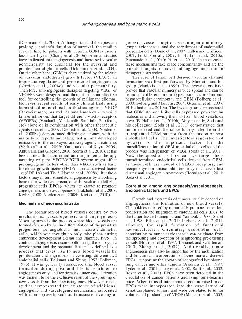
Our recent work in rat orthotopic human gliomamodel showed paradoxically increased production ofVEGF at the peripheral part of tumors, as well as theelevated expression of HIF-1α and SDF-1α, and asignificant increase in the number of dilated bloodvessels in animals that underwent two weeks of PTK787(small molecule tyrosine kinase inhibitor) treatment (Aliet al., 2010) (Fig. 1). In addition, as reported by ourgroup and other investigators, SDF-1α is one of thepotent chemo attractant for bone marrow derived EPCsdue to the presence of CXCR4 receptors in these cells(Jin et al., 2006; Arbab et al., 2008) and may be involvedin enhanced angiogenesis and invasiveness of the tumor
551Anti-angiogenesis and bone marrow cells
following treatment with VEGFRs inhibitors. Moreover,we have also reported the role of inflammatory cytokineRANTES, which also act as a chemo attractant for thesecells (Silverman et al., 2007; Janic et al., 2010). Anytherapy may cause enhanced inflammation in the tumorsites and invite more endothelial progenitor cells andpossible angiogenesis.
Therefore, it is important to gain insight into thepossible mechanisms that are activated during anti-angiogenic treatment to understand and potentiallyprevent its failure. It is of extreme importance todocument these changes in vivo and develop a noveltherapeutic approach for overcoming these paradoxiceffects. Current challenge in preventing vasculogenesis:
Current evidences from recent publications indicatethe involvement of both angiogenesis andvasculogenesis processes for glioma growth (tumorgrowth) (Dome et al., 2007; Folkins et al., 2009; Yu etal., 2010). As discussed above, most of the agents thattarget neovascularization are in fact againstangiogenesis. With emerging new insights intovasculogenesis, investigators are looking into possible
mechanisms for how bone marrow derived progenitorcells or EPCs migrate and incorporate into tumorneovascularization (Patenaude et al., 2010). One of themechanisms that has been pointed out is the involvementof SDF-1-CXCR4 axis (Jin et al., 2006; Petit et al.,2007; Shichinohe et al., 2007). SDF-1 is a chemokinethat is expressed in tumor cells and released in thecirculation following hypoxia in the tumor (with the upregulation of HIF-1α) (Moore et al., 2001; Ceradini etal., 2004; Arbab et al., 2008). In an experiment, Heissiget al. (2002) determined the mechanisms of releasinghematopoietic stem cells (HSC) and EPCs from bonemarrow. Under steady-state conditions, quiescent c-Kit+HSCs or EPCs reside in a niche in close contact withstromal cells. Membrane-bound cytokines, such asmKitL not only convey survival signals, but also supportthe adhesion of stem cells to the stroma. Increasedchemokine/cytokine such as SDF-1 and VEGF induceup-regulation of MMP-9 resulting in the release of sKitL(soluble Kit ligand). sKitL confers signals that transfer c-Kit+ HSCs or EPCs from quiescent to proliferative stateand enhances mobility of VEGFR2+ EPCs and Lin Sca-1+c-Kit+ repopulating cells, translocating them into avascular-enriched niche favoring differentiation andmobilization to the peripheral circulation. SDF-1 is a
552Anti-angiogenesis and bone marrow cells
Fig. 1. Expression of different angiogenic factors (left panel) in the vehicle- and PTK787-treated tumors from representative cases at the peripheral (P),central part of the tumors (C) and contralateral brains (B). Note the increased expression of VEGF, SDF-1α and HIF-1α at the peripheral part ofPTK787 treated tumors. Right panel shows the densitometry analysis of the blot (%ß-actin and normalized to contralateral brain). The analysis alsoconfirmed the finding of the blot. Note the patterns of VEGF, SDF-1α and HIF-1α in PTK787 treated tumors.
strong chemo-attractant for CXCR4 positive cells.Preventing interaction of SDF-1-CXCR4 is thought to bea mechanism to block vasculogenesis. AMD3100, areceptor (CXCR4) antagonist was initially developed asanti human immunodeficiency virus (HIV) drug andlater used to mobilize CD34+ HSCs cells to theperipheral circulation (Petit et al., 2007). Although
AMD3100 increased the number of peripheral CD34+ orprogenitor cells, the recent investigations pointed outthat continuous treatment with AMD3100 or similarCXCR4 receptor antagonists inhibit vasculogenesis intumors causing inhibition of tumor growth (Petit et al.,2007; Kioi et al., 2010). Similarly anti SDF-1α antibodyor siRNA techniques can be used to block the release of
553Anti-angiogenesis and bone marrow cells
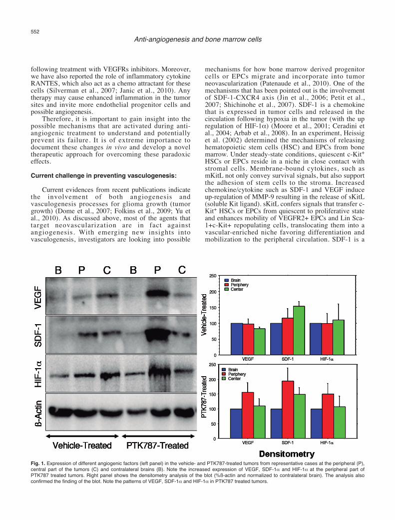
Fig. 2. Migration and accumulation of administered EPCs in PTK treated tumor. Five million In-111 labeled EPCs followed by 5 million magneticallylabeled EPCs were administered in the same rat. SPECT images were obtained on day 0, 1, 3, and 7. MRI was obtained by a clinical 3T system on day7 following last SPECT. T2WI with a TE of 35ms (A), T2*WI with a TE of 20 (B) and corresponding R2* map (C). Contrast enhanced T1WI (D) andcorresponding Ktrans (E) and tumor blood volume (F) maps. Note the low signal intensity areas on T2*WI (B, black arrows) and corresponding R2*map (C, yellow arrows) indicating accumulation of iron positive cells, which is proved by DAB enhanced Prussian blue staining (I). SPECT images ofthe tumor (G and H) obtained at 24 hours showed increased activity at the site of tumor indicating accumulation of In-111 labeled EPCs. DABenhanced Prussian blue staining showed accumulation of iron positive cells around the tumor (I). Note a few of the iron positive cells also make thelining of blood vessels (inset, black arrows).
SDF-1α and interaction for the mobilization of bonemarrow progenitor cells responsible for vasculogenesis.In vivo imaging that shows the effect of chemokinereceptor antagonist on the tumor growth andvasculogenesis will be very helpful for future clinicaltrials.Role of in vivo imaging in the detection of angio-genesis/vasculogenesis:
DCE MRI or perfusion CT has been used todetermine the vascular permeability and tumor bloodvolume, which are the indirect measurements ofneovascularization and total vascular densities (Taylor etal., 1999; Hoang et al., 2004; Preda et al., 2006;Provenzale et al., 2006; Raatschen et al., 2009).However, DCE MRI or perfusion CT cannot predict theinvolvement of bone marrow progenitor cells (BMPC) inthe neovascularization processes in tumors before orafter anti-angiogenic treatments. There has been no invivo imaging modality capable of detecting migration
and incorporation of host BMPC to the sites ofangiogenesis. Investigators have shown the migrationand accumulation of host BMPC or EPC to theimplanted tumor or in different cancers or lesions by invitro studies (Asahara et al., 1997, 1999; Yu et al., 2010).Involvement of stem cells in the formation of tumorvasculatures has been determined by in vivo imagingmodalities, however, the stem cells in questions weremanipulated ex vivo then administered in animal models(Anderson et al., 2005; Arbab et al., 2006). Differentimaging modalities are used to track the migration andincorporation of administered cells to the sites of tumors(Brenner et al., 2004; Arbab and Frank, 2008; Mani etal., 2008). We have pioneered the technique to trackadministered EPCs to the site of tumorangiogenesis/vasculogenesis by in vivo cellular MRI(Arbab et al., 2006, 2008). We have also used nuclearmedicine technique to track the migration andaccumulation of administered stem cells to the sites oftumors (Fig. 2).
In vivo determination of the involvement of host
554Anti-angiogenesis and bone marrow cells
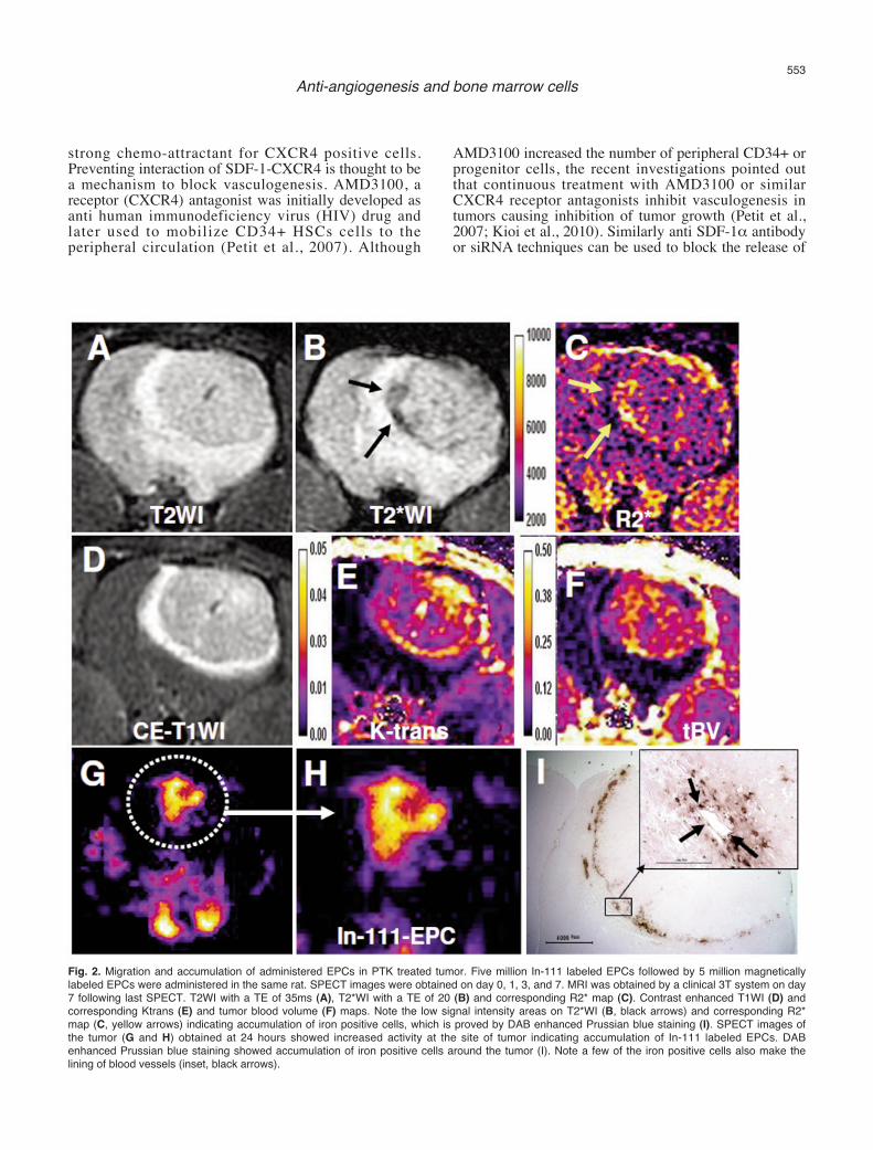
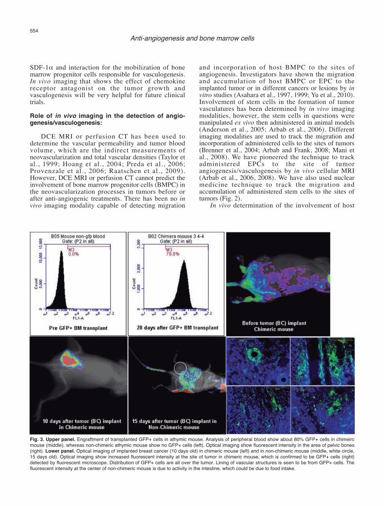
Fig. 3. Upper panel. Engraftment of transplanted GFP+ cells in athymic mouse. Analysis of peripheral blood show about 80% GFP+ cells in chimeircmouse (middle), whereas non-chimeric athymic mouse show no GFP+ cells (left). Optical imaging show fluorescent intensity in the area of pelvic bones(right). Lower panel. Optical imaging of implanted breast cancer (10 days old) in chimeric mouse (left) and in non-chimeric mouse (middle, white circle,15 days old). Optical imaging show increased fluorescent intensity at the site of tumor in chimeric mouse, which is confirmed to be GFP+ cells (right)detected by fluorescent microscope. Distribution of GFP+ cells are all over the tumor. Lining of vascular structures is seen to be from GFP+ cells. Thefluorescent intensity at the center of non-chimeric mouse is due to activity in the intestine, which could be due to food intake.

BMPC in the formation of neovascularization in tumorsis challenging. To be detected by in vivo imagingmodalities, the host bone marrow cells should carry areporter, such as fluorescent protein or genes that can betargeted later (such as luciferase or sodium iodidesymporter or specific promoter mediated activation).However, this reporter should only be present in BMPCbut not in other cell types. Making of transgenic animalmodel having such conditional gene expression wouldbe difficult. The alternate way would be to makechimeric animal models, where bone marrow cells ofrecipient animals should be replaced with bone marrowcells from animals expressing different reporters, such asGFP+ bone marrow cells (Sengupta et al., 2003; Sheikhet al., 2007; Yu et al., 2010). Recently a chimeric animalmodel has been developed in our laboratory to determinethe involvement of BMPC in the tumorneovascularization (Fig. 3). Sub-lethally irradiatedathymic mice received bone marrow cells from greenfluorescent protein positive (GFP+) transgenic mice(Schaefer et al., 2001). GFP+ bone marrow cells weretransplanted in athymic mice 24 hours following sub-lethal irradiation and the tumors were implanted after 28days when flowcytometric analysis showed more than70% engraftment of GFP+ cells. Migration andaccumulation of transplanted bone marrow cells in theimplanted breast cancer were determined by opticalimaging (Kodak, Carestream multi-spectral system,Carestream, USA) with proper excitation and emissionprofiles. Optical imaging showed gradual increase inGFP intensity in the tumors and multiple GFP+ cellslining the blood vessels and other infrastructures of thetumors were observed under fluorescent microscope.Our ongoing studies using different antiangiogenicagents will determine the involvement of bone marrowderived progenitor cells in the development of resistanceto anti-angiogenic treatments, and development ofrecurrence and distal metastases of tumors.Conclusions:
In this short review possible mechanisms for thedevelopment of resistance to anti-angiogenic treatmentsin GBM are discussed. Activation of alternateangiogenic pathways and involvement of bone marrowderived progenitor cells could be the mechanisms for theresistance and in vivo imaging should be utilized ordeveloped to determine the involvement of bone marrowcells in tumor vasculogenesis.
References
Ahluwalia M.S. and Gladson C.L. (2010). Progress on antiangiogenictherapy for patients with malignant glioma. J. Oncol. 2010, 689018.
Ali M.M., Janic B., Babajani-Feremi A., Varma N.R., Iskander A.S.,Anagli J. and Arbab A.S. (2010). Changes in vascular permeabilityand expression of different angiogenic factors following anti-angiogenic treatment in rat glioma. PLoS ONE 5, e8727.
Anderson S.A., Glod J., Arbab A.S., Noel M., Ashari P., Fine H.A. andFrank J.A. (2005). Noninvasive mr imaging of magnetically labeledstem cells to directly identify neovasculature in a glioma model.Blood 105, 420-425.
Arbab A.S. and Frank J.A. (2008). Cellular mri and its role in stem celltherapy. Regen. Med. 3, 199-215.
Arbab A.S., Pandit S.D., Anderson S.A., Yocum G.T., Bur M., FrenkelV., Khuu H.M., Read E.J. and Frank J.A. (2006). Magneticresonance imaging and confocal microscopy studies of magneticallylabeled endothelial progenitor cells trafficking to sites of tumorangiogenesis. Stem Cells 24, 671-678.
Arbab A.S., Janic B., Knight R.A., Anderson S.A., Pawelczyk E., RadA.M., Read E.J., Pandit S.D. and Frank J.A. (2008). Detection ofmigration of locally implanted ac133+ stem cells by cellular magneticresonance imaging with histological findings. FASEB J. 22, 3234-3246.
Asahara T., Murohara T., Sullivan A., Silver M., van der Zee R., Li T.,Witzenbichler B., Schatteman G. and Isner J.M. (1997). Isolation ofputative progenitor endothelial cells for angiogenesis. Science 275,964-967.
Asahara T., Masuda H., Takahashi T., Kalka C., Pastore C., Silver M.,Kearne M., Magner M. and Isner J.M. (1999). Bone marrow origin ofendothelial progenitor cells responsible for postnatal vasculogenesisin physiological and pathological neovascularization. CirculationRes. 85, 221-228.
Batchelor T.T., Sorensen A.G., di Tomaso E., Zhang W.T., Duda D.G.,Cohen K.S., Kozak K.R., Cahil l D.P., Chen P.J., Zhu M.,Ancukiewicz M., Mrugala M.M., Plotkin S., Drappatz J., Louis D.N.,Ivy P., Scadden D.T., Benner T., Loeffler J.S., Wen P.Y. and JainR.K. (2007). Azd2171, a pan-vegf receptor tyrosine kinase inhibitor,normalizes tumor vasculature and alleviates edema in glioblastomapatients. Cancer Cell 11, 83-95.
Beerepoot L.V., Mehra N., Vermaat J.S., Zonnenberg B.A., GebbinkM.F. and Voest E.E. (2004). Increased levels of viable circulatingendothelial cells are an indicator of progressive disease in cancerpatients. Ann. Oncol. 15, 139-145.
Bergers G. and Hanahan D. (2008). Modes of resistance to anti-angiogenic therapy. Nat. Rev. Cancer 8, 592-603.
Brenner W., Aicher A., Eckey T., Massoudi S., Zuhayra M., Koehl U.,Heeschen C., Kampen W.U., Zeiher A.M., Dimmeler S. and HenzeE. (2004). 111in-labeled cd34+ hematopoietic progenitor cells in arat myocardial infarction model. J. Nucl. Med. 45, 512-518.
Ceradini D.J., Kulkarni A.R., Callaghan M.J., Tepper O.M., Bastidas N.,Kleinman M.E., Capla J.M., Galiano R.D., Levine J.P. and GurtnerG.C. (2004). Progenitor cell trafficking is regulated by hypoxicgradients through hif-1 induction of sdf-1. Nat. Med. 10, 858-864.
Chang S.M., Butowski N.A., Sneed P.K. and Garner I.V. (2006).Standard treatment and experimental targeted drug therapy forrecurrent glioblastoma multiforme. Neurosurg. Focus 20, E4.
de Groot J.F., Fuller G., Kumar A.J., Piao Y., Eterovic K., Ji Y. andConrad C.A. (2010). Tumor invasion after treatment of glioblastomawith bevacizumab: Radiographic and pathologic correlation inhumans and mice. Neuro-Oncology 12, 233-242.
Dhermain F., Ducreux D., Bidault F., Bruna A., Parker F., Roujeau T.,Beaudre A., Armand J.P. and Haie-Meder C. (2005). Use of thefunctional imaging modalities in radiation therapy treatment planningin patients with glioblastoma. Bull. Cancer 92, 333-342. (in French).
Dietrich J., Norden A.D. and Wen P.Y. (2008). Emerging antiangiogenictreatments for gliomas - efficacy and safety issues. Curr. Opin.
555Anti-angiogenesis and bone marrow cells

Neurol. 21, 736-744.Dome B., Hendrix M.J.C., Paku S., Tovari J. and Timar J. (2007).
Alternative vascularization mechanisms in cancer: Pathology andtherapeutic implications. Am. J. Pathol. 170, 1-15.
El Hallani S., Boisselier B., Peglion F., Rousseau A., Colin C., Idbaih A.,Marie Y., Mokhtari K., Thomas J.-L., Eichmann A., Delattre J.-Y.,Maniotis A.J. and Sanson M. (2010a). A new alternative mechanismin glioblastoma vascularization: Tubular vasculogenic mimicry. Brain133, 973-982.
El Hallani S., Boisselier B., Peglion F., Rousseau A., Colin C., Idbaih A.,Marie Y., Mokhtari K., Thomas J.L., Eichmann A., Delattre J.Y.,Maniotis A.J. and Sanson M. (2010b). A new alternative mechanismin glioblastoma vascularization: Tubular vasculogenic mimicry. Brain133, 973-982.
Ellis L.M., Liu W., Ahmad S.A., Fan F., Jung Y.D., Shaheen R.M. andReinmuth N. (2001). Overview of angiogenesis: Biologic implicationsfor antiangiogenic therapy. Semin. Oncol. 28, 94-104.
Folberg R. and Maniotis A.J. (2004). Vasculogenic mimicry. APMIS 112,508-525.
Folberg R., Hendrix M.J. and Maniotis A.J. (2000). Vasculogenicmimicry and tumor angiogenesis. Am. J. Pathol. 156, 361-381.
Folkins C., Shaked Y., Man S., Tang T., Lee C.R., Zhu Z., Hoffman R.M.and Kerbel R.S. (2009). Glioma tumor stem-like cells promote tumorangiogenesis and vasculogenesis via vascular endothelial growthfactor and stromal-derived factor 1. Cancer Res. 69, 7243-7251.
Folkman J. (1995). Clinical applications of research on angiogenesis. N.Engl. J. Med. 333, 1757-1763.
Folkman J. and Shing Y. (1992). Angiogenesis. J. Biol. Chem. 267,10931-10934.
Gerstner E.R., Frosch M.P. and Batchelor T.T. (2010). Diffusionmagnetic resonance imaging detects pathologically confirmed,nonenhancing tumor progression in a patient with recurrentglioblastoma receiving bevacizumab. J. Clin. Oncol. 28, e91-93.
Goldbrunner R.H., Bendszus M., Wood J., Kiderlen M., Sasaki M. andTonn J.C. (2004). Ptk787/zk222584, an inhibitor of vascularendothelial growth factor receptor tyrosine kinases, decreasesglioma growth and vascularization. Neurosurgery 55, 426-432;discussion 432.
Guzman G., Cotler S.J., Lin A.Y., Maniotis A.J. and Folberg R. (2007). Apilot study of vasculogenic mimicry immunohistochemical expressionin hepatocellular carcinoma. Arch. Pathol. Lab. Med. 131, 1776-1781.
Heissig B., Hattori K., Dias S., Friedrich M., Ferris B., Hackett N.R.,Crystal R.G., Besmer P., Lyden D., Moore M.A., Werb Z. and RafiiS. (2002). Recruitment of stem and progenitor cells from the bonemarrow niche requires mmp-9 mediated release of kit-ligand. Cell109, 625-637.
Hillen F. and Griffioen A. (2007). Tumour vascularization: Sproutingangiogenesis and beyond. Cancer Metast. Rev. 26, 489-502.
Hoang B.H., Dyke J.P., Koutcher J.A., Huvos A.G., Mizobuchi H.,Mazza B.A., Gorlick R. and Healey J.H. (2004). VEGF expression inosteosarcoma correlates with vascular permeability by dynamic mri.Clin. Ortho. Relat. Res. 32-38.
Hormigo A., Ding B.S. and Rafii S. (2011). A target for antiangiogenictherapy: Vascular endothelium derived from glioblastoma. Proc.Natl. Acad. Sci. USA 108, 4271-4272.
Hotfilder M., Nowak-Gottl U. and Wolff J.E. (1997). Tumorangiogenesis:A network of cytokines. Klin Padiatr. 209, 265-270.
Janic B., Guo A.M., Iskander A.S., Varma N.R., Scicli A.G. and Arbab
A.S. (2010). Human cord blood-derived ac133+ progenitor cellspreserve endothelial progenitor characteristics after long term in vitroexpansion. PLoS ONE 5, e9173.
Jiang Y., Jahagirdar B.N., Reinhardt R.L., Schwartz R.E., Keene C.D.,Ortiz-Gonzalez X.R., Reyes M., Lenvik T., Lund T., Blackstad M., DuJ., Aldrich S., Lisberg A., Low W.C., Largaespada D.A. and VerfaillieC.M. (2002). Pluripotency of mesenchymal stem cells derived fromadult marrow. Nature. 418, 41-49.
Jin D.K., Shido K., Kopp H.G., Petit I., Shmelkov S.V., Young L.M.,Hooper A.T., Amano H., Avecilla S.T., Heissig B., Hattori K., ZhangF., Hicklin D.J., Wu Y., Zhu Z., Dunn A., Salari H., Werb Z., HackettN.R., Crystal R.G., Lyden D. and Rafii S. (2006). Cytokine-mediateddeployment of sdf-1 induces revascularization through recruitment ofcxcr4(+) hemangiocytes. Nat. Med. 12, 557-567.
Kerbel R.S. (2008). Tumor angiogenesis. N. Engl. J. Med. 358, 2039-2049.
Kioi M., Vogel H., Schultz G., Hoffman R.M., Harsh G.R. and BrownJ.M. (2010). Inhibition of vasculogenesis, but not angiogenesis,prevents the recurrence of glioblastoma after irradiation in mice. J.Clin. Invest. 120, 694-705.
Liekens S., De Clercq E. and Neyts J. (2001). Angiogenesis: Regulatorsand clinical applications. Biochem. Pharmacol. 61, 253-270.
Los M., Roodhart J.M. and Voest E.E. (2007). Target practice: Lessonsfrom phase iii trials with bevacizumab and vatalanib in the treatmentof advanced colorectal cancer. Oncologist 12, 443-450.
Lyden D., Hattori K., Dias S., Costa C., Blaikie P., Butros L., ChadburnA., Heissig B., Marks W., Witte L., Wu Y., Hicklin D., Zhu Z., HackettN.R., Crystal R.G., Moore M.A., Hajjar K.A., Manova K., Benezra R.and Rafii S. (2001). Impaired recruitment of bone-marrow-derivedendothelial and hematopoietic precursor cells blocks tumorangiogenesis and growth. Nat. Med. 7, 1194-1201.
Mancuso P., Calleri A., Cassi C., Gobbi A., Capillo M., Pruneri G.,Martinelli G. and Bertolini F. (2003). Circulating endothelial cells as anovel marker of angiogenesis. Adv. Exp. Med. Biol. 522, 83-97.
Mani V., Adler E., Briley-Saebo K.C., Bystrup A., Fuster V., Keller G.and Fayad Z.A. (2008). Serial in vivo positive contrast mri of ironoxide-labeled embryonic stem cell-derived cardiac precursor cells ina mouse model of myocardial infarction. Magn. Reson. Med. 60, 73-81.
Maniotis A.J., Folberg R., Hess A., Seftor E.A., Gardner L.M., Pe'er J.,Trent J.M., Meltzer P.S. and Hendrix M.J. (1999). Vascular channelformation by human melanoma cells in vivo and in vitro:Vasculogenic mimicry. Am. J. Pathol. 155, 739-752.
Moore M.A., Hattori K., Heissig B., Shieh J.H., Dias S., Crystal R.G. andRafii S. (2001). Mobilization of endothelial and hematopoietic stemand progenitor cells by adenovector-mediated elevation of serumlevels of sdf-1, vegf, and angiopoietin-1. Ann. NY Acad. Sci. 938,36-45.
Norden A.D., Drappatz J. and Wen P.Y. (2008a). Antiangiogenictherapy in malignant gliomas. Curr. Opin. Oncol. 20, 652-661.
Norden A.D., Drappatz J. and Wen P.Y. (2008b). Novel anti-angiogenictherapies for malignant gliomas. Lancet Neurol. 7, 1152-1160.
Norden A.D., Young G.S., Setayesh K., Muzikansky A., Klufas R., RossG.L., Ciampa A.S., Ebbeling L.G., Levy B., Drappatz J., Kesari S.and Wen P.Y. (2008c). Bevacizumab for recurrent malignantgliomas: Efficacy, toxicity, and patterns of recurrence. Neurology 70,779-787.
Pàez-Ribes M., Allen E., Hudock J., Takeda T., Okuyama H., Viñals F.,Inoue M., Bergers G., Hanahan D. and Casanovas O. (2009).
556Anti-angiogenesis and bone marrow cells
Antiangiogenic therapy elicits malignant progression of tumors toincreased local invasion and distant metastasis. Cancer Cell 15,220-231.
Patenaude A., Parker J. and Karsan A. (2010). Involvement ofendothelial progenitor cells in tumor vascularization. Microvasc. Res.79, 217-223.
Petit I., Jin D. and Rafii S. (2007). The sdf-1-cxcr4 signaling pathway: Amolecular hub modulating neo-angiogenesis. Trends Immunol. 28,299-307.
Preda A., van Vliet M., Krestin G.P., Brasch R.C. and van Dijke C.F.(2006). Magnetic resonance macromolecular agents for monitoringtumor microvessels and angiogenesis inhibition. Invest. Radiol. 41,325-331.
Provenzale J.M., York G., Moya M.G., Parks L., Choma M., Kealey S.,Cole P. and Serajuddin H. (2006). Correlation of relativepermeability and relative cerebral blood volume in high-gradecerebral neoplasms. Am. J. Roentgenol 187, 1036-1042.
Raatschen H.J., Fu Y., Brasch R.C., Pietsch H., Shames D.M. and YehB.M. (2009). In vivo monitoring of angiogenesis inhibitory treatmenteffects by dynamic contrast-enhanced computed tomography in axenograft tumor model. Invest. Radiol. 44, 265-270.
Rafii S., Lyden D., Benezra R., Hattori K. and Heissig B. (2002).Vascular and haematopoietic stem cells: Novel targets for anti-angiogenesis therapy? Nat. Rev. Cancer 2, 826-835.
Rahman R., Smith S., Rahman C. and Grundy R. (2010).Antiangiogenic therapy and mechanisms of tumor resistance inmalignant glioma. J. Oncol. 2010, 251231.
Reardon D.A., Fink K.L., Mikkelsen T., Cloughesy T.F., O'Neill A.,Plotkin S., Glantz M., Ravin P., Raizer J.J., Rich K.M., Schiff D.,Shapiro W.R., Burdette-Radoux S., Dropcho E.J., Wittemer S.M.,Nippgen J., Picard M. and Nabors L.B. (2008). Randomized phase iistudy of cilengitide, an integrin-targeting arginine-glycine-asparticacid peptide, in recurrent glioblastoma multiforme. J. Clin. Oncol. 26,5610-5617.
Remer S. and Murphy M.E. (2004). The challenges of long-termtreatment outcomes in adults with malignant gliomas. Clin. J. Oncol.Nurs. 8, 368-376.
Reyes M., Dudek A., Jahagirdar B., Koodie L., Marker P.H. andVerfaillie C.M. (2002). Origin of endothelial progenitors in humanpostnatal bone marrow. J. Clin. Invest. 109, 337-346.
Risau W. and Flamme I. (1995). Vasculogenesis. Ann. Rev. Cell Dev.Biol. 11, 73-91.
Saidi A., Javerzat S., Bellahcène A., De Vos J., Bello L., Castronovo V.,Deprez M., Loiseau H., Bikfalvi A. and Hagedorn M. (2008).Experimental anti-angiogenesis causes upregulation of genesassociated with poor survival in glioblastoma. Int. J. Cancer 122,2187-2198.
Samejima N. and Yamazaki K. (1988). A study on the vascularproliferation in tissues around the tumor in breast cancer. Jpn. J.Surg. 18, 235-242.
Schaefer B.C., Schaefer M.L., Kappler J.W., Marrack P. and Kedl R.M.(2001). Observation of antigen-dependent cd8+ t-cell/ dendritic cellinteractions in vivo. Cell Immunol. 214, 110-122.
Sengupta N., Caballero S., Mames R.N., Butler J.M., Scott E.W. andGrant M.B. (2003). The role of adult bone marrow-derived stem cellsin choroidal neovascularization. Invest. Ophthalmol. Visual Sci. 44,4908-4913.
Sheikh A.Y., Lin S.A., Cao F., Cao Y., van der Bogt K.E., Chu P., ChangC.P., Contag C.H., Robbins R.C. and Wu J.C. (2007). Molecularimaging of bone marrow mononuclear cell homing and engraftmentin ischemic myocardium. Stem Cells 25, 2677-2684.
Shi Q., Rafii S., Wu M.H., Wijelath E.S., Yu C., Ishida A., Fujita Y.,Kothari S., Mohle R., Sauvage L.R., Moore M.A., Storb R.F. andHammond W.P. (1998). Evidence for circulating bone marrow-derived endothelial cells. Blood 92, 362-367.
Shichinohe H., Kuroda S., Yano S., Hida K. and Iwasaki Y. (2007). Roleof sdf-1/cxcr4 system in survival and migration of bone marrowstromal cells after transplantation into mice cerebral infarct. BrainRes. 1183, 138-147.
Sierra J.R., Cepero V. and Giordano S. (2010). Molecular mechanismsof acquired resistance to tyrosine kinase targeted therapy. Mol.Cancer 9, 75.
Silverman M.D., Haas C.S., Rad A.M., Arbab A.S. and Koch A.E.(2007). The role of vascular cell adhesion molecule 1/ very lateactivation antigen 4 in endothelial progenitor cell recruitment torheumatoid arthrit is synovium. Arthrit is Rheum. 56, 1817-1826.
Soda Y., Marumoto T., Friedmann-Morvinski D., Soda M., Liu F.,Michiue H., Pastorino S., Yang M., Hoffman R.M., Kesari S. andVerma I.M. (2011). Transdifferentiation of glioblastoma cells intovascular endothelial cells. Proc. Natl. Acad. Sci. USA 108, 4274-4280.
Taylor J.S., Tofts P.S., Port R., Evelhoch J.L., Knopp M., Reddick W.E.,Runge V.M. and Mayr N. (1999). Mr imaging of tumormicrocirculation: Promise for the new millennium. J. Magn. Reson.Imaging 10, 903-907.
Tomanek R.J. and Schatteman G.C. (2000). Angiogenesis: Newinsights and therapeutic potential. Anat. Rec. 261, 126-135.
Verhoeff J.J., van Tellingen O., Claes A., Stalpers L.J., van Linde M.E.,Richel D.J., Leenders W.P. and van Furth W.R. (2009). Concernsabout anti-angiogenic treatment in patients with glioblastomamultiforme. BMC Cancer 9, 444.
Wong E.T. and Brem S. (2011). Taming glioblastoma by targetingangiogenesis: 3 years later. J. Clin. Oncol. 29, 124-126.
Yamanaka R. and Saya H. (2009). Molecularly targeted therapies forglioma. Ann. Neurol. 66, 717-729.
Yu L., Su B., Hollomon M., Deng Y., Facchinetti V. and Kleinerman E.S.(2010). Vasculogenesis driven by bone marrow-derived cells isessential for growth of ewing's sarcomas. Cancer Res. 70, 1334-1343.
Zhang Z.G., Zhang L., Jiang Q. and Chopp M. (2002). Bone marrow-derived endothelial progenitor cells participate in cerebralneovascularization after focal cerebral ischemia in the adult mouse.Circ. Res. 90, 284-288.
Accepted November 28, 2011
557Anti-angiogenesis and bone marrow cells