activation of metabotropic gaba receptors increases the...
TRANSCRIPT

Activation of metabotropic GABA receptors increasesthe energy barrier for vesicle fusion
Benjamin R. Rost1,2, Patrick Nicholson1,2, Gudrun Ahnert-Hilger3, Andreas Rummel4, Christian Rosenmund1,5,Joerg Breustedt1,*,` and Dietmar Schmitz1,5,6,*,`
1Neuroscience Research Centre, Universitatsmedizin Berlin, Chariteplatz 1, 10117 Berlin, Germany2Graduate Program Medical Neuroscience, Universitatsmedizin Berlin, Chariteplatz 1, 10117 Berlin, Germany3Functional Cell Biology, Centre of Anatomy, Universitatsmedizin Berlin, Chariteplatz 1, 10117 Berlin, Germany4Institut fur Toxikologie, Medizinische Hochschule Hannover, 30625 Hannover, Germany5Cluster of Excellence Neurocure, Charite, Universitatsmedizin Berlin, Chariteplatz 1, 10117 Berlin, Germany6Bernstein Centre for Computational Neuroscience, 10115 Berlin, Germany*These authors contributed equally to this work`Authors for correspondence ([email protected]; [email protected])
Accepted 9 May 2011Journal of Cell Science 124, 3066–3073� 2011. Published by The Company of Biologists Ltddoi: 10.1242/jcs.074963
SummaryNeurotransmitter release from presynaptic terminals is under the tight control of various metabotropic receptors. We report here that inaddition to the regulation of Ca2+ channel activity, metabotropic GABAB receptors (GABABRs) at murine hippocampal glutamatergic
synapses utilize an inhibitory pathway that directly targets the synaptic vesicle release machinery. Acute application of the GABABRagonist baclofen rapidly and reversibly inhibits vesicle fusion, which occurs independently of the SNAP-25 C-terminus. Usingapplications of hypertonic sucrose solutions, we find that the size of the readily releasable pool remains unchanged by GABABR
activation, but the sensitivity of primed vesicles to hypertonic stimuli appears lowered as the response amplitudes at intermediatesucrose concentrations are smaller and release kinetics are slowed. These data show that presynaptic GABABRs can inhibitneurotransmitter release directly by increasing the energy barrier for vesicle fusion.
Key words: GABAB receptor, Metabotropic, Neurotransmission, Energy barrier, SNARE, Presynaptic inhibition
IntroductionMetabotropic signalling through c-aminobutyric acid B receptors
(GABABR) has been implicated in a number of physiological and
pathophysiological processes, such as hippocampal rhythmic
activity (Scanziani, 2000), seizure disorders and anxiety and
depression (Cryan and Kaupmann, 2005). On the cellular level,
GABABRs are found to be both postsynaptically and
presynaptically localized. On the postsynaptic side, they mediate
the slow component of the inhibitory postsynaptic potential
through activation of K+ conductances (Luscher et al., 1997;
Newberry and Nicoll, 1984). On the presynaptic side, they can act
as auto- or hetero-receptors on GABAergic and glutamatergic
terminals, respectively. In both cases activation of presynaptic
GABABRs leads to a decrease in transmitter release (Bowery,
2006). The reduction of transmitter release is for the most part
achieved by the closing of voltage-dependent Ca2+ channels
(VDCCs) (Dunlap and Fischbach, 1981). This conversion of
VDCCs into the ‘reluctantly opening state’ occurs by G-protein
bc-subunit-mediated signal transduction in a membrane-delimited
pathway, as has been directly demonstrated for the calyx of Held
(Kajikawa et al., 2001; Takahashi et al., 1998).
However, there are also indications that GABABRs can
modulate transmitter release independently of VDCCs. In
hippocampal neuronal cell cultures, it has been shown that both
adenosine receptor and GABABR activation can reduce the
frequency of action potential (AP)-independent miniature
excitatory postsynaptic currents (mEPSCs) in pyramidal
neurons (Scanziani et al., 1992; Scholz and Miller, 1992).
Furthermore, these receptors can inhibit artificially induced
transmitter release evoked by ionomycin and a-latrotoxin, which
occurs independently of VDCCs (Capogna et al., 1996). It has
been argued that this form of GABABR-mediated inhibition acts
‘downstream’ of VDCCs, but a possible molecular mechanism
for this modulation in the mammalian central nervous system has
so far remained elusive. Studies in the lamprey spinal cord
provided evidence that the SNARE complex member SNAP-25 is
important for presynaptic serotinergic modulation of transmission
(Blackmer et al., 2001). These authors showed that this
modulation was largely independent of VDCCs but involved a
direct Gbc-subunit-mediated pathway acting on the SNAP-25 C-
terminus (Gerachshenko et al., 2005).
In the present study, we investigated the mechanism by which
GABABRs modulate synaptic transmission at hippocampal
pyramidal neurons. We hypothesized that a direct interference
of GABABR signalling with transmitter release at the level of the
release machinery would increase the energy barrier for vesicle
fusion (Rosenmund and Stevens, 1996). We found that
GABABRs indeed affect transmission through two G-protein
dependent pathways: besides the classical pathway involving
reduction of Ca2+-influx through VDCCs, a second, downstream
pathway leads to an increase of the energy barrier for vesicle
fusion. Notably, this form of inhibition does not require the C-
terminus of the SNARE complex member SNAP-25, as it is not
affected by Botulinum neurotoxin-A (BoNT-A) treatment.
3066 Research Article
Journ
alof
Cell
Scie
nce
ResultsActivation of GABABRs increases the energy barrier for
vesicle fusion
In order to assess the involvement of VDCCs in GABABR-
mediated modulation of transmission, we monitored AP-triggered
presynaptic Ca2+ transients in the Schaffer-collateral pathway
and recorded field excitatory postsynaptic potentials (fEPSPs) in
area CA1 in acute hippocampal slice preparations (Fig. 1A).
GABABR activation with baclofen (3–30 mM) profoundly
inhibited fEPSP amplitudes. At the same time, baclofen
reduced the presynaptic Ca2+ signals by a maximum of 28%. In
a parallel set of experiments, we reduced the Ca2+ concentration
in the external solution stepwise and monitored presynaptic Ca2+
signals or postsynaptic fEPSPs, while CaCl2 was replaced by
equimolar concentrations of MgCl2. A data fit revealed a power
function for transmitter release at CA1 synapses with an
exponent of 4.1. We correlated postsynaptic responses and
presynaptic Ca2+ signals from the baclofen experiments with the
power function and found that the baclofen effect on synaptic
transmission was greater than what would be expected if the drug
was acting exclusively on VDCCs (Fig. 1A2). We also recorded
mEPSCs from CA1 pyramidal neurons in both standard
(2.5 mM) extracellular Ca2+, as well as in Ca2+-free conditions
(i.e. 0 mM Ca2+ and EGTA in the extracellular solution, data not
shown). In both conditions, baclofen reduced the frequency of
mEPSCs to a similar degree (reduction in 2.5 mM Ca2+,
26.9¡2.6%, n56, P,0.01; in 0 mM Ca2+, 28.5¡3.9%, n57,
P,0.01), whereas the mEPSC amplitude remained unchanged. In
summary, the data from the slice experiments indicate that
GABABR activation has an additional effect downstream of the
modulation of VDCCs.
To investigate whether GABABRs trigger a signalling cascade by
acting directly on the release machinery, we used autaptic cultures
of hippocampal pyramidal neurons. Combined with techniques that
allow for rapid application of hypertonic solutions, this system
enabled us to study the efficiency of vesicle exocytosis in presence
and absence of GABABR activation (Basu et al., 2007; Gerber et al.,
2008), while bypassing Ca2+ triggering of neurotransmitter release
(Rosenmund and Stevens, 1996). We first determined the sensitivity
of autaptic EPSCs to baclofen treatment, which gave similar effects
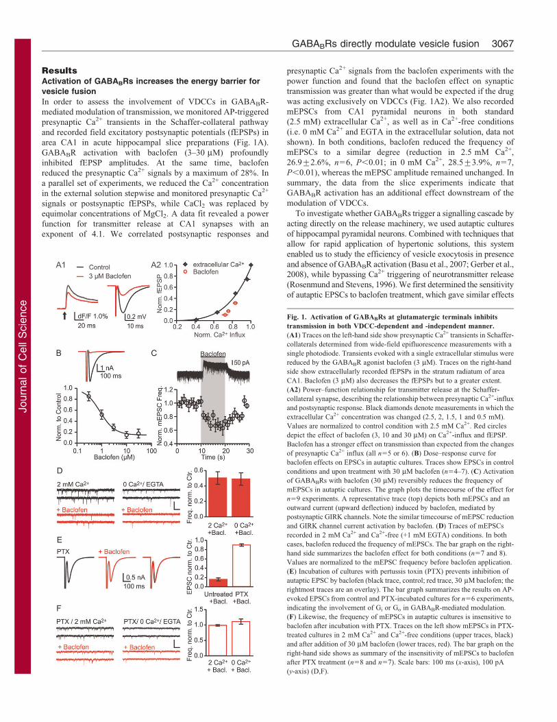
Fig. 1. Activation of GABABRs at glutamatergic terminals inhibits
transmission in both VDCC-dependent and -independent manner.
(A1) Traces on the left-hand side show presynaptic Ca2+ transients in Schaffer-
collaterals determined from wide-field epifluorescence measurements with a
single photodiode. Transients evoked with a single extracellular stimulus were
reduced by the GABABR agonist baclofen (3 mM). Traces on the right-hand
side show extracellularly recorded fEPSPs in the stratum radiatum of area
CA1. Baclofen (3 mM) also decreases the fEPSPs but to a greater extent.
(A2) Power–function relationship for transmitter release at the Schaffer-
collateral synapse, describing the relationship between presynaptic Ca2+-influx
and postsynaptic response. Black diamonds denote measurements in which the
extracellular Ca2+ concentration was changed (2.5, 2, 1.5, 1 and 0.5 mM).
Values are normalized to control condition with 2.5 mM Ca2+. Red circles
depict the effect of baclofen (3, 10 and 30 mM) on Ca2+-influx and fEPSP.
Baclofen has a stronger effect on transmission than expected from the changes
of presynaptic Ca2+ influx (all n55 or 6). (B) Dose–response curve for
baclofen effects on EPSCs in autaptic cultures. Traces show EPSCs in control
conditions and upon treatment with 30 mM baclofen (n54–7). (C) Activation
of GABABRs with baclofen (30 mM) reversibly reduces the frequency of
mEPSCs in autaptic cultures. The graph plots the timecourse of the effect for
n59 experiments. A representative trace (top) depicts both mEPSCs and an
outward current (upward deflection) induced by baclofen, mediated by
postsynaptic GIRK channels. Note the similar timecourse of mEPSC reduction
and GIRK channel current activation by baclofen. (D) Traces of mEPSCs
recorded in 2 mM Ca2+ and Ca2+-free (+1 mM EGTA) conditions. In both
cases, baclofen reduced the frequency of mEPSCs. The bar graph on the right-
hand side summarizes the baclofen effect for both conditions (n57 and 8).
Values are normalized to the mEPSC frequency before baclofen application.
(E) Incubation of cultures with pertussis toxin (PTX) prevents inhibition of
autaptic EPSC by baclofen (black trace, control; red trace, 30 mM baclofen; the
rightmost traces are an overlay). The bar graph summarizes the results on AP-
evoked EPSCs from control and PTX-incubated cultures for n56 experiments,
indicating the involvement of Gi or Go in GABABR-mediated modulation.
(F) Likewise, the frequency of mEPSCs in autaptic cultures is insensitive to
baclofen after incubation with PTX. Traces on the left show mEPSCs in PTX-
treated cultures in 2 mM Ca2+ and Ca2+-free conditions (upper traces, black)
and after addition of 30 mM baclofen (lower traces, red). The bar graph on the
right-hand side shows as summary of the insensitivity of mEPSCs to baclofen
after PTX treatment (n58 and n57). Scale bars: 100 ms (x-axis), 100 pA
(y-axis) (D,F).
GABABRs directly modulate vesicle fusion 3067
Journ
alof
Cell
Scie
nce
to those from the fEPSP recordings in hippocampal slices (Fig. 1B).
Next, we applied pulses of baclofen (30 mM) to investigate the
kinetics of the GABABR effect on neurotransmission (Fig. 1C). A
reduction of mEPSC frequency was already present within the first
second of baclofen application, which is similar to the outward
current by postsynaptic G-protein gated inward rectifier (GIRK)
channels. The speed of this effect renders phosphorylation-
dependent processes rather unlikely, as it displays comparable
latencies to the postsynaptic activation of GIRK channels, which is
mediated by Gbc subunits.
We also found that GABABR activation decreased mEPSC
frequency in Ca2+-free (+1 mM EGTA) conditions to a similar
extent as in standard extracellular Ca2+, further indicating a
signalling cascade acting downstream of VDCCs (Fig. 1D,
Table 1). It has recently been shown that activation of G-
protein bc-subunits alters vesicles fusion properties and thereby
changes the kinetics and amplitudes of EPSCs (Photowala et al.,
2006). In our recordings, we did not detect differences in the
kinetics or amplitudes of mEPSC following application of
baclofen (Table 1), which suggests that GABABR-mediated
presynaptic inhibition does not alter the fusion mode of
synaptic vesicles. We confirmed that this effect involves Gi or
Go proteins, as preincubation of cultures with 0.5 mg/ml pertussis
toxin (PTX) abolished baclofen-mediated inhibition of EPSCs
(Fig. 1E) and mEPSCs (Fig. 1F). In contrast to earlier reports
indicating that presynaptic GABA effects are independent of Gi
or Go (Dutar and Nicoll, 1988; Thompson and Gahwiler, 1992),
these results argue for a Gi or Go-dependent effect of GABABRs
Table 1. Frequency, amplitude and kinetics of mEPSCs measured in autaptic cultures in Ca2+ and Ca2+-free conditions.
2 mM Ca2+ (n57) 0 Ca2+ (n58)
Parameter Control Baclofen P-value Control Baclofen P-value
Frequency (Hz) 17.51¡3.9 8.6¡2.8 P,0.05 9.1¡2.8 5.3¡2.2 P,0.001Amplitude (pA) 21.6¡2.6 22.1¡2.9 NS 18.1¡1.4 16.9¡1.5 NSRise time (milliseconds) 0.35¡0.03 0.33¡0.02 NS 0.35¡0.02 0.35¡0.02 NSHalf width (milliseconds) 2.62¡0.10 2.59¡0.11 NS 2.62¡0.06 2.65¡0.11 NSDecay time constant
(milliseconds)4.61¡0.35 4.31¡0.19 NS 4.28¡0.21 4.35¡0.24 NS
Statistics were evaluated using paired two-tailed Student’s t-test. NS, not significant.
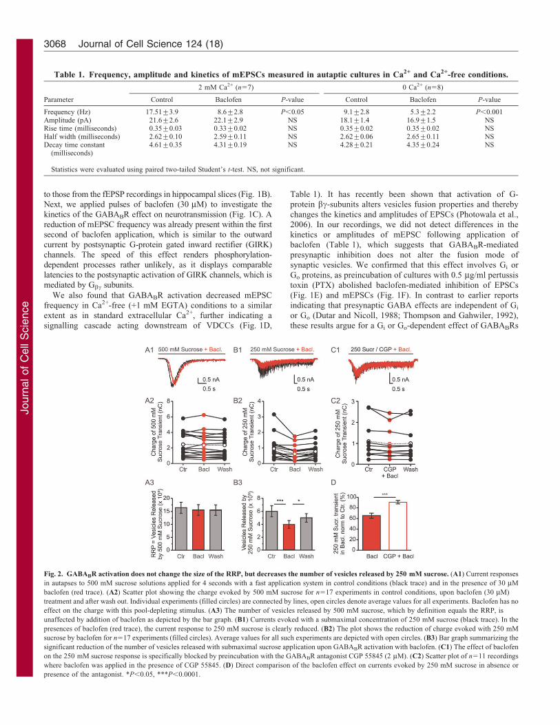
Fig. 2. GABABR activation does not change the size of the RRP, but decreases the number of vesicles released by 250 mM sucrose. (A1) Current responses
in autapses to 500 mM sucrose solutions applied for 4 seconds with a fast application system in control conditions (black trace) and in the presence of 30 mM
baclofen (red trace). (A2) Scatter plot showing the charge evoked by 500 mM sucrose for n517 experiments in control conditions, upon baclofen (30 mM)
treatment and after wash out. Individual experiments (filled circles) are connected by lines, open circles denote average values for all experiments. Baclofen has no
effect on the charge with this pool-depleting stimulus. (A3) The number of vesicles released by 500 mM sucrose, which by definition equals the RRP, is
unaffected by addition of baclofen as depicted by the bar graph. (B1) Currents evoked with a submaximal concentration of 250 mM sucrose (black trace). In the
presences of baclofen (red trace), the current response to 250 mM sucrose is clearly reduced. (B2) The plot shows the reduction of charge evoked with 250 mM
sucrose by baclofen for n517 experiments (filled circles). Average values for all such experiments are depicted with open circles. (B3) Bar graph summarizing the
significant reduction of the number of vesicles released with submaximal sucrose application upon GABABR activation with baclofen. (C1) The effect of baclofen
on the 250 mM sucrose response is specifically blocked by preincubation with the GABABR antagonist CGP 55845 (2 mM). (C2) Scatter plot of n511 recordings
where baclofen was applied in the presence of CGP 55845. (D) Direct comparison of the baclofen effect on currents evoked by 250 mM sucrose in absence or
presence of the antagonist. *P,0.05, ***P,0.0001.
Journal of Cell Science 124 (18)3068
Journ
alof
Cell
Scie
nce

on transmitter release, both at the level of VDCCs and further
downstream.
To gain further insight into the mechanism of VDCC-independent modulation of transmission by GABABRs, weused pulsed application of hypertonic solutions onto autaptic
cultures. Application of 500 mM sucrose can be used to test thereadily releasable pool (RRP) of vesicles (Rosenmund andStevens, 1996), whereas the sensitivity of the stimulated release
upon application of intermediate hypertonicity probes the energybarrier for vesicle fusion (Basu et al., 2007; Gerber et al., 2008).Application of 30 mM baclofen did not alter the size of the RRP
[Fig. 2A, 500 mM control, 16.3(¡2.2)6103 compared with +baclofen, 15.4(¡2.1)6103 vesicles; n517, P.0.05], whereasresponses to 250 mM sucrose were significantly reduced bybaclofen [Fig. 2B, 250 mM control, 5.9(¡0.8)6103 vesicles
compared with + baclofen: 3.9(¡0.6)6103; n517, P,0.001].Consequently, the fraction of vesicles released by 250 mMsucrose significantly decreased from 38.8(¡3.5)% to
26(¡2.5)% (values normalized to the RRP size determinedwith 500 mM sucrose), which amounts to a reduction of ,33%.We verified the specificity of the baclofen effect on
hypertonicity-evoked release by performing sucroseapplications in neurons preincubated with 2 mM CGP 55845, ahigh-affinity GABABR antagonist. In presence of the antagonist,
inhibition of EPSCs by 30 mM baclofen was essentially abolished(supplementary material Fig. S1A). CGP 55845 also stronglyreduced the inhibitory effect of baclofen on release evoked by250 mM sucrose [Fig. 2C1–C2, 250 mM control, 1.09(¡0.26)
nC compared with + baclofen, 0.97(¡0.22) nC; n511, P,0.05].The decrease in CGP 55845 was significantly smaller than theeffect of baclofen seen in absence of the GABABR antagonist
(Fig. 2D, P,0.001, unpaired two-sided Student’s t-test),confirming that the baclofen-induced increase of the energybarrier for vesicle fusion is specifically mediated by activation of
GABABRs.
Furthermore, we found that the peak vesicular release rate wassignificantly reduced in the presence of baclofen for both 500 and250 mM sucrose applications [Fig. 3A,B, values normalized to
500 mM control, 500 mM sucrose + baclofen, 80.5(¡3.9)%,n517, P,0.0001; 250 mM control, 13.1(¡1.1)% and 250 mMsucrose + baclofen, 7.0(¡0.6)%, n517, P,0.0001]. In addition,
the onset kinetics were significantly slower upon GABABRactivation [Fig. 3C1, 500 mM control, 0.52(¡0.01) secondscompared with + baclofen, 0.64(¡0.02) seconds, n517,
P,0.0001; 250 mM control, 0.91(¡0.06) seconds comparedwith + baclofen: 1.2(¡0.10) seconds, n517, P,0.001]. In linewith these results, the decay kinetics of the sucrose-evoked currenttransients were slower for 500 mM sucrose [Fig. 3C1, 500 mM
control 0.42(¡0.03) seconds + baclofen 0.51(¡0.02) seconds;n517, P,0.001]. As expected, co-application of the GABABRantagonist CGP 55845 blocked the decelerating effect of baclofen
on hypertonicity-evoked release for both 500 and 250 mM sucrose(supplementary material Fig. S1C). In summary, these findingsindicate that GABABR signalling directly affects vesicle release,
independently of VDCCs. As the RRP size is unaltered, it ratherseems that the fusion process itself is affected.
BoNT-A treatment does not prevent VDCC-independentinhibition
The above data strongly argue for a scenario in which GABABRactivation affects the release machinery in a way that increases
the energy demand for overcoming the fusion barrier for vesicle
release. Which specific components of the release apparatus
might be affected by GABABR activation? Considering the
Fig. 3. GABABR activation alters the kinetics of sucrose-evoked release.
(A1) Cumulative number of vesicles released in 500 mM sucrose (black curve)
from a representative experiment. Baclofen application (red curve) slows the
number of vesicles released with respect to time but leaves the total number of
released vesicles unaltered. The peak release rate is determined from the
maximum slope of the curves (straight lines in black and red, respectively). The
bar graph (A2) summarizes the significant effect of baclofen application on the
peak release rate evoked with 500 mM sucrose for n517 experiments. Values
are normalized to the release rate before baclofen application. (B1) Example
experiment demonstrating the cumulative release rate for a submaximal
stimulus with 250 mM sucrose (black curve). In the presence of baclofen (red)
both the release rate and the maximum number of released vesicles are reduced.
(B2) The effect of baclofen on the peak release rate, as determined from the
straight line fits in B1, is summarized in the bar graph for all experiments,
normalized to the release rates in 500 mM sucrose (n517). The peak release
rate is again significantly reduced by baclofen. (C1) Correlation plot showing
the effect of baclofen on the response latency and decay time constant of
sucrose-induced current transients. For 500 mM (black diamond) and 250 mM
(black circle) sucrose stimuli, addition of baclofen (red diamond and circle,
respectively) prolongs the onset latency and the decay time constant of the
current transients. (C2) Graph plotting the fraction of vesicles released from the
RRP against the peak release rate in the absence or presence of baclofen. As
GABABR activation decreases the peak release rate, it also decreases the
fraction of vesicles released from the RRP during submaximal hypertonic
stimuli (n517 for all panels). *P,0.05, ***P,0.0001.
GABABRs directly modulate vesicle fusion 3069
Journ
alof
Cell
Scie
nce

literature, a probable candidate would be a member of the
proteins forming the SNARE complex, specifically SNAP-25
(Blackmer et al., 2001; Gerachshenko et al., 2005). We incubated
cultures with BoNT-A, which cleaves nine C-terminal amino
acids from SNAP-25 and could thereby block SNAP-25-
dependent effects of baclofen. To achieve full cleavage of the
protein in neuronal cell culture, a 48-hour incubation with
500 ng/ml BoNT-A was required, as observed by western
blotting (Fig. 4A). Cleavage of SNAP-25 in autaptic neurons
resulted in complete silencing of transmission with respect to
both AP-evoked and spontaneous release (Fig. 4B,C). Moreover,
incubation with BoNT-A also prevented vesicle release with
hyperosmotic solutions (Fig. 4B). In order to test the possible
block of GABABR-mediated reduction of vesicle release by
SNAP-25 cleavage, we used 2 mM ionomycin to restore release
by introducing artificial Ca2+-permeable pores in BoNT-A-
treated cultures (Capogna et al., 1997). Application of
ionomycin led to a steady inward current with multiple
superimposed release events (Fig. 4C). All ionomycin-induced
currents were completely blocked by 10 mM NBQX [an 2-amino-
3-(5-methyl-3-oxo-1,2-oxazol-4-yl)propanoic acid (AMPA)-
receptor antagonist], confirming that this procedure evoked no
other conductances except for AMPA-receptor-mediated currents
(Fig. 4C, Fig. 5A). We calculated the total charge of release over
4 seconds before, during and after baclofen application and found
that baclofen reduced the ionomycin-evoked charge by 27% in
control cultures [Fig. 4D,E, untreated ionomycin + baclofen,
73.4(¡6.5)% of control; n511, P,0.05] and by 38% in BoNT-A
treated cultures [Fig. 4D,E, BoNT-A-treated ionomycin +
baclofen, 61.5(¡5.7)% of control; n59, P,0.05]. This effect
was not significantly different between the two conditions
(P50.2). Therefore, we conclude that GABABR activation
increases the barrier for vesicle fusion independently of SNAP-
25. This set of experiments was performed in the continuous
presence of SCH 23390 (20 mM) in order to block postsynaptic
GIRK channels activated by GABABRs (supplementary material
Fig. S2). The effectiveness of SCH 23390 was verified in all
experiments. In another series of experiments, we applied baclofen
in the presence and absence of 10 mM NBQX instead of SCH
23390. This enabled us to subtract the baclofen-induced GIRK
channel currents from the ionomycin experiments (Fig. 5A–C). In
addition, baclofen reduced the ionomycin-induced transmitter
release in control [Fig. 5B,D, 66.6(¡7.2)%, n59, P,0.001], as
Fig. 4. Cleavage of SNAP-25 with BoNT-A does not abolish Ca2+-
independent presynaptic inhibition by GABABRs. (A) Western blot
demonstrating the efficacy of SNAP-25 cleavage by BoNT-A. A 48-hour
incubation of neuronal cultures with 500 ng/ml BoNT-A is sufficient for full
cleavage of SNAP-25 (rightmost lane). The toxin cleaves the C-terminal nine
amino acids, reducing the molecular mass from 25 to 22.5 kDa. Double bands
for 24- to 72-hour incubations with 50 ng/ml BoNT-A indicate incomplete
cleavage. (B) BoNT-A treatment completely abolishes AP-triggered and
sucrose-evoked transmitter release in autaptic cultures (grey traces).
(C) Ionomycin (2 mM) was used to restore transmitter release in BoNT-A-
treated cultures. A steady inward current is superimposed with multiple
individual release events. The trace on the right-hand side shows, on an
expanded timescale, the individual ionomycin-induced currents that are
completely blocked by the AMPA receptor antagonist NBQX. (D) The upper
trace depicts the effect of baclofen applied during the steady state of
ionomycin-induced vesicle fusion in a control cell. The lower trace shows the
effect of baclofen on ionomycin-induced currents in a BoNT-A-treated
neuron. In both cases, baclofen reduced the currents to the same extent. (E)
Bar graph summarizing the reduction of ionomycin-induced currents by
baclofen. For quantification the charge was calculated over 4 seconds and
normalized to the control. Baclofen significantly reduced the ionomycin-
induced currents in control (n511, filled bars) and BoNT-A incubated
cultures (n59, open bars). The amount of reduction was not significantly
different between the two conditions. *P,0.05.
Journal of Cell Science 124 (18)3070
Journ
alof
Cell
Scie
nce
well as in BoNT-A-treated cultures [Fig. 5C,D: 75.6(¡10.8)%,
n510, P,0.05], further corroborating our evidence for a SNAP-
25-independent mode of action for GABABRs.
DiscussionIn the present study, we investigated the effects of GABABR
activation on evoked and spontaneous release from glutamatergic
hippocampal neurons and found that, in addition to the VDCC-
mediated effects, the activation of presynaptic GABABRs leads
to a substantial increase in the energy barrier for vesicle fusion.
We furthermore demonstrate that, in contrast to other synapses
studied previously, this modulation of the release machinery is
independent of the C-terminus of SNAP-25.
What are the mechanistic consequences of a release
modulation downstream of VDCCs? We addressed this
question by challenging autaptic neurons with hypertonic
solutions. Similar to high frequency ‘trains’ of APs,
applications of 500 mM sucrose allow to estimate the number
of vesicles in the RRP (Rosenmund and Stevens, 1996; Stevens
and Williams, 2007; Moulder and Mennerick, 2005). In addition,
hypertonic solutions have the advantage of providing a Ca2+-
independent stimulus to induce transmitter release, which is
indispensable when investigating VDCC-independent inhibition
of transmitter release. Hypertonic solutions provide energy to
vesicles primed for fusion, comparable to the effect of Ca2+-
binding to synaptotagmin. When the tonicity at synapses
increases, more primed vesicles reach the energy threshold for
fusion. If the energy threshold for primed vesicles is increased by
a Gi or Go-dependent mechanism, responses to subsaturating
tonicities, such as 250 mM added sucrose, will decrease in size
relative to the maximal response induced by 500 mM sucrose. In
addition, the onset of hypertonically induced transmitter release
will be delayed and the maximal release rate will decrease. We
found that this was indeed the case, as GABABR activation
substantially slows the kinetics of sucrose-evoked fusion and
reduces the total number of vesicles released by intermediate
hypertonic stimuli. With similar methods, it was previously
possible to identify mutations that change the energy barrier for
fusion independently of the priming processes further upstream in
the presynaptic vesicle cycle. For example, the gain-of-function
mutation H567K in the vesicle priming factor Munc13-1 lowers
the energy barrier for vesicle fusion, resulting in an increased
sensitivity to sucrose challenge and increased spontaneous
vesicle release, whereas it does not affect the RRP size (Basu
et al., 2007). Conversely, complexin-deficient synapses have a
normal RRP size, but show a comparable increase of the energy
barrier and reduction in spontaneous release (Xue et al., 2010), as
seen for GABABR activation in the current work. So, our results
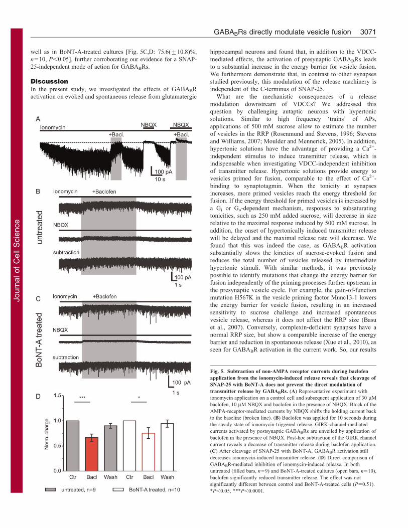
Fig. 5. Subtraction of non-AMPA receptor currents during baclofen
application from the ionomycin-induced release reveals that cleavage of
SNAP-25 with BoNT-A does not prevent the direct modulation of
transmitter release by GABABRs. (A) Representative experiment with
ionomycin application on a control cell and subsequent application of 30 mM
baclofen, 10 mM NBQX and baclofen in the presence of NBQX. Block of the
AMPA-receptor-mediated currents by NBQX shifts the holding current back
to the baseline (broken line). (B) Baclofen was applied for 10 seconds during
the steady state of ionomycin-triggered release. GIRK-channel-mediated
currents activated by postsynaptic GABABRs are unveiled by application of
baclofen in the presence of NBQX. Post-hoc subtraction of the GIRK channel
current reveals a decrease of transmitter release during baclofen application.
(C) After cleavage of SNAP-25 with BoNT-A, GABABR activation still
decreases ionomycin-induced transmitter release. (D) Direct comparison of
GABABR-mediated inhibition of ionomycin-induced release. In both
untreated (filled bars, n59) and BoNT-A-treated cultures (open bars, n510),
baclofen significantly reduced transmitter release. The effect was not
significantly different between control and BoNT-A-treated cells (P50.51).
*P,0.05, ***P,0.0001.
GABABRs directly modulate vesicle fusion 3071
Journ
alof
Cell
Scie
nce
establish a role for a direct modulatory influence on GABABRsfor the vesicular release machinery in the mammalian synapse.
The C-terminus of the SNARE complex member SNAP-25 has
been suggested to be the target for direct modulation of vesicularfusion by GPCRs, as it has been found to be directly affected,through Gbc-subunits, in serotonin-mediated modulation in the
spinal cord of lampreys (Blackmer et al., 2001; Gerachshenko et al.,2005). A similar mechanism has been proposed for noradrenaline(norepinephrine)-mediated modulation at glutamatergic fibres onto
the central amygdala in rats, also arguing for SNAP-25 as a directtarget of metabotropic signalling at mammalian synapses (Delaneyet al., 2007). In the case of the lamprey spinal cord, synapse direct
modulation was also reported to involve alterations in the openingof the vesicular fusion pore (Photowala et al., 2006; Schwartz et al.,2007). Mechanistically these studies suggested that the interactionof G-protein bc-subunits with the C-terminus of SNAP-25
competes with synaptotagmin for the binding site to the SNAREcomplex, thereby hindering tight coupling of the Ca2+ sensor to thevesicle (Blackmer et al., 2005). On the basis of three lines of
evidence, we rather exclude the SNAP-25 interaction site withsynaptotagmin as a target for G-protein bc-subunit-mediateddirect inhibition of neurotransmitter release by GABABRs. First,
we find a reduction of mEPSC frequency by GABABR activationin Ca2+-free extracellular solution, arguing against theinvolvement of the Ca2+ sensor. Second, we observe thatGABABRs cause a reduction in sucrose-evoked transmitter
release that is independent of Ca2+ (Rosenmund and Stevens,1996) and synaptotagmin-1 (Xu et al., 2009). Finally, we showthat cleavage of SNAP-25 by BoNT-A does not abolish the
inhibitory effect of GABABRs on ionomycin-evoked release. Apossible explanation for the discrepancy between our data andstudies that did show an involvement of SNAP-25 is that
metabotropic receptors might in fact utilize different pathwaysat distinct synapses and thereby possess diverse targets formodulation. The completeness of the BoNT-A block might also
affect the interpretation of results. We have taken great care toachieve complete cleavage of SNAP-25, necessitating activerestoration of release by agents such as ionomycin (Capogna et al.,1997). Therefore, we can exclude the involvement of the SNAP-
25 C-terminus in direct inhibition by GABABRs at hippocampalsynapses. However, our experiments do indicate a GABABR-mediated direct effect on the core release machinery that is
involved in the very late steps of exocytosis. A study in the calyxof Held has demonstrated that GABABR activation can directlymodulate vesicle priming, leading to a slower refilling of the RRP.
This effect, however, was observed only after prolongedstimulation paradigms, and it involved a cAMP-dependentsignalling cascade (Sakaba and Neher, 2003). Because we did
not detect any changes in the RRP size, the docking and primingsteps seem unimpaired by GABABR activation during lowfrequency activity in our preparation. So, direct inhibition ofvesicle fusion acts after priming, at a very late step of transmitter
release.
Here, we establish a role for a direct modulation of the releasemachinery during GABAergic inhibition of transmission at
glutamatergic synapses, which adds to our understanding ofmetabotropic signalling. What functional relevance could beattributed to the increased energy barrier seen in our study?
Inhibiting release while only slightly reducing Ca2+ influx mightbe advantageous in situations of higher frequency activity, duringwhich a substantial Ca2+ influx is required for accelerated
replenishing of the RRP (Wang and Kaczmarek, 1998). The exacttarget of the release machinery that is responsible for this
modulation remains to be determined and will be the focus offuture studies.
Materials and MethodsAll experiments were performed according to the rules of Berlin authorities and theanimal welfare committee of the Charite Berlin. For field potential, whole-cell andCa2+-imaging experiments, sagittal slices (300 mm) were prepared from C57/BL6mice at postnatal day 19–29. Animals were briefly anesthetized with isofluoraneand decapitated. Brains were rapidly removed and chilled in cold solutionscontaining: 87 mM NaCl, 26 mM NaHCO3, 75 mM sucrose, 25 mM glucose,2.5 mM KCl, 1.25 mM NaH2PO4, 0.5 mM CaCl2 and 7 mM MgCl2, saturatedwith 95% O2 and 5% CO2. Slices were cut on a vibratome (VT 1200S, Leica) andincubated at 34 C for 30 minutes. They were transferred into the recordingsolution containing: 119 mM NaCl, 26 mM NaHCO3, 10 mM glucose, 2.5 mMKCl, 1 mM NaH2PO4, 2.5 mM CaCl2 and 1.3 mM MgCl2, equilibrated with 95%O2 and 5% CO2 at room temperature.
For photodiode Ca2+ measurements (Gundlfinger et al., 2007; Regehr and Tank,1991), slices were placed under an upright microscope (BX51WI, Olympus)equipped with an Olympus LumPlan FI 606 0.9 NA water-immersion objective.Schaffer collaterals were locally labelled with the low-affinity Ca2+ indicatorMagnesium Green AM (100 mM, Invitrogen) dissolved in 20% Pluronic/DMSO.Epifluorescence was measured outside of the loading spot at least 30 minutes afterlabelling. Signals were low-pass filtered at 1 kHz, digitized at 5 kHz and recordedwith IGOR Pro (WaveMetrics). AP-triggered change in fluorescence intensity(DF) relative to the baseline intensity of fluorescence (F) was calculated as DF/F.
Field EPSPs (fEPSPs) were recorded with low-resistance pipettes filled withexternal solution in the stratum radiatum of CA1 using a Multiclamp 700A(Molecular Devices). Fibres were stimulated at 0.1 Hz. Data were filtered at 2 kHzand digitized at 5 kHz and recorded with IGOR. Extracellular CaCl2 was replacedby equimolar concentrations of MgCl2 to keep the total concentration of divalentcations constant.
Autaptic cultures were prepared as described previously (Bekkers and Stevens,1991; Pyott and Rosenmund, 2002). For astrocyte precultures, cortices of newbornmice (postnatal day zero, P0) were digested with trypsin. Cells were grown in T75flasks in BME medium (with 10% fetal calf serum, 1 mM Glutamax, 0.2%penicillin-streptomycin, 10 mM HEPES, 5 mM glucose and 2.5 mg/ml insulin;Invitrogen and Sigma) for 1 week. Growth permissive 200-mm spots of a 1:4collagen and poly-D-lysine mixture were printed, using a custom-made stamp, onagarose-coated coverslips in six-well plates. 26104 astrocytes from precultureswere seeded per well and grown until fully covering the microislands. Beforeplating hippocampal neurons (36103 cells per well), the medium was changed toNeurobasal A (with 2% B27, 0.2% penicillin-streptomycin; Invitrogen). To obtainneurons, P0 mouse hippocampi were digested using papain (Worthington, 20 units/ml, 1 mM L-cysteine, 0.5 mM EDTA in EBSS) for 60 minutes at 37 C.
Whole-cell voltage clamp recordings were performed with Axopatch 200A orMulticlamp 700B amplifiers using borosilicate electrodes (2–3 mV tip resistance)filled with: 146 mM K-gluconate, 17.8 mM Hepes, 1 mM EGTA, 4 mM Mg-ATP,0.3 mM Na-GTP, 12 mM creatine phosphate and 50 units per ml creatine-phosphokinase (pH adjusted to 7.3 with KOH). Data were filtered at 2–5 kHz anddigitized at 5–10 kHz. Fast applications of hypertonic solutions were performedusing procedures described previously (Pyott and Rosenmund, 2002; Rosenmundet al., 1995). Sucrose was dissolved in extracellular solution containing: 140 mMNaCl, 2.4 mM KCl, 10 mM HEPES, 10 mM glucose, 2 mM CaCl2 and 4 mMMgCl2 (pH adjusted to 7.3 with NaOH, 300 mOsm).
To determine the efficacy of Botulinum neurotoxin A (BoNT-A), protein lysatesof toxin-treated neuronal cultures were analyzed by western blotting using amonoclonal antibody directed against SNAP-25 (Synaptic Systems, Goettingen,Germany).
Data are given as means ¡ standard error. Significance was determined usingpaired Student’s t-test or single-factor ANOVA with Bonferroni’s post-hoc testwhere appropriate. The number of vesicles in the RRP was calculated bynormalizing the charge of the 500 mM sucrose transient by the mean mEPSCcharge.
We thank Anke Schonherr for excellent technical assistance. Wewould also like to thank Sarah Shoichet for help preparing themanuscript. D.S. is supported by grants from the DFG (SFB 618,SFB 665, GRK1123, EXC257), the Einstein Foundation and theBMBF (Bernstein Focus, Bernstein Centre for ComputationalNeuroscience); C.R. is supported by the DFG (EXC257).
Supplementary material available online at
http://jcs.biologists.org/lookup/suppl/doi:10.1242/jcs.074963/-/DC1
Journal of Cell Science 124 (18)3072
Journ
alof
Cell
Scie
nce
ReferencesBasu, J., Betz, A., Brose, N. and Rosenmund, C. (2007). Munc13-1 C1 domain activation
lowers the energy barrier for synaptic vesicle fusion. J. Neurosci. 27, 1200-1210.Bekkers, J. M. and Stevens, C. F. (1991). Excitatory and inhibitory autaptic currents in
isolated hippocampal neurons maintained in cell culture. Proc. Natl. Acad. Sci. USA
88, 7834-7838.Blackmer, T., Larsen, E. C., Takahashi, M., Martin, T. F., Alford, S. and Hamm,
H. E. (2001). G protein betagamma subunit-mediated presynaptic inhibition:regulation of exocytotic fusion downstream of Ca2+ entry. Science 292, 293-297.
Blackmer, T., Larsen, E. C., Bartleson, C., Kowalchyk, J. A., Yoon, E. J.,
Preininger, A. M., Alford, S., Hamm, H. E. and Martin, T. F. (2005). G proteinbetagamma directly regulates SNARE protein fusion machinery for secretory granuleexocytosis. Nat. Neurosci. 8, 421-425.
Bowery, N. G. (2006). GABAB receptor: a site of therapeutic benefit. Curr. Opin.
Pharmacol. 6, 37-43.Capogna, M., Gahwiler, B. H. and Thompson, S. M. (1996). Presynaptic inhibition of
calcium-dependent and -independent release elicited with ionomycin, gadolinium,and alpha-latrotoxin in the hippocampus. J. Neurophysiol. 75, 2017-2028.
Capogna, M., McKinney, R. A., O’Connor, V., Gahwiler, B. H. and Thompson,
S. M. (1997). Ca2+ or Sr2+ partially rescues synaptic transmission in hippocampalcultures treated with botulinum toxin A and C, but not tetanus toxin. J. Neurosci. 17,7190-7202.
Cryan, J. F. and Kaupmann, K. (2005). Don’t worry ‘B’ happy!: a role for GABAB
receptors in anxiety and depression. Trends Pharmacol. Sci. 26, 36-43.Delaney, A. J., Crane, J. W. and Sah, P. (2007). Noradrenaline modulates transmission
at a central synapse by a presynaptic mechanism. Neuron 56, 880-892.Dunlap, K. and Fischbach, G. D. (1981). Neurotransmitters decrease the calcium
conductance activated by depolarization of embryonic chick sensory neurones.J. Physiol. 317, 519-535.
Dutar, P. and Nicoll, R. A. (1988). Pre- and postsynaptic GABAB receptors in thehippocampus have different pharmacological properties. Neuron 1, 585-591.
Gerachshenko, T., Blackmer, T., Yoon, E. J., Bartleson, C., Hamm, H. E. and
Alford, S. (2005). Gbetagamma acts at the C terminus of SNAP-25 to mediatepresynaptic inhibition. Nat. Neurosci. 8, 597-605.
Gerber, S. H., Rah, J. C., Min, S. W., Liu, X., de Wit, H., Dulubova, I., Meyer, A. C.,Rizo, J., Arancillo, M., Hammer, R. E. et al. (2008). Conformational switch ofsyntaxin-1 controls synaptic vesicle fusion. Science 321, 1507-1510.
Gundlfinger, A., Bischofberger, J., Johenning, F. W., Torvinen, M., Schmitz, D. and
Breustedt, J. (2007). Adenosine modulates transmission at the hippocampal mossyfibre synapse via direct inhibition of presynaptic calcium channels. J. Physiol. 582,263-277.
Kajikawa, Y., Saitoh, N. and Takahashi, T. (2001). GTP-binding protein beta gammasubunits mediate presynaptic calcium current inhibition by GABAB receptor. Proc.
Natl. Acad. Sci. USA 98, 8054-8058.Luscher, C., Jan, L. Y., Stoffel, M., Malenka, R. C. and Nicoll, R. A. (1997). G
protein-coupled inwardly rectifying K+ channels (GIRKs) mediate postsynaptic butnot presynaptic transmitter actions in hippocampal neurons. Neuron 19, 687-695.
Moulder, K. L. and Mennerick, S. (2005). Reluctant vesicles contribute to the total
readily releasable pool in glutamatergic hippocampal neurons. J. Neurosci. 25, 3842-
3850.
Newberry, N. R. and Nicoll, R. A. (1984). Direct hyperpolarizing action of baclofen on
hippocampal pyramidal cells. Nature 308, 450-452.
Photowala, H., Blackmer, T., Schwartz, E., Hamm, H. E. and Alford, S. (2006). G
protein betagamma-subunits activated by serotonin mediate presynaptic inhibition by
regulating vesicle fusion properties. Proc. Natl. Acad. Sci. USA 103, 4281-4286.
Pyott, S. J. and Rosenmund, C. (2002). The effects of temperature on vesicular supply
and release in autaptic cultures of rat and mouse hippocampal neurons. J. Physiol.
539, 523-535.
Regehr, W. G. and Tank, D. W. (1991). Selective fura-2 loading of presynaptic
terminals and nerve cell processes by local perfusion in mammalian brain slice.
J. Neurosci. Methods 37, 111-119.
Rosenmund, C. and Stevens, C. F. (1996). Definition of the readily releasable pool of
vesicles at hippocampal synapses. Neuron 16, 1197-1207.
Rosenmund, C., Feltz, A. and Westbrook, G. L. (1995). Synaptic NMDA receptor
channels have a low open probability. J. Neurosci. 15, 2788-2795.
Sakaba, T. and Neher, E. (2003). Direct modulation of synaptic vesicle priming by
GABAB receptor activation at a glutamatergic synapse. Nature 424, 775-778.
Scanziani, M. (2000). GABA spillover activates postsynaptic GABAB receptors to
control rhythmic hippocampal activity. Neuron 25, 673-681.
Scanziani, M., Capogna, M., Gahwiler, B. H. and Thompson, S. M. (1992).
Presynaptic inhibition of miniature excitatory synaptic currents by baclofen and
adenosine in the hippocampus. Neuron 9, 919-927.
Scholz, K. P. and Miller, R. J. (1992). Inhibition of quantal transmitter release in the
absence of calcium influx by a G protein-linked adenosine receptor at hippocampal
synapses. Neuron 8, 1139-1150.
Schwartz, E. J., Blackmer, T., Gerachshenko, T. and Alford, S. (2007). Presynaptic
G-protein-coupled receptors regulate synaptic cleft glutamate via transient vesicle
fusion. J. Neurosci. 27, 5857-5868.
Stevens, C. F. and Williams, J. H. (2007). Discharge of the readily releasable pool with
action potentials at hippocampal synapses. J. Neurophysiol. 98, 3221-3229.
Takahashi, T., Kajikawa, Y. and Tsujimoto, T. (1998). G-Protein-coupled modulation
of presynaptic calcium currents and transmitter release by a GABAB receptor.
J. Neurosci. 18, 3138-3146.
Thompson, S. M. and Gahwiler, B. H. (1992). Comparison of the actions of baclofen at
pre- and postsynaptic receptors in the rat hippocampus in vitro. J. Physiol. 451, 329-345.
Wang, L. Y. and Kaczmarek, L. K. (1998). High-frequency firing helps replenish the
readily releasable pool of synaptic vesicles. Nature 394, 384-388.
Xu, J., Pang, Z. P., Shin, O. H. and Sudhof, T. C. (2009). Synaptotagmin-1 functions
as a Ca2+ sensor for spontaneous release. Nat. Neurosci. 12, 759-766.
Xue, M., Craig, T. K., Xu, J., Chao, H. T., Rizo, J. and Rosenmund, C. (2010).
Binding of the complexin N terminus to the SNARE complex potentiates synaptic-
vesicle fusogenicity. Nat. Struct. Mol. Biol. 17, 568-575.
GABABRs directly modulate vesicle fusion 3073
Journ
alof
Cell
Scie
nce