activity-based metagenomic screening and biochemical ...aem.asm.org/content/77/22/8106.full.pdf ·...
TRANSCRIPT
APPLIED AND ENVIRONMENTAL MICROBIOLOGY, Nov. 2011, p. 8106–8113 Vol. 77, No. 220099-2240/11/$12.00 doi:10.1128/AEM.05925-11Copyright © 2011, American Society for Microbiology. All Rights Reserved.
Activity-Based Metagenomic Screening and Biochemical Characterization ofBovine Ruminal Protozoan Glycoside Hydrolases�†
Seth D. Findley,1 Melanie R. Mormile,2 Andrea Sommer-Hurley,1 Xue-Cheng Zhang,1 Peter Tipton,3
Krista Arnett,3 James H. Porter,4 Monty Kerley,4 and Gary Stacey1,3*Center for Sustainable Energy, National Center for Soybean Biotechnology, Division of Plant Sciences, University of Missouri,
Columbia, Missouri1; Department of Biological Sciences, Missouri University of Science and Technology, Rolla, Missouri2;Division of Biochemistry, University of Missouri, Columbia, Missouri3; and Division of Animal Sciences,
University of Missouri, Columbia, Missouri4
Received 23 June 2011/Accepted 9 September 2011
The rumen, the foregut of herbivorous ruminant animals such as cattle, functions as a bioreactor to processcomplex plant material. Among the numerous and diverse microbes involved in ruminal digestion are theruminal protozoans, which are single-celled, ciliated eukaryotic organisms. An activity-based screen wasexecuted to identify genes encoding fibrolytic enzymes present in the metatranscriptome of a bovine ruminalprotozoan-enriched cDNA expression library. Of the four novel genes identified, two were characterized inbiochemical assays. Our results provide evidence for the effective use of functional metagenomics to retrievenovel enzymes from microbial populations that cannot be maintained in axenic cultures.
To process fibrous plant materials, the rumen harbors acollection of diverse microorganisms, including bacteria, ar-chaea, fungi, and protozoa (reviewed in references 33 and 45).While the diversity and functions of the thousands of microbialspecies of this unique ecosystem (32) are interesting from boththe evolutionary and functional perspectives, the rumen alsorepresents a rich resource of enzymes for converting lignocel-lulosic feedstocks into biofuel (35, 43) and other applications(19). A range of inexpensive, robust enzymes with a broadrange of specificities will likely be required for efficient indus-trial processing of highly complex plant polysaccharides. Iden-tification of such enzymes that microorganisms use to breakdown plant materials has been greatly facilitated by meta-genomics (42), both in the form of activity-based screens (20,52) and also through increasingly powerful, high-throughputgenomic DNA sequencing approaches (28, 57). As evident innumerous studies (28, 39, 41), metagenomics has proven to beparticularly effective for identification of carbohydrate-activegenes of fiber-adherent bacterial species of the rumen.
In addition to bacteria and archaea, the rumen also hostseukaryotic species, namely, anaerobic fungi and ciliate proto-zoans (reviewed in reference 33). Addressing the function ofruminal protozoans, in particular, has been a challenge due tothe difficulty of maintaining these organisms in axenic cultures(55). Thus, assessing the diversity and dynamics of ruminalprotozoans has been addressed historically in morphogenicstudies (reviewed in reference 12) and by molecular phyloge-netics (e.g., by using 18S rDNA markers [47]). Ruminal pro-tozoans are known to contribute to fiber degradation in their
hosts (21), and determination and characterization of theirability to directly process plant material have been addressedby diverse strategies, such as direct, biochemical detection ofspecific fibrolytic enzymes (e.g., cellulases) in extracts derivedfrom individual protozoan species (38, 54), by molecular clon-ing studies to directly identify genes encoding enzymes capableof degrading cellulose or hemicellulose (49, 50) and, mostrecently, by sequencing of protozoan-derived expressed se-quence tag (EST) libraries (41). Early studies to establish thecapacity of protozoan species to express their own enzymes fordegradation of plant material included that of Howard et al.(29), who demonstrated that Epidinium ecaudatum indeedcontains fibrolytic enzyme activity. Similarly, Bailey et al. (3)demonstrated the presence of both a hemicellulase and a xy-lobiase in E. ecaudatum by using purified cell extracts. Morerecently, Clayet et al. (8), using gel filtration of E. ecaudatumextracts, identified at least 10 distinct enzyme activities forplant cell wall degradation; their fractions contained a range ofenzymes with glycoside hydrolase (GH) activities, includingtwo distinct carboxymethylcellulases with molecular masses of23 and 45 kDa (8).
Altogether, about a dozen protozoan fibrolytic genes havebeen identified in activity-based molecular screens; a compa-rable number have been identified from informatics-basedstudies (41), predominantly in ovine and bovine rumen sys-tems. The protozoan enzyme genes characterized to date arediverse, both in terms of the individual GH domains (27)utilized as well as the combinatorial domain organization ofproteins that contain them. GH domains are modular by de-sign; they exist in individual polypeptides in variable copy num-bers and variable association with other, noncatalytic modules(e.g., carbohydrate binding domains [reviewed in reference27]). The described rumen protozoan-derived GH domaingenes primarily encode single or dual GH5 domains (cellulasesuperfamily) (49, 50, 53) or GH10 or GH11 domains (xylanase-related domains) (4, 14, 15). The combinatorial complexity offibrolytic genes thus far detected in ruminal ciliates speaks to
* Corresponding author. Mailing address: Division of Plant Sci-ences, Center for Sustainable Energy, National Center for SoybeanBiotechnology, University of Missouri, Columbia, MO 65211. Phone:(573) 884-4752. Fax: (573) 884-4799. E-mail: [email protected].
† Supplemental material for this article may be found at http://aem.asm.org/.
� Published ahead of print on 23 September 2011.
8106
on July 20, 2018 by guesthttp://aem
.asm.org/
Dow
nloaded from
the potentially diverse utilization of GH modules within theentire ruminal protozoan population (4, 14, 15, 41, 49, 50, 53).Yet, due in part to the importance of demonstrating the exis-tence of fibrolytic genes in a given protozoan species, enzymecloning studies have largely been conducted using mono-fau-nated animals, in which the host ruminant is inoculated with asingle ciliate species. Therefore, to investigate the potentialdiversity of fibrolytic enzymes in a total ciliate population, anactivity-based metagenomics screen was conducted of themetatranscriptome of protozoans in the rumen fluid derivedfrom a single, fistulated cow.
MATERIALS AND METHODS
Materials. Carboxymethyl cellulose (CMC), cellulose (fibrous, medium), ga-lactan, laminarin from Laminaria digitata, mannan from Saccharomyces cerevi-siae, and xylan from beechwood were obtained from Sigma-Aldrich. AZCL-HE-cellulose, AZCL-xylan (oat), arabinan (sugar beet), �-glucan (oat, mediumviscosity), wheat arabinoxylan (medium viscosity), and xyloglucan from tamarindseed (amyloid) were obtained from Megazyme, Inc. Reagents for the reducingsugar assay, ammonium iron(III) sulfate dodecahydrate, 3-methyl-2-benzothia-zolinone hydrazone hydrochloride hydrate, and sulfanic acid, were obtained fromSigma-Aldrich. Isopropyl �-D-1-thiogalactopyranoside (IPTG) was obtainedfrom Gold Biotechnology.
Rumen sample collection, protozoa purification, and mRNA purification. Ru-men protozoans were harvested by a procedure based on that described inreference 36, modified as follows: approximately 3 liters of total rumen content(fluid and solids) was collected from a fistulated Holstein cow maintained at theUniversity of Missouri Dairy Farm, Columbia, MO. The donor cow was fed atotal mixed ration (TMR) of a common lactation diet that consisted of alfalfahaylage, corn silage, corn, and protein, minerals, and fat-soluble vitamins addedto meet or exceed nutrient requirements. The rumen sample was collected in themorning, prior to feeding, into a prewarmed canister and transported to the labwithin 30 min. Aliquots of the sample were treated in three pulses of 30 s each,with a blender, and pressed through a double layer of cheesecloth to remove bulksolids. The resulting liquid (approximately 1 liter) was supplemented to 1%maltose and 0.5% sucrose and then flocculated anaerobically for 1 h at 39°C.After aspirating the floating feed particle layer, the liquid was dialyzed againsteight changes of 39°C Coleman anaerobic buffer (55) by using a home-made10-�m-pore-size Nitex filter cloth bag (Sefar America) with gentle agitation. Thevolume of the resulting material was �50 ml and represented a concentratedmixture of protozoa whose composition was verified by microscopic observation.Total RNA was isolated from this material using the TRIzol Plus RNA purifi-cation system (Invitrogen); mRNA was then purified using the magnetic bead-based FastTrack 2.0 mRNA isolation kit (Invitrogen).
Lambda Zap-based protozoan cDNA library construction. A Lambda ZapII-based protozoan cDNA expression library was constructed by using the ZapcDNA synthesis kit (catalog number 200401; Stratagene), starting with 5 �g ofpolyadenylated mRNA (for detailed protocols, the manual for this cDNA syn-thesis kit is available on the Agilent website). After size fractionation of totalcDNA by gel exclusion chromatography using Sepharose CL-2B gel filtrationmedium (procured from Stratagene) in a 1-ml disposable plastic pipette, cDNAfractions ranging from 0.5 to �10 kb were pooled and ligated into the preparedlambda vector (see below) and packaged using the ZAP-cDNA Gigapack IIIgold cloning kit (catalog number 200450; Stratagene), according to protocolsprovided by the manufacturer. After titer determinations, 700,000 PFU of theprimary library were amplified on plates by using standard lambda phage pro-cedures (Stratagene Zap cDNA synthesis manual) in order to generate thesecondary library, which was used for activity-based screening.
Activity-based screen for fibrolytic enzymes. To identify candidate fibrolyticenzymes for IPTG-inducible cDNA expression, plaque-based high-throughputscreening used plates containing dye-linked insoluble polysaccharide substrates(44, 51). Two classes of fibrolytic enzymes were screened: xylanases and cellu-lases, using AZCL-HE-cellulose and AZCL-xylan (oat), respectively (Mega-zyme, Inc.). The expression library was screened on petri dishes containingNZYM bottom agar supplemented with a 1� micronutrient solution (1,000�MNS; 3.0 mM H3BO3, 0.46 mM MnCl2, 0.16 mM CuSO4, 0.6 mM ZnSO4, 0.1mM NaMoO4, 0.01 mM NiSo4, 0.01 mM CoCl2) and NZYM top agarose,supplemented with 1� MNS plus 20 mM IPTG. To identify cDNA clonesencoding fibrolytic enzymes, the top agarose incorporated either AZCL-HE-cellulase or AZCL-xylan (oat) at a final concentration of 0.3% (wt/vol). For each
substrate, approximately 1,000,000 PFU were screened (10,000 PFU per 150-mmplate). Over a 2- to 5-day incubation period at 37°C, 70 clones were picked thatexhibited xylan-degrading activity and 10 clones were picked that exhibitedcellulose-degrading activity. Positives were plaque purified in three rounds ofplaque purification and then in vivo excised to generate pBluescript DNA prep-arations, using procedures described in the Stratagene Zap cDNA synthesismanual. To initially characterize the positives, rescued plasmid-borne cDNAswere sequenced with the T3 promoter primer (5�-AAT-TAA-CCC-TCA-CTA-AAG-GG-3�), which flanks the 5� end of the directionally cloned cDNA insert.Selected clones of the longer cDNA types (types 3 and 4, see below) were thencompletely sequenced with the T7 promoter primer (5�-TAA-TAC-GAC-TCA-CTA-TAG-GG-3�) in addition to custom, internal primers when required (datanot shown). To initially assess the diversity of the cDNA collection, the CAP3sequence assembly program (30), BLASTp (1), and ClustalW2 (6) were used.The sequences of the longest cDNAs of each type (see Results for analysisfindings) were deposited in GenBank (see below).
Sequence searches, alignments, and phylogeny. Protozoan and bacterial gly-coside hydrolase sequences were collected for our analysis using BLASTpsearches (31) of the GenBank nonredundant (nr) protein sequences database,using default search parameters to identify sequences with homology values of1e�50. Protein sequences were aligned using MUSCLE 3.6 (16) with a FASTAoutput format and then manually edited using Jalview (7). Majority-ruled par-simonious trees were generated using the program protpars of PHYLIP (18),with maximum likelihood branch lengths calculated using TREE-PUZZLE (46).Bootstrap values were calculated using the program seqboot of the PHYLIPpackage. All trees were viewed and printed in a pdf format using Tree Viewer(58).
Molecular cloning for expression constructs. To investigate the biochemicalproperties of representative positives (see Results section for summary of posi-tive classes; see also Fig. S1 in the supplemental material), a single-domain genefor each substrate screened was characterized: one type 1 (identified on cellulosesubstrate) and one type 2 (identified on xylan substrate). The coding sequence ofthe longest cDNA from each class was cloned into the NcoI and XhoI sites of themultiple-cloning site of the C-terminal His6 tag expression vector, pET29a(Novagen). To accomplish this, restriction sites were added to the PCRprimers and amplified DNA fragments as follows. For type I (cellulose sub-strate), the 5� primer sequence was GGG-CCA-TGG-CTT-TGG-GCT-TAA-TTT-CAA-TTTC (the NcoI site is underlined and the first codon of the cDNAis indicated by bold text) and the 3� PCR primer sequence was GGG-CTC-GAG-TTT-GGA-AAC-AGC-GGC-TTT-GTA-AG (XhoI site is underlined). For type2, the 5� primer sequence was GGG-CCA-TGG-CTT-TAA-ATT-ATG-TAT-CAT-CTA-ATA-ATT-TTC (NcoI site is underlined and the first codon of thecDNA is indicated by bold text) and the 3� PCR primer sequence was GGG-CTC-GAG-TGC-TCC-AGC-AAC-TTG-CAT-AAT (XhoI site is underlined).Coding sequences were amplified using the Platinum PCR supermix high-fidelitykit (Invitrogen) or Phusion high-fidelity DNA polymerase (New England Bio-Labs) under the conditions recommended by the manufacturers. The resultingamplicon DNAs were purified using the Wizard PCR DNA purification system(Promega) and cloned into pET29a, which was prepared by standard molecularbiology techniques. Miniprep (Promega) DNA for each type was sequenced withT7 promoter and T7 terminator primers to verify the absence of PCR-derivedmutations. The resulting expression constructs, and proteins derived from them,are designated here type 1-7.1 and type 2-8.6.
Expression and purification of recombinant enzymes. The type 1-7.1 and type2-8.6 pET29a constructs were transformed into Escherichia coli BL21(DE3) cells(Invitrogen), and expression cultures for each construct were grown at 37°C in500 ml LB broth containing 30 �g/ml kanamycin. The cultures were grown to anoptical density at 600 nm (OD600) of 0.6 to 0.8, at which point expression wasinduced by the addition of IPTG to a final concentration of 1 mM. Cultures werethen grown at 37°C for an additional 3 h. Bacterial cells were harvested bycentrifugation (10,000 � g at 4°C for 10 min) and resuspended in 20 ml ofequilibration/wash solution (50 mM sodium phosphate buffer [pH 7.0], 300 mMNaCl) supplemented with 1� Complete, EDTA-free protease inhibitor cocktail(Roche) and phenylmethylsulfonyl fluoride to a final concentration of 1 mM.Cell resuspensions were lysed using a French press, and the resulting lysates werecentrifuged at 10,000 � g at 4°C for 10 min, to remove cell debris. Recombinantproteins were then affinity purified by using Talon metal affinity resin (Clontech),according to the manufacturer’s protocols. The purified enzymes were eluted ina single-step elution with equilibration/wash buffer supplemented with 150 mMimidazole. Proteins were then concentrated and imidazole removed with anultrafiltration membrane (Vivaspin-20 column; GE Healthcare). Enzyme puritywas confirmed by SDS-PAGE (see Fig. S2 in the supplemental material), and the
VOL. 77, 2011 METAGENOMICS ANALYSIS OF RUMINAL PROTOZOAN GH ENZYMES 8107
on July 20, 2018 by guesthttp://aem
.asm.org/
Dow
nloaded from

protein concentration was determined using the Quick Start Bradford dye re-agent (Bio-Rad).
Enzyme assays. The activity of each enzyme was initially confirmed in a simple,colorimetric assay using the same insoluble substrates that were used in the platescreening procedure. Specifically, 1 mg of the respective substrate (AZCL-HE-cellulose or AZCL-xylan) was suspended in 1 ml of protein purification equili-bration buffer (300 mM NaCl, 50 mM sodium phosphate [pH 7.0]), at 37°C for30 min. Reactions were initiated by adding 50 �l of purified enzyme (0.5 to 2.2mg/ml protein), and the release of solubilized dye was visually validated. OptimalpH conditions were then preliminarily determined in assays using 550 �l of 50mM Britton-Robinson buffer (5) plus 400 �l 0.2% AZCL-labeled substrates.Reactions were initiated by adding 50 �l of purified enzyme solution (10 �gprotein/�l). After incubation at 37°C for 1 h, supernatant absorbance at 590 nmwas determined (see Fig. S3A and S4A in the supplemental material). More-precise pH optimal activity determinations were made by measuring the releaseof reducing sugars from polysaccharides by using the 3-methyl-2-benzothiazoli-none hydrazone reagent (MBTH) (2). Assays were run with either 1% CMC in50 mM sodium acetate buffer or with 1% xylan in 50 mM 3-(N-morpho-lino)propanesulfonic acid (MOPS) buffer. Assays to determine optimal tem-perature, substrate specificities, and enzymatic activities were performed in250 �l of 1% polysaccharide solutions buffered with either 50 mM MES[2-(N-morpholino)ethanesulfonic acid (pH 6.0)] or 50 mM MOPS (pH 7.4).Reactions were initiated by adding 10 �l (22 �g protein/ml) of purified enzyme.After incubation, aliquots were added to the MBTH reagent. The quantities ofreleased reducing sugars were determined using glucose, mannose, galactose,arabinose, and xylose as standards. The apparent Km and Vmax values weredetermined by fitting the rate data to the Michaelis-Menten equation (Kaleida-Graph, version 3.6; Synergy Software). Enzyme activity was assessed by measur-ing the release of reducing sugars over polysaccharide concentration ranges of0.05 to 10 mg/ml. Triplicates were collected for each time point at each substrateconcentration used throughout the analyses.
Polysaccharide analysis using HPLC. Solutions of xylan or �-glucan (250-�lvolume at 1.0 mg/ml) were treated with the type 1-7.1 or type 2-8.6 proteins (10�l of solution at 22 �g protein/ml) for 10 and 60 min. Negative controls includedxylan and �-glucan substrates without enzyme amendments. Samples were incu-bated at 45°C, and reactions were terminated by adding 10 �l of 0.5 M NaOH.The 25-�l reaction mixtures were then injected onto a DX-500 high-performanceliquid chromatography (HPLC) instrument (Dionex) equipped with a 250- by4-mm CarboPac-1 column (Dionex) at a solvent flow rate of 1 ml min�1. Thegradient system utilized 100 mM NaOH as solvent A and 100 mM NaOH, 1 MNaO-acetate as solvent B. The gradient was run at 100% solvent A for 15 min,followed by a linear gradient to 100% solvent B over 60 min. Detection was bypulsed amperometry using an ED40 electrochemical detector (Dionex) (25).
Nucleotide sequence accession numbers. The sequences of the longest cDNAsof each type were deposited in GenBank and assigned accession numbers asfollows: type 1, JN635693; type 2, JN635694; type 3, JN635695; type 4, JN635696.
RESULTS
Classification of protozoan metagenomic cDNAs. Sixty-three clones positive for glycoside hydrolase activity were se-quenced: 60 were identified on xylan substrate and three wereidentified on cellulose substrate. Sequencing of the 5� end ofeach cDNA generated approximately 800 bp of sequence foreach clone; analysis of these sequences permitted classification
of the cDNAs into four types, which are discussed in detailbelow. Because 8 of the 63 cDNAs likely represent aberrantclones (see discussion below and Fig. S1 in the supplementalmaterial), they were omitted in our overviews, which were thusrestricted to 55 clones (Table 1 and Fig. 1).
Cellulase positives. The three type 1 cDNAs were isolatedon cellulose indicator plates. Each type 1 cDNA encoded asingle GH5 domain-containing protein (cellulase superfamily)(26); all three cDNA sequences were identical, suggesting thatthey were independent isolates of the same, amplified cDNA.
Xylanase positives. Type 2 through type 4 cDNAs wereisolated on xylan indicator plates. Sequence analysis of thesepositives with xylanase activity sorted into three distinct classes(Fig. 1A): the type 2 cDNA encodes a protein with a singleGH10 domain. The type 3 cDNA encodes a partial, N-terminalGH11 domain in addition to a second, full-length GH11 do-main; thus, it is unlikely to be a full-length cDNA. The type 4cDNAs encode a protein with two complete GH11 domains.Among the type 4 clones, DNA sequencing identified 13 dif-ferent 5� ends. The longest cDNA encodes an open readingframe (ORF) that contains two GH11 domains, whereas theshortest cDNAs encode at least the C-terminal GH11 domain.In addition to these intact type 4 cDNAs, eight cDNAs likelyrepresent aberrant, deletion forms of the full-length type 4cDNA (see Fig. S1 in the supplemental material).
The sequences of the two-domain GH11-positive types (type3 and type 4) were then compared through DNA and polypep-tide alignments, which indicated that they represent highlysimilar, yet distinct genes. The DNA alignments (data notshown) between the overlapping 1,118 bp of the two cDNAsshowed 94.6% identity, with nine gaps, most of which were inthe 3�-untranslated regions of the two cDNAs. A ClustalWprotein alignment (Fig. 1B) indicated an overall identity be-tween the two proteins of 92.5%, with two gaps. The bestconservation (Fig. 1C) was between the first GH11 domain(domain I) of each protein and between the second GH11domain (domain II) of each protein. In contrast, comparingthe first GH11 domain of each protein to the GH11 domain ofother protein revealed lower conservation, which may indicatea diversification of substrate specificity between the two do-mains.
A phylogenetic analysis on the full-length peptide sequencesof each positive type was performed (Fig. 2). The phylogenetictree was generated by maximum parsimony analysis of theretrieved amino acid sequences and the closest related se-quences in the NCBI protein database. The closest related
TABLE 1. Summary of metagenomic screen positivesa
cDNAtype Substrate CDS length
(aa) GH domain(s) Pfam Best match Species % identity/% similarity
Total no. ofcDNAs
sequenced
No. of cDNAswith unique
5� ends
1 Cellulose 498 GH5 00150 CAH69214 E. ecaudatum 83/91 3 12 Xylan 346 GH10 00331 CAL91981 E. ecaudatum 82/91 1 13 Xylan 351 GH11, GH11 00457 CAL91983 E. ecaudatum 67/74 2 14 Xylan 462 GH11, GH11 00457 CAL91983 E. ecaudatum 70/78 49 13
a Four types of cDNAs were recovered from the activity-based screens, which utilized either a cellulose- or xylan-based dye-linked substrate. The length of the codingregion (CDS), in amino acids (aa), of the longest cDNA for the given type is shown. The indicated GH domain(s) was detected by BLASTp homology search; the Pfamassignment for the respective GH domain is also reported. Best match indicates the GenBank (protein) accession number for the best hits, which were all derived fromE. ecaudatum.
8108 FINDLEY ET AL. APPL. ENVIRON. MICROBIOL.
on July 20, 2018 by guesthttp://aem
.asm.org/
Dow
nloaded from

amino acid sequences for all of the searched sequences origi-nated from another protozoan. The 498-residue type 1-7.1(cellulose substrate) ORF shares 83% identity and 91% simi-larity over 496 residues with a GH5-containing protein se-quence (CAH6914) identified from E. ecaudatum. In additionto the GH5-homologous domain (residues 64 to 335, identified
as Pfam00150), this ORF also contains a C-terminal 163-ami-no-acid sequence (residues 336 to 498) with no significant hitfrom either BLASTp or psi-Blast searches. Additional analysiswould be required to determine the potential role of this noveldomain in carbohydrate binding, noncatalytic stabilization, etc.The type 2-8.6 sequence (putative xylanase) is most closelyrelated to a GH10 domain-containing protein from E. ecauda-tum, whereas the dual-domain type 3 and 4 proteins are mostclosely related to a different dual GH11 domain sequence, alsoidentified in E. ecaudatum. Thus, the phylogenetic analysissuggested that the amino acid sequences identified in this studywere of protozoan, not bacterial, origin. In further support ofthis, cDNAs for the shorter types (types 1 to 3), as well as thefull-length sequence of the type 4 cDNA, have poly(A) tracts;types 1 to 3 also had a typical eukaryotic upstream polyade-nylation signal (AATAAA). As has been reported for numer-ous other ruminal protozoan genes (14, 15, 17, 37), codonusage analysis (data not shown) indicated a strong bias for Aand T nucleotides in the first and third positions, as was alsoreflected by the G�C content (32 to 36%) of the nucleotidesequences of all four types analyzed. Notably, the closest non-protozoan homolog for each type included Ruminococcus spe-cies, a group that includes anaerobic, cellulolytic bacteria.
Biochemical characterization of type 1-7.1. The type 1-7.1-positive sample was identified as a possible cellulase due to itsactivity on cellulose indicator plates and its GH5 domain ho-mology. While many characterized cellulases are typically ac-tive against carboxymethyl cellulose (an artificial substrate),the recombinant enzyme derived from our library had 85 timeshigher activity against xyloglucan (896.06 14.98 U/mg) thanagainst CMC (10.45 2.03 U/mg) and 32 times the activityagainst �-glucan (334.29 13.92 U/mg) (Fig. 3). It also exhib-ited minimal activity against arabinoxylan (24.39 3.64 U/mg)and xylan (19.09 6.49 U/mg). Furthermore, no activity wasdetected against arabinan, galactan, laminarin, or mannan(Fig. 3). When the type 1-7.1 amino acid sequence was com-pared to its closest BLASTp match (CAH69214) (48), a cellu-lase identified in E. ecaudatum, it exhibited additional differ-ences. The pH optimum for the cellulase from E. ecaudatum isreported to be 8.3 (53), whereas the optimum for type 1-7.1 is5.9 (Table 2; see also Fig. S3A and B in the supplementalmaterial). The apparent Km, Vmax, and Kcat values and theKcat/Km ratio for �-glucan are 0.83 mg/ml, 97.7 �mol/min/mg,7.4 s�1, and 8.9 ml mg�1 s�1, and for xyloglucan the values are0.19 mg/ml, 179.1 �mol/min/mg, 13.6 s�1, and 71.6 ml mg�1
s�1, respectively (Table 3). The Km value for xyloglucan wasslightly lower than the Km value for �-glucan, indicating aslightly higher affinity for this substrate. Moreover, the Vmax
value for xyloglucan was 2.3 times higher than for �-glucan.The enzyme turnover rate, Kcat, and the catalytic efficiency(Kcat/Km) were also higher for xyloglucan. It is possible thatthis enzyme might be considered a specific xyloglucanase, be-cause its activity against xyloglucan is more than 10 timeshigher than its activity against CMC (24). However, furtheranalyses are required for confirmation. Xyloglucanases havebeen also identified in fungi and bacteria (22, 23, 24, 40, 56).Due to its broad pH and temperature tolerances (Table 2; seealso Fig. S3A and C in the supplemental material), our xylo-glucanase could be useful in industrial degradation of hemi-celluloses from plant biomass.
FIG. 1. Summary of metagenomic screen positives. (A) Overalldomain organization of the polypeptide encoded by the longest cDNAof each type. The numbers adjacent to the diagrams indicate aminoacid residues. The type 1 cDNA encodes a protein with a single,N-terminal GH5 domain and a C-terminal domain of unknown func-tion. The type 2 cDNA encodes a protein with a single GH10 domain.The type 3 (partial) cDNA encodes a protein with a partial N-terminalGH11 domain and a second C-terminal GH11 domain, whereas thetype 4 cDNA encodes a protein with two GH11 domains. (B) Align-ment between type 3 and 4 protein sequences, with the GH11 domainsindicated by boxes. (C) Domain comparison between type 3 and 4proteins. Domain I and domain II refer to the first and second GH11domains, as indicated in panel B. The first number indicates the per-cent identity, whereas the second number (in parentheses) indicatesthe percent similarity. “Inter” refers to the interdomain sequence.
VOL. 77, 2011 METAGENOMICS ANALYSIS OF RUMINAL PROTOZOAN GH ENZYMES 8109
on July 20, 2018 by guesthttp://aem
.asm.org/
Dow
nloaded from

Biochemical characterization of type 2-8.6. The closestBLASTp match for type 2-8.6 was also a GH10 domain-con-taining enzyme. Typically, members of this GH tend to havelow pI values (9). In contrast, type 2-8.6 has a calculated pIvalue of 6.35 (Table 2), a value that is higher than those
reported for GH10 enzymes but a lower value than is typicallyobserved for GH11 enzymes (which tend to be high values [9]).Although the enzyme had detectable activity against xylan(95.62 0.38 U/mg), it possessed a higher activity againstarabinoxylan (584.39 2.07 U/mg) (Fig. 3). The apparent Km,
FIG. 2. Phylogenetic topology of rumen protozoan and bacterial glycoside hydrolases. A majority-ruled parsimony tree with maximumlikelihood branch lengths was calculated using full-length amino acid sequences. Bootstrap values of 1,000 independent trees larger than 60 arelabeled on each branch. Major clades are delimited by solid horizontal lines. Sequences identified in this study are shown in white in a black box.Protozoan sequences are shown in bold italics. GenBank accession numbers for each sequence are given within parentheses.
8110 FINDLEY ET AL. APPL. ENVIRON. MICROBIOL.
on July 20, 2018 by guesthttp://aem
.asm.org/
Dow
nloaded from

Vmax, Kcat, and Kcat/Km values for xylan are 6.95 mg/ml, 14.5�mol/min/mg, 1.1 s�1, and 0.2 ml mg�1 s�1, and for arabinoxy-lan they are 0.14 mg/ml, 117.3 �mol/min/mg, 8.9 s�1, and 63.6ml mg�1 s�1, respectively (Table 2). While enzymes that pos-sess activity against arabinoxylan have been characterized inbacteria, fungi, and plants (11), no protozoan enzymes havebeen identified to date. To confirm the complete enzymatichydrolysis of arabinoxylan, further studies need to be com-pleted to determine the reaction products and structure of theenzyme (10).
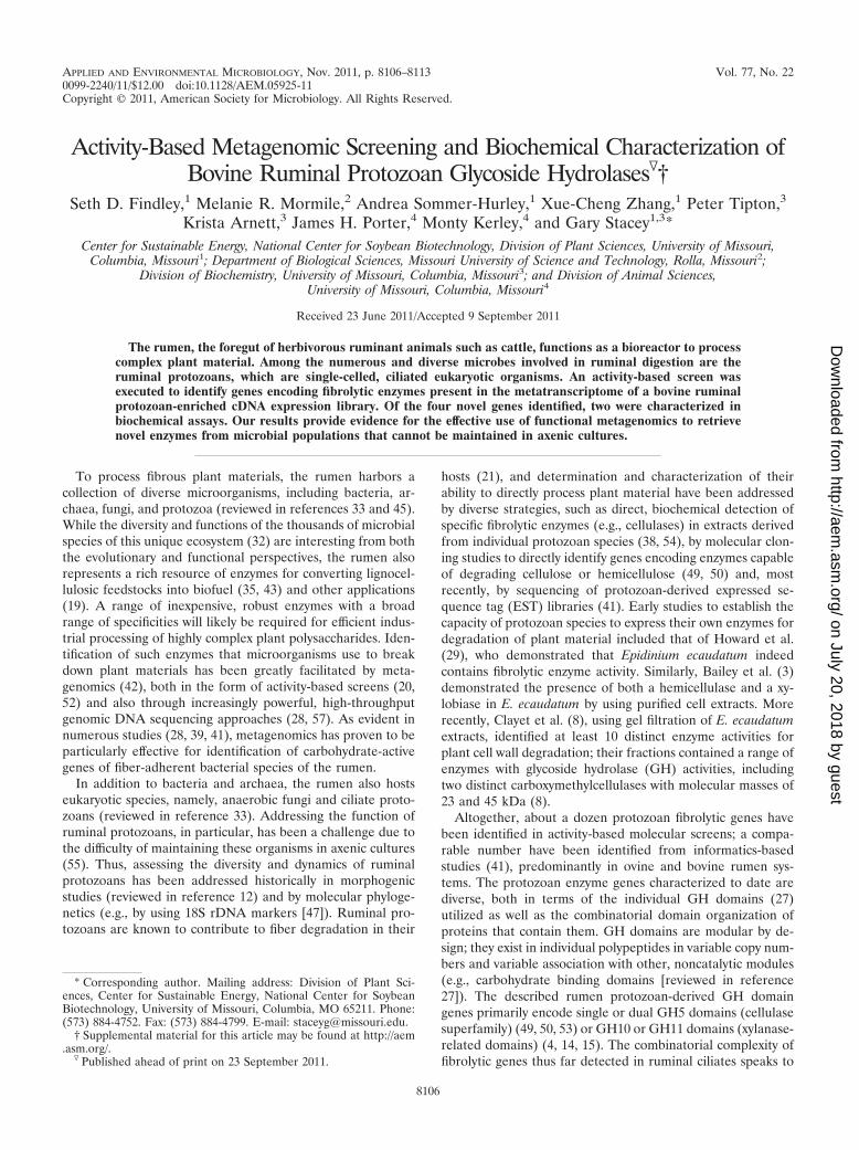
Mode of action of the type 1-7.1 and type 2-8.6 enzymes.Fibrolytic enzymes are essential for the digestion of cellulosicbiomass in the ruminant diet. A suite of enzymes is required toproduce a variety of free sugars, (exoenzymes), as well asoligosaccharides (endoenzymes) for metabolism by rumen bac-teria and subsequent digestion by the ruminant host. TheHPLC analysis of hydrolysis products resulting from exposureof �-glucan to type 1-7.1 and xylan exposure to type 2-8.6indicated that each tested enzyme employs an endo-type ofcleavage (Fig. 4). Compared to chromatographs of �-glucan(Fig. 4A) and xylan (Fig. 4C), each of the treated substrateshad multiple peaks prior to the polysaccharide peaks (Fig. 4Band D), indicating that a range of oligosaccharides was gener-ated by the actions of the endoglycoside hydrolases. It shouldbe noted that similar chromatographs were obtained for boththe 10- and 60-min exposures. If the enzymes were exo-acting,the primary products would be either glucose (from �-glucan)
or xylose (from xylan). Both glucose and xylose formed a singlepeak at 4 min on the chromatographs (data not shown). Therewas no peak at the 4-min point for glucose when �-glucan wasexposed to type 1-7.1, yet multiple peaks were present as thechromatographic run proceeded (Fig. 4B). There was a peak at4 min, indicating xylose production after xylan exposure to type2-8.6, as well as multiple peaks at later time points, indicat-ing the presence of oligoxylose chains (Fig. 4D). These dataare consistent with the fact that many GH5 and GH10 en-zymes are known to be endo-�-1,4-glucanases and endo-�-1,4-xylanases (9).
DISCUSSION
An activity-based metagenomic screen was executed with theaim of assessing the diversity of fibrolytic enzymes encoded bythe metatranscriptome of protozoans present in bovine rumenfluid. Using just two substrates, a cellulose and a hemicellulose,four genes with diverse GH domains and modular organiza-tions were identified. Phylogenetic analysis of these genes re-vealed the closest homologs to be protozoan and the closestnonprotozoan homologs to be most closely related to Gram-positive bacteria. These observations support the hypothesisthat lignocellulose-degrading genes were acquired by protozo-ans from ruminal bacteria by horizontal gene transfer (15, 41).There was close homology of the positive sequences obtained
TABLE 2. Basic biochemical properties of type 1-7.1 and type 2-8.6recombinant proteins
PropertyValue for protein type
1-7.1 2-8.6
GH family 5 10Molecular mass (kDa) 56.0 39.2pI (calculated) 4.56 6.35pH activity range 5.0–8.0 5.0–9.0pH optimum 5.9 7.4Temp activity range (°C) 4–60 25–60Temp optimum (°C) 50 45
FIG. 3. Substrate specificity analysis for type 1-7.1 and type 2-8.6 recombinant proteins. Substrate specificity assays were performed in triplicatein 250 �l of 1% polysaccharide solution buffered with either 50 mM MES (pH 6.0; optimum conditions for type 1-7.1) or 50 mM MOPS (pH 7.4;optimum conditions for type 2-8.6). Reactions were initiated by adding 10 �l (22 g of protein/ml) of purified enzyme.
TABLE 3. Kinetic data for type 1-7.1 and type 2-8.6recombinant proteins
Type andsubstrate
Apparent Km(mg/ml)
Vmax (�mol/min/mg) Kcat (s�1)
Catalyticefficiency(Kcat/Km
ml/mg/s�)
Type 1-7.1�-Glucan 0.83 97.7 7.4 8.9Xyloglucan 0.19 179.1 13.6 71.6
Type 2-8.6Xylan 6.95 14.5 1.1 0.2Arabinoxylan 0.14 117.3 8.9 63.6
VOL. 77, 2011 METAGENOMICS ANALYSIS OF RUMINAL PROTOZOAN GH ENZYMES 8111
on July 20, 2018 by guesthttp://aem
.asm.org/
Dow
nloaded from
in our study (Fig. 1B) to E. ecaudatum genes identified in aprevious protozoan EST sequencing study (41). The putativecellulases and xylanases identified by Ricard et al. (41) were allderived from one (the Entodiniophorphids) of the two majorgroups of rumen ciliates. The fact that each of the gene typesidentified in our bovine rumen screens had potential homologsin protozoan species originating from sheep rumen raises thepossibly that the genes represent orthologs derived from re-lated ciliate species in the two different hosts. The fact thatnone of our positives exhibited novel domain organization(relative to other identified ciliate GH genes) may indicate thatruminal ciliates acquired only a limited repertoire of bacterialfibrolytic genes.
One strength of activity-based screening is its ability to di-rectly recover genes encoding biocatalysts for specific sub-strates (e.g., cellulose). In our current study, two enzymes wereidentified, expressed, and biochemically characterized. The pu-tative xyloglucanse possesses a high specific activity towardtamarind xyloglucan (896.06 U/mg of protein) (Fig. 3). In com-parison, previously characterized xyloglucanses from fungalsources ranged in their activity from 45 to 98 U/mg of protein(24). Our xyloglucanse also had a lower apparent Km value(0.19 mg/ml) (Table 3) than a xyloglucan-specific endo-�-1,4-glucanase gene (xeg5A), isolated from bovine rumen micro-flora and expressed in E. coli (Km, 3.61 mg/ml) (56) and oneisolated from the fungus Aspergillus aculeatus (Km, 3.6 mg/ml)
(40). Similarly, our putative arabinoxylanase had a lower ap-parent Km (0.14 mg arabinoxylan/ml) (Table 3) than a GH10xylanase isolated from Penicillium funiculosum (3.7 mg/ml)(34). In addition, our putative arabinoxylanase had a higherspecific activity (584.39 U/mg) (Fig. 3) than the P. funiculosumxylanase (106.4 U/mg) (34) on arabinoxylan, as well as a higherspecific activity against beechwood xylan (type 2-8.6, 95.62U/mg; P. funiculosum xylanase, 60.1 U/mg) (34).
The powerful and rapid, activity-based metagenomics ap-proach does have its own technical and efficiency obstacles (asdiscussed in reference 52), however, as evidenced by our re-covery of hybrid/deletion cDNA clones. Thus, more direct,sequence-based metagenomics (e.g., single-cell-based genomicsequencing [57]), in combination with molecular phylogenetics(13, 48) may represent the more attractive technique for char-acterizing the population dynamics, functions, and fibrolyticgenes of ciliate ruminal protozoa.
ACKNOWLEDGMENT
We thank the University of Missouri Mizzou Advantage Programfor funding.
REFERENCES
1. Altschul, S. F., et al. 1997. Gapped BLAST and PSI-BLAST: a new gener-ation of protein database search programs. Nucleic Acids Res. 25:3389–3402.
2. Anthon, G. E., and D. M. Barrett. 2002. Determination of reducing sugarswith 3-methyl-2-benzothiazolinonehydrazone. Anal. Biochem. 305:287–289.
FIG. 4. HPLC results for cleavage product analysis of type 1-7.1 and type 2-8.6 recombinant proteins. (B and D) HPLC results for hydrolysisproducts of �-glucan after exposure to type 1-7.1 (B) and xylan after exposure to type 2-8.6 (D) for 60 min. (A and C) Polysaccharides �-glucan(A) and xylan (C), without addition of enzyme, were used as controls. The glucose and xylose standard peaks were detected at 4 min (data notshown). Intensity is reported in microcoulombs (�C).
8112 FINDLEY ET AL. APPL. ENVIRON. MICROBIOL.
on July 20, 2018 by guesthttp://aem
.asm.org/
Dow
nloaded from
3. Bailey, R. W., R. T. Clarke, and D. E. Wright. 1962. Carbohydrases of therumen ciliate Epidinium ecaudatum (Crawley). Biochem. J. 83:517–523.
4. Bera-Maillet, C., E. Devillard, M. Cezette, J. P. Jouany, and E. Forano. 2005.Xylanases and carboxymethylcellulases of the rumen protozoa Polyplastronmultivesiculatum, Eudiplodinium maggii and Entodinium sp. FEMS Micro-biol. Lett. 244:149–156.
5. Britton, H. T. S., and R. A. Robinson. 1931. Universal buffer solutions andthe dissociation constant of veronal. J. Chem. Soc. 1931:1456–1462.
6. Chenna, R., et al. 2003. Multiple sequence alignment with the Clustal seriesof programs. Nucleic Acids Res. 31:3497–3500.
7. Clamp, M., J. Cuff, S. M. Searle, and G. J. Barton. 2004. The Jalview Javaalignment editor. Bioinformatics 20:426–427.
8. Clayet, F., J. Senaud, and J. Bohatier. 1992. Chromatographic separation ofsome cell wall polysaccharide-degrading enzymes of the sheep rumen ciliateEpidinium caudatum. Ann. Zootech. 41:81.
9. Collins, T., C. Gerday, and G. Feller. 2005. Xylanases, xylanase families andextremophilic xylanases. FEMS Microbiol. Rev. 29:3–23.
10. Correia, M. A., et al. 2011. Structure and function of an arabinoxylan-specificxylanase. J. Biol. Chem. 286:22510–22520.
11. Courtin, C. M., and J. A. Delcour. 2002. Arabinoxylans and endoxylanases inwheat flour bread-making. J. Cereal Sci. 35:225–243.
12. Dehority, B. A. 1993. Laboratory manual for classification and morphology ofruminal ciliate protozoa. CRC Press, Boca Raton, FL.
13. Deng, W., D. Xi, H. Mao, and M. Wanapat. 2008. The use of moleculartechniques based on ribosomal RNA and DNA for rumen microbial ecosys-tem studies: a review. Mol. Biol. Rep. 35:265–274.
14. Devillard, E., et al. 2003. Characterization of XYN10B, a modular xylanasefrom the ruminal protozoan Polyplastron multivesiculatum, with a family 22carbohydrate-binding module that binds to cellulose. Biochem. J. 373:495–503.
15. Devillard, E., et al. 1999. A xylanase produced by the rumen anaerobicprotozoan Polyplastron multivesiculatum shows close sequence similarity tofamily 11 xylanases from gram-positive bacteria. FEMS Microbiol. Lett.181:145–152.
16. Edgar, R. C. 2004. MUSCLE: multiple sequence alignment with high accu-racy and high throughput. Nucleic Acids Res. 32:1792–1797.
17. Eschenlauer, S. C., et al. 1998. Phylogenetic position and codon usage of twocentrin genes from the rumen ciliate protozoan, Entodinium caudatum.FEMS Microbiol. Lett. 166:147–154.
18. Felsenstein, J. 2000. PHYLIP (Phylogeny Inference Package). Departmentof Genetics, University of Washington, Seattle, WA.
19. Fernandez-Arrojo, L., M. E. Guazzaroni, N. Lopez-Cortes, A. Beloqui, andM. Ferrer. 2010. Metagenomic era for biocatalyst identification. Curr. Opin.Biotechnol. 21:725–733.
20. Ferrer, M., et al. 2005. Novel hydrolase diversity retrieved from a meta-genome library of bovine rumen microflora. Environ. Microbiol. 7:1996–2010.
21. Gijzen, H. J., H. J. Lubberding, M. J. T. Gerhardus, and G. D. Vogels. 1988.Contribution of rumen protozoa to fibre degradation and cellulase activity invitro. FEMS Microbiol. Lett. 53:35–43.
22. Gilbert, H. J., H. Stalbrand, and H. Brumer. 2008. How the walls comecrumbling down: recent structural biochemistry of plant polysaccharide deg-radation. Curr. Opin. Plant Biol. 11:338–348.
23. Gloster, T. M., et al. 2007. Characterization and three-dimensional struc-tures of two distinct bacterial xyloglucanases from families GH5 and GH12.J. Biol. Chem. 282:19177–19189.
24. Grishutin, S. G., et al. 2004. Specific xyloglucanases as a new class of poly-saccharide-degrading enzymes. Biochim. Biophys. Acta 1674:268–281.
25. Hausalo, T. 1995. Analysis of wood and pulp carbohydrates by anion ex-change chromatography with pulse amperometric detection, p. 131–136.Proceedings of the 8th International Symposium on Wood Pulping Chem-istry, vol. III. Helsinki, Finland. Tappi Press, Norcross, GA.
26. Henrissat, B. 1991. A classification of glycosyl hydrolases based on aminoacid sequence similarities. Biochem. J. 280:309–316.
27. Henrissat, B., and G. J. Davies. 2000. Glycoside hydrolases and glycosyl-transferases. Families, modules, and implications for genomics. PlantPhysiol. 124:1515–1519.
28. Hess, M., et al. 2011. Metagenomic discovery of biomass-degrading genesand genomes from cow rumen. Science 331:463–467.
29. Howard, B. H., G. Jones, and M. R. Purdom. 1960. The pentosanases ofsome rumen bacteria. Biochem. J. 74:173–180.
30. Huang, X., and A. Madan. 1999. CAP3: a DNA sequence assembly program.Genome Res. 9:868–877.
31. Karlin, S., and S. F. Altschul. 1990. Methods for assessing the statistical
significance of molecular sequence features by using general scoringschemes. Proc. Natl. Acad. Sci. U. S. A. 87:2264–2268.
32. Kim, M., M. Morrison, and Z. Yu. 2011. Status of the phylogenetic diversitycensus of ruminal microbiomes. FEMS Microbiol. Ecol. 76:49–63.
33. Krause, D. O., et al. 2003. Opportunities to improve fiber degradation in therumen: microbiology, ecology, and genomics. FEMS Microbiol. Rev. 27:663–693.
34. Lafond, M., et al. 2011. GH10 xylanase D from Penicillium funiculosum:biochemical studies and xylooligosaccharide production. Microb. Cell Fact10:20.
35. Li, L. L., S. R. McCorkle, S. Monchy, S. Taghavi, and D. van der Lelie. 2009.Bioprospecting metagenomes: glycosyl hydrolases for converting biomass.Biotechnol. Biofuels 2:10.
36. Martin, C., A. G. Williams, and B. Michalet-Doreau. 1994. Isolation andcharacteristics of the protozoal and bacterial fractions from bovine ruminalcontents. J. Anim. Sci. 72:2962–2968.
37. McEwan, N. R., S. C. Eschenlauer, R. E. Calza, R. J. Wallace, and C. J.Newbold. 2000. The 3� untranslated region of messages in the rumen pro-tozoan Entodinium caudatum. Protist 151:139–146.
38. Michalowski, T., K. Rybicka, K. Wereszka, and A. Kasperowicz. 2001. Abilityof the rumen ciliate Epidinium ecaudatum to digest and use crystallinecellulose and xylan for in vitro growth. Acta Protozool. 40:203–210.
39. Palackal, N., et al. 2007. A multifunctional hybrid glycosyl hydrolase discov-ered in an uncultured microbial consortium from ruminant gut. Appl. Mi-crobiol. Biotechnol. 74:113–124.
40. Pauly, M., et al. 1999. A xyloglucan-specific endo-beta-1,4-glucanase fromAspergillus aculeatus: expression cloning in yeast, purification and character-ization of the recombinant enzyme. Glycobiology 9:93–100.
41. Ricard, G., et al. 2006. Horizontal gene transfer from bacteria to rumenciliates indicates adaptation to their anaerobic, carbohydrates-rich environ-ment. BMC Genomics 7:22.
42. Riesenfeld, C. S., P. D. Schloss, and J. Handelsman. 2004. Metagenomics:genomic analysis of microbial communities. Annu. Rev. Genet. 38:525–552.
43. Rubin, E. M. 2008. Genomics of cellulosic biofuels. Nature 454:841–845.44. Ruijssenaars, H. J., and S. Hartmans. 2001. Plate screening methods for the
detection of polysaccharase-producing microorganisms. Appl. Microbiol.Biotechnol. 55:143–149.
45. Russell, J. B., and J. L. Rychlik. 2001. Factors that alter rumen microbialecology. Science 292:1119–1122.
46. Schmidt, H. A., K. Strimmer, M. Vingron, and A. von Haeseler. 2002.TREE-PUZZLE: maximum likelihood phylogenetic analysis using quartetsand parallel computing. Bioinformatics 18:502–504.
47. Shin, E. C., et al. 2004. Phylogenetic analysis of protozoa in the rumencontents of cow based on the 18S rDNA sequences. J. Appl. Microbiol.97:378–383.
48. Sylvester, J. T., S. K. Karnati, Z. Yu, M. Morrison, and J. L. Firkins. 2004.Development of an assay to quantify rumen ciliate protozoal biomass in cowsusing real-time PCR. J. Nutr. 134:3378–3384.
49. Takenaka, A., C. G. D’Silva, H. Kudo, H. Itabashi, and K. J. Cheng. 1999.Molecular cloning, expression, and characterization of an endo-beta-1,4-glucanase cDNA from Epidinium caudatum. J. Gen. Appl. Microbiol.45:57–61.
50. Takenaka, A., K. Tajima, M. Mitsumori, and H. Kajikawa. 2004. Fiberdigestion by rumen ciliate protozoa. Microbes Environ. 19:203–210.
51. Ten, L. N., W. T. Im, Z. Aslam, L. Larina, and S. T. Lee. 2007. Novelinsoluble dye-labeled substrates for screening inulin-degrading microorgan-isms. J. Microbiol. Methods 69:353–357.
52. Uchiyama, T., and K. Miyazaki. 2009. Functional metagenomics for enzymediscovery: challenges to efficient screening. Curr. Opin. Biotechnol. 20:616–622.
53. Wereszka, K., et al. 2004. A cellulase produced by the rumen anaerobicprotozoan Epidinium ecaudatum has an unusual pH optimum. Endocytobio-sis Cell Res. 15:561–569.
54. Williams, A. G., and G. S. Coleman. 1985. Hemicellulose-degrading enzymesin rumen ciliate protozoa. Curr. Microbiol. 12:85–90.
55. Williams, A. G., and G. S. Coleman. 1992. The rumen protozoa. Springer-Verlag, New York, NY.
56. Wong, D. D., V. J. Chan, A. A. McCormack, and S. B. Batt. 2010. A novelxyloglucan-specific endo-beta-1,4-glucanase: biochemical properties and in-hibition studies. Appl. Microbiol. Biotechnol. 86:1463–1471.
57. Woyke, T., et al. 2009. Assembling the marine metagenome, one cell at atime. PLoS One 4:e5299.
58. Zmasek, C. M., and S. R. Eddy. 2001. ATV: display and manipulation ofannotated phylogenetic trees. Bioinformatics 17:383–384.
VOL. 77, 2011 METAGENOMICS ANALYSIS OF RUMINAL PROTOZOAN GH ENZYMES 8113
on July 20, 2018 by guesthttp://aem
.asm.org/
Dow
nloaded from