acute cholecystitis jane chunwen teng d.o. 12/5/12
TRANSCRIPT

ACUTE CHOLECYSTITIS
Jane Chunwen Teng D.O. 12/5/12
ACUTE CHOLECYSTITIS
Acute Cholecystitis: symptoms of RUQ pain, fever,
and leukocytosis associated with gallbladder
inflammation that is usually related to gallstone
disease.
Acalculous Cholecystitis: Clinically identical to
acute cholecystitis but is not associated with
gallstones, and usually occurs in critically ill pts. It
accounts for approximately 10 % of cases of acute
cholecystitis and is associated with high morbidity
and mortality.
CHRONIC CHOLECYSTITIS
Chronic inflammatory cell infiltration of the
gallballdder seen on histopathology.
Almost invariably associated with the presence of
gallstones and is thought to be the result of
mechanical irritation or recurrent attacks of acute
cholecystitis leading to fibrosis and thickening of the
gallbladder.
PATHOGENESIS
In contrast to biliary colic, the development of acute
cholecystitis is not fully explained by cystic duct obstruction
alone.
Studies in animals have demonstrates that ligation of the
cystic duct does not result in acute cholecystitis.
However, acute cholecystitis can be produced
experimentally by blockade of the cystic duct followed by
deliberate irritation of the gallbladder mucosa.
PATHOGENESIS
Lysolecithin (normally absent in bile) maybe release
following trauma of the gallbladder wall from an impacted
gallstone.
Inflammatory mediators (e.g. PGE2 and 6 keto PG F1
alpha) synthesized by inflamed human gallbladder
microsomes increased four times above normal.
Prolonged impaction of stones in the cystic duct can lead
to hydrops.
CLINICAL MANIFESTATION
RUQ or epigastrium abdominal pain, may radiate
to the right shoulder or back. Pain is usually steady
and sever.
Nausea, vomiting, and anorexia.
Hx of fatty food ingestion about one hour or more
before the initial onset of pain.
PHYSICAL EXAM
Pt usually are ill appearing, febrile, tachycardic
and lie still on the examining table because any
movement can aggravate the pain.
Abdominal exam usually demonstrates voluntary
and involuntary guarding.

PHYSICAL EXAM
Positive “Murphy’s sign” While palpating the area
of the gallbladder fossa just beneath the liver edge,
the patient is asked to inspire deeply, causing the
gallbladder to descend toward the examining
fingers. Pt with acute cholecystitis commonly
experience increased discomfort and may have
associated inspiratory arrest.
Using cholescintigraphy as the gold standard, the
sensitivity and specificity of a positive Murphy’s sign
were 97 and 48 %, respectively.
COMPLICATION
Left untreated, symptoms of cholecystitis may
abate within 7-10 days.
Complication can occur at alarmingly high rate,
including the development of gallbladder gangrene
(up to 20%) and subsequent perforation (2%). Other
complications include cholecystoenteric fistula,
gallstone ileus, emphysematous cholecystitis.
DIAGNOSIS
Confirmation of the diagnosis must be based upon
a combination of physical findings, laboratory
studies, and imaging tests.
The most accurate physical findings were a
positive Murphy sign (positive likelihood ratio 2.8,
95% CI 0.8 to 8.6) and right upper quadrant
tenderness (negative LR 0.4, 95% CI 0.2 to 1.1)
Lab: CBC shows leukocytosis with a left shift.
DIAGNOSIS
Elevation in the serum total bilirubin and alkaline
phosphatase concentrations are NOT common in
uncomplicated cholecystitis, since biliary obstruction
is limited to the gallbladder; if present, they should
raise the concerns about complicating conditions
such as cholangitis, choledocholithiasis, or the
Mirizzi syndrome (a gallstone impacted in the distal
cystic duct causing extrinsic compression of the
common bile duct)
DIAGNOSIS
There have been reports of mild elevation of serum
aminotransferase and amylase, along with
hyperbilirubinemia and jaundice. These
abnormalities maybe due to the passage of small
stones, sludge, or pus.
IMAGING TESTS
Ultrasonograpy is usually the first test obtained
and can often establish the diagnosis.
Nuclear cholescintigraphy may be useful in cases
in which the diagnosis remains uncertain after
ultrasounography.

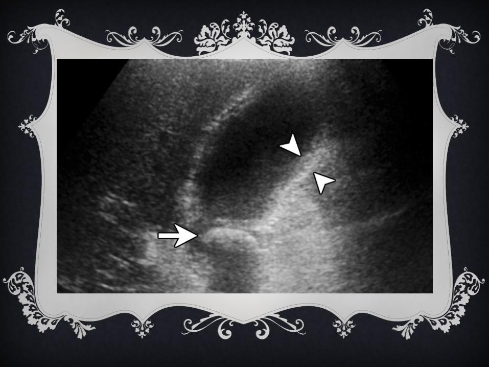
ULTRASOUND DIAGNOSIS
Gallbladder wall thickening (>4-5 mm) or edema (double wall sign)
A “sonographic Murphy’s sign”, which is similar to the Murphy’s
sign elicited during abdominal palpation, except that the positive
response is observed during palpation with the ultrasound transducer.
A particularly informative systematic review summarized the results
of 30 studies of ultrasonography for gallstones and acute
cholecystitis. Adjusted sensitivity and specificity for diagnosis of
acute cholecystitis were 88% and 80% respectively.

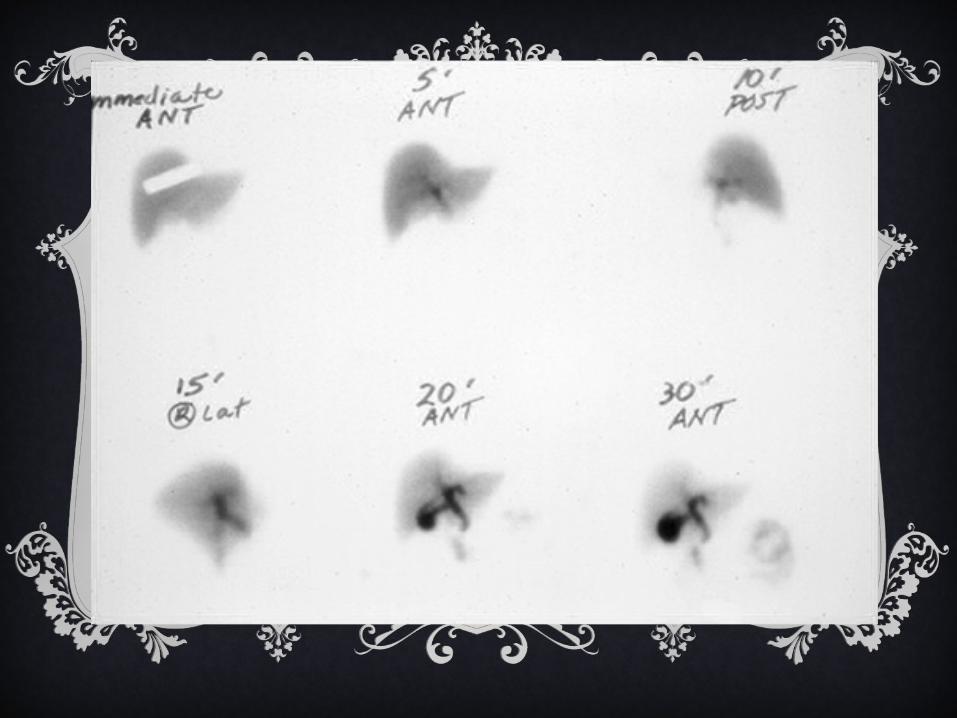
CHOLESCINTIGRAPHY (HIDA SCAN)
Indicated if the diagnosis remain uncertain following
ultrasonography.
Technetium labeled hepatic iminodiacetic acid (HIDA) is
injected intravenously and is then taken up selectively by
hepatocytes and excreted into bile.
If the cystic duct is patent, this agent will enter the
gallbladder, leading to its visualization without the need
for concentration.
CHOLESCINTIGRAPHY (HIDA SCAN)
HIDA scan is also useful demonstrating patency of the
common bile duct and ampulla.
Visualization of the contrast within the common bile duct,
gallbladder, and small bowel occurs within 30 to 60 mins.
The test is positive if the gallbladder does not visualize,
which is invariably due to cystic duct obstruction, usually
from edema associated with acute cholecystitis or an
obstructing stone.

CHOLESCINTIGRAPHY (HIDA SCAN)
Cholescintigraphy has a sensitivity and specificity of
approximately 97 and 90 %, respectively.
Cystic duct obstruction with a stone or tumor in the absence
of acute cholecystitis can cause a false positive test.
Other conditions that can cause false positive results include:
- Severe liver disease, Fasting pt receiving TPN, Biliary
sphincterotomy and Hyperbilirubinemia.

MORPHINE CHOLESCINTIGRAPHY
A modified version of the HIDA scan, in which pts are
given IV morphine during the exam.
Morphine increases sphincter of Oddi pressure, thereby
causing a more favorable pressure gradient for the
radioactive tracer to enter the cystic duct.
This modification is thought to be particularly useful in
critically ill pts, in whom standard HIDA scans may be
associated with false positive results.
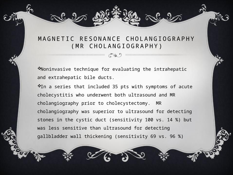
M A G N E T I C R E S O N A N C E C H O L A N G I O G R A P H Y ( M R C H O L A N G I O G R A P H Y )
Noninvasive technique for evaluating the intrahepatic and
extrahepatic bile ducts.
In a series that included 35 pts with symptoms of acute
cholecystitis who underwent both ultrasound and MR
cholangiography prior to cholecystectomy. MR cholangiography
was superior to ultrasound for detecting stones in the cystic duct
(sensitivity 100 vs. 14 %) but was less sensitive than ultrasound
for detecting gallbladder wall thickening (sensitivity 69 vs. 96 %)
CT
Abdominal CT is usually unnecessary in the diagnosis of acute
cholecystitis, although it can easily demonstrate gallbladder wall
edema associated with acute cholecystitis.
Other CT findings include pericholecystic stranding and fluid,
and high-attenuation bile.
However, CT may fail to detect gallstones because many stones
are isodense with bile.
CT can be useful when complications of acute cholecystitis are
suspected or when other diagnosis are considered.
TREATMENT
Pts diagnosed with acute cholecystitis required hospital admission
for IV hydration, correction of electrolyte disorders, and pain
control (IM ketorolac 30-60 mg adjusted for age and renal function).
Pts should be kept NPO and those who are vomiting may need
NGT placement.
The guidelines of the Infectious Diseases Society of America
(IDSA) recommend that antimicrobial therapy be instituted if
infection is suspected on the basis of lab (>12,5000 WBC) or clinical
findings (temp ≥38.5 degree) , and radiographic findings .

TREATMENT
Routine antibiotics are also recommended in pts of
advanced age or who have diabetes or
immunodeficiency, and for prophylaxis in patients
undergoing cholecystectomy to reduce septic
complications even when infection is not suspected.
Empiric antibiotic therapy should induce activity
against the most common pathogens.
TREATMENT
In a study of 467 pts, including a control group of 42
pts, including a control group of 42 with normal biliary
tress, positive bile cultures were found in 22 % pts with
symptomatic gallstones and 46 & of pts with acute
cholecystitis. The most frequent isolates from the
gallbladder or common bile duct were
- E.Coli (12%), Klebsiella (11%) , and Enterobacter (9%).

TREATMENT
Empiric antibiotic Treatment for gram negative and anaeronic
bateria
1st choice:
Monotherapy with a beta-lactam or beta-lactamase inhibitor (e.g.
pipercillin tazobactam 3.375 or 4.5 g IV q6hr or ticarcillin-
clauvulanate 3.1 g q4hr)
Or Combination of 3rd generation cephalosporin PLUS
metronidazole (ceftriaxone 1g q 24 hr or 2g q 12 hr for CNS
infection and metronidazole 500mg q8hr)
TREATMETN
The duration of antibiotic therapy is tailored to clinical
improvement.
For pts requiring prompt surgical intervention, antibiotics may
be warranted for 24 to 48 hours following cholecystectomy,
although longer or shorter courses may be appropriate depending
on individual circumstances.
Pts for whom surgical intervention is initially deferred ay
warrant antibiotic therapy over 48 to 72 hours pending resolution
of clinical signs and symptoms.

TIMING FOR SURGERY
Although there is consensus that incidentally discovered asymptomatic
gallstones should not be treated. Once a pt develops symptoms or
complications related to gallstones (such as biliary colics or acute
cholecystis), treatment to eliminate the gallstones should be
recommended, because the likelihood of subsequent symptoms or
complications is high.
The National Cooperative Gallstone Study, a trial of nonsurgical
treatment with chenodiol for biliary tract pain, demonstrated that the
risk for recurrent symptoms was approximately 70 % during the two yrs
following initial presentation.
TIMING FOR SURGERY
The selection of treatment and timing of definitive
therapy for acute cholecystitis depends upon the
severity of symptoms and the pts overall risk of
surgery.
The aim of definitive therapy is to eliminate the
precipitating cause of acute cholecystitis (ie,
gallstones in the case of calculous cholecystitis) to
prevent recurrent attacks.

TREATMENT
The benefit of prompt surgical intervention was also
illustrated in a subsequent study of 29,818 Medicare
pts with acute cholecystitis. Compared to pts who
underwent cholecystectomy in the initial
hospitalization, pts who were discharged without
surgery were more likely to require readmission (38
vs. 4 %) and had higher mortality (hazard ratio 1.56,
95% CI, 1.47-1.65) over the following two yrs.
TREATMENT
Lows-risk pts - The physical status scale
established by the American Society of
Anesthesiologists (ASA) is commonly used to
determine the risk of surgery.
Although previously considered to be at higher
risk, pts with DM who do not have substantial
microvascular or macrovascular disease have an
outcome after acute cholecystitis similar to the
nondiabetic population.

TREATMENT
The ASA physical status classification system is a system for
assessing the fitness of patients before surgery .
1. A normal healthy patient.
2. A patient with mild systemic disease.
3. A patient with severe systemic disease.
4. A patient with severe systemic disease that is a constant threat to life.
5. A moribund. patient who is not expected to survive without the surgery
6. A declared brain-dead patient whose organs are being removed for
donor purposes.
TREATMENT IN LOW RISK PTS
Immediate Cholecystectomy is preferred for pts who are at low
risk (ASA class I and II)
Several studies have indicated that cholecystectomy performed
for low surgical risk pts during the initial hospitalization can
reduce morbidity and costs.
Early Surgery is also easier to perform as local inflammation
increases 72 hr past the initial onset of symptoms making
dissection less precise, increasing the severity of surgical
complications, and open conversion more likely.

TREATMENT IN HIGH RISK PTS
High-risk pts - Pts who are in ASA classes III, IV, or V have a surgical
mortality ranging from 5-27%, and are considered high-risk for
cholecystectomy.
This category generally includes pts with severe chronic illnesses, such
as cardiovascular or pulmonary disease, or advanced malignancy.
High risk pts, or pts who present late in the course of their disease
process(>3-5 days), who continue to have severe symptoms and show no
appreciable improvement despite one to two days of medical
management require further intervention.
TREATMENT IN HIGH RISK PTS
Gallbladder drainage by percutaneous cholecystostomy
in conjunction of antibiotic is the initial treatment of
choice for high risk pts.
The goal of cholecystostomy is to drain purulent material
from the obstructed gallbladder.
Tube decompression of the gallbladder allows for
resolution of edema which often “opens” up the
obstructed cystic duct.
TREATMENT IN HIGH RISK PTS
Endoscopic transpapillary gallbladder drainage has also been
reported in pts with acute cholecystitis in whom percutaneous
approaches are contraindicated or anatomically impossible.
A limitation of the technique is that it can be technically
challenging to place a guidewire and drainage tube into the
gallbladder.
In addition, this procedures carries all the inherent
complications of ERCP.
TREATMENT – SURGERY
When the acute cholecystits has resolved, pts who are surgical
candidates should undergo cholecystectomy.
Surgery may also be required when the pt does not improve
following percutaneous drainage, which suggests that the
gallbladder has already progressed to gangrene.
Pts who are particularly unstable will benefit from open
cholecystostomy tube drainage achieved through a limited
lapratomy. This can be performed at the bedside in the ICU
setting if necessary.
NONSURGICAL TREATMENT
Pts who stabilize but continue to be at high risk for
surgery can be considered for percutaneous
gallstone extraction with or without mechanical
lithotripsy.
REFERENCE
http://www.uptodate.com/contents/pathogenesis-cli
nical-features-and-diagnosis-of-acute-cholecystitis?
source=search_result&search=acute+cholecystitis
&selectedTitle=2%
7E53
http://www.uptodate.com/contents/treatment-of-ac
ute-cholecystitis?source=search_result&search=ac
ute+cholecystitis&selectedTitle=1%
7E53