acute coronary syndromes 2016 - university of … acs… · acute coronary syndromes . 2016 ......
TRANSCRIPT
Morton J. Kern MD, MSCAI, FACC, FAHA Chief of Medicine, VA Long Beach HSC
Professor of Medicine University California Irvine
Acute Coronary Syndromes
2016
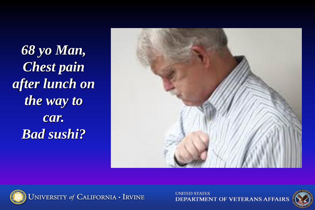
68 yo Man, Chest pain
after lunch on the way to
car. Bad sushi?
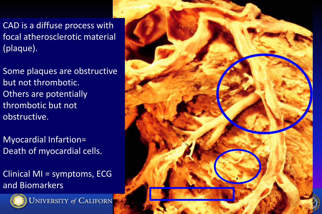
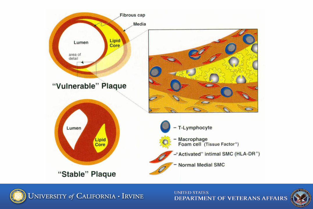
CAD is a diffuse process with focal atherosclerotic material (plaque). Some plaques are obstructive but not thrombotic. Others are potentially thrombotic but not obstructive. Myocardial Infartion= Death of myocardial cells. Clinical MI = symptoms, ECG and Biomarkers
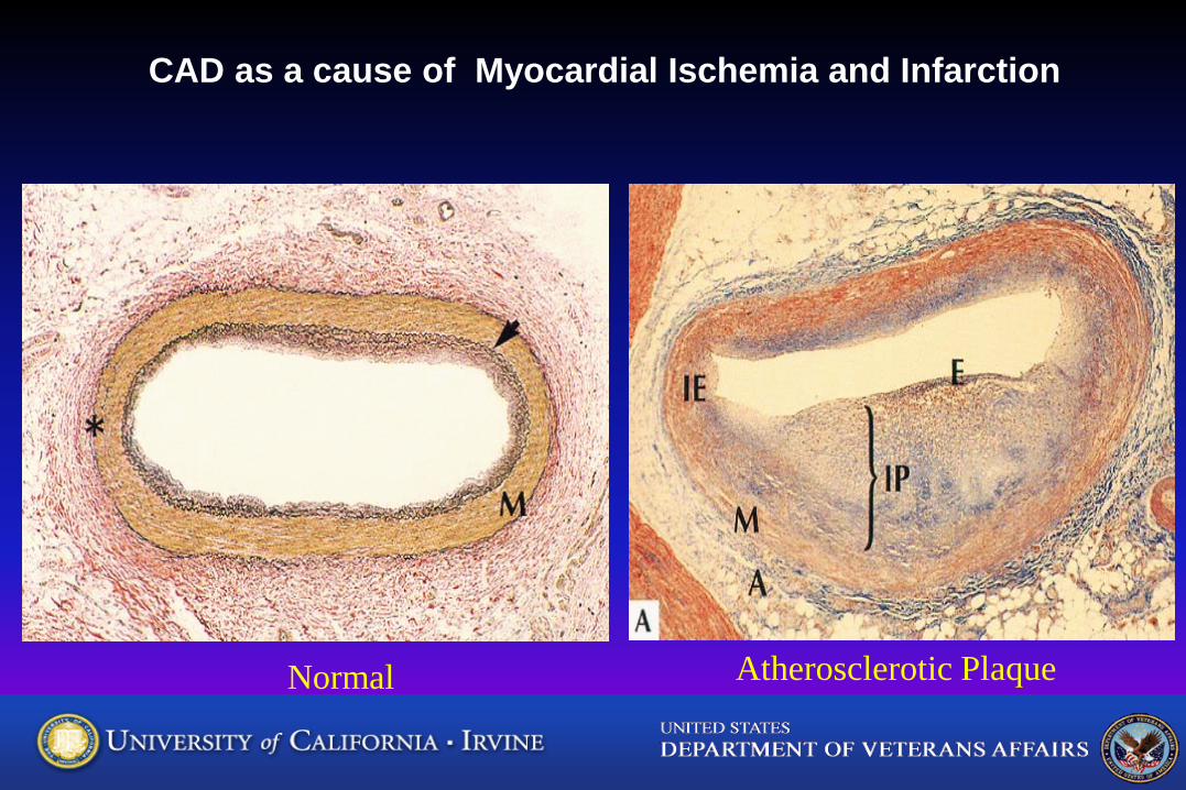
Normal Atherosclerotic Plaque
CAD as a cause of Myocardial Ischemia and Infarction
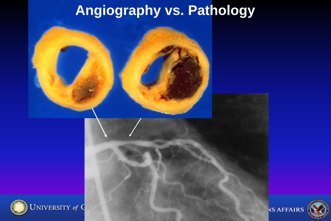
Angiography vs. Pathology
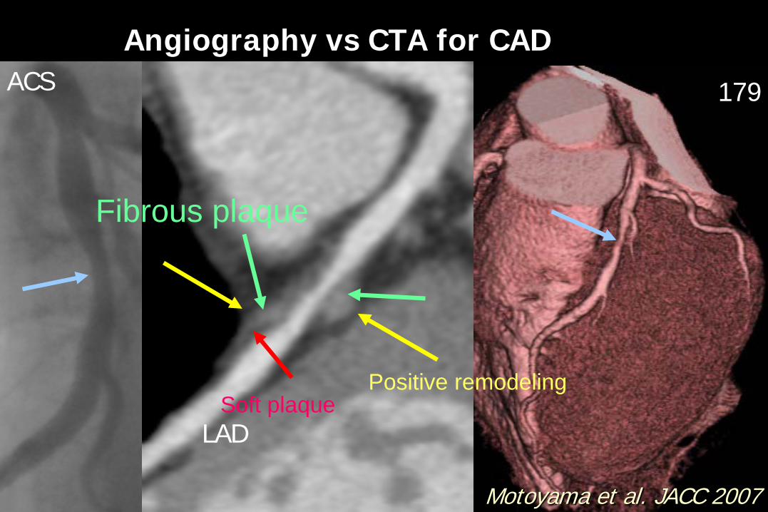
179 ACS
LAD
Angiography vs CTA for CAD
Motoyama et al. JACC 2007
Fibrous plaque
Positive remodeling Soft plaque
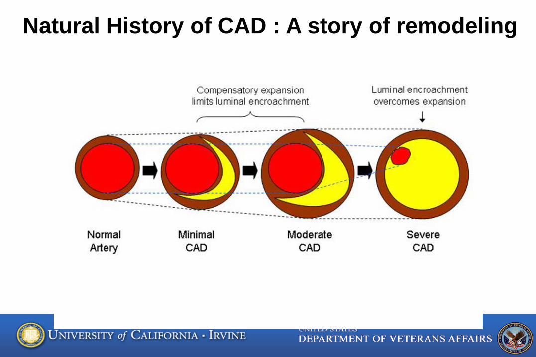
Natural History of CAD : A story of remodeling
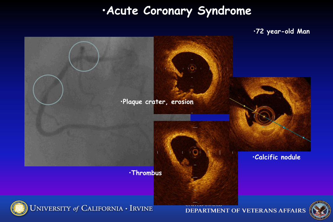
•Acute Coronary Syndrome •72 year-old Man
•Plaque crater, erosion
•Thrombus
•Calcific nodule
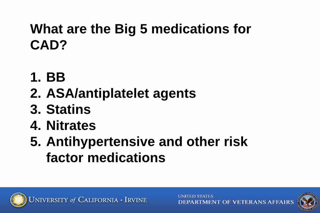
What are the Big 5 medications for CAD? 1. BB 2. ASA/antiplatelet agents 3. Statins 4. Nitrates 5. Antihypertensive and other risk
factor medications
Braunwald’s Heart Disease, 7th Edition
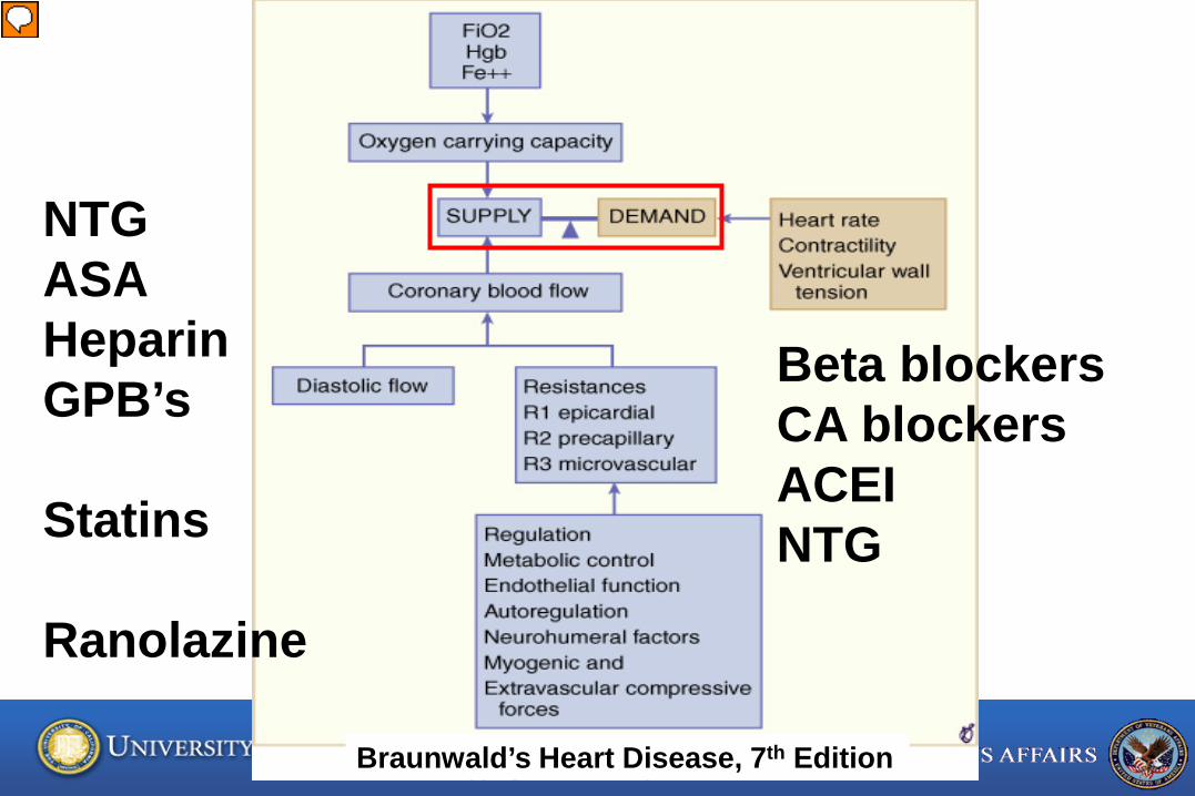
Beta blockers CA blockers ACEI NTG
NTG ASA Heparin GPB’s Statins Ranolazine
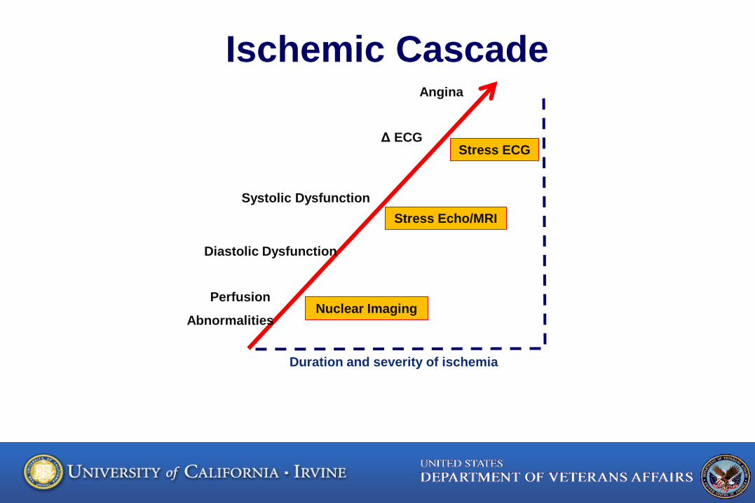
Perfusion
Abnormalities
Systolic Dysfunction
Δ ECG
Angina
Diastolic Dysfunction
Duration and severity of ischemia
Nuclear Imaging
Stress Echo/MRI
Stress ECG
Ischemic Cascade
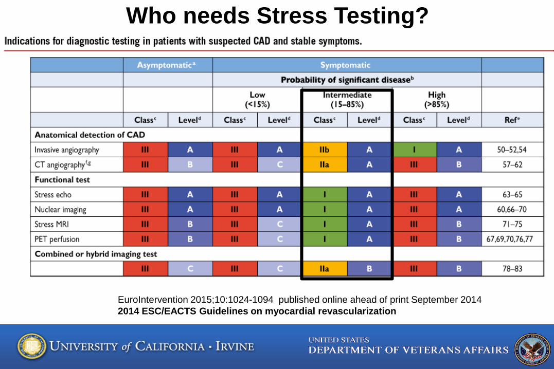
EuroIntervention 2015;10:1024-1094 published online ahead of print September 2014 2014 ESC/EACTS Guidelines on myocardial revascularization
Who needs Stress Testing?
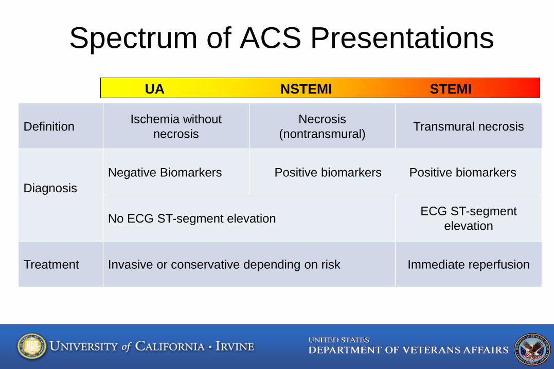
Spectrum of ACS Presentations
Definition Ischemia without necrosis
Necrosis (nontransmural) Transmural necrosis
Diagnosis
Negative Biomarkers Positive biomarkers Positive biomarkers
No ECG ST-segment elevation ECG ST-segment elevation
Treatment Invasive or conservative depending on risk Immediate reperfusion
UA NSTEMI STEMI
Roger VL, Go AS, Lloyd-Jones DM, et al.. Circulation. 2011;123:e18-e209.
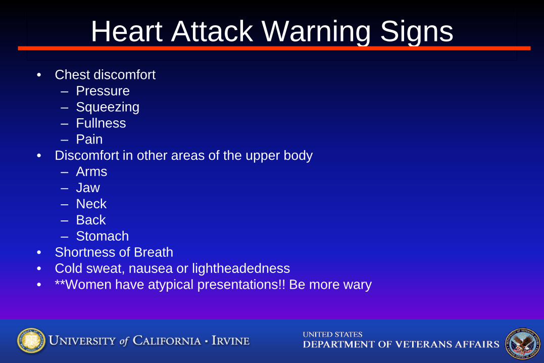
Heart Attack Warning Signs • Chest discomfort
– Pressure – Squeezing – Fullness – Pain
• Discomfort in other areas of the upper body – Arms – Jaw – Neck – Back – Stomach
• Shortness of Breath • Cold sweat, nausea or lightheadedness • **Women have atypical presentations!! Be more wary
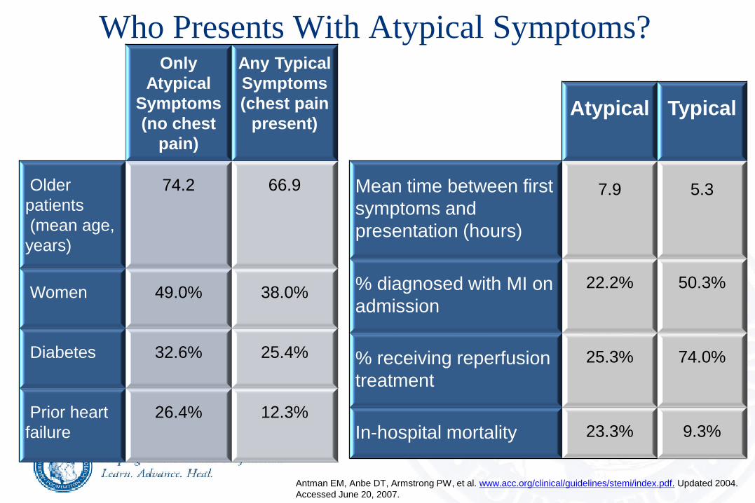
Who Presents With Atypical Symptoms?
Only Atypical
Symptoms (no chest
pain)
Any Typical Symptoms (chest pain
present)
Older patients (mean age, years)
74.2
66.9
Women
49.0%
38.0%
Diabetes
32.6%
25.4%
Prior heart failure
26.4%
12.3%
Antman EM, Anbe DT, Armstrong PW, et al. www.acc.org/clinical/guidelines/stemi/index.pdf. Updated 2004. Accessed June 20, 2007.
Atypical
Typical
Mean time between first symptoms and presentation (hours)
7.9
5.3
% diagnosed with MI on admission
22.2%
50.3%
% receiving reperfusion treatment
25.3%
74.0%
In-hospital mortality
23.3%
9.3%
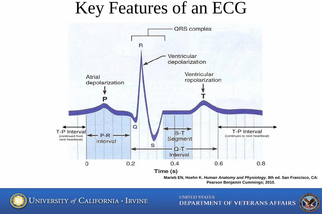
Key Features of an ECG
P-R T-P Interval
(continues to next heartbeat) T-P Interval
(continued from next heartbeat)
Marieb EN, Hoehn K. Human Anatomy and Physiology. 8th ed. San Francisco, CA: Pearson Benjamin Cummings; 2010.
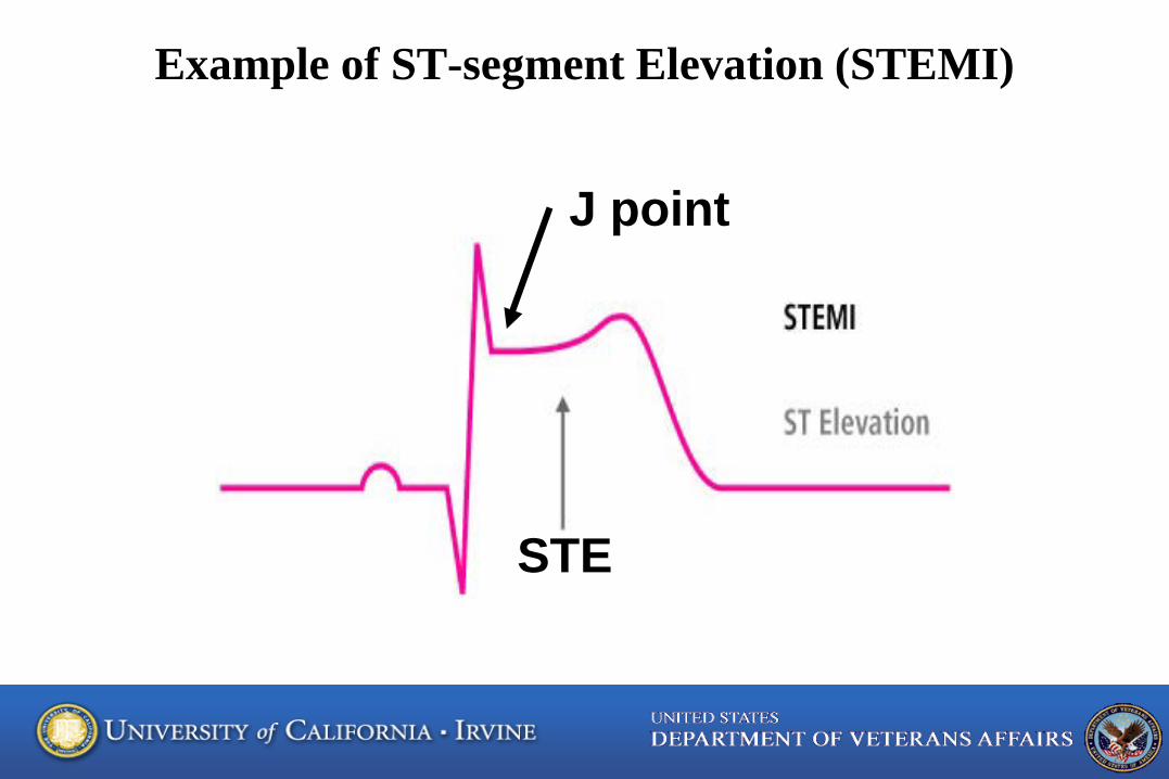
Example of ST-segment Elevation (STEMI)
J point
STE
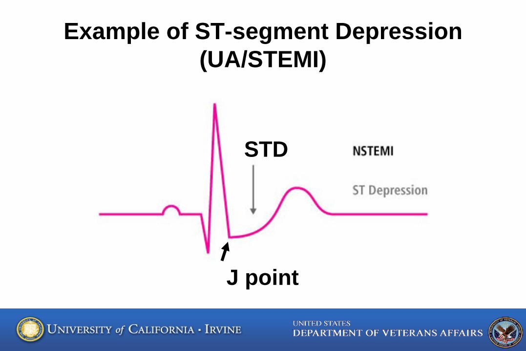
Example of ST-segment Depression (UA/STEMI)
J point
STD
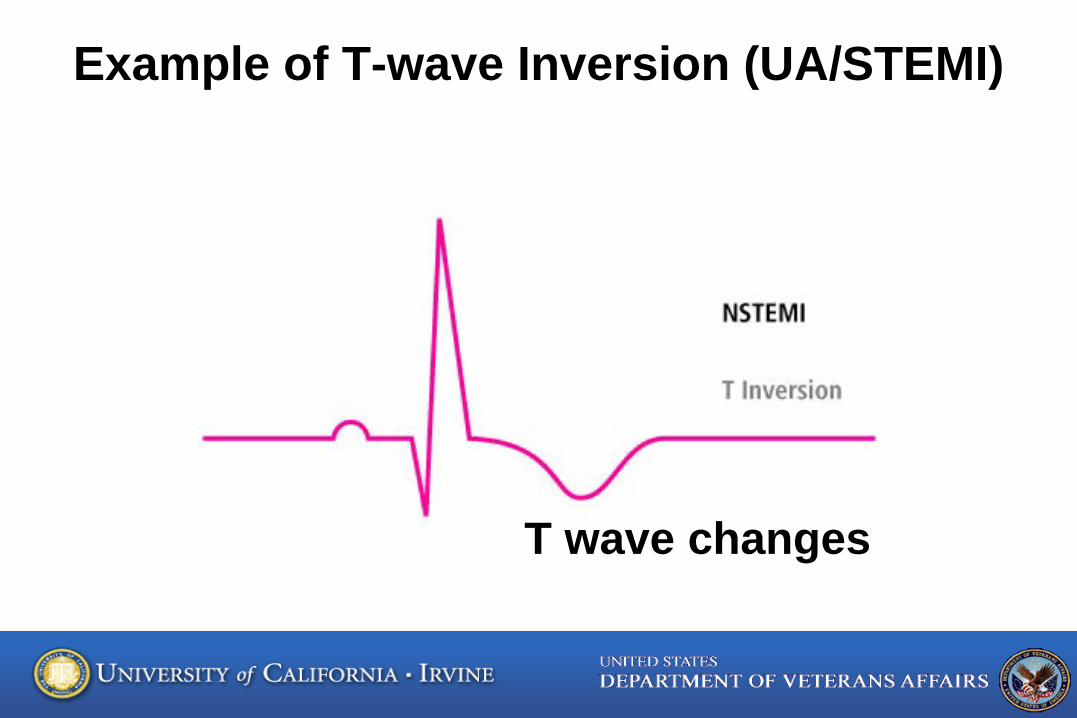
Example of T-wave Inversion (UA/STEMI)
T wave changes
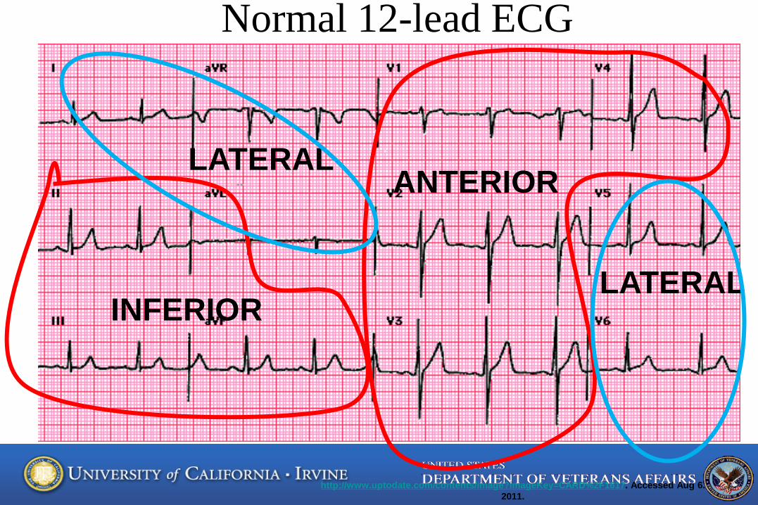
Normal 12-lead ECG
http://www.uptodate.com/contents/image?imageKey=CARD%2F1617. Accessed Aug 6. 2011.
INFERIOR
ANTERIOR LATERAL
LATERAL
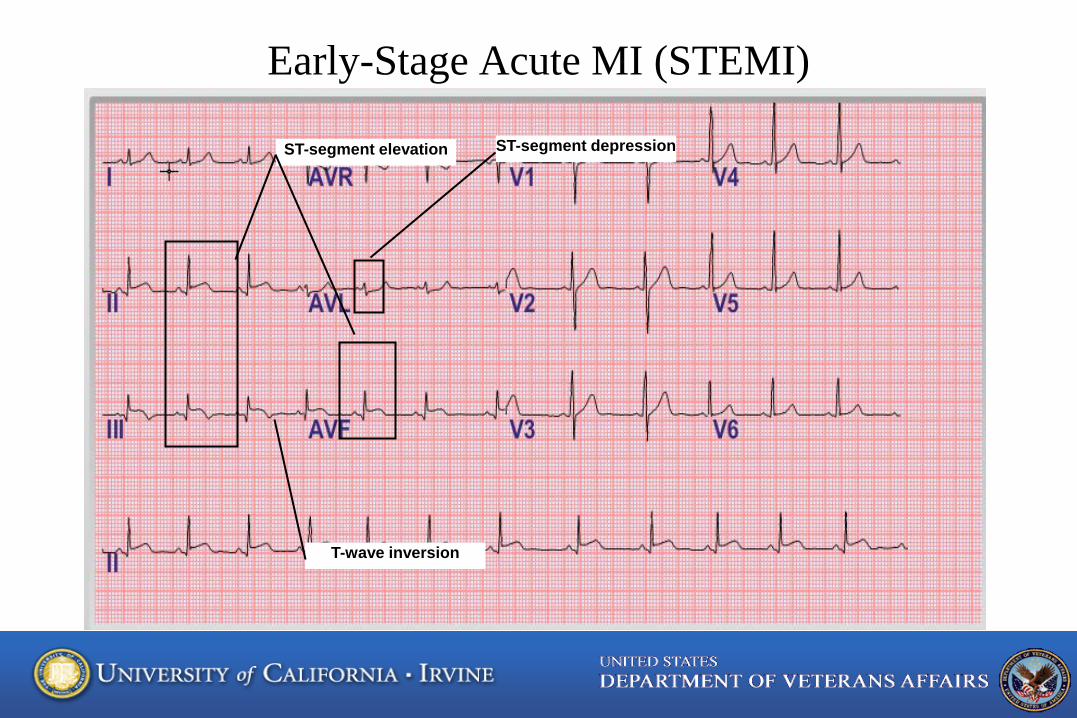
Early-Stage Acute MI (STEMI)
ST-segment elevation ST-segment depression
T-wave inversion
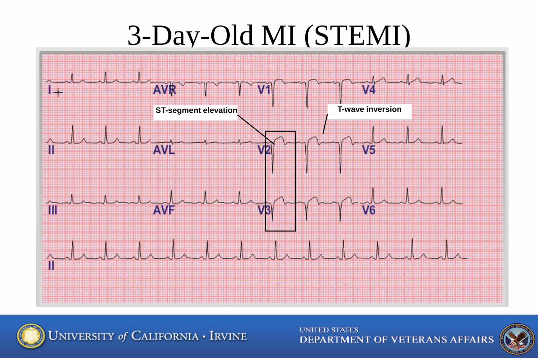
3-Day-Old MI (STEMI)
ST-segment elevation T-wave inversion
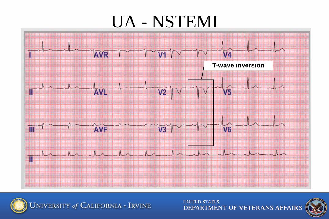
UA - NSTEMI
T-wave inversion
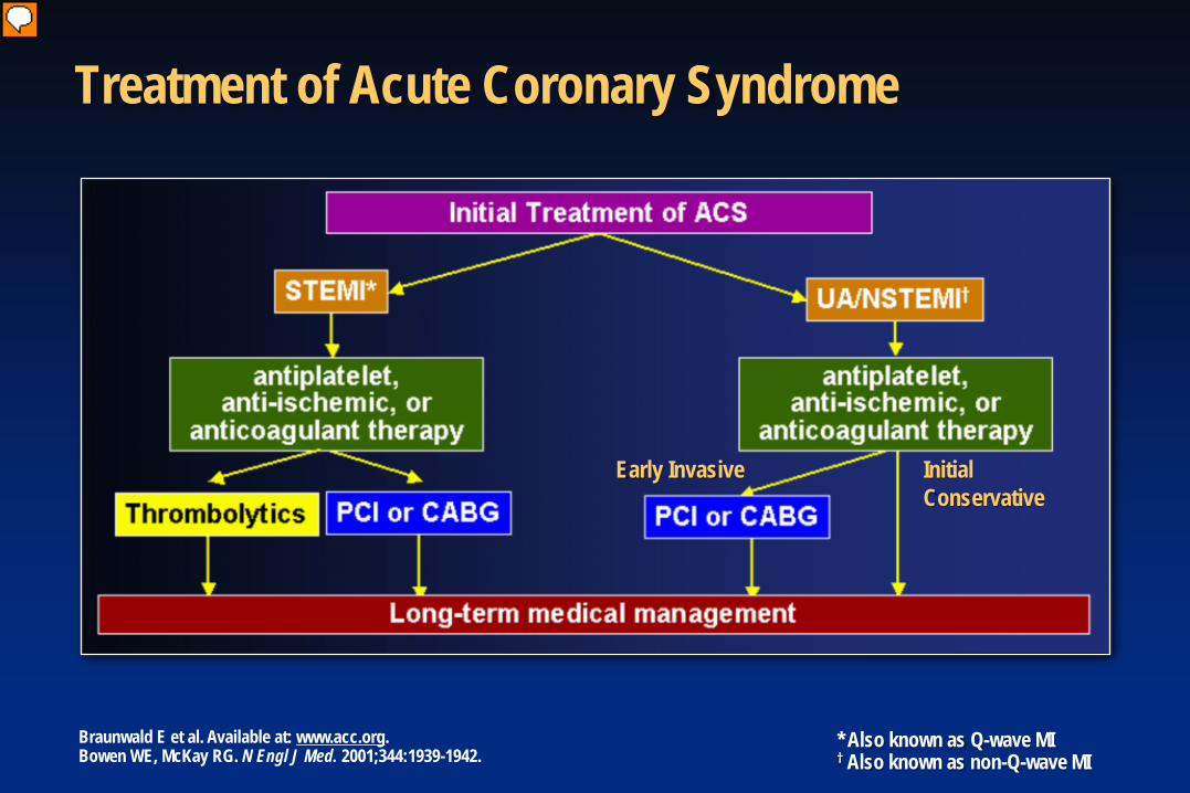
Early Invasive Initial Conservative
Braunwald E et al. Available at: www.acc.org. Bowen WE, McKay RG. N Engl J Med. 2001;344:1939-1942.
* Also known as Q-wave MI † Also known as non-Q-wave MI
Treatment of Acute Coronary Syndrome
Thygesen K et al. Circulation 2007; available at: http://circ.ahajournals.org.
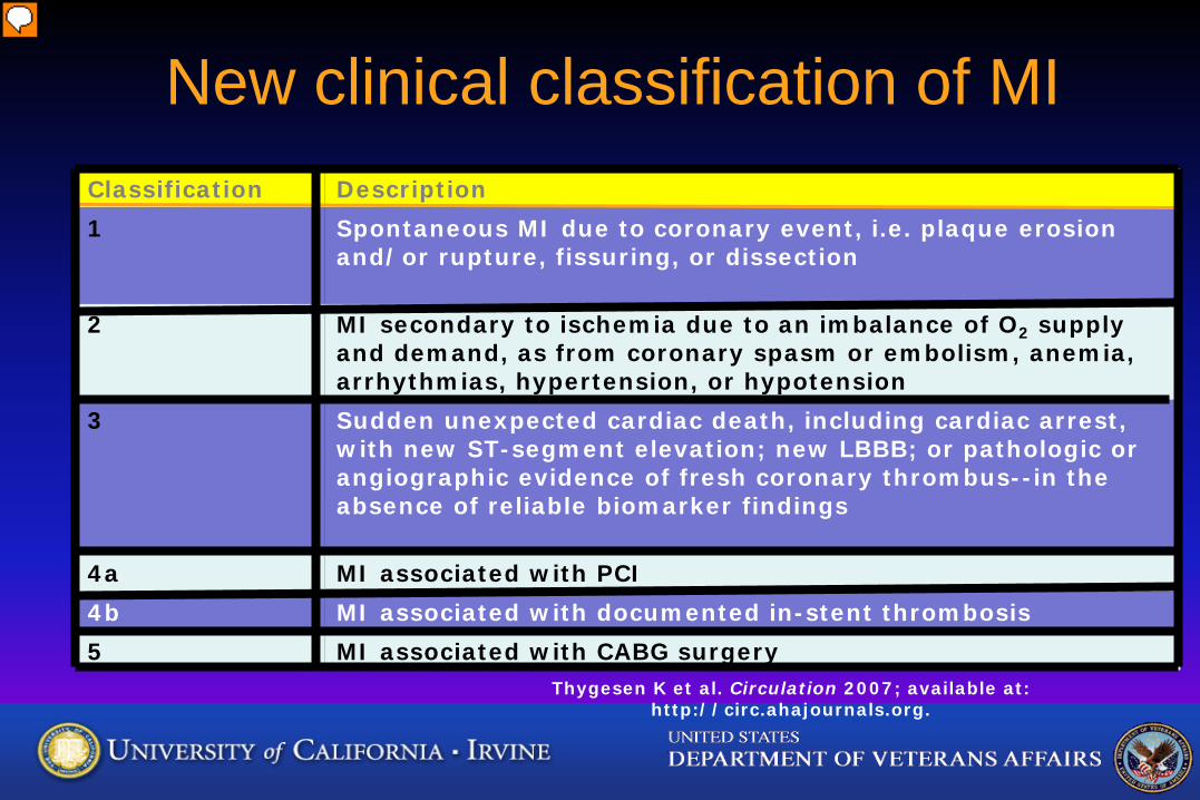
New clinical classification of MI Classification Description 1 Spontaneous MI due to coronary event, i.e. plaque erosion
and/or rupture, fissuring, or dissection
2 MI secondary to ischemia due to an imbalance of O2 supply and demand, as from coronary spasm or embolism, anemia, arrhythmias, hypertension, or hypotension
3 Sudden unexpected cardiac death, including cardiac arrest, with new ST-segment elevation; new LBBB; or pathologic or angiographic evidence of fresh coronary thrombus--in the absence of reliable biomarker findings
4a MI associated with PCI 4b MI associated with documented in-stent thrombosis 5 MI associated with CABG surgery
Thygesen, K. et al. Circulation 2007;116:2634-2653
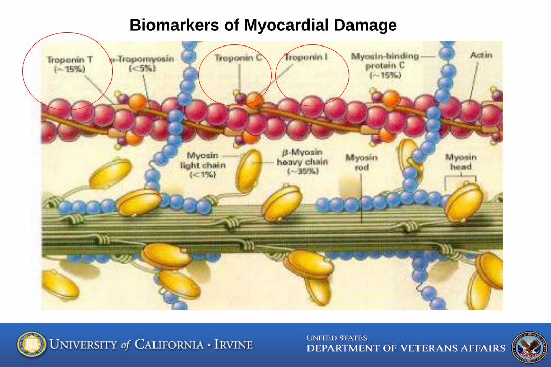
Biomarkers of Myocardial Damage
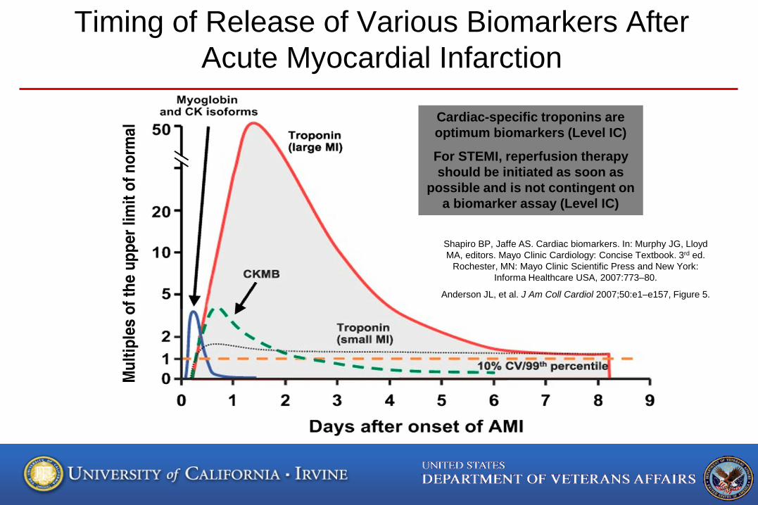
Timing of Release of Various Biomarkers After Acute Myocardial Infarction
Shapiro BP, Jaffe AS. Cardiac biomarkers. In: Murphy JG, Lloyd MA, editors. Mayo Clinic Cardiology: Concise Textbook. 3rd ed.
Rochester, MN: Mayo Clinic Scientific Press and New York: Informa Healthcare USA, 2007:773–80.
Anderson JL, et al. J Am Coll Cardiol 2007;50:e1–e157, Figure 5.
Cardiac-specific troponins are optimum biomarkers (Level IC)
For STEMI, reperfusion therapy should be initiated as soon as
possible and is not contingent on a biomarker assay (Level IC)
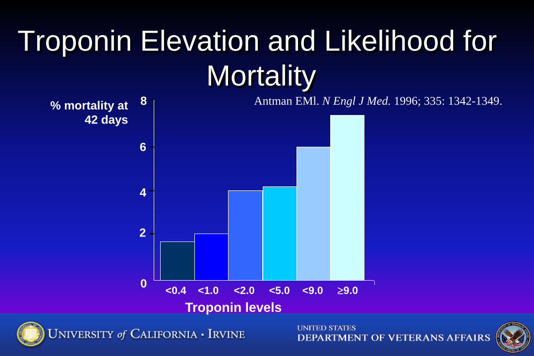
Troponin Elevation and Likelihood for Mortality
Antman EMl. N Engl J Med. 1996; 335: 1342-1349. % mortality at 42 days
<0.4 <1.0 <2.0 <5.0 <9.0 ≥9.0
2
4
6
8
0
Troponin levels
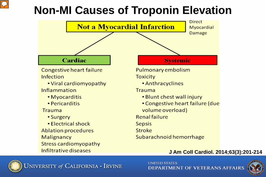
Non-MI Causes of Troponin Elevation
J Am Coll Cardiol. 2014;63(3):201-214
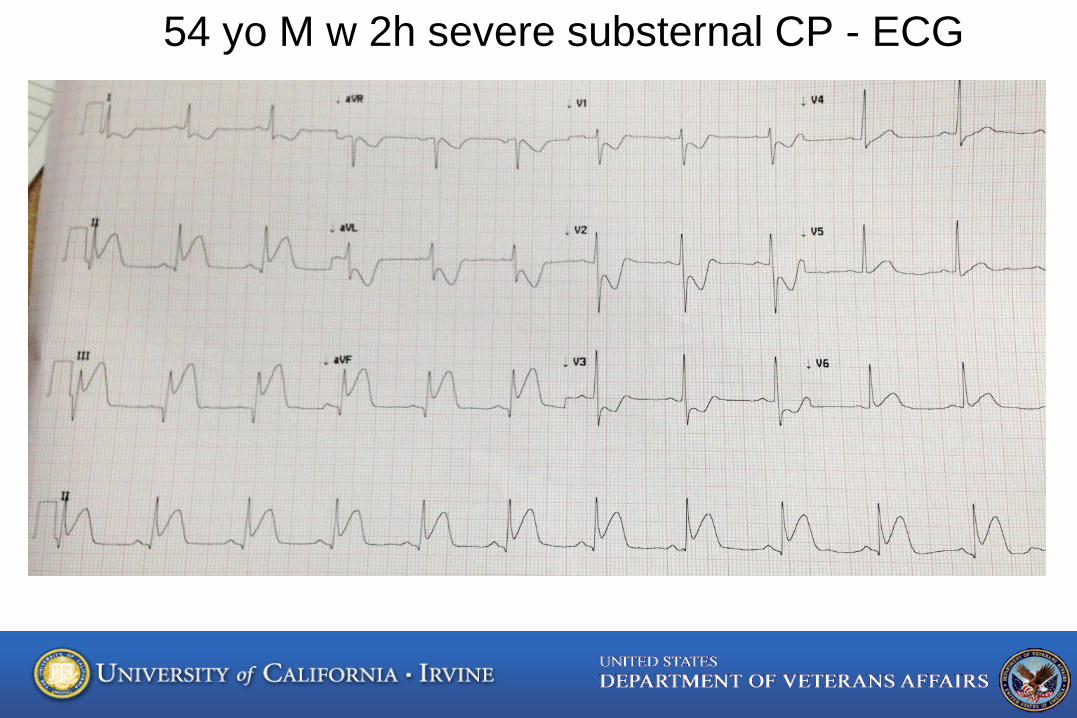
54 yo M w 2h severe substernal CP - ECG
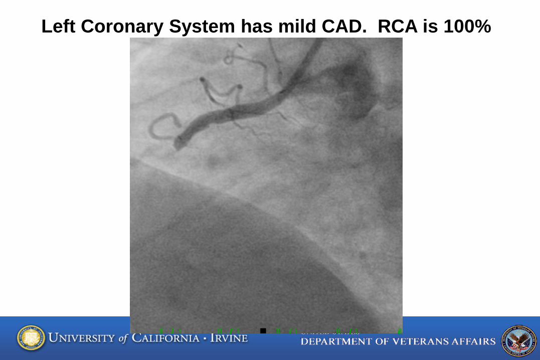
Left Coronary System has mild CAD. RCA is 100%
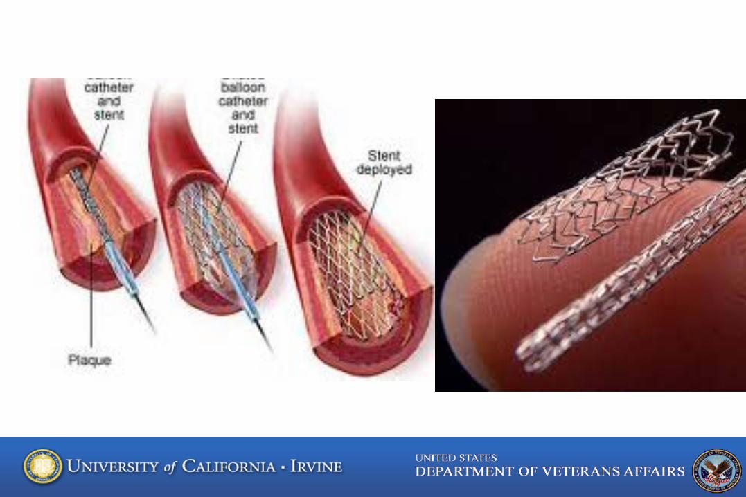
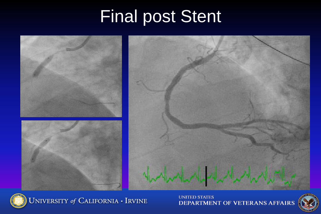
Final post Stent
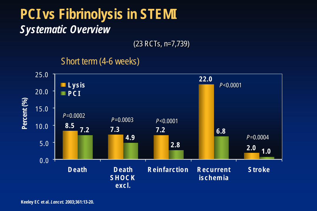
PCI vs Fibrinolysis in STEMI Systematic Overview
Short term (4-6 weeks)
Keeley EC et al. Lancet. 2003;361:13-20.
P=0.0002 P=0.0003 P<0.0001
P<0.0001
P=0.0004
(23 RCTs, n=7,739)
8 . 5 7 . 3 7 . 2
2 2 . 0
2 . 0
7 . 2 4 . 9
2 . 8
6 . 8
1 . 0 0 . 0
5 . 0
1 0 . 0
1 5 . 0
2 0 . 0
2 5 . 0
D e a t h D e a t h S H O C K
e x c l .
R e i n f a r c t i o n R e c u r r e n t i s c h e m i a
S t r o k e
Perc
ent (
%)
L y s i s P C I
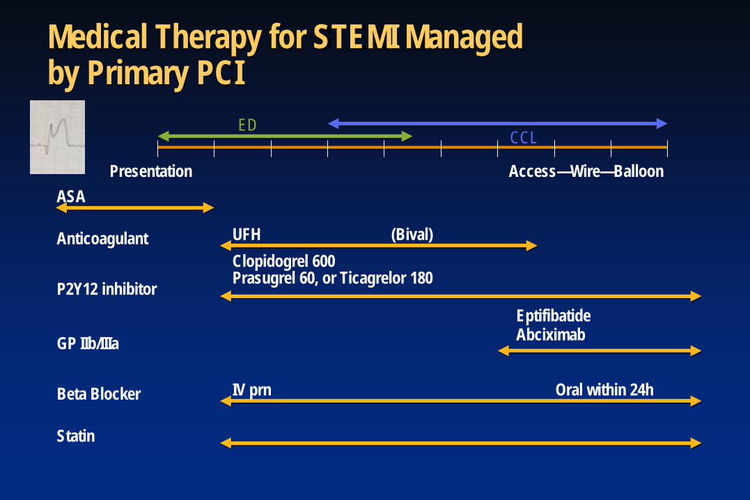
Medical Therapy for STEMI Managed by Primary PCI
ASA
Anticoagulant UFH (Bival)
P2Y12 inhibitor Clopidogrel 600 Prasugrel 60, or Ticagrelor 180
Beta Blocker IV prn Oral within 24h
GP IIb/IIIa Eptifibatide Abciximab
Statin
Presentation Access—Wire—Balloon
ED CCL
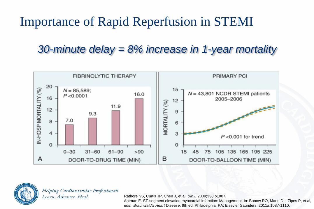
Importance of Rapid Reperfusion in STEMI
30-minute delay = 8% increase in 1-year mortality
Rathore SS, Curtis JP, Chen J, et al. BMJ. 2009;338:b1807. Antman E. ST-segment elevation myocardial infarction: Management. In: Bonow RO, Mann DL, Zipes P, et al, eds. Braunwald's Heart Disease. 9th ed. Philadelphia, PA: Elsevier Saunders; 2011a:1087-1110.
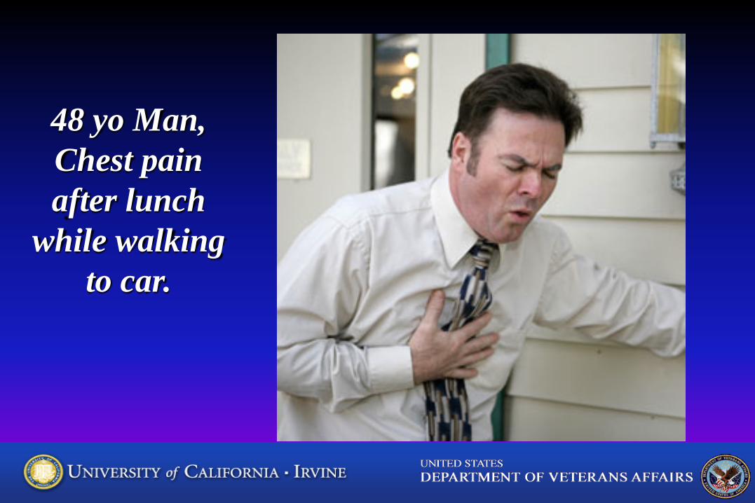
48 yo Man, Chest pain after lunch
while walking to car.
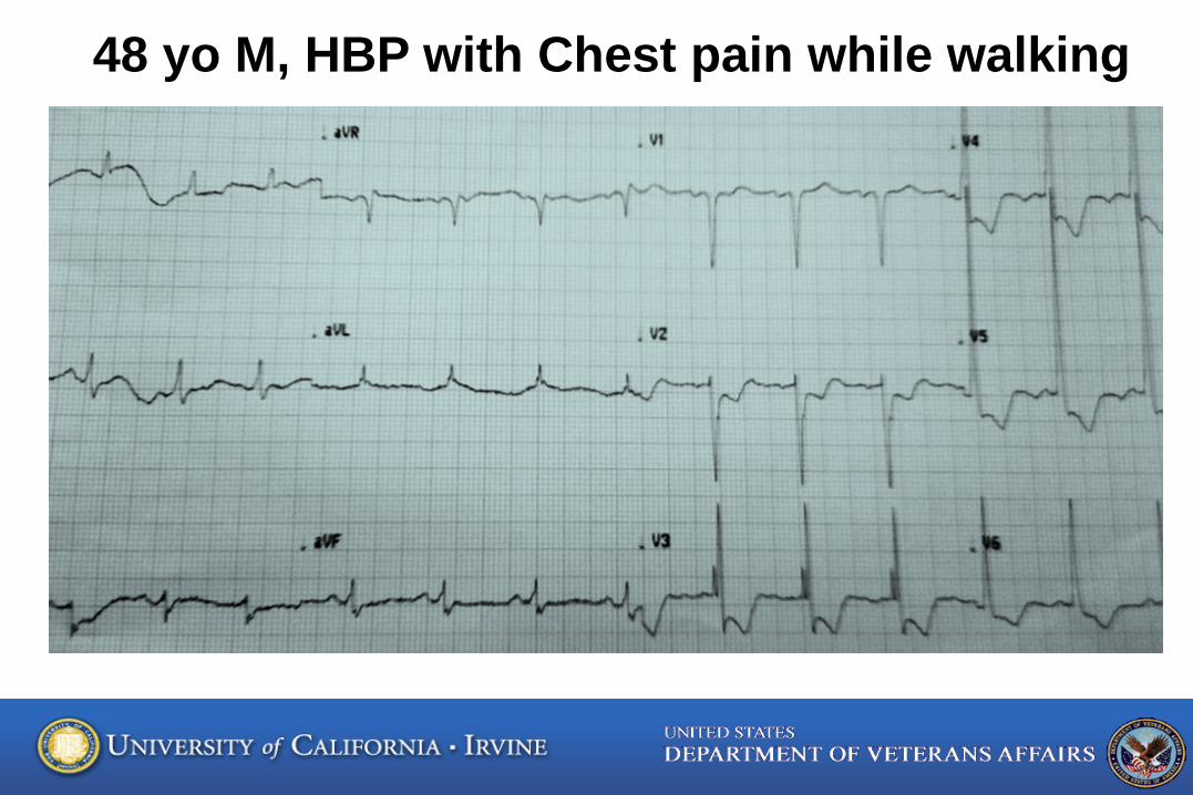
48 yo M, HBP with Chest pain while walking
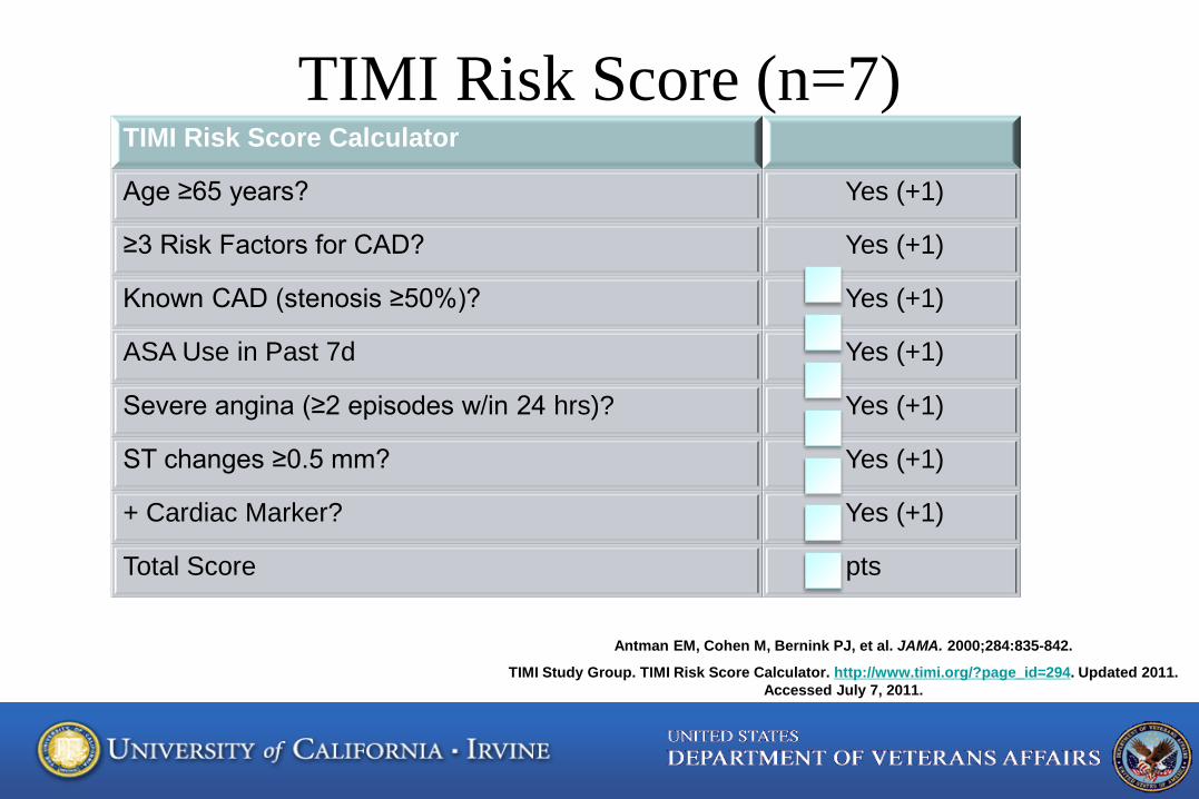
TIMI Risk Score (n=7) TIMI Risk Score Calculator
Age ≥65 years? Yes (+1)
≥3 Risk Factors for CAD? Yes (+1)
Known CAD (stenosis ≥50%)? Yes (+1)
ASA Use in Past 7d Yes (+1)
Severe angina (≥2 episodes w/in 24 hrs)? Yes (+1)
ST changes ≥0.5 mm? Yes (+1)
+ Cardiac Marker? Yes (+1)
Total Score pts
Antman EM, Cohen M, Bernink PJ, et al. JAMA. 2000;284:835-842.
TIMI Study Group. TIMI Risk Score Calculator. http://www.timi.org/?page_id=294. Updated 2011. Accessed July 7, 2011.

What does TIMI RISK mean?
Increasing TIMI RISK 0/1 to 5/7 increases risk of death, MI, urgent revascularization within 14 days 5% to 41%.
Antman EM et al. TIMI 11B, JAMA 2000;284:835-842
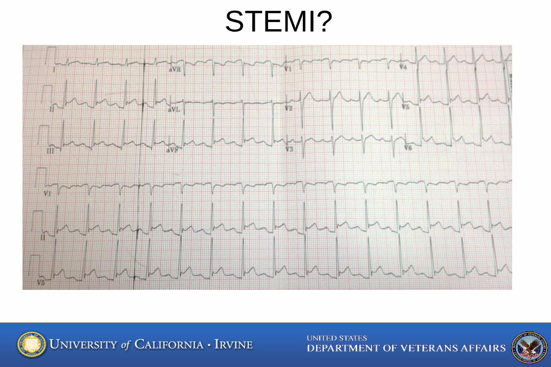
STEMI?
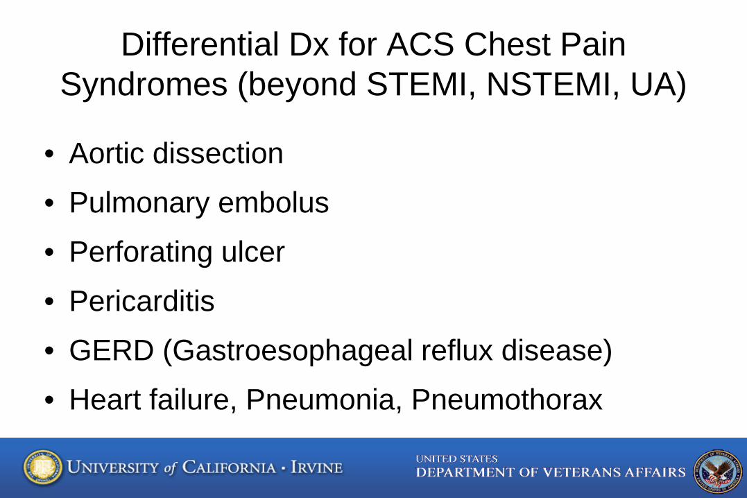
Differential Dx for ACS Chest Pain Syndromes (beyond STEMI, NSTEMI, UA)
• Aortic dissection
• Pulmonary embolus
• Perforating ulcer
• Pericarditis
• GERD (Gastroesophageal reflux disease)
• Heart failure, Pneumonia, Pneumothorax