acute mental status changes in the intensive care unit danagra georgia ikossi, md stanford general...
TRANSCRIPT
Acute Mental Status Changes in the Intensive Care Unit
Danagra Georgia Ikossi, MD
Stanford General Surgery Resident
10/24/2006

Just because you’re nuts, it doesn’t mean you’re not sick… the ongoing search for organic causes
• Brief review of Delirium, Seizures and Stroke
• “ICU Psychosis”– How do you know if they’re confused? (J. Am. Ger. Soc. 2005)
– Why do they become delirious? (Critical Care 2001)
– Does delirium portend a poor outcome? (JAMA 2004)
– Geriatrics: Delirium plus dementia, what to do? (J. Am. Ger. Soc. 2005)

Disorders of Mentation
• Abnormalities of mental function– Conciousness:
• Arousal (awake?)
• Awareness (responsive?)
– Cognition:• Orientation (accurate perception of experiences)
• Judgment and Reasoning (ability to process data and generate meaningful information)
• Memory (ability to store and retrieve information)

• Levels of Conciousness
– Awake: aroused and aware
– Somnolent: easily aroused and aware
– Stuporous: aroused with difficulty, impaired awareness
– Comatose: unarousable and unaware
– Vegetative state: aroused but unaware

Etiology of depressed level of consciousness
In non head injured patients • SMASHED
• Substrate deficiencies (glucose, thiamine)
• Meningoencephalitis or Mental illness (malingering, psychogenic coma)
• Alcohol or Accident (CVA)
• Seizures
• Hyper-capnia, -glycemia, -thyroid, -thermia OR Hypo-xia, -tension, -thyroid, -thermia
• Electrolyte abnormalities (hyperNa, hypoNa, hyperCa) and Encephalopathies
• Drugs

Verbal
Oriented 5
Confused 4
Inappropriate 3
Incomprehensible 2
None 1
Eye Opening
Spontaneous 4
To Speech 3
To Pain 2
None 1
Motor
Obeys Commands 6
Localizes 5
Withdraws 4
Abnormal Flexion 3
Abnormal Extension 2
None 1
Glascow Coma Scale: GCS
Max 15Min 3“T” denotes intubation

Predictive value of GCS – at 1 hour: GCS <6, 70% will not regain “satisfactory
neurologic recovery”
– At 3 days, GCS<6, 100% negative outcome

• Septic Encephalopahthy– Can be caused by any infection aside from CNS
infections
– Early sign of sepsis
– Advanced cases progress to multiple abscesses throughout brain matter
– Similar biochemical changes to hepatic encephalopathy• Increased aromatic amino acids, decreased branched
chain amino acids in plasma

Delirium
• Most common mental disorder in the hospitalized geriatric patient
• Up to 87% of elderly pts
• As many as 75% are not recognized by the physician caring for the patient
• Characterized by: acute mental status change and inattention and disorganized thought or altered level of consciousness -- Hallmark: acute onset and fluctuating clinical course
• Most often drug related (40%) - but all other organic causes must be ruled out

DSM-IV Diagnosis of DeliriumA. Reduced ability to maintain and shift attention to external stimuliB. Disorganized thinking, as indicated by rambling, irrelevant, or incoherent
speechC. At least two of the following:
1. Reduced level of consciousness2. Perceptual disturbances: misinterpretations, illusions, or hallucinations3. Disturbance of sleep–wake cycle with insomnia or daytime sleepiness4. Increased or decreased psychomotor activity5. Disorientation to time, place, or person6. Memory impairment
D. Abrupt onset of symptoms (hours to days), with daily fluctuationE. Either one of the following:
1. Evidence from history, physical examination, or laboratory tests of specific organic etiologic factor(s)2. Exclusion of non-organic mental disorders when no etiologic organic factor can be identified

Delirium
• Hypoactive delirium: – Characterized by lethargy rather than agitation– Most common form in the elderly
• Dementia and Delerium:– Both have attention deficits and disordered thought– Dementia is not acute and is not fluctuating– 75% of delirium in hospital is superimposed on
dementia– Hospitalization can cause transient or permanent
decompensation in the functioning of a patient with preexisting dementia
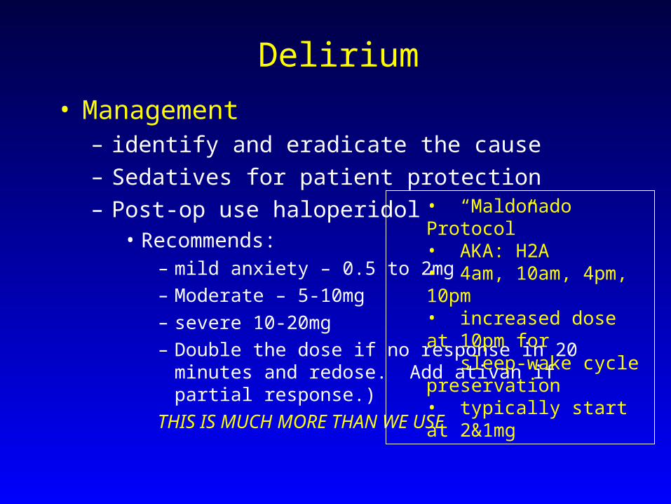
Delirium
• Management– identify and eradicate the cause
– Sedatives for patient protection
– Post-op use haloperidol• Recommends:
– mild anxiety – 0.5 to 2mg
– Moderate – 5-10mg
– severe 10-20mg
– Double the dose if no response in 20 minutes and redose. Add ativan if partial response.)
THIS IS MUCH MORE THAN WE USE
• “Maldonado Protocol”• AKA: H2A • 4am, 10am, 4pm, 10pm • increased dose at 10pm for sleep-wake cycle preservation • typically start at 2&1mg

Important to differentiate Delirium from DTs• Delirium Tremens
– Alcohol withdrawal
– Do not use haldol (lowers seizure threshold)
– Benzodiazepines are primary treatment
– Clonidine (alpha-2-agonist) for associated hypertension (also eases withdrawal centrally) \
– Valium: Onset 1-2 min, lasts as long as 12 hrs (active metabolite)
• 10/10/10 (q8 hrs x 3)
– Ativan: Slow onset (5-15 min) and longest duration (10-20hrs)
– Versed: Fast onset, short acting
• Lipid soluble, prolonged sedation if used long term

Cocaine Related Delirium
• Treated like Delirium Tremens• Benzos, not haldol

Who becomes delirious?• Prospective analysis of over 800 ICU patients in
Turkish hospital• 11% rate of DSM diagnosis of delirium• Collected clinical data and performed stepwise
conditional logistic regression to identify predictors of development of delirium (compared to controls)– Infection, fever, hypotension, anemia, and “respiratory
diseases”. – Hypocalcemia, hyponatremia, uremia, increased
hepatic enzymes, hyperamylasemia, hyperbilirubinemia, metabolic acidosis
Aldemir et al Critical Care 2001

Delirium, Dementia or Both?
• Delirium is a risk factor for increased ICU and Hospital length of stay
• In the geriatric population, becomes difficult to differentiate between underlying dementia and delirium
• Group at Brown did a prospective study of 118 patients in ICU
• Baseline dementia diagnosis given by family on Blessed Dementia Scale
• Delirium diagnosed by CAM and CAM-ICU scales
Ely et al JAGS, May 2003

Blessed-Dementia Scale
– ActivityOne point for each, unless otherwise indicated.
– CHANGES IN PERFORMANCE OF EVERYDAY ACTIVITIES– Inability to perform household tasks – Inability to cope with small sums of money – Inability to remember shortlist of items; for example, in shopping list – Inability to find way about indoors – Inability to find way about familiar streets – more…
– CHANGES IN HABITS– Eating – Dressing – Sphincter control – – CHANGES IN PERSONALITY, INTERESTS, DRIVE– Increased rigidity – Increased egocentricity – Impairment of regard of feeling for others – Coarsening of affect
– More….

CAM ICU SCORE
1. Acute Onset or Fluctuating Course Absent Present acute change in mental status from baseline? OR did the abnormal behavior fluctuate during the past 24
hours?2. Inattention Absent Present Did the patient have difficulty focusing attention as evidenced by scores less than 8 on either the auditory
or visual component of the Attention Screening Examination (ASE)?
3. Disorganized Thinking Absent Present Does the patient have disorganized or incoherent thinking as evidenced by incorrect answers to 2 or more
of the following 4 questions and/or demonstrate an inability to follow commands? Questions (Alternate Set A and Set B): 2 sets of logic questions (does a stone float? Does a leaf float?)
4. Altered Level of Consciousness Absent Present Is the patient’s level of consciousness anything other than alert (e.g. vigilant, lethargic or stuporous), or is
VAMASS < or > 3 (and not decreased due to sedation)? – Alert: Looks around spontaneously, fully aware of environment, interacts appropriately. – Vigilant: Hyperalert. – Lethargic: Drowsy but easily aroused. Unaware of some elements in the environment, or no appropriate
spontaneous interaction with interviewer. Becomes fully aware and appropriate with minimal noxious stimulation. – Stupor: Becomes incompletely aware with strong noxious stimulation. Can be aroused only by vigorous and
repeated stimuli. As soon as stimulus removed, subject lapses back into unresponsive state.
Overall CAM ICU Score: If 1 + 2, and either 3 or 4 is present, patient has delirium. Yes No

• 30% of pts had baseline dementia
• 14% were depressed
• 31% had delirium on first interview
• 70% had delirium sometime during hospitalization
• Most ICU delirium persisted after leaving ICU
• Patients with dementia had 2.4x risk of developing delirium during hospital stay compared to matched pts without delirium

Delirium and mortality
• 275 patients over 1 year, prospectively enrolled, CAM-ICU and Richmond Agitation-Sedation scale used
• 81% delirious at some point during ICU stay• Compared to well matched controls:• Increased mortality (34% vs 15%)• Increased length of stay (by 10 days on average)• Adjusted Hazard Ratios: 3.4 for mortality and 2.0
for LOS

Perspective on ICU Psychosis
• Until the 1990s, ICU pts were sedated and paralyzed and the changes in mental status went unrecognized
• Once the deleterious effects of longterm paralysis and sedation were realized, there was a decrease in the use of paralytics and sedatives
• It was realized that patients had changes in mental status
• Risk factors include: preexisting mental illness, severity of illness, advanced age, medical comorbidity, sleep deprivation and medications
Polderman Critical Care 2005

• ‘ICU psychosis’ was almost ‘normal’consequence of prolonged ICU stay
• Diagnosis is challenging with hypoactive delirium (more common)
• Many intensivists use a “wait and see” approach to treatment
• Others use Haldol liberally – beware the side effects, EPS

• Authors suggest:
– Basic prevention: Avoid sleep deprivation, increase cognitive stimulation, talk to the patient, play music, early mobilization, avoid dehydration, electrolyte disturbances, and hypoxia
– High index of suspicion, frequent screening
– Treatment should be more prompt (prevent sequelae)
– Stop offending drugs (benzos and narcotics misused to treat “confusion”)
– Treat with antipsychotics – drug of choice remains haloperidol
• Monitor for prolonged QT
• Interacts with multiple othe drugs common in ICU
– Neuroleptics not well studied in the ICU may be helpful in non-agtated delerium (risperdol, olanzapine, ziprasidone)
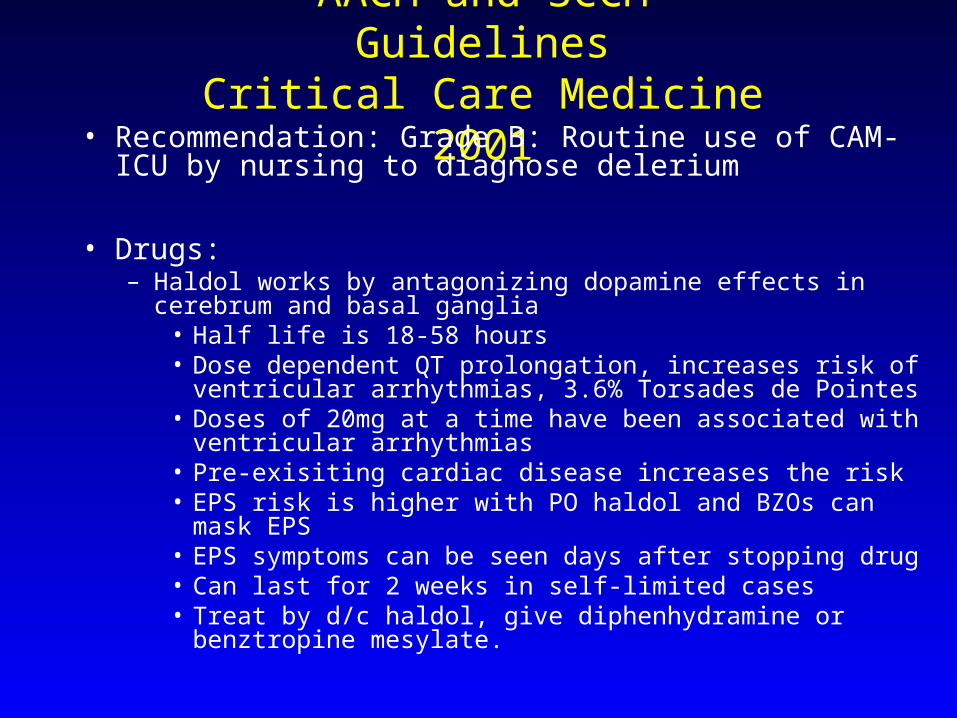
AACM and SCCM GuidelinesCritical Care Medicine 2001
• Recommendation: Grade B: Routine use of CAM-ICU by nursing to diagnose delerium
• Drugs: – Haldol works by antagonizing dopamine effects in cerebrum and basal
ganglia• Half life is 18-58 hours• Dose dependent QT prolongation, increases risk of ventricular
arrhythmias, 3.6% Torsades de Pointes• Doses of 20mg at a time have been associated with ventricular
arrhythmias• Pre-exisiting cardiac disease increases the risk• EPS risk is higher with PO haldol and BZOs can mask EPS• EPS symptoms can be seen days after stopping drug• Can last for 2 weeks in self-limited cases• Treat by d/c haldol, give diphenhydramine or benztropine
mesylate.

• Haldol also associated with 50% of neuroleptic malignant cases
• Chlorpromazine more anticholinergic, hypotensive effects
• Droperidol gives frightening dreams and hypotension by direct vasodilation
– Recommendation: Grade C: Haldol for chemical treatment of delirium
AACM and SCCM GuidelinesCritical Care Medicine 2001

• Recommendation: Grade B: non-pharmacologic methods to increase and improve sleep with sedative/hypnotics as adjuncts.
• Titrate the environmental stimuli• Sleep environment should be assessed• Ear plugs help• Single bed rooms, quiet time• Day/night lighting and noise levels• Relaxation techniques
– deep breathing exercises– music therapy– massage for 5-10 minutes
AACM and SCCM GuidelinesCritical Care Medicine 2001

Seizures• Second most common neurologic complication in
ICU
• Movements– Tonic contractions (sustained contractions)
– Atonic contraction (no movement)
– Clonic contraction (periodic contractions with regular frequency and amplitude)
– Myoclonus (periodic contractions with irregular amplitude and frequency)
– Automatisms (lipsmacking, chewing, etc)

• Generalized Seizures– Symetric and syncrhonous electrical discharge of the entire cerebral
cortex
– May or may not be accompanied by muscular contraction (if none, absence or petit-mal)
• Partial Seizures– Electrical discharges that are confined to a restricted part of cortex
– Simple partial (does not impair consciousness)
– Complex partial (does impair consciousness)
• Temporal lobe seizures: motionless stare and automatisms
• Epilepsia partialis continua: persistent tonic-clonic movements of facial and limb muscles unilaterally
• Status Epilepticus– more than 30 minutes of continuous seizure activity
– 2 or more sequential seizures without intervening consciousness

New Onset Seizures• Drug intoxication (amphetamies, cocaine, phenocyclidine, cipro, imipenam,
lidocaine, PCN, theophylline, TCA)
• Drug withdrawal (EtOH, BZO, Barbiturates, Opiates)
• Infection (Meningoencephalitis, abscess)
• Ischemia (focal or diffuse)
• Space occupying lesion (tumors or bleeds)
• Metabolic derrangement (hepatic encephalopathy, uremia, hypo-glycemia, -
natremia, -calcemia)

• Evaluation:– Examination looking for lateralizing signs
– Review of medications
– Imaging (CT)
– Procedural diagnostics (LP, labs, blood cultures)
• Management: – BZO
– Valium 0.2mg/kg IV stops 80% of seizures within 5 min, effect lasts 30 min
– Ativan 0.1mg/kg is as effective and lasts 12-24hrs
– Dilantin 20mg/kg following valium, aim for 20mg/l therapeutic serum level

Stroke
• Acute neurologic disorder• Nontraumatic brain injury, vascular origin• Focal findings (not global)• Persists for more than 24 hours• 80% ischemic, 20% of which are embolic
– Most thrombi are mural, LA, LV, DVT with PFO
• TIA: transient ischemic attack, focal deficits resolve in less than 24 hours (ischemia rather than infarction)
• Minor Stroke = RIND (reversible ischemic neurologic deficit) resolves within 3 weeks of event
• Major Stroke = deficits persist for more than 3 weeks

• Evaluation: common things you’ll see at the bedside– Full neuro exam, looking for focal deficits
– Seixures in 10% of cases, focal and within first 24 hours
– Fever in 50% of strokes (not with TIA) – look for other sources
– Coma and LOC are not common – more likely hemorrhage, massive infarct with edema, brainstem infarction, seizure (absence) or postictal state
– Aphasia – Left MCA distribution
– Weakness in contralateral limbs (can also have other metabolic causes)

Diagnostic Studies
• Time is brain• Coags, Chemistries: hypoglycemia, hyponatremia,
ARF• ECG: Afib? • CT head: 70% sensitivity for infarct, 90% for
hemorrhage - critical to distinguish btwn these• Better if after 24 hours for infarct• MRI: more sensitive esp for brainstem and
cerebellar strokes

Diagnostics and Treatment
• ICP: monitoring not recommended routinely– Elevate HOB 30 degrees
– Do not use measures that will decrease CBF
– minimize suctioning ( HTN)
– Do not hyperventilate (reduces CBF)
– Steroids not recommended
– Hyperosmolar therapy can be used if edema is severe (Mannitol, HTS)