acute respiratory distress syndrome requiring
TRANSCRIPT

Received 10/16/2019 Review began 11/01/2019 Review ended 11/06/2019 Published 11/12/2019
© Copyright 2019Kundu et al. This is an open access articledistributed under the terms of theCreative Commons Attribution LicenseCC-BY 3.0., which permits unrestricteduse, distribution, and reproduction in anymedium, provided the original author andsource are credited.
Acute Respiratory Distress Syndrome RequiringExtracorporeal Membrane Oxygenation as theInitial Presentation of Anti-neutrophillicCytoplasmic Auto-antibody Positive VasculitisSuhali Kundu , Shaurya Sharma , Ramandeep Minhas , Joshua Scheers-Masters , Paul C. Saunders
1. Internal Medicine, Maimonides Medical Center, Brooklyn, USA 2. Diagnostic Radiology, SUNY Downstate MedicalCenter, Brooklyn, USA 3. Rheumatology, Maimonides Medical Center, Brooklyn, USA 4. Cardiothoracic Surgery,Maimonides Medical Center, Brooklyn, USA
Corresponding author: Suhali Kundu, [email protected]
AbstractAcute respiratory distress syndrome (ARDS) is a life-threatening inflammatory state of lung injury that canrequire acute interventions including mechanical ventilation as well as emergent veno-venousextracorporeal membrane oxygenation (VV-ECMO) for management. Etiologies of ARDS are not clearlydiscernible in certain cases and can vary from sepsis, pneumonia, trauma and intoxication. Anti-nuclearcytoplasmic auto-antibody (ANCA)-associated vasculitis (AAV) is a group of several conditions that can havepulmonary complications including ARDS.
We present a case where the primary manifestation of myeloperoxidase (MPO)-ANCA positive vasculitis wasARDS, in order to highlight the importance of investigating rare vasculitides as the underlying cause ofARDS and the importance of ECMO as an early life-saving intervention for the management of ARDS.
Categories: Cardiac/Thoracic/Vascular Surgery, Nephrology, RheumatologyKeywords: ards, extracorporeal membrane oxygenation (ecmo), mpo/p-anca, p-anca vasculitis, pauci immuneglomerulonephritis, renal biopsy, critical care, cardiothoracic surgery, icu, young
IntroductionPulmonary vasculitides are rare heterogenous disease entities characterized by vessel inflammation anddestruction. Diagnosis of these disorders is arduous because of their variegated clinical presentation. Thereare several conditions classified under the general heading of anti-nuclear cytoplasmic autoantibody(ANCA)-associated vasculitis (AAV) including the following: microscopic polyangiitis (MPA), granulomatosiswith polyangiitis (GPA) and eosinophilic granulomatosis with polyangiitis (Churg-Strauss syndrome). Theincidence of AAV is 15-20 cases per million per year, a prevalence of 90-300 cases per million [1]. Lunglesions are an important feature of AAV. These disease conditions affect multiple organs including thekidneys, lungs, joints, eyes, heart, nervous system and skin [2, 3]. For our interest, this article primarilyfocuses on the involvement of the lungs. Manifestations differ depending on the specific condition. Forexample, the hallmark feature for Churg-Strauss is asthma whereas GPA presents with upper and/or lowerrespiratory tract lesions. MPA most frequently manifests with pulmonary fibrosis and alveolar hemorrhage[2]. Pulmonary involvement is less frequent in MPA than either GPA or Churg-Strauss syndrome. Around 10-30% of patients will develop diffuse alveolar hemorrhage, and although rare, lung involvement in AAV maymanifest to acute respiratory distress syndrome (ARDS) [1]. Acute respiratory distress syndrome is a processof non-hydrostatic pulmonary edema with hypoxemia [4]. This paper examines one such case of severeARDS as the primary manifestation of AAV in a young man.
Case PresentationA 33-year-old man with no significant past medical history presents to the emergency department (ED) withthe chief complaint of progressively worsening shortness of breath over 24 hours. He works at a constructionsite and was not wearing a protective mask while being exposed to cement dust. He was brought in withconcerns of inhalation injury. On arrival, he was found to be in severe respiratory distress requiringsupplemental oxygen via a nasal cannula, which was escalated to a non-rebreather facemask and further toBiPAP (BiLevel positive airway pressure) due to worsening oxygenation. He remained hypercarbic andhypoxemic on repeat blood gas analysis despite non-invasive ventilation and required intubation for severehypoxic respiratory failure secondary to ARDS (Figure 1) in the intensive care unit.
1 1 2, 1 3 4
Open Access CaseReport DOI: 10.7759/cureus.6135
How to cite this articleKundu S, Sharma S, Minhas R, et al. (November 12, 2019) Acute Respiratory Distress Syndrome Requiring Extracorporeal MembraneOxygenation as the Initial Presentation of Anti-neutrophillic Cytoplasmic Auto-antibody Positive Vasculitis. Cureus 11(11): e6135. DOI10.7759/cureus.6135
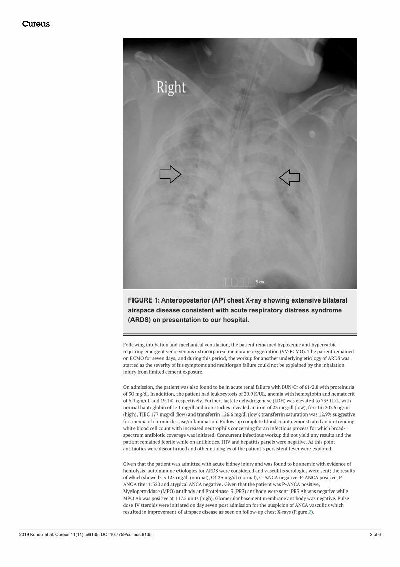
FIGURE 1: Anteroposterior (AP) chest X-ray showing extensive bilateralairspace disease consistent with acute respiratory distress syndrome(ARDS) on presentation to our hospital.
Following intubation and mechanical ventilation, the patient remained hypoxemic and hypercarbicrequiring emergent veno-venous extracorporeal membrane oxygenation (VV-ECMO). The patient remainedon ECMO for seven days, and during this period, the workup for another underlying etiology of ARDS wasstarted as the severity of his symptoms and multiorgan failure could not be explained by the inhalationinjury from limited cement exposure.
On admission, the patient was also found to be in acute renal failure with BUN/Cr of 61/2.8 with proteinuriaof 30 mg/dl. In addition, the patient had leukocytosis of 20.9 K/UL, anemia with hemoglobin and hematocritof 6.1 gm/dL and 19.1%, respectively. Further, lactate dehydrogenase (LDH) was elevated to 735 IU/L, withnormal haptoglobin of 151 mg/dl and iron studies revealed an iron of 23 mcg/dl (low), ferritin 207.6 ng/ml(high), TIBC 177 mcg/dl (low) and transferrin 126.6 mg/dl (low); transferrin saturation was 12.9% suggestivefor anemia of chronic disease/inflammation. Follow-up complete blood count demonstrated an up-trendingwhite blood cell count with increased neutrophils concerning for an infectious process for which broad-spectrum antibiotic coverage was initiated. Concurrent infectious workup did not yield any results and thepatient remained febrile while on antibiotics. HIV and hepatitis panels were negative. At this pointantibiotics were discontinued and other etiologies of the patient’s persistent fever were explored.
Given that the patient was admitted with acute kidney injury and was found to be anemic with evidence ofhemolysis, autoimmune etiologies for ARDS were considered and vasculitis serologies were sent; the resultsof which showed C3 125 mg/dl (normal), C4 25 mg/dl (normal), C-ANCA negative, P-ANCA positive, P-ANCA titer 1:320 and atypical ANCA negative. Given that the patient was P-ANCA positive,Myeloperoxidase (MPO) antibody and Proteinase-3 (PR3) antibody were sent; PR3 Ab was negative whileMPO Ab was positive at 117.5 units (high). Glomerular basement membrane antibody was negative. Pulsedose IV steroids were initiated on day seven post admission for the suspicion of ANCA vasculitis whichresulted in improvement of airspace disease as seen on follow-up chest X-rays (Figure 2).
2019 Kundu et al. Cureus 11(11): e6135. DOI 10.7759/cureus.6135 2 of 6

FIGURE 2: Anteroposterior (AP) chest X-ray showing interval decreasein bilateral lung infiltrates seven days after initiation of extra-corporealmembrane oxygenation (ECMO) and addition of pulse-dose steroids.
Following three days of IV steroids, oral steroids were continued for the remainder of the hospital course. CTof the chest was repeated at 10 days post VV-ECMO which demonstrated improvement of his extensive lungdisease with an evidence of a small amount of residual basilar interstitial lung disease (Figure 3).
2019 Kundu et al. Cureus 11(11): e6135. DOI 10.7759/cureus.6135 3 of 6

FIGURE 3: CT chest without contrast: Acute respiratory distresssyndrome (ARDS) in ANCA-associated vasculitis (AAV), 10 days postVV-ECMO with radiographic improvement of lung disease and evidenceof residual basal interstitial lung disease (ILD).ANCA: Anti-nuclear cytoplasmic auto-antibody; VV-ECMO: Veno-venous extracorporeal membraneoxygenation.
Renal biopsy was obtained on day 12 post admission, which revealed pauci-immune focal necrotizingglomerulonephritis, with 22% crescents (Figure 4). The patient was diagnosed with MPO antibody positiveANCA vasculitis with interstitial lung disease (ILD)/Alveolitis and pauci-immune glomerulonephritis. Inaddition to steroids, we also started Rituximab 12 days post admission following confirmatory pathologyresults. The patient was discharged 20 days post admission and continues to be on maintenance Rituximabtherapy. The patient remained clinically stable and asymptomatic on daily PO Prednisone, and receivedRituximab 500 mg intravenously every six months for maintenance therapy. To date, our patient hascompleted four cycles of Rituximab treatment with improvement of renal function (as depicted bynormalization of his serum creatinine and proteinuria). The patient has shown remarkable clinicalimprovement on follow-up appointments with negligible respiratory symptoms.
FIGURE 4: Renal biopsy of our patient with pauci-immune focalnecrotizing glomerulonephritis showing crescent formation withinglomeruli (arrows). Image courtesy of nephroCORE labs.
2019 Kundu et al. Cureus 11(11): e6135. DOI 10.7759/cureus.6135 4 of 6

DiscussionPulmonary involvement in AAV is well documented in the literature but is variegated in its manifestationmaking it difficult to diagnose and treat. For example, MPA is characterized primarily by ILD, while GPA haspulmonary manifestations that include cavitary masses, nodules, and airway stenosis. Both MPA and GPAmay present with alveolar hemorrhage syndrome. The diagnosis of AAV as a cause of lung disease ischallenging because of the wide differential diagnosis, including infections, adverse drug injury andidiopathic ILD [5-7]. In a study with 140 patients with AAV who underwent chest CT, Mohammad et al.showed that 80% had pulmonary abnormalities and patients with PR3-ANCA positivity demonstratedcentral airway disease while MPO-ANCA demonstrated usual interstitial pneumonitis (UIP) patterns. It hasalso been shown that there is predominance of MPO-ANCA positivity in patients with AAV and ILD [8, 9].
Of the mentioned pulmonary manifestations that occur in AAV, new onset ARDS may be the primary anddevastating presentation of undiagnosed AAV. Early intervention for ARDS is lifesaving and for our caseimmediate ECMO intervention was highly beneficial. ECMO served not only to drastically improve hisrespiratory function, but also served as a useful tool to stabilize the patient until a definitive diagnosis to theetiology of his ARDS could be established (Table 1).
Loscar et al. highlighted a case in 1997 of GPA in a 19-year-old female whose initial presentation was ARDSand sepsis requiring ECMO intervention [10]. Further studies have shown that routine screening for ANCAantibodies in patients with ARDS can rapidly establish a diagnosis of AAV and early intervention even inpatients requiring ECMO is favorable [3, 11].
ConclusionsThe importance of our case lies in the recognition that pulmonary injury, in its most severe form of ARDS,may be the initial presentation of anti-nuclear cytoplasmic auto-antibody (ANCA)-associated vasculitis(AAV). With astute clinical judgement, these rare causes may well be an important differential diagnosis tobe considered in patients with ARDS of unknown etiology.
Appendices AECC definition Berlin criteria Kigali modification of Berlin criteria
Timing Acute onset Within one week of a known clinical insult ornew or worsening respiratory symptoms
Within one week of a known clinical insult ornew or worsening respiratory symptoms
OxygenationPaO2/FiO2 ≤200 mmHg(dentified as acute lunginjury if ≤300 mmHg)
Mild: PaO2/FiO2 >200 mmHg but ≤300mmHg; Moderate: PaO2/FiO2 >100 mmHgbut ≤200 mmHg; Severe: PaO2/FiO2 ≤100mmHg
SpO2/FiO2 ≤315
PEEPrequirement None
Minimum 5 cmH2O PEEP required byinvasive mechanical ventilation (noninvasiveacceptable for mild ARDS)
No PEEP requirement, consistent with AECCdefinition
Chestimaging
Bilateral infiltrates seenon frontal chestradiograph
Bilateral opacities not fully explained byeffusions, lobar/lung collapse or nodules bychest radiograph or CT
Bilateral opacities not fully explained byeffusions, lobar/lung collapse or nodules bychest radiograph or ultrasound
Origin ofoedema
Pulmonary artery wedgepressure <18 mmHgwhen measured or noevidence of left atrialhypertension
Respiratory failure not fully explained bycardiac failure or fluid overload (needobjective assessment, such asechocardiography, to exclude hydrostaticoedema if no risk factor present)
Respiratory failure not fully explained bycardiac failure or fluid overload (needobjective assessment, such asechocardiography, to exclude hydrostaticoedema if no risk factor present)
TABLE 1: American-European Consensus Conference (AECC), Berlin and Kigali criteria for acuterespiratory distress syndrome (ARDS).Referenced from [12].
PEEP: Positive end-expiratory pressure; PaO2: Arterial oxygen tension; FiO2: Inspiratory oxygen fraction; SpO2: Arterial oxygen saturation measuredby pulse oximetry; CT: Computed tomography.
2019 Kundu et al. Cureus 11(11): e6135. DOI 10.7759/cureus.6135 5 of 6

Additional InformationDisclosuresHuman subjects: Consent was obtained by all participants in this study. Conflicts of interest: Incompliance with the ICMJE uniform disclosure form, all authors declare the following: Payment/servicesinfo: All authors have declared that no financial support was received from any organization for thesubmitted work. Financial relationships: All authors have declared that they have no financialrelationships at present or within the previous three years with any organizations that might have aninterest in the submitted work. Other relationships: All authors have declared that there are no otherrelationships or activities that could appear to have influenced the submitted work.
References1. Frankel SK, Schwarz MI: The pulmonary vasculitides. Am J Respir Crit Care Med. 2012, 186:216-223.
10.1164/rccm.201203-0539CI2. Homma S, Suzuki A, Sato K: Pulmonary involvement in ANCA-associated vasculitis from the view of the
pulmonologist. Clin Exp Nephrol. 2013, 17:667-671. 10.1007/s10157-012-0710-73. Dolch M, Irlbeck M, Wessely M, Rau S, Frey L, Schönermarck U: Acute respiratory distress syndrome (ARDS)
as primary manifestation in ANCA-associated vasculitis. La Presse Médicale. 2013, 42:752-753.10.1016/j.lpm.2013.02.237
4. Bernard GR, Artigas A, Brigham KL, et al.: The American-European Consensus Conference on ARDS.Definitions, mechanisms, relevant outcomes, and clinical trial coordination. Am J Respir Crit Care Med.1994, 149:818. 10.1164/ajrccm.149.3.7509706
5. Roden AC, Camus P: Iatrogenic pulmonary lesions. Semin Diagn Pathol. 2018, 35:260-271.10.1053/j.semdp.2018.03.002
6. Schwarz MI, Fontenot AP: Drug-induced diffuse alveolar hemorrhage syndromes and vasculitis . Clin ChestMed. 2004, 25:133-140. 10.1016/S0272-5231(03)00139-4
7. Gildea TR: Pulmonary disease in small-vessel vasculitis . Cleve Clin J Med. 2012, 79:27-30.10.3949/ccjm.79.s3.06
8. Mohammad AJ, Mortensen KH, Babar J, et al.: Pulmonary involvement in antineutrophil cytoplasmicantibodies (ANCA)-associated vasculitis: the influence of ANCA subtype. J Rheumatol. 2017, 44:1458-1467.10.3899/jrheum.161224
9. Katsumata Y, Kawaguchi Y, Yamanaka H: Interstitial lung disease with ANCA-associated vasculitis . ClinMed Insights Circ Respir Pulm Med. 2015, 9:51-56. 10.4137/CCRPM.S23314
10. Loscar M, Hummel T, Haller M, et al.: ARDS and Wegener granulomatosis. (Article in German) .Anaesthesist. 1997, 46:969-973. 10.1007/s001010050494
11. ter Maaten JC, Franssen CF, Gans RO, van Schijndel RJ, Hoorntje SJ: Respiratory failure in ANCA-associatedvasculitis. Chest. 1996, 110:357-362. 10.1378/chest.110.2.357
12. Confalonieri M, Salton F, Fabiano F: Acute respiratory distress syndrome. Eur Respir Rev. 2017, 26:160116.10.1183/16000617.0116-2016
2019 Kundu et al. Cureus 11(11): e6135. DOI 10.7759/cureus.6135 6 of 6