adaptations and deficits in the vestibulo-ocular reflex after ... · abstract: palsy of a nerve...
TRANSCRIPT
111
Ann. N.Y. Acad. Sci. 1004: 111–121 (2003). © 2003 New York Academy of Sciences.doi: 10.1196/annals.1303.070
Adaptations and Deficits in the Vestibulo-Ocular Reflex after Peripheral Ocular Motor Palsies
JAMES A. SHARPE,a,b DOUGLAS TWEED,a,c AND AGNES M.F. WONGa,b
aDivision of Neurology, and Departments of bOphthalmology, and cPhysiology, University Health Network, University of Toronto, Toronto, Ontario, Canada
ABSTRACT: Palsy of a nerve might be expected to lower vestibulo-ocular reflex(VOR) responses in its fields of motion, but effects of peripheral neuromuscu-lar disease were unknown. We recorded the VOR during sinusoidal head rota-tions in yaw, pitch, and roll at 0.5–2 Hz and static torsional gain in 43 patientswith unilateral nerve palsies. Sixth nerve palsy (n = 21) reduced both abductionand adduction VOR gains in darkness. In light, horizontal visually enhancedVOR (VVOR) gains were normal in moderate and mild palsy. In severe palsy,horizontal VVOR gains remained low in the paretic eye when it was fixating,whereas gains in the nonparetic eye became higher than normal. Third nervepalsy (n = 10) decreased VOR and VVOR gains during abduction, adduction,elevation, depression, extorsion, and intorsion. Fourth nerve palsy (n = 13) re-duced VOR gains of the paretic eye during intorsion, extorsion, elevation, de-pression, abduction, and adduction, but in light vertical and horizontal VVORgains were normal. In the nonparetic eye, all gains were normal. Reduced VORgains in the direction of paretic muscles and also in the direction of their antag-onists, together with normal gains in the nonparetic eye, indicate a selective ad-justment to the antagonists of paretic muscles. Increase of VVOR gains tonormal in the paretic eye, when used for fixation, without conjugate increase ingains in the occluded nonparetic eye, provides further evidence of selective ad-aptation for the paretic eye. Motions of the eyes after nerve palsies indicate mo-nocular VOR adaptation in three dimensions.
KEYWORDS: adaptation; nerve palsies; sixth nerve palsy; third nerve palsy;fourth nerve palsy; static torsional vestibulo-ocular reflex; angular vestibulo-ocular reflex; torsional vestibulo-ocular reflex; vertical vestibulo-ocular reflex;horizontal vestibulo-ocular reflex; Hering’s law; diplopia
INTRODUCTION
Assessment of strabismus emphasizes static deviations and little information isavailable about the effects of paralytic strabismus on eye-movement dynamics suchas during the vestibulo-ocular reflex (VOR). Adaptive changes in the VOR occur in
Address for correspondence: Dr. James A. Sharpe, Division of Neurology, University HealthNetwork TWH, WW 5-440, 399 Bathurst Street, Toronto, Ontario M5T 2S8, Canada. Fax: 416-603-5596.
112 ANNALS NEW YORK ACADEMY OF SCIENCES
response to different visual stimuli.1,2 Disconjugate VOR adaptation has been elic-ited in monkeys in response to anisometropic prisms3 and experimental weakeningof the horizontal rectus muscles.4,5 We examined the angular VOR in patients withunilateral peripheral sixth, third, and fourth nerve palsies to determine effects of pal-sies in different directions on the VOR and its adaptation, if any, in each eye to mo-nocular palsies. We identified changes in the actions of antagonists to pareticmuscles that indicate monocular adaptations to peripheral neuromuscular deficits.
METHODS
Patients with unilateral peripheral palsy of the sixth (n = 21), third (n = 10), orfourth (n = 13) ocular motor nerves were recruited from the NeuroophthalmologyCenter at the University Health Network.6–8 The duration and age of onset of diplo-pia, the presence or absence of risk factors for ischemia (diabetes mellitus and hy-pertension), and associated neurologic symptoms and signs were determined. Themagnitude of strabismus was measured objectively using the prism and cover testand subjectively using the Maddox rod and prism test. Appropriate tests were per-formed to rule out myasthenia gravis, thyroid ophthalmopathy, other orbital diseas-es, or intracranial lesions. Ranges of duction were estimated as the estimatedpercentage of the normal abduction in the other eye. On the basis of the duction de-fect, patients with sixth nerve palsies were classified into three groups: mild (81–95% of normal range of abduction), moderate (51–80%), and severe (≤50%). Fifteennormal subjects of similar ages served as controls (mean age, 52 years; SD, 15 years;median age, 58 years; age range 19–69; 8 women).
The angular dynamic VOR was measured in darkness while patients made activesinusoidal head on body rotations in yaw and pitch at approximately 0.5 and 2 Hz,and in roll at approximately 0.5, 1, and 2 Hz at amplitudes of approximately ±10°from orbital midposition. The dynamic visually enhanced VOR (VVOR) was mea-sured while they fixated on a stationary laser target 1 M from the naision, with oneeye occluded, fixating in turn with the paretic and nonparetic eye. To measure thestatic torsional VVOR, we had patients fixate on the center target with one eye oc-cluded as we measured their ocular responses to static head rolls of approximately30o toward each shoulder, as measured with a head search coil. Static torsional gainis defined as change in torsional eye position divided by change in head position dur-ing maintained head roll. The test then was repeated with the other eye fixating andthe fellow eye occluded, and also in total darkness (VOR).
Eye movements were recorded by three-dimensional binocular magnetic scleralsearch coils (Skalar Instrumentation, Delft, the Netherlands) using 1.83-m-diameterfield coils arranged in a cube (CNC Engineering, Seattle, WA). There was minimalcross talk; large horizontal and vertical movements produced deflections in the tor-sional channel of less than 4% of the amplitude of the horizontal and vertical move-ment. Any coil slippage was assessed by offsets in torsional eye position signalduring testing. Consistency of calibrated positions after each eye movement provid-ed evidence that the coil did not slip on the eye. Eye and head position signals werefiltered with a bandwidth of 0 to 90 Hz, and digitized at 200 Hz.
Fast phases of vestibular nystagmus were identified9 and positions between 80 msbefore and after fast phases were removed, the gaps being replaced with quadratic
113SHARPE et al.: ADAPTATIONS AND DEFICITS IN THE VOR
fits. The offset due to the fast phase then was removed, and the ongoing slow phasewas interpolated to yield a cumulative trace of eye position.
Using a least squares sinusoidal fit,10 we fitted eye and head positions with onecycle, and phases and amplitudes and amplitude gains were computed. We also plot-ted head velocity against eye velocity and performed a linear regression for each di-rection. The slopes of the fitted lines were the gains, and the results were comparableto those computed by the least squares sinusoidal fit technique.
RESULTS
Of 21 patients with sixth nerve palsy, six had severe, seven had moderate, andeight had mild palsy; the duration of symptoms ranged from 2 weeks to 96 months(mean, 16 months). For 10 patients with third nerve palsies, the duration of symp-toms ranged from 1 week to 50 months (mean, 18 months). In 13 patients with fourthnerve palsies, duration of symptoms ranged from 1 week to 132 months (mean, 35months). VOR function was tested at one point in the course of their palsies and ex-pressed as changes from normal, rather than serial intrasubject changes. Any recov-ery toward normal values was not assessed. Abnormalities are interpreted as deficitsor adaptation to those deficits.
Horizontal VOR
Sixth nerve palsy. In all patients, horizontal VOR gains in darkness were de-creased in the paretic eye in both abduction and adduction and remained normal inthe nonparetic eye in both directions (FIG. 1). In light, horizontal VVOR gains werenormal in both eyes in moderate and mild palsy. During active head movement, nor-mal persons have both VOR (in darkness) and the VVOR gains approximating uni-ty.11 In severe palsy, horizontal VVOR gains were low in the paretic eye in bothdirections, during viewing with either eye, whereas those in the nonparetic eye werehigher than normal (>1.0) when the paretic eye viewed (FIG. 1). In light and dark-ness, mean phase differences between the eye and head positions approximated180°, designated as zero phase shift.
Third nerve palsy. Horizontal VOR and VVOR gains of the paretic eye were de-creased during both abduction and adduction.
Fourth nerve palsy. In darkness, horizontal VOR gains of the paretic eye were re-duced symmetrically during both abduction and adduction (P < 0.01), whereas gainsof the nonparetic eye were normal. In light, during paretic or nonparetic eye viewing,horizontal VVOR gains of both the paretic and the nonparetic eyes were normal (P <0.05). Neither eye showed any significant phase shift from zero in light or in darkness.
Vertical VOR
Sixth nerve palsy. Vertical VOR and VVOR gains were normal in severe and mildpalsies.
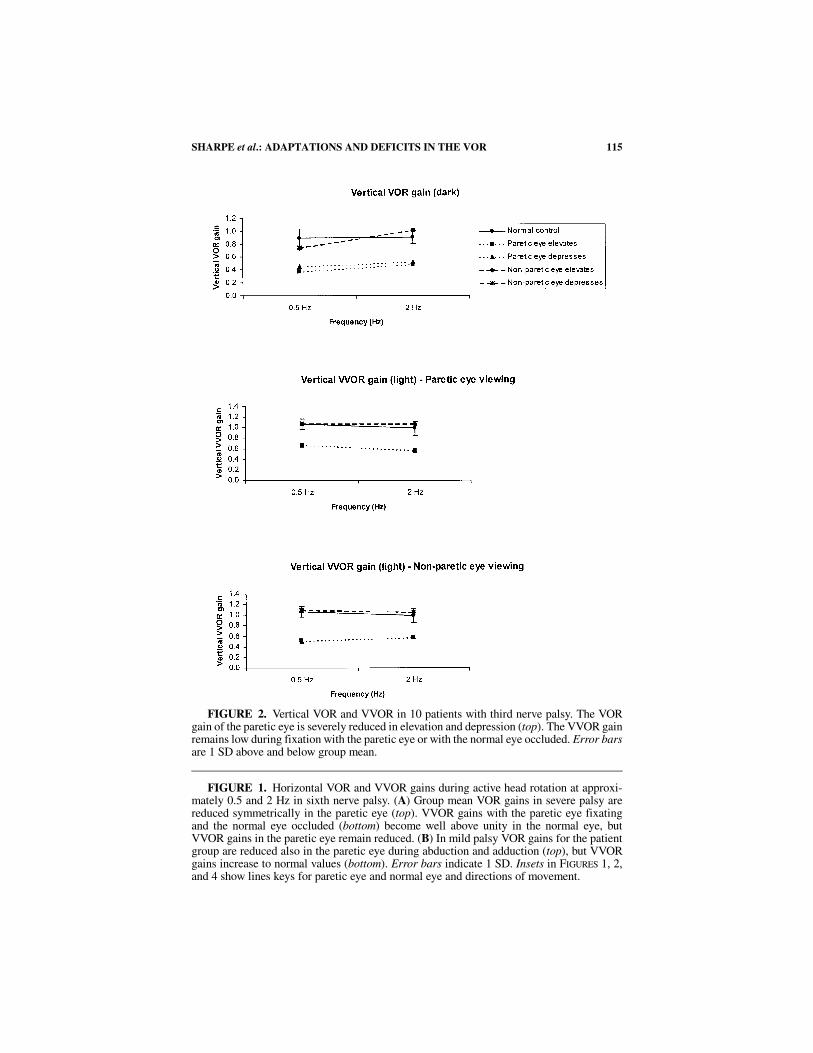
Third nerve palsy. In darkness (FIG. 2, top graph), vertical VOR gains of the pareticeye were reduced (P < 0.01) symmetrically during both elevation and depression,whereas gains of the nonparetic eye were normal. In light, during paretic eye or non-paretic eye viewing (FIG. 2, middle and bottom graphs), vertical VVOR gains of the

114 ANNALS NEW YORK ACADEMY OF SCIENCES
FIG
UR
E1.
See
foll
owin
g pa
ge fo
r le
gend
.
115SHARPE et al.: ADAPTATIONS AND DEFICITS IN THE VOR
FIGURE 1. Horizontal VOR and VVOR gains during active head rotation at approxi-mately 0.5 and 2 Hz in sixth nerve palsy. (A) Group mean VOR gains in severe palsy arereduced symmetrically in the paretic eye (top). VVOR gains with the paretic eye fixatingand the normal eye occluded (bottom) become well above unity in the normal eye, butVVOR gains in the paretic eye remain reduced. (B) In mild palsy VOR gains for the patientgroup are reduced also in the paretic eye during abduction and adduction (top), but VVORgains increase to normal values (bottom). Error bars indicate 1 SD. Insets in FIGURES 1, 2,and 4 show lines keys for paretic eye and normal eye and directions of movement.
FIGURE 2. Vertical VOR and VVOR in 10 patients with third nerve palsy. The VORgain of the paretic eye is severely reduced in elevation and depression (top). The VVOR gainremains low during fixation with the paretic eye or with the normal eye occluded. Error barsare 1 SD above and below group mean.
116 ANNALS NEW YORK ACADEMY OF SCIENCES
paretic eye remained reduced (P < 0.05), whereas gains in the nonparetic eye were nor-mal. Neither eye showed any significant phase shift from zero in light or in darkness.
Fourth nerve palsy. In darkness, vertical VOR gains of the paretic eye were sym-metrically reduced during both depression and elevation (P < 0.05), whereas gainsof the nonparetic eye were normal. In light, during paretic eye and nonparetic eyeviewing, vertical VVOR gains of both the paretic and the nonparetic eyes were nor-mal (P < 0.05). Neither eye showed any significant phase shift from zero in light orin darkness.
Torsional VOR, Dynamic and Static
Sixth nerve palsy. Dynamic torsional VOR and VVOR gains were significantlyreduced during head roll in both the paretic and nonparetic eyes when compared withnormal controls (P < 0.05). Neither eye showed any significant phase shift from zeroduring vertical or torsional rotation. Static torsional gains were reduced in 19 (90%)of the 21 patients in light and in dark (Z-tests, P < 0.05). Static torsional VOR andVVOR gains of each eye were conjugate between the two eyes in all patients and didnot differ during right eye or left eye viewing.
Third nerve palsy. In darkness dynamic torsional VOR gains of the paretic eyewere reduced during both intorsion and extorsion (P < 0.01), whereas gains of thenonparetic eye were normal. In light, and during viewing with either eye, torsionalVVOR gains of the paretic eye remained reduced (P < 0.01), whereas gains in the
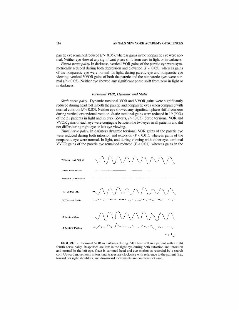
FIGURE 3. Torsional VOR in darkness during 2-Hz head roll in a patient with a rightfourth nerve palsy. Responses are low in the right eye during both extortion and intorsionand normal in the left eye. Gaze is summed head and eye motion as recorded by a searchcoil. Upward movements in torsional traces are clockwise with reference to the patient (i.e.,toward her right shoulder), and downward movements are counterclockwise.
117SHARPE et al.: ADAPTATIONS AND DEFICITS IN THE VOR
nonparetic eye were normal.7 In normal persons, viewing a foveal target does not ap-preciably raise torsional VVOR gains of above torsional gains in darkness.12 Neithereye showed any significant phase shift from zero in light or in darkness. Static tor-sional VOR and VVOR gains of the paretic eye were reduced during intorsion andextorsion (P < 0.05) but were normal in the nonparetic eye.
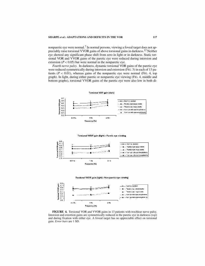
Fourth nerve palsy. In darkness, dynamic torsional VOR gains of the paretic eyewere reduced symmetrically during intorsion and extorsion (FIG. 3) in each of 13 pa-tients (P < 0.01), whereas gains of the nonparetic eye were normal (FIG. 4, topgraph). In light, during either paretic or nonparetic eye viewing (FIG. 4, middle andbottom graphs), torsional VVOR gains of the paretic eye were also low in both di-
FIGURE 4. Torsional VOR and VVOR gains in 13 patients with trochlear nerve palsy.Intorsion and extortion gains are symmetrically reduced in the paretic eye in darkness (top)and during fixation with either eye. A foveal target has no appreciable effect on torsionalgain. Error bars are 1 SD.

118 ANNALS NEW YORK ACADEMY OF SCIENCES
rections (P < 0.05), whereas those of the nonparetic eye remained normal.8 In lightand in darkness, the mean phase was zero. Static torsional VOR and VVOR gains ofthe paretic eye were reduced during intorsion (P < 0.05) and normal during extorsionof the paretic eye. In the nonparetic eye, static torsional VOR and VVOR gains werenormal.
DISCUSSION
During head rotation in darkness, angular VOR gains are reduced during move-ment in the directions of actions of paretic muscles as anticipated from their palsy.However, dynamic gains are also reduced in the fields of actions of their antagonistmuscles. VOR gains in the nonparetic eye remain normal, indicating a selective ad-justment of the paretic eye, specifically to the antagonists of paretic muscles. Inlight, visual input increases gain of the paretic eye when it is used for fixation, pro-viding further evidence of selective adaptation in the paretic eye. Torsional dynamicVOR and VVOR gains of the paretic eye are reduced for both extortion and intorsionin third and fourth nerve palsies. Motion of the eyes after nerve palsies exemplifiesmonocular adaptation of the VOR in three dimensions.
Sixth Nerve Palsy
In darkness, horizontal VOR gains are reduced during abduction of the pareticeye in all patients, as anticipated in sixth nerve palsy. Gains are also reduced duringadduction of the paretic eye (FIG. 1), suggesting that innervation to the medial rectushas changed. After severe palsy, vision does not increase abducting or adducting hor-izontal VVOR gains to normal in the paretic eye but causes secondary increase inVVOR gains to values above unity in the nonparetic eye, when the paretic eye fix-ates. To adopt a conventional term from strabismology, this is a secondary change inthe VOR, occurring when the paretic eye fixates. In mild and moderate palsy, visionenhances the VOR in the paretic eye but causes no change in the nonparetic eye, sug-gesting a monocular readjustment of innervation selectively to the paretic eye. Ver-tical VOR and VVOR gains are normal, indicating that the lateral rectus does nothave significant vertical actions through the ±10° excursions that we tested.
Reduced torsional VOR gains in the paretic eye can be explained by the esotropiain sixth nerve palsy. Torsional VOR gain normally varies with vergence.13,14 We at-tribute the reduced torsional gains in the paretic eye to the mechanism that normallylowers it during convergence.6 The low torsional gains in the nonparetic eye may bean adaptation to reduce torsional disparity between the two eyes.
Third Nerve Palsy
Adducting VOR gains are reduced, as anticipated from medial rectus palsy. Ab-ducting gains are also reduced; the reduction is attributed to an adaptive decrease ininnervation to the lateral rectus, to achieve symmetry of the horizontal VOR. Tor-sional VOR gains are reduced during extorsion from palsy of the inferior obliquemuscle. Gains are also reduced during intorsion,7 which may be explained by anadaptive decrease in innervation to the superior oblique, to restore symmetry of thetorsional VOR.

119SHARPE et al.: ADAPTATIONS AND DEFICITS IN THE VOR
Fourth Nerve Palsy
During head rotation in darkness, VOR gains are reduced during intorsion, de-pression, and abduction of the paretic eye, as anticipated from paresis of the superioroblique muscle. VOR gains during extorsion (FIG. 3), elevation, and adduction of theparetic eye also are reduced, whereas VOR gains in the nonparetic eye remain nor-mal, indicating a selective central adjustment of innervation to the paretic eye. Inlight, torsional VVOR gains in the paretic eye remained reduced (FIG. 4). Visual in-put increases vertical and horizontal VVOR gains to normal in the paretic eye, with-out a conjugate increase in VVOR gains in the nonparetic eye,8 providing furtherevidence of selective adaptation in the paretic eye.
Patients selected a preferred eye for viewing during the course of their palsies.We did not control the eye of habitual fixation, which was probably the nonpareticeye in most cases, although they may have switched from one eye to the other at theirwhim or subconscious choice. The adaptation might have differed if patients pre-dominantly used the paretic eye for fixation.
Changes in Antagonists of Paretic Agonist Muscles
Without the decreased VOR gains in the direction of action of antagonists ofparetic muscles, the VOR would be asymmetric in the paretic eye. The asymmetrywould drive the paretic eye further into direction of action of the antagonist witheach cycle of head rotation, resulting in increasing position disparity between thetwo eyes and more diplopia (FIG. 5). The brain might adopt any one of four strategiesto prevent this disparity. First, it might increase its innervation to the paretic agonistto increase VOR gain of the paretic eye, but this strategy is limited by the palsy itself.Second, the brain might generate saccades in direction of paresis to correct for thelow agonist VOR gains. However, during activation of the paretic agonist, if com-mon premotor signals are sent to motoneurons of the yolked agonist in the other eye,unwanted saccades would appear conjugately in the nonparetic eye, driving its foveaoff its target. Third, the brain might attempt to prevent asymmetry of the VOR by
FIGURE 5. Schematic of consequence of asymmetry of VOR in unilateral sixth nervepalsy. Without adaptation to achieve symmetry, the paretic eye would move further into ad-duction with each cycle of head motion.

120 ANNALS NEW YORK ACADEMY OF SCIENCES
decreasing antagonist gains in the paretic eye. Hering15 suggested that the brain con-trols gaze by two systems, one for conjugate movements and the other for vergence.However, if common and conjugate premotor signals were sent to motoneurons toboth eyes, the yolked antagonist gain in the other (normal) eye would be reduced aswell. For example, in the case of a left lateral rectus weakness from a left sixth nervepalsy, any adaptive reduction in innervation to the left medial rectus muscle wouldbe accompanied by reduced innervation to the right lateral rectus muscle, in accordwith Hering’s proposal or “law.” It was not. Fourth, the brain could selectively re-duce VOR gains during action of the antagonist of the paretic muscle by reducing itsinnervation. This is apparently the strategy that the brain uses, in violation ofHering’s law.
Changes in normal orbital plant mechanics might contribute to the decreasedVOR gains of the paretic eye in the direction of the antagonists to paretic muscles.The relative contribution of agonist contraction and antagonist relaxation varies withorbital position,16 and it may be altered when one muscle of an agonist–antagonistpair is palsied. Contracture is characterized by muscle shortening and stiffening as aresult of decreased number of sarcomeres.17 If the reduction of VOR gains in bothdirections were caused by changes in extraocular muscle mechanics, one would ex-pect VOR gains to be subnormal during rotation or in light (VVOR) as well as indarkness, and that the peak velocities of nystagmus quick phases would be reducedin each direction. However, our results indicate that although VOR gains were de-creased, VVOR gains could increase to normal values in light. In addition, althoughVOR gains were reduced in each direction, peak velocities of saccades in the antag-onist’s direction of action were normal (data not shown here). Our results provideevidence that decrease in VOR gains is not the result of changes in mechanical prop-erties of the orbital plant but is caused by a functional central adaptation to the palsy.
Proprioceptive signals from extraocular muscles18 might contribute to VOR ad-aptation. Proprioceptive afferent fibers project via the ophthalmic branch of thetrigeminal nerve to the spinal trigeminal nucleus, but a portion also may enter via theocular motor nerves.19 Although visual information plays a massively dominant rolein the control of VOR, altered proprioceptive inflow from a shortened (slack) antag-onist or a palsied muscle might participate in the monocular adaptations that weidentified after peripheral nerve palsies. Binocular disparity of retinal images that in-creases during head motion and asymmetry of retinal image slip when the VOR isimbalanced by palsy of a muscle appears to be the visual drive for monocular adap-tation to reduce image slip and diplopia.
ACKNOWLEDGMENTS
This work was supported by Canadian Institutes of Health Research grants MT15362 and ME 5504.
REFERENCES
1. GONSHOR, A. & G. MELVILL JONES. 1976. Extreme vestibulo-ocular adaptation inducedby prolonged optical reversal of vision. J. Physiol. (Lond.) 256: 381–414.

121SHARPE et al.: ADAPTATIONS AND DEFICITS IN THE VOR
2. YAGI, T., M. SHIMIZU, S. SEKINE & T. KAMIO. 1981.New neurootological test fordetecting cerebellar dysfunction. Vestibulo-ocular reflex changes with horizontalvision-reversal prisms. Ann. Otol. Rhinol. Laryngol. 90: 276–280.
3. OOHIRA, A. & D.S. ZEE. 1992. Disconjugate ocular motor adaptation in rhesus monkey.Vision Res. 32: 489–497.
4. SNOW, R., J. HORE & T. VILIS. 1985. Adaptation of saccadic and vestibulo-ocular sys-tems after extraocular muscle tenectomy. Invest. Ophthalmol. Visual Sci. 26: 924–931.
5. VIRRE, E., C. WERNER & T. VILIS. 1988. Monocular adaptation of the saccadic systemand vestibulo-ocular reflex. Invest. Ophthalmol. Visual Sci. 29: 1339–1347.
6. WONG, A.M.F., D. TWEED & J.A. SHARPE. 2002. Adaptations and deficits in thevestibulo-ocular reflex after sixth nerve palsy. Invest. Ophthalmol. Visual Sci. 43:99–111.
7. WONG, A.M.F. & J.A. SHARPE. 2002. Adaptations and deficits in the vestibulo-ocularreflex after third nerve palsy. Arch. Ophthalmol. 120: 360–368.
8. WONG, A.M.F., J.A. SHARPE & D. TWEED. 2002. The vestibulo-ocular reflex in fourthnerve palsy: deficits and adaptations. Vision Res. 42: 2205–2218.
9. RANALLI, P.J. & J.A. SHARPE. 1988. Vertical vestibulo-ocular reflex, smooth pursuitand eye-head tracking dysfunction in internuclear ophthalmoplegia. Brain 111:1299–1317.
10. SOKOLNIKOFF, I.S. & E.S. SOKOLNIKOFF. 1941. Higher Mathematics for Engineers andPhysicists. McGraw Hill. New York.
11. KIM, J.S. & J.A. SHARPE. 2001. The vertical vestibulo-ocular reflex and its interactionwith vision during active head motion: effects of aging. J. Vestib. Res. 11: 3–12.
12. MORROW, M.J. & J.A. SHARPE. 1993. The effects of head and trunk position on tor-sional vestibular and optokinetic eye movements in humans. Exp. Brain Res. 95:144–150.
13. TWEED, D., M. FETTER, D. SIEVERING, et al. 1994. Rotational kinematics of the humanvestibuloocular reflex. II. Velocity steps. J. Neurophysiol. 72: 2480–2489.
14. MISSLISCH, H., D. TWEED & B.J.M. HESS. 2001. Stereopsis outweighs gravity in thecontrol of the eyes. J. Neurosci. 21: RC126.
15. HERING, E. 1868. Die Lehre vom binokularen Sehen. Wilhelm Englemann. Leipzig. 16. COLLINS, C.C. 1975. The human ocular system. In Basic Mechanisms of Ocular Motil-
ity and their Clinical Implications. G. Lennerstrand & P. Bach-y-Rita, Eds.: 145–180. Pergamon Press, New York.
17. SCOTT, A.B. 1994. Change of eye muscle sarcomeres according to eye position. J.Pediatr. Ophthalmol. Strabismus 31: 85–88.
18. HAYMAN, M.R., J.P. DONALDSON & I.M. DONALDSON. 1995. The primary afferent path-way of extraocular muscle proprioception in the pigeon. Neuroscience 69: 671–683.
19. GENTLE, A. & G. RUSKELL. 1997. Pathway of the primary afferent nerve fibers servingproprioception in monkey extraocular muscles. Ophthalmic Physiol. Opt. 17: 225–231.