addison’s disease: a rare case report - international ... · addison’s disease: a rare case...
TRANSCRIPT
268International Journal of Scientific Study | May 2016 | Vol 4 | Issue 2
Addison’s Disease: A Rare Case ReportSanil Parekh1, Prashant Melmane2, Archana Bhate3, Amir Khan1, Ashish Sarode1
1Post-graduate student, Department of General Medicine, Dr. D. Y. Patil Hospital and University, Navi Mumbai, Maharashtra, India, 2Assosciate
Professor, Department of General Medicine, Dr. D. Y. Patil Hospital and University, Navi Mumbai, Maharashtra, India, 3Head, Department of General Medicine, Dr. D. Y. Patil Hospital and University, Navi Mumbai, Maharashtra, India
secondary or tertiary causes of adrenocortical insufficiency are not included in the term – “Addison’s disease.” Primary adrenal insufficiency can be a life-threatening disorder particularly in stressful situations, since cortisol secretion cannot be increased on demand at all.1 Often, there is a delay in diagnosis due to lack of suspicion on account of the subtle nature of the signs and symptoms in many cases and partly also due to the delay in visiting experts,3 leading to increased morbidity and mortality.
CASE REPORT
A 32-year-old Indian male presented to the outpatient department with complaints of 2-3 episodes of vomiting and pain in abdomen, following excessive binge drinking, the night before hospitalization. No history of fever, diarrhea, disorientation, breathlessness, loss of consciousness. The patient also complained of generalized weakness, giddiness, apathy, and lethargy since 5-6 days. A history of similar complaints on and off for the past few years, wherein the local doctor treated him on the lines of a psychiatric disorder, mainly depressive disorder. Moreover, there is a history of weight loss (about 9 kgs in the last 18 months). On enquiry, history of hyperpigmentation of skin mainly elbows, knees, lips, and oral mucosa over the last 2 years. Furthermore, decreased libido, disturbed sleep habits, mood changes but normal bowel habits. No history of visual disturbances, headaches.
INTRODUCTION
Addison’s disease is a rare endocrine disease.1 Addison’s disease has an incidence of 0.8 per million and a prevalence of 40-110 per million in the USA and European countries.2 No data on incidence and prevalence is available from India.
Addison’s disease or primary hypoadrenalismis caused by a total or near total destruction or dysfunction of one or both adrenal cortices. This results in decreased secretion of the adrenal cortical hormones-cortisol, aldosterone and androgens. The disease is characterized by weight loss, muscle weakness, fatigue, low blood pressure, and sometimes darkening of the skin in both exposed and non-exposed parts of the body. It can mimic a gastrointestinal disorder or a psychiatric disease, especially depression.3
A deficiency of Adrenocroticotrophic hormone (ACTH) can also produce hypocortisolism, but this is known as secondary adrenal insufficiency. Addison’s disease is a term restricted to primary adrenocortical insufficiency. Other
Case Report
AbstractAddison’s disease is a rare endocrine disease. Addison’s disease mainly refers to primary adrenocortical insufficiency which is to be distinguished from other forms of secondary adrenal insufficiency which may result from pituitary or hypothalamic diseases, which results in reduced secretion of Adrenocroticotrophic hormone and hence reduced glucocorticoid activity only, unlike primary insufficiency, in which there is reduced mineralocorticoid activity as well. This is a report of a case of Addison’s disease (autoimmune) as seen in a 32-year-old male, who presented with features mimicking gastrointestinal complaints secondary to alcohol intake, and features of depression. This case highlights the prevalence of this condition in the community, and also the need for physicians to keep a high index of suspicion for Addison’s disease.
Key words: Addison’s disease, Adrenocroticotrophic hormone, Autoimmune, Cortisol
Access this article online
www.ijss-sn.com
Month of Submission : 03-2016 Month of Peer Review : 04-2016 Month of Acceptance : 05-2016 Month of Publishing : 05-2016
Corresponding Author: Dr. Sanil Parekh, E-2203, Palm Residency, Sector 4, Nerul, Navi Mumbai, Maharashtra, India. Phone: +91-9820368538. E-mail: [email protected]
DOI: 10.17354/ijss/2016/299
Parekh, et al.: A Case of Addison’s Disease
269 International Journal of Scientific Study | May 2016 | Vol 4 | Issue 2
No past history of tuberculosis, diabetes mellitus, thyroid disorders, alcoholic liver disease or any other comorbid illness. History of frequent visits to nearby doctor for similar complaints. Furthermore, patient regularly consumes alcohol about 3-4 times a week (about 60 ml/day). Other medical history revealed no significant abnormality. A provisional diagnosis of alcohol-induced gastritis was made.
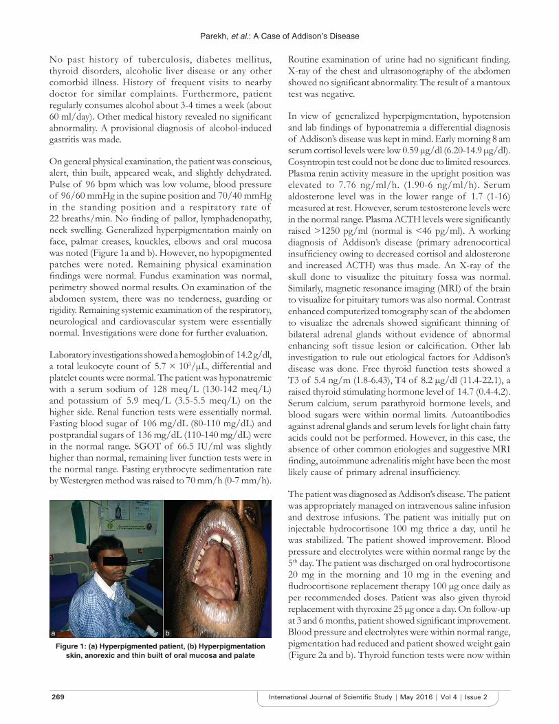
On general physical examination, the patient was conscious, alert, thin built, appeared weak, and slightly dehydrated. Pulse of 96 bpm which was low volume, blood pressure of 96/60 mmHg in the supine position and 70/40 mmHg in the standing position and a respiratory rate of 22 breaths/min. No finding of pallor, lymphadenopathy, neck swelling. Generalized hyperpigmentation mainly on face, palmar creases, knuckles, elbows and oral mucosa was noted (Figure 1a and b). However, no hypopigmented patches were noted. Remaining physical examination findings were normal. Fundus examination was normal, perimetry showed normal results. On examination of the abdomen system, there was no tenderness, guarding or rigidity. Remaining systemic examination of the respiratory, neurological and cardiovascular system were essentially normal. Investigations were done for further evaluation.
Laboratory investigations showed a hemoglobin of 14.2 g/dl, a total leukocyte count of 5.7 × 10³/µL, differential and platelet counts were normal. The patient was hyponatremic with a serum sodium of 128 meq/L (130-142 meq/L) and potassium of 5.9 meq/L (3.5-5.5 meq/L) on the higher side. Renal function tests were essentially normal. Fasting blood sugar of 106 mg/dL (80-110 mg/dL) and postprandial sugars of 136 mg/dL (110-140 mg/dL) were in the normal range. SGOT of 66.5 IU/ml was slightly higher than normal, remaining liver function tests were in the normal range. Fasting erythrocyte sedimentation rate by Westergren method was raised to 70 mm/h (0-7 mm/h).
Routine examination of urine had no significant finding. X-ray of the chest and ultrasonography of the abdomen showed no significant abnormality. The result of a mantoux test was negative.
In view of generalized hyperpigmentation, hypotension and lab findings of hyponatremia a differential diagnosis of Addison’s disease was kept in mind. Early morning 8 am serum cortisol levels were low 0.59 µg/dl (6.20-14.9 µg/dl). Cosyntropin test could not be done due to limited resources. Plasma renin activity measure in the upright position was elevated to 7.76 ng/ml/h. (1.90-6 ng/ml/h). Serum aldosterone level was in the lower range of 1.7 (1-16) measured at rest. However, serum testosterone levels were in the normal range. Plasma ACTH levels were significantly raised >1250 pg/ml (normal is <46 pg/ml). A working diagnosis of Addison’s disease (primary adrenocortical insufficiency owing to decreased cortisol and aldosterone and increased ACTH) was thus made. An X-ray of the skull done to visualize the pituitary fossa was normal. Similarly, magnetic resonance imaging (MRI) of the brain to visualize for pituitary tumors was also normal. Contrast enhanced computerized tomography scan of the abdomen to visualize the adrenals showed significant thinning of bilateral adrenal glands without evidence of abnormal enhancing soft tissue lesion or calcification. Other lab investigation to rule out etiological factors for Addison’s disease was done. Free thyroid function tests showed a T3 of 5.4 ng/m (1.8-6.43), T4 of 8.2 µg/dl (11.4-22.1), a raised thyroid stimulating hormone level of 14.7 (0.4-4.2). Serum calcium, serum parathyroid hormone levels, and blood sugars were within normal limits. Autoantibodies against adrenal glands and serum levels for light chain fatty acids could not be performed. However, in this case, the absence of other common etiologies and suggestive MRI finding, autoimmune adrenalitis might have been the most likely cause of primary adrenal insufficiency.
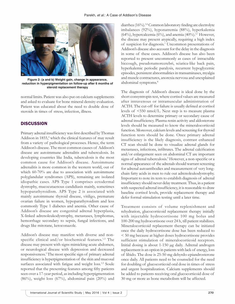
The patient was diagnosed as Addison’s disease. The patient was appropriately managed on intravenous saline infusion and dextrose infusions. The patient was initially put on injectable hydrocortisone 100 mg thrice a day, until he was stabilized. The patient showed improvement. Blood pressure and electrolytes were within normal range by the 5th day. The patient was discharged on oral hydrocortisone 20 mg in the morning and 10 mg in the evening and fludrocortisone replacement therapy 100 µg once daily as per recommended doses. Patient was also given thyroid replacement with thyroxine 25 µg once a day. On follow-up at 3 and 6 months, patient showed significant improvement. Blood pressure and electrolytes were within normal range, pigmentation had reduced and patient showed weight gain (Figure 2a and b). Thyroid function tests were now within
Figure 1: (a) Hyperpigmented patient, (b) Hyperpigmentation skin, anorexic and thin built of oral mucosa and palate
a b
Parekh, et al.: A Case of Addison’s Disease
270International Journal of Scientific Study | May 2016 | Vol 4 | Issue 2
normal limits. Patient was also put on calcium supplements and asked to evaluate for bone mineral density evaluation. Patient was educated about the need to double dose of steroids in times of stress, infection, illness.
DISCUSSION
Primary adrenal insufficiency was first described by Thomas Addison in 1855,4 which the clinical features of may result from a variety of pathological processes. Hence, the term Addison’s disease. The most common causes of Addison’s disease are autoimmune adrenalitis and tuberculosis. In developing countries like India, tuberculosis is the most common cause for Addison’s disease. Autoimmune adrenalitis is more common in the western world, out of which 60-70% are due to association with autoimmune polyglandular syndromes (APS), remaining are isolated idiopathic cases. APS Type 1 comprises ectodermal dystrophy, mucocutaenous candidiasis mainly, sometimes hypoparathyroidism. APS Type 2 is associated with mainly autoimmune thyroid disease, vitiligo, premature ovarian failure in women, hypoparathyroidism and less commonly Type 1 diabetes and anemia. Other cause of Addison’s disease are congenital adrenal hyperplasia, X-linked adrenoleukodystrophy, metastases, lymphomas, hemorrhage secondary to sepsis, fungal infections, and drugs like mitotane, ketoconazole.
Addison’s disease may manifest with diverse and non-specific clinical and/or biochemical features.1,5 The disease may present with signs mimicking acute abdomen, or neurological disease with depression and decreased responsiveness.3 The most specific sign of primary adrenal insufficiency is hyperpigmentation of the skin and mucosal surfaces associated with fatigue and weight loss.1,5 Soule reported that the presenting features among fifty patients seen over a 17-year period, as including hyperpigmentation (86%), weight loss (67%), abdominal pain (20%) and
diarrhea (16%).1,6 Common laboratory finding are electrolyte imbalances (92%), hyponatremia (88%), hyperkalemia (64%), hypercalcemia (6%), and anemia (40%).2,7 However, the disease may present atypically, requiring a high index of suspicion for diagnosis.1 Uncommon presentations of Addison’s disease also account for the delay in the diagnosis in some of these cases. Addison’s disease has also been reported to present uncommonly as cases of intractable hiccough, pseudotumorcerebri, sciatica-like back pain, hyperkalemic periodic paralysis, recurrent hypoglycemic episodes, persistent abnormalities in transaminases, myalgia and muscle contractures, anorexia nervosa and unexplained abdominal symptoms.8
The diagnosis of Addison’s disease is ideal done by the short consyntropin test, where cortisol values are measured after intravenous or intramuscular administration of ACTH. The cut-off for failure is usually defined at cortisol levels of <550 nmol/L. Next step is to measure plasma ACTH levels to determine primary or secondary cause of adrenal insufficiency. Plasma renin activity and aldosterone levels should be measured to know the mineralocorticoid function. Moreover, calcium levels and screening for thyroid function tests should be done. Once primary adrenal insufficiency is the likely diagnosis, contrast enhanced CT scan should be done to visualize adrenal glands for metastases, infections, infiltrates. The adrenal calcification and/or enlargement seen on abdominal CT are important signs of adrenal tuberculosis.9 However, a non-specific or a normal appearance of the adrenals should warrant screening for adrenal autoantibodies and measurement of very long-chain fatty acids in men to rule out adrenoleukodystrophy. Important to note its tests to establish diagnosis of adrenal insufficiency should never delay treatment. Thus, in a patient with suspected adrenal insufficiency, it is reasonable to draw baseline cortisol levels, provide replacement therapy and defer formal stimulation testing until a later time.
Treatment consists of volume replenishment and rehydration, glucocorticoid replacement therapy initially with injectable hydrocortisone 100 mg bolus and 100-200 mg hydrocortisone over 24 h, till patient stabilizes. Mineralocorticoid replacement therapy can be initiated once the daily hydrocortisone dose has been reduced to < 50 mg because at higher doses hydrocortisone provides sufficient stimulation of mineralocorticoid receptors. Initial dosing is about 1-150 µg daily. Adrenal androgen replacement is an option in patients with lack of energy, loss of libido. The dose is 25-50 mg dehyrdo-epiandrosterone once daily. All patients need to be counseled for the need for doubling of glucocorticoid oral dose in times of stress and urgent hospitalization. Calcium supplements should be added to patients receiving oral glucocorticoid dose of 30 mg or more as bone metabolism will be affected.
Figure 2: (a and b) Weight gain, change in appearance, reduction in hyperpigmentation on follow-up after 6 months of
steroid replacement therapy
a b
Parekh, et al.: A Case of Addison’s Disease
271 International Journal of Scientific Study | May 2016 | Vol 4 | Issue 2
CONCLUSION
This case presented with features of the gastrointestinal disease and depression. The presence of hyperpigmentation, hypotension along with electrolyte abnormalities of hyponatremia and hyperkalemia led to the suspicion of Addison’s disease. Addison’s disease although rare does occur in the community. Diagnosis is usually late, leading to increased morbidity and mortality. A survey of patients with Addison’s disease who are members of the National Adrenal Disease Foundation revealed that 60% had sought medical attention from two or more physicians before the correct diagnosis was ever considered.3 No figures are available on the number of undiagnosed patients succumbing to adrenal insufficiency. Thus, physicians should keep a high index of suspicion for adrenal insufficiency in unexplained illness.
REFERENCES
1. OelkersW.Adrenalinsufficiency.NEnglJMed1996;335:1206-12.2. PaulMS.Theadrenalcortex.In:LarsenRP,KronenbergHM,MelmedS,
Polonsky KS, editors. Williams Textbook of Endocrinology. 10th ed. Philadelphia,PA:W.B.SaundersPublication;2003.p.525-32.
3. TenS,NewM,MaclarenN.Clinicalreview130:Addison’sdisease2001.JClinEndocrinolMetab2001;86:2909-22.
4. Hiatt JR, Hiatt N. The conquest of Addison’s disease. Am J Surg1997;174:280-3.
5. AlebiosuCO,OdusanO.Addison’sdisease:Acasereport.AnnAfrMed2003;2:85-7.
6. SouleS.Addison’sdiseaseinAfrica–Ateachinghospitalexperience.ClinEndocrinol(Oxf)1999;50:115-20.
7. Shulman DI, Palmert MR, Kemp SF; Lawson Wilkins Drug andTherapeuticsCommittee. Adrenalinsufficiency:Stillacauseofmorbidityanddeathinchildhood.Pediatrics2007;119:484-94.
8. Choudhary S, Alam A, Dewan V, Yadav D, Dubey NK. An UnusualpresentationofAddison’sdisease:Acase report.ClinPediatrEndocrinol2011;20:57-60.
9. WangYX,ChenCR,HeGX,TangAR.CTfindingsofadrenalglandsinpatientswithtuberculousAddison'sdisease.JBelgeRadiol1998;81:226-8.
How to cite this article: Parekh S, Melmane P, Bhate A, Khan A, Sarode A. Addison’s Disease: A Rare Case Report. Int J Sci Stud 2016;4(2):268-271.
Source of Support: Nil, Conflict of Interest: None declared.