adductome-based identification of biomarkers for lipid peroxidation · 2017-05-18 · lipid...
TRANSCRIPT

Adductome-based identification of biomarkers for lipidperoxidationReceived for publication, October 9, 2016, and in revised form, March 1, 2017 Published, Papers in Press, March 24, 2017, DOI 10.1074/jbc.M116.762609
Takahiro Shibata‡§, Kazuma Shimizu‡, Keita Hirano‡, Fumie Nakashima‡, Ryosuke Kikuchi¶, Tadashi Matsushita�,and Koji Uchida‡**1
From the ‡Graduate School of Bioagricultural Sciences, Nagoya University, Nagoya 464-8601,§PRESTO, Japan Science andTechnology Agency, Kawaguchi, Saitama 332-0012, the Departments of ¶Medical Technique and �Clinical Laboratory and BloodTransfusion, Nagoya University Hospital, Nagoya 466-8560, and the **Graduate School of Agricultural and Life Sciences,The University of Tokyo, Tokyo 113-8657, Japan
Edited by Dennis R. Voelker
Lipid peroxidation is an endogenous source of aldehydes thatgives rise to covalent modification of proteins in various patho-physiological states. In this study, a strategy for the comprehen-sive detection and comparison of adducts was applied to find abiomarker for lipid peroxidation-modified proteins in vivo. Thisadductome approach utilized liquid chromatography with elec-trospray ionization tandem mass spectrometry (LC-ESI-MS/MS) methods designed to detect the specific product ions frompositively ionized adducts in a selected reaction monitoringmode. Using this procedure, we comprehensively analyzedlysine and histidine adducts generated in the in vitro oxidizedlow-density lipoproteins (LDL) and observed a prominentincrease in several adducts, including a major lysine adduct.Based on the high resolution ESI-MS of the adduct and on theLC-ESI-MS/MS analysis of the synthetic adduct candidates, themajor lysine adduct detected in the oxidized LDL was identifiedas N�-(8-carboxyoctanyl)lysine (COL). Strikingly, a significantlyhigher amount of COL was detected in the sera from atheroscle-rosis-prone mice and from patients with hyperlipidemia com-pared with the controls. These data not only offer structuralinsights into protein modification by lipid peroxidation prod-ucts but also provide a platform for the discovery of biomarkersfor human diseases.
Lipid peroxidation in tissues represents a degradation pro-cess, which is the consequence of the production and the prop-agation of free radical reactions primarily involving membranepolyunsaturated fatty acids (PUFAs), and has been implicatedin the pathogenesis of various diseases, including atherosclero-sis, cancer, rheumatoid arthritis, and diabetes, as well as aging(1). Lipid peroxidation leads to the generation of a broad arrayof different products with diverse and potent biological activi-
ties. Among them are a variety of different aldehydes. Theprimary products of the lipid peroxidation reaction, lipidhydroperoxides, can undergo carbon-carbon bond cleavage viaalkoxyl radicals in the presence of transition metals giving riseto the formation of short-chain unesterified aldehydes of 3–9carbons in length, and a second class of aldehydes still esterifiedto the parent lipid (2). These reactive aldehydes are consideredimportant mediators of cellular injury due to their ability tocovalently modify biomolecules, which can disrupt essentialcellular functions and cause mutations (2). Indeed, the covalentadduction of aldehydes to apolipoprotein B in low-density lipo-proteins (LDL) has been strongly implicated in the mechanismby which LDL is converted into an atherogenic form that istaken up by macrophages, leading to the formation of foamcells.
Based on many reports concerning the chemical and immu-nochemical detection of lipid peroxidation-derived aldehydeadducts in human diseases, there is no doubt that the steady-state levels of lipid peroxidation products increase underpathophysiological conditions associated with oxidative stress.Considerable progress has recently been made towardunderstanding the mechanisms of action of lipid peroxida-tion products. However, there are intrinsic difficulties asso-ciated with measuring reactive molecules. Like other reac-tive species, such as reactive oxygen species, many lipidperoxidation products can readily react with biomolecules toform conjugates, whereas only a few studies have addressedthe concentration of the aldehyde adducts in vitro and invivo. This may be partly due to the fact that many adductsgenerated in the modified proteins are unstable under thestrong acid conditions of conventional acid hydrolysis. Inaddition, because of technical constraints, the covalent mod-ification of proteins by aldehydes has been individually stud-ied, and comprehensive analysis of protein modification hasrarely been performed.
To simultaneously detect a variety of known and unknownadducts in protein samples, we adapted a mass spectrometry-based method for the comprehensive analysis of adducts(“adductome”). We expected that, although adductomeanalysis is semiquantitative, it would help to provide a com-prehensive picture of the adducts. Our strategy for theadductome analysis is illustrated in Fig. 1. This method isbased on the fact that aldehydes form adducts with specific
This work was supported in part by Grant-in-aid for Scientific Research (A)26252018 (to K. U.), Grant-in-aid for Scientific Research on Innovative Areas“Oxygen Biology: a new criterion for integrated understanding of life”26111011 (to K. U.), and the Japan Science and Technology AgencyPRESTO program (JPMJPR1334 to T. S.) of the Ministry of Education, Sci-ences, Sports, Technology (MEXT), Japan. The authors declare that theyhave no conflicts of interest with the contents of this article.
1 To whom correspondence should be addressed: Graduate School ofAgricultural and Life Sciences, University of Tokyo, Tokyo 113-8657,Japan. Tel.: 81-3-5841-5127; Fax: 81-3-5841-8026; E-mail: [email protected].
crosARTICLE
J. Biol. Chem. (2017) 292(20) 8223–8235 8223© 2017 by The American Society for Biochemistry and Molecular Biology, Inc. Published in the U.S.A.
by guest on August 6, 2020
http://ww
w.jbc.org/
Dow
nloaded from
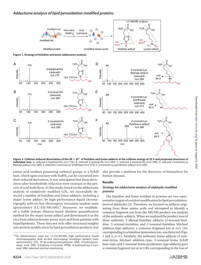
amino acid residues possessing carbonyl groups or a Schiffbase, which upon reaction with NaBH4 can be converted intotheir reduced derivatives. It was anticipated that these deriv-atives after borohydride reduction were resistant to the pro-cess of acid hydrolysis. In this study, based on the adductomeanalysis of oxidatively modified LDL, we successfully de-tected a number of histidine and lysine adducts, including amajor lysine adduct, by high-performance-liquid chroma-tography with on-line electrospray ionization-tandem massspectrometry (LC-ESI-MS/MS).2 Moreover, we establish-ed a stable isotope dilution-based absolute quantificationmethod for the major lysine adduct and determined it in thesera from atherosclerosis-prone mice and from patients withhyperlipidemia. These data not only offer structural insightsinto protein modification by lipid peroxidation products, but
also provide a platform for the discovery of biomarkers forhuman diseases.
Results
Strategy for adductome analysis of aldehyde-modifiedproteins
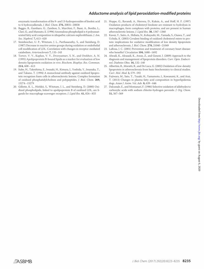
The histidine and lysine residues in proteins are two repre-sentative targets of covalent modification by lipid peroxidation-derived aldehydes (3). Therefore, we focused on adducts origi-nating from these amino acids and attempted to identify acommon fragment ion from the MS/MS product-ion analysisof the authentic adducts. When we analyzed the product ions ofthree authentic 2-alkenal-histidine adducts (2-hexenal-histi-dine, 2-octenal-histidine, and 2-nonenal-histidine Michaeladdition-type adducts), a common fragment ion at m/z 110,corresponding to a histidine immonium ion, was detected (Figs.1 and 2, A–C). Similarly, the authentic lysine adducts (2-non-enal-lysine Michael addition-type, 2-nonenal-lysine Schiffbase-type, and 2-nonenal-lysine pyridinium-type adducts) gavea common fragment ion at m/z 84, corresponding to the loss of
2 The abbreviations used are: LC-ESI-MS/MS, high performance liquidchromatography with on-line electrospray ionization tandem massspectrometry; COL, N�-(8-carboxyoctanyl)lysine; HNA, 4-hydroxynon-anoic acid; HNE, 4-hydroxy-2-nonenal; HPNE, 4-hydroperoxy-2-non-enal; SRM, selected reaction monitoring.
Figure 1. Strategy of histidine and lysine adductome analysis.
Figure 2. Collision-induced dissociation of the [M � H]� of histidine and lysine adducts at the collision energy of 25 V and proposed structures ofindividual ions. A, reduced 2-heptenal-His (m/z 270). B, reduced 2-octenal-His (m/z 284). C, reduced 2-nonenal-His (m/z 298). D, reduced 2-nonenal-LysMichael adduct (m/z 289). E, reduced 2-nonenal-Lys Schiff base (m/z 273). F, 2-nonenal-Lys pyridinium adduct (m/z 389).
Adductome analysis of lipid peroxidation-modified proteins
8224 J. Biol. Chem. (2017) 292(20) 8223–8235
by guest on August 6, 2020
http://ww
w.jbc.org/
Dow
nloaded from

NH3 from the lysine immonium ion (Figs. 1 and 2, D–F). It wasexpected that these characteristic common fragment ionsmight allow the comprehensive analysis of histidine and lysineadducts using LC-ESI-MS/MS. To test the validity of this pro-cedure, native and modified BSA with the lipid peroxidation-derived aldehydes, such as 2-alkenals, 4-hydroxy-2-alkenals,and 4-hydroperoxy-2-nonenal, were treated with NaBH4 to sta-bilize the adducts, hydrolyzed under the conditions of conven-tional acidic hydrolysis, and then subjected to the LC-ESI-MS/MS analyses. As shown in Fig. 3, the MS data could bevisualized as a two-dimensional image, in which the x axis rep-resents the LC retention time, and the y axis represents themass-to-charge ratio (m/z) for the individual detected adducts.
Adductome analysis of oxidized LDL
An important part of the pathogenesis of atherosclerosis hasbeen implicated by the oxidative modification of low-densitylipoproteins (LDL) (4, 5). The modification of LDL involves theperoxidation of PUFAs included in the LDL particle along withthe appearance of lipid peroxidation products, such as alde-hydes (2, 6). Hence, we applied the adductome analysis to thenative and Cu2�-oxidized LDL to simultaneously detect a vari-ety of histidine and lysine adducts. Fig. 4A shows the adduc-tome maps of the putative histidine adducts generated in thenative and oxidized LDL. The adductome maps are shown witha color gradient encoding the relative abundance from blue(low) to red (high). Product identification was tentatively made
Figure 3. Adductome maps of known histidine adducts (A) and lysine adducts (B). The specific product ions from positively ionized histidine and lysine adductswere analyzed by LC-ESI-MS/MS in the SRM mode transmitting the [M � H]� �110 (for histidine adduct, A) or [M � H]� �84 (for lysine adduct, B) transition.
Figure 4. Adductome analysis of histidine adducts detected in the oxidized LDL. A, adductome maps of histidine adducts detected in the control (left) orCu2�-oxidized LDL (right). The adductome maps are shown with a color gradient encoding the relative abundance from blue (low) to red (high). The known histidineadducts are indicated by arrows labeled H1–H6. B, chemical structure of histidine adducts (H1–H6) detected in the Cu2�-oxidized LDL.
Adductome analysis of lipid peroxidation-modified proteins
J. Biol. Chem. (2017) 292(20) 8223–8235 8225
by guest on August 6, 2020
http://ww
w.jbc.org/
Dow
nloaded from

by comparison of the retention time and the mass-to-chargeratio of each adduct detected in the aldehyde-modified proteins(Fig. 3). The most abundant product H1 was suggested to beidentical to the reduced form of the 4-hydroxy-2-nonenal(HNE)-histidine Michael adduct (Fig. 4B). In addition, prod-ucts H2, H3, H4, and H5 were putatively identified as thereduced form of 2-octenal-histidine, 2-nonenal-histidine, 2-decanal-histidine, and 2-undecanal-histidine Michael addi-tion-type adducts, respectively (Fig. 4B). The adductome mapssuggested that these 2-alkenal-histidine adducts might berelatively minor products. It was also suggested that productH6 was one of the major histidine adducts with an M � H� at310, representing an increase of m/z 155. This change in themolecular weight was previously observed in an aldehyde-oxidized product, N�-4-hydroxynonanoic acid-lysine, origi-
nating from the reaction of lysine with 4-hydroperoxy-2-nonenal (HPNE), the 4-hydroperoxy analog of HNE (7).Indeed, the hydroxynonanoic acid-histidine adduct wasdetected as one of the major products in the reaction ofhistidine with HPNE (data not shown). Thus, it was revealedthat histidine residues could be converted to the HNE- andHPNE-derived adducts during oxidative modification of theLDL (Fig. 5).
In contrast, the profile of lysine adducts generated in theoxidized LDL was much more complex than that of the histi-dine adducts (Fig. 6). In contrast to the histidine adducts, the4-hydroxy-2-alkenal-derived lysine adducts were barelydetected in the oxidized LDL. At least six minor products wereputatively identified as the 2-alkenal-lysine adducts, such asthe acrolein-lysine (K1), 2-heptenal-lysine (K2), 2-hexenal-
Figure 5. Proposed mechanism for the formation of HNE-His and HNA-lactone-His adducts.
Figure 6. Adductome analysis of lysine adducts detected in the oxidized LDL. A, adductome maps of lysine adducts detected in the control (left) orCu2�-oxidized LDL (right). The adductome maps are shown with a color gradient encoding the relative abundance from blue (low) to red (high). The knownlysine adducts are indicated by arrows labeled K1–K7. The adduct K8 indicates a putative adduct that was detected at a high level. B, chemical structure of lysineadducts (K1–K7) detected in the Cu2�-oxidized LDL.
Adductome analysis of lipid peroxidation-modified proteins
8226 J. Biol. Chem. (2017) 292(20) 8223–8235
by guest on August 6, 2020
http://ww
w.jbc.org/
Dow
nloaded from

lysine (K3), 2-octenal-lysine (K4 and K5), and 2-noxenal-lysine (K6) adducts. In addition, an HPNE-derived N�-4-hy-droxynonanoic acid-lysine adduct (HNA-lactone-Lys) wasalso detected as the minor product (K7). In addition to theseminor products, we detected K8 as one of the most abundantlysine adducts generated in the oxidized LDL. However, theproduct showed no identity to any of the putative adductsdetected in the modified proteins with authentic aldehydes(Fig. 3B). Therefore, we sought to identify the adduct K8.
Identification of a major lysine adduct
The LC-MS of the most abundant lysine adduct K8 in thepositive ion mode showed a molecular ion at m/z of 303 ([M �H]�), corresponding to a 157-Da increase in the mass value ofthe unmodified lysine. The high resolution ESI-MS showed amolecular ion peak at m/z 303.22783, [M � H]�, correspondingto the molecular formula of C15H31N2O4. These data suggestthat the adducted moiety may have the molecular formula ofC9H17O2. Because this moiety was likely to be originated fromlipid peroxidation, the structure of the adduct was speculated tobe either N�-(1-carboxyoctan-2-yl)lysine, a Michael additionadduct of lysine with 2-nonenoic acid, or N�-(8-carboxyocta-nyl)lysine, a Schiff base adduct of lysine with 9-oxononanoicacid (Fig. 7A). To confirm the structure, both adducts werechemically prepared and analyzed by LC-ESI-MS/MS. Thedata revealed that K8 was indistinguishable from N�-(8-car-
boxyoctanyl)lysine (hereinafter referred to as “COL”) (Fig. 7, Band C).
Identification of a lipid-derived aldehyde responsible for theformation of COL
We next sought to identify the lipid-derived aldehyde re-sponsible for the formation of COL in the oxidized LDL. Thepresence of a carboxyl group suggested that it might have orig-inated from the reaction of lysine with aldehydes still esterifiedto the parent molecules of lipid esters, namely core aldehydes.Hence, we attempted to identify an oxidized fatty acid moiety inthe core aldehydes. The most likely candidates might be the9-oxo-7-nonenoic and 9-oxononanoic acids. Upon reactionwith lysine, they form distinct Schiff base adducts but could beconverted to the same product (COL) via reduction withNaBH4 (Fig. 8A). Hence, to determine which adducts are actu-ally formed in the oxidized LDL, we performed the deuteriumincorporation experiments using sodium borodeuteride(NaBD4). It was anticipated that reduction with NaBD4 allowedus to discriminate between the 9-oxononanoic acid-lysine and9-oxo-7-nonenoic acid-lysine adducts; upon the NaBD4 reduc-tion of the 9-oxo-7-nonenoic acid-lysine adduct (m/z 299), themolecular mass of COL increased by 6 Da (m/z 305), whereasthe reduction of the 9-oxononanoic acid-lysine adduct (m/z301) resulted in an increase of 3 Da (m/z 304) (Fig. 8A). Thisapproach using NaBD4 was then applied to the oxidized
Figure 7. Identification of COL adduct as a major lysine adduct in oxidized LDL. A, chemical structure of N�-(1-carboxyoctan-2-yl)-lysine (left) and COLadduct (right). B, LC-ESI-MS/MS analysis of authentic COL adduct. C, co-injection experiment on the LC-ESI-MS/MS of synthetic COL adduct with the acid-hydrolyzed sample from oxidized LDL.
Adductome analysis of lipid peroxidation-modified proteins
J. Biol. Chem. (2017) 292(20) 8223–8235 8227
by guest on August 6, 2020
http://ww
w.jbc.org/
Dow
nloaded from

LDL. The oxidized LDL was reduced with NaBD4, hydro-lyzed, and analyzed by LC-ESI-MS/MS for the detection ofCOL. As shown in Fig. 8B, both deuterided COLs weredetected; however, a monodeuterided product was detectedmuch more prominently than the dideuterided product.These data strongly suggest that COL generated in the oxi-dized LDL mainly originated from the 9-oxononanoic acid-lysine adduct.
Determination of COL in the oxidized LDL
We then sought to determine whether COL could be a bio-marker for the lipid peroxidation modification of proteins invivo. To this end, we established a highly sensitive and specificmethod for the measurement of COL using LC-ESI-MS/MScoupled with a stable isotope dilution method. The collision-induced dissociation of COL produced relevant daughter ionsat m/z 156, 130, and 84 (Fig. 9, A and B). The amount of COLwas quantified by the ratio of the peak area of the target prod-ucts and of the stable isotope-labeled internal standard (Fig. 9,C and D). We then attempted to detect COL in the Cu2�-oxi-dized LDL using LC-ESI-MS/MS. When the isolated humanplasma LDL (1 mg/ml) was incubated at 37 °C with Cu2� (5�M), the formation of COL steadily increased up to 4 h andthereafter decreased (Fig. 9, E and F). Strikingly, the maximumyield of the adduct was 36 mol/mol LDL. This exceptionallyhigh yield was in good agreement with the observation thatCOL was detected as one of the most abundant lysine adductsin the adductome analysis (Fig. 6). In contrast, the yields of themost abundant histidine adducts, i.e. the HNE-histidine andHPNE-histidine (HNA-lactone-His) adducts, were less than 5mol/mol LDL (Figs. 10 and 11). COL was also detected in theminimally oxidized LDL with isolated lipoxygenase (2.40 �0.10 mol/mol LDL).
Determination of COL in the sera from spontaneouslyhyperlipidemic mice and hyperlipidemia patients
To evaluate the clinical utility of COL as a biomarker forhuman diseases, we determined the adduct in the sera from
apoE-deficient C.KOR/StmSlc-Apoesh mice, spontaneouslyhyperlipidemic mice with a genetic background of BALB/c. Weconfirmed that the total cholesterol and triglyceride levels weresignificantly increased in the hyperlipidemic mice comparedwith that in the wild-type controls, although there were no sig-nificant differences in the glucose level and body weight (Table1). The sera from the control BALB/c and hyperlipidemic micewere analyzed for COL using a stable isotope dilution-basedLC-ESI-MS/MS technique. As shown in Fig. 12, A and B, con-sistent with the importance of the oxidation in the hyperlipi-demic mice, the COL levels in the sera of hyperlipidemic micewere significantly higher than those of the normal mice. Theaverage amount of COL in the control and hyperlipidemic micewas about 473 and 748 pmol/mg serum protein, respectively(Fig. 12B). In addition, we separated lipoprotein fractions frompooled fresh sera of hyperlipidemic mice and analyzed COL byLC-ESI-MS/MS. The amount of COL in the lipoprotein frac-tions was about 90.1 pmol/mg protein, whereas the adduct inlipoprotein-depleted fractions was only about 3.6 pmol/mgprotein (Fig. 12C).
We then measured COL in the sera from patients with hyper-lipidemia. Hyperlipidemia was defined as the total cholesterollevel �220 mg/dl, an LDL level �140 mg/dl, and triglycerides�150 mg/dl (Table 2). Fourteen hyperlipidemia patients meet-ing these specific criteria and 14 healthy individuals wereanalyzed to measure the COL. The COL was detected in boththe normal and hyperlipidemia individuals, but significantlyhigher amounts were present in the hyperlipidemia group(normal (n � 14), 3.94 � 0.39 pmol/mg protein; hyperlipi-demia (n � 14), 5.36 � 0.21 pmol/mg protein (mean � S.E.),p � 0.01 by Student’s unpaired t test) (Fig. 12D). In addition,a significant but moderate correlation between the levels ofCOL and those of LDL was observed (Fig. 12E). This findingis consistent with the data that the exceptionally high yieldof COL was detected as one of the most abundant lysineadducts in the adductome analysis of the in vitro-oxidizedLDL. These clinical data suggest that COL could be a candi-
Figure 8. Deuterium incorporation experiments using NaBD4. A, reduction of 9-oxo-7-nonenoic acid-Lys and 9-oxononanoic acid-Lys Schiff base adductsby NaBD4. B, LC-ESI-MS/MS analysis of the NaBD4-reduced oxidized LDL. The oxidized LDL was analyzed by LC-ESI-MS/MS in the SRM mode (upper, 305 � 84;middle, 304 � 84; lower, 303 � 84) following NaBD4 reduction and acid hydrolysis.
Adductome analysis of lipid peroxidation-modified proteins
8228 J. Biol. Chem. (2017) 292(20) 8223–8235
by guest on August 6, 2020
http://ww
w.jbc.org/
Dow
nloaded from

date biomarker for hyperlipidemia-related diseases, such asatherosclerosis.
Discussion
In this study, to discover new biomarkers for the systematicdetection and monitoring of lipid peroxidation-specific prod-ucts, we adapted a mass spectrometry-based adductomeapproach. The major advantage of this approach was to facili-tate the visualization of putative adduct patterns and their rel-ative levels of incidence, differentiating between various modesof modification and assessing the contributions of a given modeto a particular disease state. By comparing adduct spots acrossparallel samples, this approach may lead to the identification ofunique adduct spots capable of distinguishing different sam-ples, a feature that has the potential use for biomarker discov-ery. The adductome approach was first applied by Matsuda andco-workers (8) for a survey of DNA adducts with lipid peroxi-dation products. They designed a strategy based on the princi-
ple that DNA adducts are prone to lose 2-deoxyribose frompositively ionized 2-deoxynucleoside adducts during the frag-mentation process, i.e. fragment ion peaks showing a loss of the2-deoxyribose moiety from a precursor ion in the MS spectrumwere presumed to be derived from the DNA adducts. Using thisprocedure, they performed a comprehensive analysis of theDNA adducts generated in human tissues (9 –11).
Based on the fact that the generation of covalently modifiedproteins with lipid peroxidation products is associated with anumber of pathological conditions (12), we expected that theadductome approach might also be useful for the discovery ofnew biomarkers for the lipid peroxidation modification of pro-teins. Taking advantage of the fact that the authentic histidineand lysine adducts gave the specific fragment ions that wereobserved at m/z 110 and 84, respectively, we analyzed modifiedproteins with the lipid peroxidation-derived aldehydes, such as2-alkenals, 4-hydroxy-2-alkenals, and 4-hydroperoxy-2-non-enal, and we demonstrated that the data obtained from analyz-
Figure 9. Quantification of COL adduct in Cu2�-oxidized LDL. A and B, collision-induced dissociation of the [M � H]� of COL adduct at the collision energyof 25 V. A, proposed structures of individual ions. C, LC-ESI-MS/MS analysis of [13C6,15N3]COL adduct. Upper, SRM for [13C6,15N3]COL adduct (m/z 311 � 90); lower,SRM for COL adduct (m/z 303 84). D, calibration curves for COL adduct. E and F, time-dependent formation of COL adduct in the Cu2�-oxidized LDL. The LDLswere analyzed by LC-ESI-MS/MS in the SRM mode following NaBH4 reduction and acid hydrolysis.
Adductome analysis of lipid peroxidation-modified proteins
J. Biol. Chem. (2017) 292(20) 8223–8235 8229
by guest on August 6, 2020
http://ww
w.jbc.org/
Dow
nloaded from

ing the complex adduct mixtures by LC-ESI-MS/MS could bevisualized as a two-dimensional plot (Fig. 3). The identificationof these fragment ions and the visualization of the adduct pat-terns were crucial because they allowed the comprehensive andcomparative analysis of histidine and lysine adducts for theanalysis of more complex biological samples.
The oxidative modification of LDL in the artery wall has beenimplicated as one of the major physiologically relevant mecha-nisms for the pathogenesis of atherosclerosis. There is wide-spread evidence supporting the role for lipid peroxidation inthe molecular mechanism of the formation of the oxidized LDLas a pathogenic factor. Many of the lipid peroxidation productsexhibit a high reactivity with proteins, generating a variety ofinter- and intramolecular covalent adducts. Significant frac-tions of the lysine and histidine residues are modified duringthe Cu2�-catalyzed oxidation of the LDL (13). The PUFA-de-rived reactive aldehydes are thought to be responsible for suchmodifications by formation of adducts to the imidazole groupsof the histidine residues and the �-amino groups of the lysineresidues. However, a large fraction of lysine residues modifiedduring the LDL oxidation remains unquantified and evenunidentified. Because of the extensively recognized biological
Figure 10. Collision-induced dissociation of the [M � H]� of reduced HNE-histidine and HNA-lactone-histidine adducts at the collision energy of 25V. A–D, reduced HNE-His adducts (m/z 314). E–H, HNA-lactone-His adduct (m/z 310). Proposed structures of individual ions (A and E). C, LC-ESI-MS/MS analysisof 15N3-reduced HNE-His adduct. Upper, SRM for 15N3-reduced HNE-His (m/z 317 � 113); lower, SRM for reduced HNE-His (m/z 314 � 110). D, calibration curvesfor reduced HNE-His adduct. G, LC-ESI-MS/MS analysis of [15N3]HNA-lactone-His adduct. Upper, SRM for [15N3]HNA-lactone-His (m/z 313 � 113); lower, SRM forreduced HNA-lactone-His (m/z 310 � 110). H, calibration curves for HNA-lactone-His adduct.
Figure 11. Quantification of histidine adducts in Cu2�-oxidized LDL. Time-dependent formation of reduced HNE-His (A and C) and HNA-lactone-Hisadducts (B and C) in the Cu2�-oxidized LDL. The LDLs were analyzed by LC-ESI-MS/MS in the SRM mode following NaBH4 reduction and acid hydrolysis.
Table 1Fasting serum biochemistry in BALB/c and spontaneously hyperlipi-demic miceData are shown as mean � S.E. *, p � 0.05; ***, p � 0.005.
BALB/c(n � 20)
Hyperlipidemicmice (n � 15)
COL (pmol/mg protein) 473.2 � 27.3 748.3 � 107.9*Total cholesterol (mg/dl) 90.8 � 2.4 1018 � 24***Triglyceride (mg/dl) 36.3 � 1.9 161.2 � 9.9***Glucose (mg/dl) 30.4 � 3.1 27.4 � 2.2Body weight (g) 23.1 � 0.3 23.5 � 0.3
Adductome analysis of lipid peroxidation-modified proteins
8230 J. Biol. Chem. (2017) 292(20) 8223–8235
by guest on August 6, 2020
http://ww
w.jbc.org/
Dow
nloaded from

effects of the oxidized LDL, identification and quantification ofthe adducts, especially the lysine adducts, are essential for clar-ifying the role of the oxidized LDL in the pathogenesis of ath-erosclerosis and other diseases. Hence, to find a biomarker forassessing the lipid peroxidation modification of protein in vivo,we analyzed the oxidized LDL by the adductome approach andidentified 14 putative adducts (6 histidine and 8 lysine adducts)as biomarker candidates (Figs. 4 and 6). Although the identitiesof many other formed products remain to be determined, theadductome analysis of the in vitro oxidized LDL enabled theidentification of the majority of the major histidine and lysineadducts.
The histidine adductome of the oxidized LDL revealed thatHNE is mainly responsible for modification of the histidineresidues in the LDL apoB (Fig. 4). This result is consistent withthe previous finding that the yield of the HNE-histidine adductsgenerated in the oxidized LDL was about 6 mol/mol LDL,accounting for nearly 70% of the histidine residues lost (14).The adductome analysis of the oxidized LDL also allowed
detection of a histidine adduct with a five-membered lactonering (H6). The origin was speculated to be an HPNE adduct,4-hydroxynonenoic acid-histidine. HPNE indeed reacted by aMichael addition mechanism with the imidazole ring of a his-tidine analog to generate the 4-hydroxynonenoic acid-histidineadduct, which was further converted to the lactone ring-con-taining product after acid hydrolysis (Fig. 5). Other adductsidentified by the adductome included the reduced form of the2-alkenal-histidine adducts, such as 2-octenal-histidine, 2-non-enal-histidine, 2-decanal-histidine, and 2-undecanal-histidineMichael addition-type adducts (Fig. 4B).
Based on the lysine adductome analysis of the oxidized LDL,we putatively identified COL as a potential biomarker for thelipid peroxidation modification of proteins. Strikingly, themaximum yield of COL was 36 mol/mol LDL, which is probablythe most abundant among the adducts that had been detectedin the in vitro oxidized LDL. In addition, COL was also detectedin the lipoxygenase-catalyzed minimally oxidized LDL, sug-gesting that COL could be formed under a biologically relevantlipid oxidation state. Using the deuterium incorporation exper-iments (Fig. 8), we identified 9-oxononanoic acid as a source ofCOL. It has been reported that this aldehyde can be formed bythe Hock cleavage reaction during the peroxidation of linoleicacid (15), the most abundant fatty acid at the sn-2 position ofthe membrane phospholipids (16). The presence of a carboxylgroup suggested that the origin of COL might be lysine adductswith phospholipid and/or cholesteryl ester core aldehydes. Sev-eral previous studies have indeed demonstrated the covalentbinding of oxidized phospholipids to lysine residues of the LDLapoB (17, 18). Itabe et al. (19) reported that oxidized phospho-
Figure 12. Quantification of COL adduct in vivo. A and B, formation of COL adduct in the sera from control and spontaneously hyperlipidemic mice. *, p �0.05. C, formation of COL adduct in lipoproteins factions from spontaneously hyperlipidemic mice. ***, p � 0.005. D, formation of COL adduct in the sera fromnormal subjects and patients with hyperlipidemia. **, p � 0.01. E, correlation between the levels of COL and those of LDL.
Table 2Serum biochemistry in control and hyperlipidemia patientsData are shown as mean � S.E. **, p � 0.01; ***, p � 0.005.
Control(n � 14)
Hyperlipidemia(n � 14)
Female/male 6/8 5/9COL(pmol/mg protein) 3.94 � 0.39 5.36 � 0.21**Age 67.3 � 2.7 66.1 � 2.2LDL (mg/dl) 89.2 � 5.0 151.9 � 2.9***Triglyceride (mg/dl) 95.1 � 7.7 168.6 � 12.7***Total cholesterol (mg/dl) 167.4 � 6.99 246.6 � 6.4***Glucose (mg/dl) 90.9 � 2.6 168.9 � 13.8***
Adductome analysis of lipid peroxidation-modified proteins
J. Biol. Chem. (2017) 292(20) 8223–8235 8231
by guest on August 6, 2020
http://ww
w.jbc.org/
Dow
nloaded from

lipids form complexes with lysine residues on proteins due tothe presence of 9-oxononanoylphosphatidylcholine. Gillotte etal. (20) proved by measuring the phosphorus incorporated intothe LDL apoB that the majority of adducts generated in theoxidized LDL was attributed to oxidized phospholipids. Inaddition to the adduction of oxidized phospholipids to apoB,the formation of the lysine Schiff-base adduct with oxidizedcholesteryl esters during the LDL oxidation was also confirmedby LC-MS analysis (21) and by an immunochemical procedure(22).
Hyperlipidemia is a major risk factor for atherosclerotic car-diovascular disease (23). It is caused by impaired lipid metabo-lism and is marked by elevation of the serum total cholesterol,triglycerides, low-density lipoprotein cholesterol, and relativereduction of high-density lipoprotein cholesterol (24). Lipid-lowering drugs, such as statins, fibrates, and nicotinic acid, arecommonly used for the treatment of hyperlipidemia. Using LC-ESI-MS/MS coupled with a stable isotope dilution method, asignificantly higher amount of COL was detected in the serafrom the atherosclerosis-prone, spontaneously hyperlipidemicmice. In addition, significantly higher amounts of COL werepresent in the patients with hyperlipidemia. Interestingly, theCOL level in the sera from the mice was 100-fold higher thanthat of the human samples (Fig. 12). Although we have no datato explain the difference, this might be associated with the dif-ferences in the composition and/or metabolism of lipoproteinsbetween mouse and human.
A correlation between the COL and LDL (Fig. 12) suggeststhat COL may arise from the peroxidation of lipoproteins, suchas LDL. This speculation is also supported by our followingobservations: (i) the exceptionally high yield of COL wasdetected as one of the most abundant lysine adducts in theadductome analysis of the in vitro-oxidized LDL (Fig. 6), and (ii)COL was mainly detected in the lipoprotein fractions from seraof hyperlipidemic mice (Fig. 12). The oxidative modification ofLDL-associated lipids is directly involved in the initiation of theatherosclerotic process, and the LDL quality directly influencesthe cardiovascular risk (25). Thus, COL may serve as a usefulbiochemical index of lipoprotein peroxidation and/or LDLquality in vivo. COL measurements may also facilitate a varietyof investigations into the pathophysiology of both atheroscle-rosis- and age-related complications. These would include clin-ical studies aimed at elucidating the benefit of strict LDL con-trol in preventing hyperlipidemia and atherosclerosis, as well asexperimental investigations of the role of the lipid peroxidationmodification of proteins in the pathogenesis of atherosclerosis.We also attempted to detect COL in the athero-prone legionsfrom hyperlipidemic mice. Although COL was detectable inboth control and athero-prone legions, a significant differencebetween control and athero-prone legions was not observed(data not shown). The COL levels in the tissues were extremelylow (�10 pmol/mg protein or less) in comparison with thosedetected in the sera from hyperlipidemic mice (�700 pmol/mgprotein) (Fig. 12). Although details remain unclear, these datasuggest the presence of a specific production mechanism ofCOL in the circulating system and the utility and importance ofCOL as a serum biomarker for hyperlipidemia.
We hypothesized that if the lipid content is higher, moreadducts are likely to be formed even when there is a “normallevel” of lipid peroxidation. To confirm this, we normalized theamounts of COL to the triglyceride content. However, the nor-malization showed a significant decline of the COL levels in thehyperlipidemic mice compared with the control mice (data notshown). Similar results were also obtained in the hyperlipi-demic patients (data not shown). These results suggest thatalthough hyperlipidemia results in increased accumulation oflipids and thereby elevated plasma lipid peroxidation productlevels, which in turn are responsible for the increase in COL, theindividual lipids in the hyperlipidemic mice are less prone tolipid peroxidation. It may be associated with the fact that fattyacid composition can be changed in both plasma and liver sam-ples in hyperlipidemia mice. In fact, the increase in the percent-ages of myristic acid (C14:0), palmitoleic acid (C16:1), and oleicacid (C18:1) has been reported in hyperlipidemia (26).
To examine the effect of high-fat diet on the COL adductformation, we attempted to analyze the COL level in sera froma high-fat diet-fed hyperlipidemic mice. Contrary to expecta-tions, the COL adduct was significantly reduced in the high-fatdiet-fed hyperlipidemic mice compared with control diet-fedhyperlipidemic mice.3 These results may be ascribed to the highamounts of saturated and monounsaturated fatty acids (�89%)and relatively low amounts of polyunsaturated fatty acids,including linoleic acid, a precursor of COL, in the high-fat diet.This can be the reason why the COL level was significantlyreduced in the sera from the high-fat diet-fed hyperlipidemicmice. Although these results are very interesting, we would liketo investigate more about this phenomenon in our future study.
In summary, to assess the totality of the covalent modifica-tion of histidine and lysine residues in protein, we developed anovel method for the comprehensive analysis of histidine andlysine modification based on LC-ESI-MS/MS. The adductomeanalysis of the oxidized LDL in this study allowed the identifi-cation of the most abundant lysine adduct, COL. In addition, asignificantly higher amount of COL was detected in the serafrom the atherosclerosis-prone mice and from the patients withhyperlipidemia, compared with the controls. These data notonly offer structural insights into protein modification by lipidperoxidation products but also provide a platform for the dis-covery of biomarkers for human diseases.
Experimental procedures
Materials
HPNE was prepared by the autoxidation of (3Z)-nonenal asdescribed previously (7). All of the other reagents used in thestudy were of analytical grade and obtained from commercialsources.
Preparation aldehyde-modified protein
Human serum albumin or BSA (1.0 mg/ml) was incubatedwith 1.0 mM each aldehyde in PBS at 37 °C for 24 h under atmo-spheric oxygen.
3 T. Shibata, K. Shimizu, and K. Uchida, unpublished observations.
Adductome analysis of lipid peroxidation-modified proteins
8232 J. Biol. Chem. (2017) 292(20) 8223–8235
by guest on August 6, 2020
http://ww
w.jbc.org/
Dow
nloaded from

In vitro peroxidation of LDL
LDL (1.019 –1.063 g/ml) was prepared from the plasma ofhealthy humans by sequential ultracentrifugation and thenextensively dialyzed against PBS (pH 7.4) containing 100 �M
EDTA at 4 °C. The LDL used for the oxidative modification byCu2� was dialyzed against a 1000-fold volume of PBS at 4 °C. Itwas sterilized with a Mille-GV filter (Millipore) after dialysis.The protein concentration of LDL was measured using thebicinchoninic acid protein assay reagent (Thermo Fisher Scien-tific). The oxidation of LDL (1 mg of protein/ml) by 5 �M Cu2�
was carried out at 37 °C under air in PBS (pH 7.4). Enzymati-cally modified LDL was prepared using soybean lipoxygenase(Sigma). LDL (1 mg of protein/ml) was incubated with or with-out lipoxygenase (1000 units/ml) for 8 h at 37 °C in PBS (pH 7.4)containing EDTA (1 mM).
Sample reduction and hydrolysis
The protein samples were reduced with 100 mM NaBH4 atroom temperature for 3 h and then treated with an equal vol-ume of 20% trichloroacetic acid on ice for 1 h. After centrifuga-tion at 4000 � g for 30 min at 4 °C, the proteins were hydrolyzedin vacuo with 2 ml of 6 N HCl for 24 h at 110 °C.
Protein adductome analysis using LC-ESI-MS/MS
Acid-hydrolyzed protein used for the adductome analysiswas redissolved in ethanol and then subjected to the proteinadductome analysis using a TQD triple stage quadrupole massspectrometer (Waters) equipped with an ACQUITY ultra-per-formance LC system (Waters). The sample injection volumes of10 �l each were separated on a Develosil HB-C30-UG 3-�mcolumn (100 � 2.0 mm) (Nomura Chemical) at the flow rate of0.3 ml/min. A discontinuous gradient was used by solvent A(H2O containing 0.1% formic acid) with solvent B (methanol) asfollows: 1% B at 0 min, 1% B at 1 min, 99% B at 15 min, and 99%B at 20 min. Selected reaction monitoring (SRM) was per-formed in the positive ion mode using nitrogen as the nebuliz-ing gas. The experimental conditions were as follows: ionsource temperature, 120 °C; desolvation temperature, 350 °C;cone voltage, 25 V; collision energy, 25 eV; desolvation gas flowrate, 700 liters/h; cone gas flow rate, 50 liters/h; collision gas,argon. The strategy was designed to detect the product ion (m/z84.0, for lysine adducts; m/z 110.0, for histidine adducts) frompositively ionized lysine and histidine adducts by monitoringthe samples transmitting their [M � H]� �84.0 (for lysineadducts) and [M � H]� �110.0 (for His adducts) transitions.
Intra- and inter-assay variation of adductome analysis
To confirm the accuracy and reproducibility of adductomeanalysis, the intra-assay precision was determined in fiverepeats within one LC-MS/MS run using the same sample fromoxidized human LDL. Inter-assay variation was investigated inthree independent experimental runs performed on 3 days. Theaverage variations in inter-assay experiments for known Hisadducts (four adducts) and Lys adducts (seven adducts) were9.0 and 9.6%, respectively. The average variations in intra-assayexperiments for known His adducts (four adducts) and Lysadducts (seven adducts) were 3.3 and 7.8%, respectively.
Quantification of HNE-histidine and HNA-lactone-histidineadducts by LC-ESI-MS/MS
Mass spectrometric analyses were performed using anACQUITY TQD system (Waters) equipped with an ESI probeand interfaced with a UPLC system (Waters). The sample injec-tion volumes of 10 �l each were separated on a Waters BEHC18 1.7-�m column (100 � 2.1 mm) at a flow rate of 0.3ml/min. A discontinuous gradient was used by solvent A (H2Ocontaining 0.1% formic acid) with solvent B (acetonitrile con-taining 0.1% formic acid) as follows: 5% B at 0 min, 95% B at 6min, and 95% B at 7 min. Mass spectrometric analyses wereperformed on line using ESI-MS/MS in the positive ion modewith the SRM mode (cone potential 30 eV/collision energy 30eV for HNE-histidine; cone potential 35 eV/collision energy 30eV for HNA-lactone-His). The monitored SRM transitionswere as follows: [15N3]HNE-histidine, m/z 317.0 � 110.0, andHNE-histidine, m/z 314.0 � 110.0; [15N3]HNA-lactone-histi-dine, m/z 313.0 � 113.0, and HNA-lactone-histidine, m/z310.0 � 110.0. The amounts of the adducts were quantified bythe ratio of the peak area of the target adducts and of the stableisotope. QuanLynx software (Waters) was used to create thestandard curves and to calculate the adduct concentrations.
Preparation of COL
The 9-oxononanoic acid was prepared using the Grubbs cat-alyst second generation (Sigma). To a solution of 7-octenoicacid (2.5 mmol, 1 eq) and acrolein (7.4 mmol, 3 eq) in dry oxy-gen-free CH2Cl2 (12.5 ml) was added the Grubbs catalyst sec-ond generation (21.2 mg, 0.01 eq). After stirring for 3 h at roomtemperature, the solvent was evaporated, and the residue waspurified by column chromatography (hexane/ethyl acetate,50:50) to give the 9-oxononanoic acid. N�-Acetyl-L-lysine (10mM) was incubated with oxononanoic acid (10 mM) in PBS at37 °C for 24 h. After incubation, the resulting reaction solutionwas reduced with 100 mM NaBH4 at room temperature for 3 hand then hydrolyzed with HCl for 24 h at 110 °C. After acidhydrolysis, the COL was purified by reverse-phase HPLC.
Preparation of N�-(1-carboxyoctan-2-yl)-lysine
The N�-(1-carboxyoctan-2-yl)-lysine adduct was preparedby the oxidation of the trans-2-nonenal-lysine Michael adductas described previously (27). Briefly, trans-2-nonenal-treatedHSA was treated with NaClO2 for oxidation of the aldehydegroup to the carboxylic acid.
Quantification of COL adduct by LC-ESI-MS/MS
The sample injection volumes of 10 �l each were separatedon a Develosil HB-C30-UG 3-�m column (100 � 2.0 mm)(Nomura Chemical) at the flow rate of 0.3 ml/min. A discontin-uous gradient was used by solvent A (H2O containing 0.1%formic acid) with solvent B (methanol) as follows: 1% B at 0 min,99% B at 6 min. Mass spectrometric analyses were performedon line using ESI-MS/MS in the positive ion mode with theSRM mode (cone potential 40 eV/collision energy 30 eV). Themonitored SRM transitions were as follows: [13C6,15N2]COL,m/z 339.0 � 90.0; COL, m/z 331.0 � 84.0. The amounts of theadducts were quantified by the ratio of the peak area of the
Adductome analysis of lipid peroxidation-modified proteins
J. Biol. Chem. (2017) 292(20) 8223–8235 8233
by guest on August 6, 2020
http://ww
w.jbc.org/
Dow
nloaded from

target adducts and of the stable isotope. QuanLynx software(Waters) was used to create the standard curves and to calcu-late the adduct concentrations. The intra-assay precision wasdetermined in five repeats within one LC-MS/MS run using thehydrolyzed sample from serum of a single hyperlipidemicmouse. The variations in inter-assay experiments for the COLadduct was 5.3%.
Animal experiments
Male BALB/c and male spontaneously hyperlipidemicmice (C.KOR/StmSlc-Apoeshl), a line of apoE-deficient spon-taneously hyperlipidemic mice with a genetic background ofBALB/c, were purchased from Japan SLC (Hamamatsu, Japan).The mice were housed in a temperature-controlled pathogen-free room with light from 7:00 to 19:00 h (daytime) and hadfree access to standard food and water. All procedures wereapproved by the Animal Experiment Committee in the Gradu-ate School of Bioagricultural Sciences, Nagoya University.Twenty BALB/c and 20 spontaneously hyperlipidemic micewere used as serum donors at 10 weeks of age. Among them,five of the hyperlipidemic mice, which were housed in the samecage, were excluded because of significant loss of body weight.Blood was collected from the tail vein and allowed to stand for2 h at room temperature to coagulate the blood, after which thesera were collected by centrifugation at 3500 rpm for 10 minand stored at 80 °C until used.
The mouse sera (10 –30 �l, 1 mg of protein/sample) werereduced with 100 mM NaBH4 at room temperature for 3 h andthen treated with an equal volume of 20% trichloroacetic acidon ice for 1 h. After centrifugation at 4000 � g for 30 min at 4 °C,the proteins mixed with the internal standard (1 nmol) werehydrolyzed in vacuo with 2 ml of 6 N HCl for 24 h at 110 °C. Theresulting hydrolysates were dissolved with 400 �l of ethanoland analyzed by LC-MS/MS.
Plasma/serum glucose, triglyceride, low density lipoprotein,and total cholesterol were examined by the Nagoya UniversityClinical Laboratory. The lipoprotein fraction from sera of hy-perlipidemic mice was precipitated with dextran sulfate andcalcium chloride. The 10% dextran sulfate solution (4 �l) and 1M CaCl2 (100 �l) were added in 100 �l of pooled fresh sera fromfour hyperlipidemic mice (male, 9 weeks old). After incubationfor 10 min at room temperature with mixing, the samples werecentrifuged at 10,000 � g for 10 min at 4 °C. The resultingprecipitate was used as a lipoprotein fraction.
Human serum samples
Serum samples were obtained from 14 healthy individualsand 14 patients with hyperlipidemia who underwent diagnosticevaluation at the Nagoya University Hospital (Nagoya, Japan).This study was approved by the Ethical Committee of theNagoya University School of Medicine. Patients were consid-ered to have this lipid disorder based on clinical and biochem-ical criteria as follows: high total cholesterol (�220 mg/dl), trig-lyceride (�150 mg/dl), and LDL (�140 mg/dl) levels at thebaseline.
The human frozen sera (10 –30 �l, 1 mg of protein/sample)were thawed and reduced with 100 mM NaBH4 at room tem-perature for 3 h and then treated with an equal volume of 20%
trichloroacetic acid on ice for 1 h. After centrifugation at4000 � g for 30 min at 4 °C, the proteins mixed with the internalstandard (1 nmol) were hydrolyzed in vacuo with 2 ml of 6 NHCl for 24 h at 110 °C. The resulting hydrolysates were dis-solved with 400 �l of ethanol and analyzed by LC-MS/MS.
Author contributions—K. U. designed research; T. S., K. S., K. H.,F. N., and R. K. performed research; R. K., and T. M. contributedhuman samples; T. S., R. K., T. M., and K. U. analyzed data; T. S. andK. U. wrote the paper.
Acknowledgments—We thank Yuki Hondoh and Emi Inuzuka fortheir excellent editorial and technical support.
References1. Gutteridge, J. M., and Halliwell, B. (1990) The measurement and mecha-
nism of lipid peroxidation in biological systems. Trends Biochem. Sci. 15,129 –135
2. Esterbauer, H., Schaur, R. J., and Zollner, H. (1991) Chemistry and bio-chemistry of 4-hydroxynonenal, malonaldehyde and related aldehydes.Free Radic. Biol. Med. 11, 81–128
3. Uchida, K. (2003) Histidine and lysine as targets of oxidative modification.Amino Acids 25, 249 –257
4. Steinberg, D., Parthasarathy, S., Carew, T. E., Khoo, J. C., and Witztum,J. L. (1989) Beyond cholesterol. Modifications of low-density lipoproteinthat increase its atherogenicity. N. Engl. J. Med. 320, 915–924
5. Steinberg, D. (1995) Role of oxidized LDL and antioxidants in atheroscle-rosis. Adv. Exp. Med. Biol. 369, 39 – 48
6. Jürgens, G., Lang, J., and Esterbauer, H. (1986) Modification of humanlow-density lipoprotein by the lipid peroxidation product 4-hydroxynon-enal. Biochim. Biophys. Acta 875, 103–114
7. Shimozu, Y., Hirano, K., Shibata, T., Shibata, N., and Uchida, K. (2011)4-Hydroperoxy-2-nonenal is not just an intermediate but a reactive mol-ecule that covalently modifies proteins to generate unique intramolecularoxidation products. J. Biol. Chem. 286, 29313–29324
8. Kanaly, R. A., Hanaoka, T., Sugimura, H., Toda, H., Matsui, S., and Mat-suda, T. (2006) Development of the adductome approach to detect DNAdamage in humans. Antioxid. Redox Signal. 8, 993–1001
9. Kanaly, R. A., Matsui, S., Hanaoka, T., and Matsuda, T. (2007) Applicationof the adductome approach to assess intertissue DNA damage variationsin human lung and esophagus. Mutat. Res. 625, 83–93
10. Chou, P.-H., Kageyama, S., Matsuda, S., Kanemoto, K., Sasada, Y., Oka, M.,Shinmura, K., Mori, H., Kawai, K., Kasai, H., Sugimura, H., and Matsuda,T. (2010) Detection of lipid peroxidation-induced DNA adducts caused by4-oxo-2(E)-nonenal and 4-oxo-2(E)-hexenal in human autopsy tissues.Chem. Res. Toxicol. 23, 1442–1448
11. Matsuda, T., Tao, H., Goto, M., Yamada, H., Suzuki, M., Wu, Y., Xiao, N.,He, Q., Guo, W., Cai, Z., Kurabe, N., Ishino, K., Matsushima, Y., Shinmura,K., Konno, H., et al. (2013) Lipid peroxidation-induced DNA adducts inhuman gastric mucosa. Carcinogenesis 34, 121–127
12. Stadtman, E. R., and Levine, R. L. (2000) Protein oxidation. Ann. N.Y.Acad. Sci. 899, 191–208
13. Uchida, K., Toyokuni, S., Nishikawa, K., Kawakishi, S., Oda, H., Hiai, H.,and Stadtman, E. R. (1994) Michael addition-type 4-hydroxy-2-nonenaladducts in modified low-density lipoproteins: markers for atherosclerosis.Biochemistry 33, 12487–12494
14. Kumano-Kuramochi, M., Shimozu, Y., Wakita, C., Ohnishi-Ka-meyama, M., Shibata, T., Matsunaga, S., Takano-Ishikawa, Y., Wa-tanabe, J., Goto, M., Xie, Q., Komba, S., Uchida, K., and Machida, S.(2012) Identification of 4-hydroxy-2-nonenal-histidine adducts thatserve as ligands for human lectin-like oxidized LDL receptor-1.Biochem. J. 442, 171–180
15. Schneider, C., Tallman, K. A., Porter, N. A., and Brash, A. R. (2001) Twodistinct pathways of formation of 4-hydroxynonenal. Mechanisms of non-
Adductome analysis of lipid peroxidation-modified proteins
8234 J. Biol. Chem. (2017) 292(20) 8223–8235
by guest on August 6, 2020
http://ww
w.jbc.org/
Dow
nloaded from
enzymatic transformation of the 9- and 13-hydroperoxides of linoleic acidto 4-hydroxyalkenals. J. Biol. Chem. 276, 20831–20838
16. Baggio, B., Gambaro, G., Zambon, S., Marchini, F., Bassi, A., Bordin, L.,Clari, G., and Manzato, E. (1996) Anomalous phospholipid n-6 polyunsat-urated fatty acid composition in idiopathic calcium nephrolithiasis. J. Am.Soc. Nephrol. 7, 613– 620
17. Steinbrecher, U. P., Witztum, J. L., Parthasarathy, S., and Steinberg, D.(1987) Decrease in reactive amino groups during oxidation or endothelialcell modification of LDL. Correlation with changes in receptor-mediatedcatabolism. Arteriosclerosis 7, 135–143
18. Tertov, V. V., Kaplun, V. V., Dvoryantsev, S. N., and Orekhov, A. N.(1995) Apolipoprotein B-bound lipids as a marker for evaluation of lowdensity lipoprotein oxidation in vivo. Biochem. Biophys. Res. Commun.214, 608 – 613
19. Itabe, H., Takeshima, E., Iwasaki, H., Kimura, J., Yoshida, Y., Imanaka, T.,and Takano, T. (1994) A monoclonal antibody against oxidized lipopro-tein recognizes foam cells in atherosclerotic lesions. Complex formationof oxidized phosphatidylcholines and polypeptides. J. Biol. Chem. 269,15274 –15279
20. Gillotte, K. L., Hörkkö, S., Witztum, J. L., and Steinberg, D. (2000) Oxi-dized phospholipids, linked to apolipoprotein B of oxidized LDL, are li-gands for macrophage scavenger receptors. J. Lipid Res. 41, 824 – 833
21. Hoppe, G., Ravandi, A., Herrera, D., Kuksis, A., and Hoff, H. F. (1997)Oxidation products of cholesteryl linoleate are resistant to hydrolysis inmacrophages, form complexes with proteins, and are present in humanatherosclerotic lesions. J. Lipid Res. 38, 1347–1360
22. Kawai, Y., Saito, A., Shibata, N., Kobayashi, M., Yamada, S., Osawa, T., andUchida, K. (2003) Covalent binding of oxidized cholesteryl esters to pro-tein: implications for oxidative modification of low density lipoproteinand atherosclerosis. J. Biol. Chem. 278, 21040 –21049
23. LaRosa, J. C. (2001) Prevention and treatment of coronary heart disease:who benefits? Circulation 104, 1688 –1692
24. Alwaili, K., Alrasadi, K., Awan, Z., and Genest, J. (2009) Approach to thediagnosis and management of lipoprotein disorders. Curr. Opin. Endocri-nol. Diabetes Obes. 16, 132–140
25. Albertini, R., Moratti, R., and De Luca, G. (2002) Oxidation of low-densitylipoprotein in atherosclerosis from basic biochemistry to clinical studies.Curr. Mol. Med. 2, 579 –592
26. Fujiwara, M., Sato, T., Tazaki, H., Yamamoto, I., Kawasumi, K., and Arai,T. (2013) Changes in plasma fatty acid composition in hyperlipidemiadogs. Asian J. Anim. Vet. Adv. 8, 639 – 646
27. Dalcanale, E., and Montanari, F. (1986) Selective oxidation of aldehydes tocarboxylic acids with sodium chlorite-hydrogen peroxide. J. Org. Chem.51, 567–569
Adductome analysis of lipid peroxidation-modified proteins
J. Biol. Chem. (2017) 292(20) 8223–8235 8235
by guest on August 6, 2020
http://ww
w.jbc.org/
Dow
nloaded from

Tadashi Matsushita and Koji UchidaTakahiro Shibata, Kazuma Shimizu, Keita Hirano, Fumie Nakashima, Ryosuke Kikuchi,
Adductome-based identification of biomarkers for lipid peroxidation
doi: 10.1074/jbc.M116.762609 originally published online March 24, 20172017, 292:8223-8235.J. Biol. Chem.
10.1074/jbc.M116.762609Access the most updated version of this article at doi:
Alerts:
When a correction for this article is posted•
When this article is cited•
to choose from all of JBC's e-mail alertsClick here
http://www.jbc.org/content/292/20/8223.full.html#ref-list-1
This article cites 27 references, 10 of which can be accessed free at
by guest on August 6, 2020
http://ww
w.jbc.org/
Dow
nloaded from