adenovirus infection in raptors · adenoviruses of mixed host origin is explained with several,...
TRANSCRIPT

CASE REPORT
ADENOVIRUS INFECTION IN RAPTORS
Petra Zsivanovits DMedVet MRCVSa, Deborah J. Monks BVSc (Hons) MACVSc (Avian Health) CertZooMed MRCVSa, Neil A. Forbes BVetMed
CBiol MIBiol Dip ECAMS FRCVSa, Mári Benkő DMedVet PhDb, Krisztina Ursu MRCVSb,d, Rüdiger Raue DMedVetc
Abstract: Although previously described as a cause of mortality, adenoviral infection remains an under-reported disease in raptors. Previous reports of adenovirus infection in raptors have described clinical signs, including haemorrhages, haemorrhagic enteritis, neurologic abnormalities and sudden death. This report describes adenoviral outbreaks in two separate collections at similar times, involving a Harris hawk (Parabuteo unicinctus), a Bengal eagle owl (Bubo bengalensis), and a Verreaux’s eagle owl (Bubo lacteus). The outbreaks were diagnosed by necropsy, histologic examination, and PCR. Virus isolation and electron microscopy were unsuccessful. PCR with consensus primers resulted in amplicons of specific sizes. DNA sequencing identified the detected virus as a member of the genus Siadenovirus. To the authors’ knowledge this is the first report of adenovirus infection in the above mentioned species. This report highlights several aspects of adenoviral infection – clinical and histologic presentation of this novel member of the genus siadenovirus, the difficulty of diagnosis, especially in the initial stages of an outbreak, the sporadic nature of deaths within a collection and the appearance of lastly examines innovative prevention strategies warranting further research.
Keywords: Adenovirus, Parabuteo unicinctus, Bubo bengalensis, Bubo lacteus, intranuclear inclusion bodies, ventricular ulceration, raptors
INTRODUCTION Adenoviruses are non-enveloped double-stranded DNA viruses, 70-90 nm in diameter. They replicate in the cell nucleus, forming basophilic intranuclear inclusion bodies. Formerly, two genera, Mastadenovirus and Aviadenovirus existed, and within the aviadenoviruses, numerous serotypes were differentiated based on virus neutralisation testing, common group antigens or cytopathogenicity and were categorised as groups I to III (RITCHIE 1995; GERLACH 1994). However, recently, two additional genera have been recognised, based on genetic and phylogenetic analysis (BENKŐ et al. 2004; DAVISON et al. 2003). Mastadenoviruses affect mammals. Aviadenoviruses (formerly group I) represent the largest number of virus types and have been isolated from numerous bird species including poultry and parrots. One novel genus, Siadenovirus, consists of ‘Turkey haemorrhagic enteritis virus’ (‘Turkey adenovirus type 3’), ‘Marble spleen disease virus’ and ‘Chicken splenomegaly virus’, together with an adenovirus isolated from a frog (DAVISON and HARRACH 2002). The former three virus serotypes cause

different diseases in different host species but are indistinguishable on serology. Former group III Aviadenovirus, the so called ‘Egg drop syndrome ’76 – virus’ (‘Duck adenovirus type 1’) together with multiple adenoviruses isolated from reptiles, birds, ruminants and a marsupial form the remaining, fourth genus Atadenovirus (BENKŐ and HARRACH 1998). A fifth genus is proposed for the classification of a sturgeon adenovirus (BENKŐ et al. 2004). Initial molecular evidence regarding the evolution of adenovirus hypothesises the existence of five major clusters of adenovirus corresponding to the five major classes of vertebrates, and the occurrence of genera comprising adenoviruses of mixed host origin is explained with several, inter-class host swithches (BENKŐ et al. 2004, DAVISON et al. 2003). Generally, adenoviruses are opportunistic pathogenes, depending on triggering factors such as immunosuppression by diverse bacterial, fungal, viral or parasitic pathogens to cause clinical disease. Most strains are host-specific, while others can infect various species of birds (RITCHIE 1995). However, there are also some highly virulent strains. Adenoviruses are themselves thought to facilitate secondary infections by affecting lymphoreticular tissues (GERLACH 1994; RITCHIE 1995).
There have been sporadic reports about adenovirus infections in raptors with varying clinical signs. A free-ranging Goshawk (Accipiter gentiles) was found with central nervous signs (STEHLE 1965, cited by GERLACH 1994). Lesions consistent with adenovirus such as haemorrhagic enteritis and splenomegaly were reported in juvenile and adult American kestrels (Falco sparverius) (SILEO et al. 1983) and in a Tawny frogmouth (Podargus strigoides) (REECE and PASS 1985). A free-ranging Merlin (Falco columbarius) was diagnosed with adenoviral hepatitis in the USA (SCHELLING et al. 1989). Fatal adenovirus infections were reported in Mauritius kestrels (Falco punctatus) (FORBES et al. 1997). A study in Germany using agar gel precipitation tests found 4% of Common buzzards (Buteo buteo) to be seropositive for adenovirus (FROLICH et al. 2002).
The present paper describes adenoviral outbreaks in two raptor collections. In one collection Harris hawks (Parabuteo unicinctus) were primarily affected, while in the second collection a Bengal eagle owl (Bubo bengalensis) and a Verreaux’s eagle owl (Bubo lacteus) died with clinical signs consistent with adenoviral infection. For diagnostic purpose, the results of clinical presentation, necropsy, histologic examination, attempts for virus isolation and electron microscopy, and PCR were considered. This report emphases the difficulties in diagnosing adenoviral infection, particularly in the acute stages. To the best of the authors’ knowledge this is the first report of adenovirus infection in a Harris hawk, a Bengal eagle owl or a Verreaux’s eagle owl in peer-reviewed papers.
CASE REPORT The Harris hawk belonged to a clutch of four fledglings, 20-weeks of age, that were kept in a large aviary with their parents. The first fledgling died acutely with no premonitory signs. The second and third fledgling died in similar circumstances within the next 20 days. The forth fledging died acutely eight days later, and the death of this bird was preceded by approximately ten minutes of fitting. The owls described in this report belonged to a different collection. The 3-year-old European eagle owl showed 24 hours of increasing depression and the 1-year-old Bengal eagle owl showed 48 hours of depression and anorexia prior to death. In the period of Harris hawk mortality, two 1-year-old Red kites (Milvus milvus) died in the same collection. Those deaths were also acute with no premonitory signs. Necropsies on those two Red kites as well as the first three Harris hawks showed carcasses in good body condition with petechial haemorrhages and inflammation of the ventricular wall and the myocardium, pancreatic congestion and haemorrhages and moderate splenomegaly. Histologic findings of the kites and the first three dead Harris hawks were non-specific with severe acute multifocal to coalescent myocardial necrosis and haemorrhage, and moderate to severe acute lymphoid depletion in the bursa and spleen. Inclusion bodies were not detected. Necropsy findings of the fourth Harris hawk and both Eagle owls included hepatomegaly; splenomegaly; proventricular and ventricular dilation, ulceration and erythema; and renomegaly. The Harris Hawk had a Syngamus spp. infestation, while the Verreaux’s eagle owl had a suspected protozoan infection. Histological findings of all three birds consisted of hepatic necrosis, hepatitis, splenic necrosis, proventricular and ventricular ulceration and necrosis. Besides the above-mentioned organs, basophilic inclusion bodies were also seen in the pancreas and the kidneys of the Verreaux’s eagle owl. Basophilic intranuclear inclusion bodies in histologic examination were only found in those three birds. Detailed information of the necropsy findings of these three birds are shown in Table 1, and details of the histologic findings are shown in Table 2. Virus isolation on chicken embryo liver cells and electron microscopy of pooled tissue samples (liver, spleen, ventriculus, kidney, heart) of the Harris hawk and the Eagle owls were negative for adenovirus. PCR was performed on pooled tissue samples in two laboratories, one in Leipzigc, Germany, and one in Budapestb, Hungary. Both laboratories carried out aviadenovirus-specific PCR, capable of detecting the 12 fowl adenovirus serotypes (former group I) using hexon gene-targeting primers (RAUE et al. 2005a and 2005b), even in a nested system (MEULEMANS et al. 2001). These tests were negative in both laboratories. The Budapest laboratory also performed nested PCR with a primer targeting the viral DNA-polymerase gene (WELLEHAN et al. 2004). This system detected adenoviral DNA in tissue samples of all three carcasses submitted. DNA sequencing of the PCR products revealed that all three birds were infected with the same virus. Alignment of the amino acid sequences classified the virus as a member of the novel genus, Siadenovirus.

On the phylogenetic tree (based on the results of distance matrix analysis), the virus was grouped with siadenoviruses, in a common branch with the ‘Turkey adenovirus 3’, and with the frog siadenovirus representing a separate branch. Consensus PCR for circovirus and polyomavirus and a nested PCR for herpesvirus were also performed on the pooled tissue samples in the Leipzig laboratory and no viral DNA was detected. The consensus PCR for circovirus targeted a Rep- gene (RAUE, unpublished), the consensus PCR for polyomavirus targeted a VP 1- gene and was partly performed in a nested system (JOHNE et al. 2003), and the nested PCR system for herpesvirus targeted DNA polymerase genes (VANDEVANTER et al. 1996).
DISCUSSION The large basophilic intranuclear inclusion bodies coupled with the distribution in epithelial and lymphoreticular tissue, karyomegaly, hepatic and splenic necrosis and ventriculitis combine to generate a histological pattern that is highly suggestive of adenoviral infection in the presented cases. Ventricular ulceration has so far not been described as a main feature in raptors infected with adenovirus. However, ventricular erosions and dilation are found in poultry with adenovirus infection (NAKAMURA et al. 2002; ONO et al. 2003). Intranuclear inclusion bodies were identified in liver, spleen, ventriculus, pancreas, small intestine and kidneys. A similar pattern with intranuclear inclusion bodies in hepatocytes, cells from the pancreas, proventriculus, spleen, lung, enterocytes and bone marrow has also been observed in poultry infected with adenovirus (ONO et al. 2003; NAKAMURA et al. 2002; RITCHIE, 1994).
The first three Harris hawks and the Red kites did not show inclusion bodies on histology, but gross lesions consistent with any acute bacterial or viral infection. However, based on the similar presentation on necropsy and the clustering of deaths, adenovirus infection as the cause of death in these birds is highly suspicious. The clinical course of this outbreak with non-specific initial necropsy findings such as haemorrhages and inflammation of internal organs, and intranuclear inclusion bodies becoming more apparent during the course of infection is consistent with a previous report (FORBES et al. 1997). Experimental infections of broilers with avian adenovirus showed that the frequency of intranuclear inclusions was greatest after five days post-inoculation and that histologic lesions such as ventricular erosions worsened seven to nine days after inoculation (NAKAMURA et al. 2002). On histology the initial presentation of the birds described in this case report resembled a septic process. The progression of clinical and histologic findings is noteworthy and highlights how difficult it can be to make a diagnosis of ‘adenovirus’ infection when only a single bird is affected and dies following a peracute or acute infection. It demonstrates the potential need of continuing serial necropsy and histological examinations, preferably with PCR, of subsequent losses within an outbreak to elucidate the aetiology of an infection within a collection.

Virus isolation as well as electron microscopic examination were negative for adenovirus in the Harris hawk and the two Eagle owls examined. It is notoriously difficult to isolate adenoviruses infecting raptors on chicken embryo liver cells (GOUGH; GERLACH, personal communication, 2004). Atadenoviruses and siadenoviruses show generally poor in vitro replication ability. The frog siadenovirus could only be propagated on turtle heart cells (DAVISON et al. 2000), while ‘Turkey adenovirus type 3’ can be propagated in embryos or young birds, but not in conventional cell lines (PITCOVSKI et al. 1998). Electron microscopy requires a virus particle concentration of greater than one million virus particles per millilitre of sample to give positive results (RITCHIE 1994). Therefore, negative results of virus isolation and electron microscopy do not exclude adenoviral infection. PCR is a rapid and sensitive way to screen clinical samples for the presence of microbial nucleic acid. It also facilitates the acquisition of DNA templates for sequencing. Genetic and phylogenetic analyses identify gene region homologues in all genera (‘genus-common genes’) or specific to a certain genus (’genus-specific genes’). Most genus-common genes are located centrally in the adenoviral genome while most genus-specific genes are found near the end of the genome (DAVISON et al. 2003). Initially, detection of adenoviral DNA in different clinical samples has been attempted with hexon gene-specific primers (KISS et al. 1995). Hexon is the major component of the adenovirus capsid and has several highly conserved regions. Nonetheless, because of its variations in members of the different genera, it was not possible to find hexon-specific PCR primers that would be feasible in the whole virus family. For conventional aviadenoviruses, several systems have been described (RAUE and HESS, 1998, MEULEMANS et al. 2001, RAUE et al. 2005a and 2005b). PCR with those hexon-gene primers gave negative results in the present cases. Consensus, highly degenerated primers for the detection of a short fragment of the adenoviral DNA-polymerase gene have been successfully used for the amplification of a non-cultivable atadenovirus in the intestinal content of brushtail possums (Trichosurus vulpecula) (THOMSON et al. 2002). An even more sensitive nested PCR system, targeting the neighbouring, very conserved fragment of the DNA-polymerase gene, has also been described (WELLEHAN et al. 2004) and used for the analysis of different atadenoviruses present in different lizard species. In the Budapest laboratory, this nested PCR system was used to generate DNA fragments for sequencing. The taxonomic place of this likely new adenovirus type is as yet preliminary, and needs further confirmation with the use of siadenovirus-specific primers. Inclusion bodies similar to adenoviral inclusion can be induced by other viruses, namely polyomavirus, herpesvirus and circovirus (RITCHIE 1994). Therefore, further PCR tests were performed on the tissue samples of the Harris hawk and the Eagle owls to exclude those viruses.
It is interesting that adenovirus was identified in Harris hawks and Eagle owls, representing species that are often considered as less susceptible to a number of bacterial or viral diseases, compared with less ‘hardy’ species such as American kestrels or Mauritius kestrels. One report states a high susceptibility of Mauritius kestrels to infection with adenovirus group I (aviadenovirus) (FORBES et al. 1997). However, during the outbreaks there were no deaths in any kestrel species also kept in the collection. It is known

that different serotypes of adenovirus express different pathogenicity and can pose a threat to different host-species (RITCHIE 1994, GERLACH 1994, FORBES et al. 1997). Certain adenoviruses, especially members of the genera atadenovirus and siadenovirus have proved to be highly pathogenic, being capable of infecting multiple host species and causing experimentally reproducible specific diseases (BENKŐ et al. 2004). The supposedly higher pathogenicity of siadenovirus involved in these cases compared to aviadenovirus serotypes might explain the development of disease in these new host species. It is also possible that the endoparasite infections found in some of the birds immunocompromised them such that the adenovirus infection could become clinically apparent. There is some suggestion that the virus itself may contribute to immunosuppression by damaging lymphoid tissues (RITHCIE 1995). Adenovirus has a considerable tenacity in the environment (MC FERRAN 1991). Interestingly all the deaths described in the report occurred within a period of four weeks. Transmission is thought to occur through the oral route or inhalation (GERLACH 1994). Free-ranging pigeons and waterfowl may serve as a source of virus (TAKASE et al. 1990). However, the two collections are 78 km apart from each other and to our knowledge there were no reports about other deaths in birds due to adenovirus infection in the proximity of those collections. Day-old chicks infected with adenovirus have been found responsible for fatal adenovirus outbreaks in Mauritius kestrels (FORBES et al. 1997). The two collections described in this report were using the same food source to purchase their day-old-chicks. No further birds died in either collection after both were advised to feed mammalian-derived food items to their birds for the next few weeks. Prevention of adenovirus infection is difficult. Simply avoiding feeding avian-derived food is relatively reliable, but this is difficult for larger collections with respect to practicability and costs. However, when confronted with unexplained deaths within a collection of raptors, the immediate halt of feeding all avian-derived food is recommended. As wild pigeons or waterfowl can also serve as source of adenovirus infection, strict hygiene including the prohibition of any contact with wild birds or their faeces are necessary to help prevent disease outbreaks in collection of birds of prey. Serology or molecular biological techniques such as PCR to screen for adenovirus in day-old chicks or other avian-derived food items may present an interesting alternative in order to minimise adenovirus infections in the future. Serology can be used to detect latently and clinically affected birds (RITCHIE 1995). Serology includes agar-gel diffusion, ELISA, virus neutralisation testing and haemagglutination testing. The variety of adenovirus serotypes and the cyclic pattern of antibody levels and virus shedding must be considered. The virus can be found in asymptomatic birds without causing problems until there is an episode of immunosuppression. Virus can also be detected in faeces or pharyngeal secretions. Depending on the serological test birds can be screened for general adenovirus infection (agar gel diffusion, ELISA) or for antibodies to specific strains of adenovirus (virus neutralisation tests, haemagglutination inhibition tests) (RITCHIE 1995). One could consider

further research to establish for example an ELISA that can detect family specific adenoviral antigens for diagnosis of adenovirus infection. Screening for antibodies in day-old chicks is less reliable as there is simply no time for seroconversion. Any antibodies found are likely to be maternal antibodies. When establishing PCR tests to screen for adenoviral infection, primers to common adenovirus antigens would have to be considered to increase its sensitivity due to the extensive variety of different serotypes PCR testing to detect adenovirus in turkeys, fowl and pigeons are described in literature (HESS et al. 1999; RAUE et al. 2002; XIE et al. 1999; JIANG et al. 1999). If a sufficiently common adenoviral antigen could be found, then vaccination of raptors might be an interesting strategy. Attenuated-live adenovirus vaccines are described for pheasants, turkeys and poultry (RITCHIE 1995). Research in poultry showed that maternally derived antibodies do not prevent infection but reduce clinical signs and mortality (RITCHIE 1995). Without a common antigen, the practicality of vaccination would be hindered by the existence of different serotypes of adenovirus. In poultry there are serotype-specific vaccines available, providing protection only against that specific adenoviral serotype (FINGERUT et al. 2003; GERLACH, personal communication, 2004). Molecular biological techniques such as PCR may be used to primarily identify the genus of the adenovirus affecting a collection. Siadenoviruses as identified in the present cases show several highly conserved gene regions and several identical gene regions with the ‘Turkey adenovirus type 3’. A vaccination is available against Turkey haemorrhagic enteritis (RITCHIE 1995). There is need for more research investigating the development of protective antibodies in raptors vaccinated with a modified ‘Turkey adenovirus type 3’- vaccine, against other members of the genus siadenovirus and the potential of cross-protection in different raptor species. CONCLUSION This report describing adenovirus infection in a Harris hawk, a Bengal eagle owl and a Verreaux’s eagle owl demonstrates that adenovirus can affect several raptor species, depending of factors causing immunosuppression or the genera of adenovirus involved. The described birds were infected with a likely new adenovirus serotype of the genus siadenovirus. As the infection can often only be diagnosed late in the course of disease, it might be easily missed, even with histologic examination. Therefore, PCR testing is recommended. Adenovirus infection in raptors presents another interesting field for further research. The need of screening tools for hens and / or day-old chicks prior to feeding, the identification of different genera or serotypes affecting different raptor species or protecting raptors via vaccination can be considered. CITATION INDEX 1. BENKŐ M and HARRACH B. A proposal for a new (third) genus within the
family Adenoviridae. Archives of Virology 1998; 143 (4). 829-837.

2. BENKŐ M, HARRACH B, BOTH GW, RUSSELL WC, ADAIR BM, ÁDÁM
É, DE JONG JC, HESS M, JOHNSON M, KAJON A, KIDD AH, LEHMKUHL HD, LI QG, MAUTNER V, PRING-AKERBLOM P, WADELL G. Adenoviridae. In: FAUQUET CM, MAJO MA, MANILOFF J, DESSELBERGER U, BALL LA (eds): Virus taxonomy, VIIIth report of the International Committee on Taxonomy of Viruses. Elsevier/Academic Press, London, Great Britain 2004; 213-228.
3. DAVISON AJ, WRIGHT KM, HARRACH B. DNA sequence of frog
adenovirus. Journal of General Virology 2000; 81(10). 2431-2439. 4. DAVISON AJ, BENKŐ M, HARRACH B. Genetic content and evolution of
adenoviruses. Journal of General Virology 2003; 84: 2895-2908. 5. DAVISON AJ and HARRACH B. Siadenovirus. Adenoviridae. In: TIDONA
CA and DARAI G (eds.): The Springer Index of Viruses. Springer-Verlag, Berlin, Heidelberg, New York 2002; 29-33.
6. FINGERUT E, GUTTER B, GALLILI G, MICHAEL A, PITCOVSKI J. A
subunit vaccine against the adenovirus egg-drop syndrome using part of its fiber protein. Vaccine 2003; 21(21-22): 2761-2766.
7. FORBES NA, SIMPSON GN, HIGGINS RJ, GOUGH RE. Adenovirus
infection in Mauritius kestrels (Falco punctatus). Journal of Avian Medicine and Surgery 1997; 11(1): 31-33.
8. FROLICH K, PRUSAS C, SCHETTLER E, HAFEZ HM. Antibodies to
adenovirus in free-living Common buzzards from Germany. Journal for Wildlife Diseases 2002; 38(3): 633-636.
9. GERLACH H. Viruses. In: RITHCIE BW, HARRISION GJ, HARRISON LR
(eds): Avian Medicine: Principles and application. Lake Worth, Florida, USA: Wingers Publishing 1994; 862-948.
10. HESS M, RAUE R, HAFEZ HM. PCR for specific detection of
haemorrhagic enteritis virus of turkeys, an avian adenovirus. Journal of Virological Methods 1999; 81(1-2): 199-203.
11. JIANG P, OJKIC D, TUBOLY T, HUBER P, NAGY E. Application of the
polymerase chain reaction to detect fowl adenoviruses. Canadian Journal of Veterinary Research 1999; 63(2): 124-128.
12. JOHNE R, ENDERLEIN D, NIEPER H, MÜLLER H. Novel polyomavirus
detected in the feces of a Chimpanzee by nested broad-spectrum PCR. Journal of Virology 2005; in press.
13. KISS I, MATIZ K, ALLARD A, WADELL G, BENKŐ M. Detection of
homologous DNA sequences in animal adenoviruses by polymerase chain reaction. Acta Veterinaria Hungarica 1996; 44(2): 243-251.

14. MC FERRAN JB. Adenovirus infection. In: CALNEK BW et al. (eds):
Diseases of poultry. 9th edition. Ames, Iowa State, USA: University Press 1991; 552-582.
15. MEULEMANS G, BOSCHMANS M, VAN DEN BERG TP.
DECAESSTECKER M. Polymerase chain reaction combined with restriction enzyme analysis for detection and differentiation of fowl adenoviruses. Avian Pathology; 30: 655-660.
16. NAKAMURA K, OHYAMA T, YAMADA M, ABE T, TANAKA H, MASE M.
Experimental gizzard erosions in specific-pathogen-free chicks by serotype 1 group I avian adenoviruses from broilers. Avian Diseases 2002; 46(4): 893-900.
17. ONO M, OKDA Y, YAZAWA S, IMAI Y, SHIBATA I, SATO S, OKADA K.
Adenovirus gizzard erosion in commercial broiler chickens. Veterinary Pathology 2003; 40(3): 294-303.
18. PITCOVSKI J, MUALEM M, REI-KOREN Z, KRISPEL S, SHMUELI E,
PERETZ Y, GUTTLER B, GALLILI GE, MICHAEL A, GOLDBERG D. The complete DNA sequence and genome organization of the avian adenovirus, hemorrhagic enteritis virus. Virology 1998; 249(2): 307-315.
19. RAUE R and HESS M. Hexon based PCRs combined with restriction
enzyme analysis for rapid detection and differentiation of fowl adenoviruses and egg drop syndrome virus. Journal of Virological Methods 1998; 249(2): 37-315.
20. RAUE R, HAFEZ HM, HESS M. A fiber gene-based polymerase chain
reaction for specific detection of pigeon adenovirus. Avian Pathology 2002; 31(1): 95-99.
21. RAUE R, FREICK M, REINHARDT B, SCHMIDT V, JOHNE R, KALETA
EF, MÜLLER H. A comprehensive study on a disease complex associated with pigeon circovirus infection, young pigeon disease – Part 2. Avian Pathology 2005a; submitted.
22. RAUE R, GERLACH H, MÜLLER H. Phylogenetic analysis of the hexon
loop 1 region of an adenovirus from psittacine birds supports the existence of a new psittacine adenovirus (PsAdV). Archives of Virology 2005b; submitted.
23. REECE RL and PASS DA. Inclusion body hepatitis in a Tawny frogmouth
(Podargus strigoides). Australian Veterinary Journal 1985; 62:426. 24. RITCHIE BW. Adenoviridae. In: RITCHIE BW (ed): Avian viruses –
Function and control. Lake Worth, Florida, USA: Wingers Publishing 1995; 313-334.

25. SCHELLING SH, GARLICK DS, ALROY J. Adenoviral hepatitis in a Merlin (Falco columbarius). Veterinary Pathology 1989; 26: 529-530.
26. SILEO L, FRANSON JC, GRAHAM DL. Hemorrhagic enteritis in captive
American kestrels (Falco sparverius). Journal of Wildlife Diseases) 1983; 19: 244-247.
27. STEHLE S. Krankheiten bei Greifvögeln (Accipitres) und bei Eulen (Sträges) mit Ausnahme der parasitären Erkrankungen. Tiermedizinische Dissertation. Tierärztliche Hochschule Hannover, Germany. 1965. Cited by GERLACH H. Viruses. In: RITCHIE BW, HARRISON GJ, HARRISON LR (eds): Avian Medicine: Principles and application. Lake Worth, Florida, USA: Wingers Publishing 1994; 862-948.
28. TAKASW N, YOSHINAGA N, EGASHIRA T. Avian adenovirus isolated
from pigeons affected with inclusion body hepatitis. Japanese Journal of Veterinary Science 1990; 52: 201-215.
29. THOMSON D, MEERS J, HARRACH B. Molecular confirmation of an
adenovirus in brushtail possums (Trichosurus vulpecula). Virus Research 2002; 83(1-2): 189-195.
30. VANDEVANTER DR, WARRENER P, BENNETT L, SCHULTZ ER,
COULTER S, GARBER RL, ROSE TM. Detection and analysis of diverse herpesviral species by consensus primer PCR. Journal of Clinical Microbiology 1996; 34: 1666-1671.
31. WELLEHAN JFX, JOHNSON AJ, HARRACH B, BENKŐ M, PRESSIER
AP, JOHNSON CM, GARNER MM, CHILDRESS A, JACOBSON ER. Detection and analysis of six lizard adenoviruses by consensus primer PCR provides further evidence of a reptilian origin for the Atadenoviruses. Journal of Virology 2004; 78(23): 13366-13369.
32. XIE Z, FADL AA, GIRSHICK T, KHAN MI. Detection of avian adenovirus
by polymerase chain reaction. Avian Diseases 1999; 43(1): 98-105. AUTHORS’ ADDRESSES aPetra Zsivanovits DMedVet MRCVS Great Western Referrals, Avian and Exotic Department Unit 10, Berkshire House, County Park Estate, Shrivenham Road Swindon, SN1 2NR, Great Britain e-mail: [email protected] bVeterinary Medical Research Institute, Hungarian Academy of Science, Budapest, Hungary dCentral Veterinary Institute, Budapest, Hungary cInstitute for Virology, Faculty of Veterinary Medicine University of Leipzig, Germany
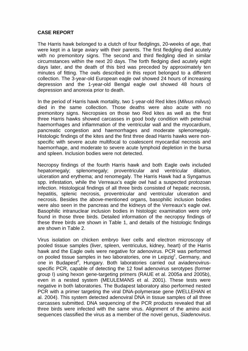
Title captions: Figure 1 (A and B). Picture A shows oesophageal (X) and proventricular (Y) dilation in a Bengal eagle owl (Bubo bengalensis) that died of adenovirus infection. PCR identified an adenovirus of the novel genus Siadenovirus. Picture B shows myocardial haemorrhage in a Red kite (Milvus milvus), also highly suspicious to have died of adenoviral infection. Figure 2. This picture shows basophilic intranuclear inclusion bodies in the liver of a Harris hawk (Parabuteo unicinctus). These inclusion are consistent with adenovirus infection. As similar inclusions can be found with other viruses such as circovirus, polyomavirus or herpesvirus further tests to confirm the diagnosis are recommended. In this case PCR identified an adenovirus of the genus Siadenovirus. Figure 3. Phylogenitic tree (based on distance matrix analysis of partial sequences of the DNA-polymerase genes) presenting the separation of five clusters (ProtDist – Dayhoff PAM 001 matrix), Fitch-Margoliash – global rearrangement, Treeview). Members of the various genera are indicated by different letter fonts (Mastadenoviruses – small letters, Aviadenoviruses – capital letters, Siadenoviruses – boldface, Atadenoviruses – underlined, and the fish adenovirus – italics). The newly identified siadenovirus (marked in boldface and underlined) in the present cases forms a separate branch together with the ‘Turkey adenovirus type 3’. Figure 4. Alignment of the partial amino acid sequences comparing the four genera of adenovirus (plus one pending for the sturgeon adenovirus). Genera are separated by lines. The conserved residues are shaded in light grey and the common amino acid sequences within the siadenoviruses are shaded in dark grey. The newly identified siadenovirus is printed in boldface.

Table 1. Necropsy findings of the three raptors infected with adenovirus (only abnormal findings are described) Findings Harris Hawk Bengal eagle owl Verreaux’s eagle
owl Liver Moderate
hepatomegaly, white mottling
Mild hepatomegaly, visceral gout
Moderate hepatomegaly, mottled
Spleen Moderate splenomegaly, mottled
Mild splenomegaly, visceral gout
Proventriculus Severe dilation Ventriculus Severe dilation,
ulceration in pyloric area, non-attached cuticle
Ulceration with erythema
Intestines Duodenum severely dilated
Caecae with yellow cores, moderately dilated, protozoan infection
Lungs / air sacs
Congestion, Syngamus trachea in bronchi / air sac
Moderate airsacculitis, congestion
Mild airsacculitis
Heart Pericarditis, epicarditis, visceral gout
Kidneys Cranial poles with severe renomegaly
Severe renomegaly, visceral gout
Cranial poles with moderate renomegaly, mottled

Table 2. Histology findings of the three raptors infected with adenovirus (only abnormal findings are described) Findings Harris Hawk Bengal eagle owl Verreaux’s eagle owl Liver Severe acute multifocal to
coalescent necrosis with occasional intranuclear basophilic inclusions, occasionally hepatocytes with karyomegaly
Minimal subacute non-suppurative hepatitis
Minimal multifocal acute necrotising hepatitis, disseminated epithelial and lymphoreticular cell nuclear atypia with the formation of presumptive intranuclear inclusion bodies
Spleen Severe acute multifocal to coalescent necrosis, intranuclear basophilic inclusions within reticular cells with karyomegaly and marginated chromatin
Severe acute congestion Disseminated epithelial and lymphoreticular cell nuclear atypia with the formation of presumptive intranuclear inclusion bodies
Proventriculus Mild acute to subacute multifocal necrotising proventriculitis
Ventriculus Subacute focally extensive suppurative serositis
Severe acute to subacute diffuse necrotising venticulitis, intranuclear inclusion bodies
Severe multifocal subacute ulcerative ventriculitis, mucosal epithelial necrosis
Intestines / pancreas
Disseminated epithelial and lymphoreticular cell nuclear atypia with the formation of presumptive intranuclear inclusion bodies
Lungs / air sacs
Intrabronchial nematode egg, presumptively Syngamus trachea
Heart Epicardial haemorrhage Kidneys Moderate multifocal tubular necrosis
with urate deposition Disseminated epithelial and lymphoreticular cell nuclear atypia with the formation of presumptive intranuclear inclusion bodies

Figure 1 (A and B). Picture A shows oesphageal (X) and proventricular (Y) dilation n a Bengal eagle owl (Bubo bengalensis) that died of adenovirus infection. PCR identified an adenovirus of the novel genus Siadenovirus. Picture B shows myocardial haemorrhage in a Red kite (Milvus milvus), also highly suspicious to have died of adenoviral infection.
XY
A
XY
A
BB

Figure 2. This picture shows basophilic intranuclear inclusion bodies in the liver of a Harris hawk (Parabuteo unicinctus). These inclusion are consistent with adenovirus infection. As similar inclusions can be found with other viruses such as circovirus, polyomavirus or herpesvirus further tests to confirm the diagnosis are recommended. In this case PCR identified an adenovirus of the genus Siadenovirus.

Figure 3. Phylogenitic tree (based on distance matrix analysis of partial sequences of the DNA-polymerase genes) presenting the separation of five clusters (ProtDist – Dayhoff PAM 001 matrix), Fitch-Margoliash – global rearrangement, Treeview). Members of the various genera are indicated by different letter fonts (Mastadenoviruses – small letters, Aviadenoviruses – capital letters, Siadenoviruses – boldface, Atadenoviruses – underlined, and the fish adenovirus – italics). The newly identified siadenovirus (marked in boldface and underlined) in the present cases forms a separate branch together with the ‘Turkey adenovirus type 3’.

0.1
sturgeon tokay gecko
blue skink
corn snake gilamonster
leopard geckodragon
chameleon duck-1
sheep-7 cattle-Rus
cattle-4 frog-1
turkey-3 raptor
CHICKEN-9 CHICKEN-1
mouse-1 cattle-3
dog-2 dog-1 swine-3 tree shrew
swine-5 cattle-2
cattle-1 monkey-1
man-40monkey-3
man-7chimp-21 man-35man-11
chimp-25 man-4
man-12 man-17
man-2man-5
man-1
PARROT

Figure 4. Alignment of the partial amino acid sequences comparing the four genera of adenovirus (plus one pending for the sturgeon adenovirus). Genera are separated by lines. The conserved residues are shaded in light grey and the common amino acid sequences within the siadenoviruses are shaded in dark grey. The newly identified siadenovirus is printed in boldface. man-1 SALTHPMPWGPPLNPYERALAARAWQQALDLQGCKIDYFDARLLPGIFTVDADPPDETQLDPLPPFCSRKGGRLCWTNERLRGEVATSVDL man-2 SALTHPMPWGPPLNPYERALAARAWQQALDLQGCKIDYFDARLLPGVFTVDADPPDETQLDPLPPFCSRKGGRLCWTNERLRGEVATSVDL man-5 SALTHPMPWGPPLNPYERALAARAWQQALDLQGCKIDYFDARLLPGVFTVDADPPDETQLDPLPPFCSRKGGRLCWTNERLRGEVATSVDL man-17 SALTHPMPWGPPLNPYERAMAAREWQMALDDASSKIDYFDKKLCPGIFTIDADPPDEHLLDVLPPFCSRKGGRLCWTNEPLRGEVATSVDL man-4 SALTHPMPWGTPLSPYERALAVREWQASLDDLGTCISYFDPDLLPGIFTIDADPPDELMLDPLPPFCSRKGGRLCWTNEPLRGEVATSVDL chimp-25 SALTHPMPWGTPLSPYERALAVREWQAALDDLATSISYFDPDLLPGIFTIDADPPDEVMLDPLPPFCSRKGGRLCWTNEPLRGEVATSVDL chimp-21 SALTHPMPWGTPLNPYERALAAREWQMALDD-PAHISYFDKDLLPGIFTMDADPPDELMLDPLPPFCSRKGGRLCWTNEPLRGEVATSVDL man-11 SALTHPMPWGSPLNPYERALAAREWQMALDD-PTPISYFDKDLLPGIFTMDADPPDELMLDPLPPFCSRKGGRLCWTNEPLRGEVATSVDL man-35 SALTHPMPWGSPLNPYERALAAREWQMALDD-PTPISYFDKDLLPGIFTMDADPPDELMLDPLPPFCSRKGGRLCWTNEPLRGEVATSVDL man-7 SALTHPMPWGTPLNPYERALAVREWQMTLDD-PATISYFDKDLLPGIFTIDADPPDEFMLDPLPPFCSRKGGRLCWTNEPLRGEVATTVDL man-12 SALTHPMPWGPPLNPYERALAVRQWQVALENYTCKIDYFDKNLCPGIFTIDADPPDENQLDVLPPFCSRKGGRLAWTNESLRGEVVTSVDL man-40 SALTHPMPWGFPLNPYERALAVRDWEHALLQVGTPIDYFNRTLLPGIFTIDADPPPENLLDVLPPLCSRKGGRLCWTNEPLRGEVVTSVDL monkey-3 SALTHPMPWGAPLSPYERALAVRDWETALRRPGHQIDYFDKHLLPAIFTIDADPPDERLLDPLPPFCSRKGGRLCWTNEPLRGEVATSIDV monkey-1 SALTHPMPWGPPLNPYERALAVKKWEDALQDTDTEIDYFNKILLPGIFTIDADPPPANLLDPLPPFCSRKGGRLCWTNEPLRGEVATSVDL dog-1 SALTHPFPAGSPLNPYERAVAIKAYEHKMQE-HKTISYFDEDLLPGIFTIDADPPAEEFLDVLPPFCSRKGGRLCWTNEPLRGEVTTSIDV dog-2 SALTHPFPAGSPLNPYERALAIKAYEQKMLN-HKTISYFDKDLLPGIFTIDADPPAEEFLDVLPPFCSRKGGRLCWTNEPLRGEIATSIDV cattle-1 SALTHPFPAGQPLNPFERAVAASDWSRRLSAHGSRIDYFDDTLLPGIFTVDADPPDELFLDELPPFCSRKGGRLCWTNEPLRGEVATSVDM cattle-2 SALTHPFPSGQPLNPYERALAATEWIRKLENLEQKIDYFDECLLPGIFTIDADPPDELFLDELPPFCSRKGGRLCWANEPLRGEVATSIDL cattle-3 SALTHPFPAGKPLNPFDRALAIKNWQDRLTQLHRPIDYFDRTLLPAIFTIDADPPPEAFLDVIPPFCSRKGGRLCWTNETLRGEVVTCLDA swine-5 SALTHPMPSGSPLNPFERALAVAVWEDQLKSVGQKMDYFDEKLLPGIFTIDADPPDESFLDVLPPFCSRKGGRLCWTNEPLRGEVATSVDV swine-3 SALTHPFPAGQPLNPFDRALAARRWQDRLDG-PEPLSYFDPDLLPALFTIDADPPDEDQLDVLPPYCSRKGGRLCWTNEPLRGEVATSVDV tree shrew SALTHPFPSGRPLNPFDRALAVKNWEMRLKN-SQTIDYFTPHLLPGIFTIDADPPSETYLDVLPPFCSRKGGRLCWTNESLRGEVATSVDI mouse-1 SALTHPMPSGWPLEPKARAEALADWTKHLSN-SAPISYFNTCLLHGIVLIDADPPCETQLDVLPPFCSRKGGRLCWTNEPLRGEITTTIDV cattle-4 SALTHPMPYGRTLNPFEANTSIDEMQNMLDS-SEVLSYFDPRIKAMIVVADCEPPTLEYLDVLPPLCSKKSGKLCWTNEPLINETVTSIDL cattle-Rus SALTHPMPYGRTLNPFEANIAIDELQSMLDS-SVILSYFDSGIKAMVVVADCEPPTLEYLDVLPPLCSKKSGKLCWTNGPLVNETLTSIDL sheep-7 SALTHPLPYGKTLNAFEANAQIDYFQELLQR-KEKIDYFDNSIKPMIVVADCEPPSLDYLDVLPPLCSKKSGKLCWSNETLINEVLTSIDL duck-1 SALTHPMPFGRTEDPLTASISIKTFQDKLDS-PAKLSYFGESIKPMIVYADCYPPPLEHVDVLPPLCSRKSGRLCWTNEPLLGEVVTTIDL corn snake SALSHPMPYGPTLSPFDSAVAIAEFQRKLDG-QSELSYFDPDIFPMIVVADAFPPSLHCLDVLPPLCSKRSGKLCWTNEPLLGEVLTTVDL dragon SALSHPMPCGRTLPPLDASIEIRRFQDKLDK-PHKISYFDPNLKPMIVAADCIPPPLNELDVLPPLCSKASGRLCWTNEPLVGEVLTSIDL chameleon SALSHPMPSGTTESPTDAALSIAYFQDLLDK-PDQISYFSQ-VKPMIVLADCYPPALARLDVLPPLCSRRSGKLCWTNEVLTAEALTTVDL leopard gecko SALSHPMPYGTTLSPFDSSKAMASFQALLDG-KDCLSYFDPRILPMIVKVDCFPPPLYHLDTLPPLCSKKSGRLCWTNEPLLGEVITTVDI tokay gecko SALTHPMPFGLPCEPFTANIHIRQFQYLLDEVGKPISYFDERIKPMIVAADCFPPSIKELDVLPPMCTRKGGKLCWTNESLHMEILTSVDL blue skink SALTHPMPSGIPLDPFTSSIAIRKFQNKLDE-PSTISYFDPDIFPMVVVADCSPPPLEQLDVLPPLCSKKSGRLCWTNEPLEVETLTTIDL gilamonster SALSHPMPYGLTLSPLDASVAMARFQDKLDS-TEKLSFFDKNILPMIVKADCFPPPLYHLDVLPPLCSKKSGRLCWTNEPLLGEVLTTVDI raptor SALTHPMPYGIPVGEKERLEEIKKFTNLLSR-RDKISYFNQGIKPMIVTVNAFPPPTELLDPLPPLCSKKSGKLCWTNEPLNNEVVTSIDI frog-1 SALTHPMPFGVPLSQKEKNQEIHILQSKLQN-EKTLNYFDPEIKPMIISISAFPPPVEYLTNIPPICSRKSGRLCWTNEALYDETVTIVDV turkey-3 SALTHPMPYGFPIGEKERNNEITKLNEKLKKTKTKLSYFTD-IKPMVVMIDAIPPPPEHLDPLPPLCSRQSGKLCWTNEILKNEIVTSIDI chicken-1 SALTHPMPHGMPLDPKFTAQHVEELNRLLTN-ESHLSYFDARIKPSILKIEAYPPPPEMLDPLPPICSRRGGRLVWTNEALYDEVVTVIDI chicken-9 SALTHPMPHGMPLDPKFTAKHVDELNELMSS-ERPISYFDHRIKPCILKVEAYPPPAEHLDTLPPICSRRGGRLVWTNEALYDEVITVIDI parrot SALTHPMPHGIPLDPHHTVTHVNLLNIILASGERPISYFDPRIKPAILKTDAFPPPPEMLDVLPPICSRRGGRLVWTNEPLHDEVITIIDI sturgeon SALTHPMPYGYPLEPLEASIHIKLFQELLDR-PEDISYFNDTVKPMILSIDAHPPNINYLDTLPPLCSRQSGRLCWTNEALISEIVTSLDC