adipose-derived stem cells combined with neuregulin-1 delivery
TRANSCRIPT

1
Adipose-derived stem cells combined with Neuregulin-1
delivery systems for heart tissue engineering
P. Díaz-Herráez1†, E. Garbayo1†, T. Simón-Yarza1, F.R. Formiga1, F. Prosper2, M.J. Blanco-
Prieto1*
†P. Díaz-Herráez and E. Garbayo contribute equally to this manuscript.
1 Department of Pharmacy and Pharmaceutical Technology, School of Pharmacy, University of
Navarra, Pamplona, Spain.
2 Hematology, Cardiology and Cell Therapy, Clínica Universidad de Navarra and Foundation
for Applied Medical Research, University of Navarra, Pamplona, Spain.
Address for correspondence: Maria J. Blanco-Prieto, Department of Pharmacy and
Pharmaceutical Technology, School of Pharmacy, University of Navarra, Irunlarrea 1, E-
31080 Pamplona, Spain. Tel.: +34 948 425600 x 6519; fax: +34 948 425649 e-mail:

2
Abstract:
Myocardial infarction (MI) is the leading cause of death worldwide and extensive
research has therefore been performed to find a cure. Neuregulin-1 (NRG) is a growth
factor involved in cardiac repair after MI. We previously described how biocompatible
and biodegradable microparticles, which are able to release NRG in a sustained manner,
represent a valuable approach to avoid problems related to the short half-life after
systemic administration of proteins. The effectiveness of this strategy could be
improved by combining NRG with several cytokines involved in cardiac regeneration.
The present study investigates the potential feasibility of using NRG-releasing particle
scaffold combined with adipose derived stem cells (ADSC) as a multiple growth factor
delivery-based tissue engineering strategy for implantation in the infarcted myocardium.
NRG-releasing particle scaffolds with a suitable size for intramyocardial implantation
were prepared by TROMS. Next, ADSC were adhered to particle scaffolds and their
potential for heart administration was assessed in a MI rat model. NRG was
successfully encapsulated reaching encapsulation efficiencies of 92.58 ± 3.84 %. NRG
maintained its biological activity after the microencapsulation process. ADSC cells
adhered efficiently to particle scaffolds within a few hours. The ADSC-cytokine
delivery system developed proved to be compatible with intramyocardial administration
in terms of injectability through a 23-gauge needle and tissue response. Interestingly,
ADSC-scaffolds were present in the peri-infarted tissue two weeks after implantation.
This proof of concept study provides important evidence required for future
effectiveness studies and for the translation of this approach.
Keywords: Particle scaffold, PLGA Microparticles, ADSC, NRG-1, Myocardial
infarction, Cardiac repair

3
1 Introduction:
Cardiovascular diseases cause more than 17 million deaths each year according to
the latest report of the World Health Organization (available in
http://www.who.int/cardiovascular_diseases), constituting the greatest health risk in
western countries [1]. Despite the advances in pharmacological treatment, a major
improvement able to repair the massive loss of cardiomyocytes after a myocardial
infarction (MI) has not yet been reached, heart transplantation being the only real option
for severe cases. Due to this situation new approaches have been explored in the last
few years [2-5]. One of these strategies is the use of growth factors (GF). GF are
thought to benefit the damaged heart through direct effects in the myocardium and by
stimulating and mobilizing progenitor cells [6]. However, GF protein administration
presents serious limitations due to their short in vivo half life, physical and chemical
instability, and the low oral bioavailability of these macromolecules [7]. The use of drug
delivery systems (DDS) that encapsulate GF might overcome these drawbacks.
Microparticles (MP), one of these DDS, could protect proteins from degradation and
ensure sustained release among time [8]. Recently, our group explored new therapeutic
strategies for MI treatment, based on the use of polymeric MP that release different GF
involved in cardiac angiogenesis and neovascularization [8-11]. Neuregulin-1 (NRG)
deserves special attention in heart regeneration because it is involved in cardiac repair
after MI [12]. This protein plays a crucial role in the adult cardiovascular system by
inducing sarcomere membrane organization and integrity [13], cell survival [14, 15] and
angiogenesis [16]. We recently proved that NRG-releasing MP promoted cardiac repair
and improved cardiac performance [11]. NRG-releasing MP effectiveness could be
improved by combining this protein with several other cytokines involved in cardiac
regeneration. This could be achieved by preparing a polymer-based GF delivery system
4
that allows the release of multiple factors [6]. However, to date GF delivery systems
have not demonstrated the ability to deliver cocktail of factors with distinct kinetics
[17]. This aspect, besides the limitation that GF dose and timing are crucial for helping
regeneration, makes it difficult to co-administer different GFs [6, 7]. The combination
of NRG-releasing MP with cytokines secreted by stem cells (SC), capable of responding
to the host environment, opens up a possible solution to that drawback. Moreover, MP
possess many features that make them suitable for use as cardiac scaffolds. In particular,
they are biodegradable, biocompatible, non-toxic and, importantly, they can provide
structural support for cell survival and differentiation [18-24].
Among the different SC sources, adipose-derived stem cells (ADSC) have shown
promising results in cardiac repair [25-28]. They are good candidates for cell therapy
studies because of their easy isolation from the stromal vascular fraction [29-32] and
their extensive differentiation potential. In addition, ADSCs are able to secrete
angiogenic and/or anti-apoptotic factors [33], such as granulocyte-macrophage colony
stimulating factor (GM-CSF), vascular endothelial growth factor (VEGF), hepatocyte
growth factor (HGF), fibroblast growth factor (bFGF), and transforming growth factor-
β (TGF-β) [31].
For all of these reasons, the primary purpose of this work was to investigate the
potential feasibility of NRG-releasing particle scaffold combined with ADSC as a multi
GF delivery-based tissue engineering strategy for the ischemic heart. To this end, NRG-
releasing delivery system was prepared using Total Recirculation One-Machine System
(TROMS), a technique based on the multiple emulsion solvent evaporation method
which is suitable for the encapsulation of labile molecules like cytokines and GFs [8,
34]. We primarily investigated the physical characteristics of the particle scaffold such

5
as morphology or size. Then, NRG-releasing particle scaffolds were combined with
ADSC and flow properties such as dispersability and injectability of the ADSC particle
scaffold suspension were analyzed to avoid complications during their administration.
The myocardial response to ADSC combined with NRG-releasing particle scaffold was
finally evaluated using a MI rat model to ensure safety and biocompatibility
requirements.

6
2.1 Materials Poly(lactic-co-glycolic acid) (PLGA) with monomer ratio (lactic acid/glycolic
acid) of 50:50 Resomer® RG 503H (Mw: 34 kDa) was provided by Boehringer-
Ingelheim (Ingelheim, Germany). Polyethylene glycol (PEG; Mw: 400), human serum
albumin (HAS), bovine serum albumin (BSA), dimethylsulfoxide (DMSO),
carboxymethyl-cellulose, mannitol, polysorbate 80, sodium azide and rhodamine B
isothiocyanate were provided by Sigma-Aldrich (Barcelona, Spain). Dichloromethane
and acetone were obtained from Panreac Quimica S.A. (Barcelona, Spain).
Poly(vinylalcohol) (PVA) 88% hydrolyzed (Mw: 125,000) was obtained from
Polysciences, Inc. (Warington, USA). Collagen I of rat tail 3mg/mL, Minimum
Essential Medium Alpha (α-MEM) Medium, 0.05% Trypsin-EDTA, Heat inactivated
Fetal Bovine Serum (FBS), Phosphate Buffered Saline pH 7.2 (PBS) and Dulbecco´s
Modified Eagle Medium (DMEM) were provided by Gibco-Invitrogen (Carlsbad, CA,
USA). ADSC cells were obtained from inguinal adipose tissue of male Sprague-Dawley
transgenic rats. H9c2 cells were provided by ATCC. Poly D-Lysine 1 mg/ml (PDL) was
provided by Merck-Millipore (Darmstadt, Germany). rh Neuregulin-1b-iso (NRG) was
provided by EuroBioSciences (Friesoythe, Germany). 3-(4,5-dimethylthiazol-2-yl)-5-
(3-carboxymethoxyphenyl)-2-(4-sulfophenyl)-2H tetrazolium (MTS) was purchased
from Promega (Madison, USA). Goat polyclonal anti-human NRG-1 antibody (sc-1793)
and horseradish-peroxidase-conjugated donkey anti-goat IgG (sc-2020) were purchased
from Santa Cruz Biotechnology (Santa Cruz, CA, USA).

7
2.2 Preparation of NRG-releasing particle scaffold
NRG-releasing PLGA particle scaffolds were prepared by the emulsion solvent
evaporation method using TROMS as previously described [11] with minor
modifications. In order to obtain batches with the defined particle size the following
TROMS parameters were adjusted: pumping flow, recirculation times to obtain both
W1/O and W1/O/W2 emulsions, and inner diameters of the needles used to prepare the
emulsions. Briefly, the organic phase (O) composed of 100 mg of PLGA dissolved in 4
ml of a dichloromethane/acetone mixture (ratio 3:1) was injected into the inner aqueous
phase (W1) containing 200 μg of NRG, 5 mg of HSA and 5 μl of PEG 400 dissolved in
200 μl of phosphate-buffered saline (PBS pH 7.9). Next, the inner emulsion (W1/O) was
recirculated through the system under a turbulent regime maintained by a pumping flow
through a needle. After this homogenization step, the W1/O emulsion was injected into
the outer aqueous phase (W2) composed of 20 ml of a 0.5% w/v PVA solution. The
turbulent injection through a second needle resulted in the formation of a multiple
emulsion (W1/O/W2), which was allowed to circulate through the system to become
homogeneous. The multiple emulsion was stirred for 3 h to allow solvent evaporation.
Particle scaffolds were washed three times with ultrapure water by consecutive
centrifugations at 4 °C (20,000×g, 10 min). NRG-releasing particle scaffolds were
lyophilized for 48h without cryoprotective agents (Virtis Genesis 12 EL, Gardines,
NY). The conditions of freeze drying were -50ºC to +15ºC over 2 days. After complete
lyophilization, the vials were sealed under vacuum and stored at -20°C until use.
Unloaded particle scaffolds were prepared in the same manner without adding NRG.
For fluorescence-labeled formulation, rhodamine B isothiocyanate (0.5 mg/mL) was
added to the inner aqueous phase and particle scaffolds were prepared as described.

8
2.3 NRG-releasing PLGA particle scaffold characterization
2.3.1 Particle size analysis
The mean particle size and size distribution were examined by laser
diffractometry using a Mastersizer® (Malvern Instruments, Malvern, UK). Particle
scaffolds were dispersed in ultrapure water and analyzed under continuous stirring. The
average particle size was expressed as the volume mean diameter in micrometers.
2.3.2 Drug content
The amount of NRG encapsulated in the particle scaffold was determined by
dissolving 0.5 mg of lyophilized loaded particles in 25 µL of DMSO, and was
quantified using western blot. After electrophoresis and transference, the membranes
were blocked with 5% nonfat dried milk in TBS plus 0.05% Tween 20 for 2 h, then
incubated overnight at 4ºC with primary antibody goat IgG-NRG-1β-IGGF2 (sc-1793)
1:50. After several washes the membranes incubated with antigoat IG-HRP (sc-2020)
1:2000 secondary antibody for 2 h. Immunoreactive bands were, after several washes,
visualized using LumiLight plus western blotting substrate (Roche Diagnostics,
Mannheim, Germany). The quantifications were determined by ImageQuant RT ECL.
Sample values were quantified using a blotting standard curve with known amounts of
NRG.
2.3.3 In vitro bioactivity assay
The bioactivity of NRG released from particle scaffolds was evaluated in vitro by
determining the proliferative capacity of H9c2 cells after NRG treatment. H9c2 cells
obtained from embryonic BD1X rat heart tissue were cultured in DMEM medium
supplemented with 10% FBS, 1% glutamine and 1% penicillin/streptomycin at 37 °C

9
under 5% CO2/95% air [35-37]. Cells were subcultured when 60% confluency was
achieved. In order to quantify cell proliferation after NRG stimulation, cells were
seeded in 96-well tissue culture plates at a density of 2 × 10³ cells/well. After 24 h,
medium was removed and the cells were incubated with 150 ng/mL of NRG released
from particle scaffolds over 24 h, which had previously been quantified by western blot,
with 150 ng/mL of free NRG or medium alone as control. Culture medium
supplementation was modified for these experiments by reducing the FBS to 5 % in the
culture medium. Treatments were removed every day, and fresh treatment was added to
the cells. After three days of treatments, the number of viable cells was determined by
MTT assay. Statistics were calculated with Prism 5.0 software (Graphpad Software Inc.,
San Diego, CA, USA). The differences among treatment groups were assessed by
ANOVA with a Tukey post hoc correction when the values measured were normally
distributed
2.4 Isolation and culture of ADSC cells
ADSC cells were obtained by in vitro culture of the stromal vascular fraction
(SVF) isolated from inguinal adipose tissue of male Sprague-Dawley transgenical rats
that expressed the green fluorescent protein (GFP), as previously described [38]. ADSC
cells were cultured in α-MEM medium supplemented with 10% FBS, 1 ng/mL bFGF
and 1% penicillin/streptomycin. Cells were subcultured when 80% confluence was
reached.
2.5 Adhesion of ADSC cells to particle scaffold
To favor cell attachment to the MP surface, the particle scaffold was overlaid with
0.5 μg/cm2 of type 1 collagen and/or PDL [18]. Particle scaffold coating was performed
in 15 mL falcon tubes. Scaffolds were re-suspended in DPBS and the mixture was

10
sonicated until the particles were completely dispersed in the liquid. Then, coating
solutions were added to the falcon tube and mixed with the particles under rotation at 37
ºC for 2 h. Coated particle scaffolds were washed 3 times with distilled sterile water and
lyophilized for long term storage. For ADSC adhesion, coated MP were resuspended
with complete α-MEM medium, and were ultrasounded and briefly vortexed prior to
addition of 2,5 X 10⁵ or 5 X 10⁵ cells. The mixture was then gently flushed and plated
in Costar® Ultra Low Cluster Flat Bottom Sterile Polystyrene Plate. Plates were
incubated at 37 ºC for 4 h. At different times cells were observed to study the evolution
of the adhesion.
2.6 Determination of dispersability and injectability of ADSC adhering to particle
scaffold
Particles with adhered ADSC cells were dispersed in two resuspension medium
consisting of: (1) 0.1% (w/v) carboxymethyl-cellulose, 0.8% (w/v) polysorbate 80 and
0.8% (w/v) mannitol in PBS, pH 7.4 and (2) 0.4% (w/v) carboxymethyl-cellulose, 3.2%
(w/v) polysorbate 80 and 3.2% (w/v) mannitol in PBS, pH 7.4. The dispersing media
were sterilized by autoclaving prior to use. The suspension injectability was assessed by
the ability of the particles combined with ADSC to pass through different needles (23,
24, 25, 27 and 29 gauge (G) needles).
2.7 In vivo studies using chronic myocardial infarction model
All animal procedures were approved by the University of Navarra Institutional
Committee on Care and Use of Laboratory Animals as well as the European
Community Council Directive Ref. 86/609/EEC.

11
2.7.1 Induction of myocardial infarction
A total of 26 female Sprague-Dawley rats (Harlan-IBERICA, Spain) underwent
permanent occlusion of the left anterior descending coronary artery, as previously
described [11]. Briefly, rats were anesthetized with 4% isoflurane in an induction
chamber and supported with a mechanical ventilator. Prior to surgery, animals received
analgesic drug ketoprofen 5 mg/Kg subcutaneously. The rats were then intubated and
1.5–2% isoflurane was maintained for continuous anesthesia. The heart was accessed
through a left thoracotomy through the fourth intercostal space, and the left anterior
descending coronary artery was permanently occluded 2–3 mm distal from its origin.
The chest was then closed in layers and rats allowed to recover on a heating pad.
2.7.2 Intramyocardial implantation
Seven days post-myocardial infarction, rats were placed into the following
groups: (Group 1) 2.5 x 10⁵ ADSCs-0.75 mg of particle scaffold coated with 0.5
µg/cm2 of collagen in 100 µL of resuspension medium, (Group 2) 5 x 10⁵ ADSCs-0.75
mg of particle scaffold coated with 0.5 µg/cm2 of collagen in 100 µL of resuspension
medium, (Group 3) 5 x 10⁵ ADSCs-0.75 mg of particle scaffold coated with 0.5 µg/cm2
of mixture of collagen and poly-D-lysine (1:1) in 100 µL of resuspension medium,
(Group 4) 5 x 10⁵ ADSCs-0.75 mg of NRG-releasing particle scaffold coated with 0.5
µg/cm2 of mixture of collagen and poly-D-lysine (1:1) in 100 µL of resuspension
medium and (Group 5) 100 µL of resuspension medium. Two animals in groups 1, 2
and 3 were injected with rhodamine B fluorescent particle scaffolds to visualize
particles by fluorescent microscopy. All the treatments were injected in 4 sites of the
border zone surrounding the infarct zone using a 23 G needle. After treatment injection,
the chest was closed and rats were allowed to recover on a heating pad.

12
2.7.3 Histological assessment of myocardial tissue after treatment implantation
Two weeks after treatment implantation, animals were sacrificed and hearts were
collected for histology. After being harvested, the hearts were perfused-fixed in 4%
paraformaldehyde at 4 °C, and sliced in three 4-mm-thick segments from apex to base.
The hearts were dehydrated in ethanol 70% at 4 °C, embedded in paraffin and 5-μm-
sections were cut. Hematoxylin–eosin (HE) staining was performed to visualize tissue
structure and to study tissue retention of implanted treatment.
2.7.4 Verification of NRG-releasing particle scaffold retention in the infarcted tissue
and ADSC cell fate
Fluorescence microscopy was used to evaluate tissue retention of rhodamine
particle scaffold and the fate of the ADSCs cells.
13
3 Results and Discussion
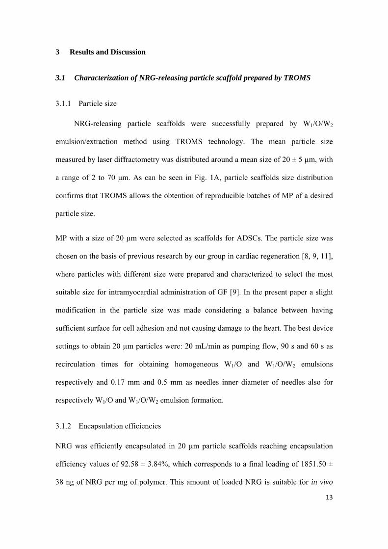
3.1 Characterization of NRG-releasing particle scaffold prepared by TROMS
3.1.1 Particle size
NRG-releasing particle scaffolds were successfully prepared by W1/O/W2
emulsion/extraction method using TROMS technology. The mean particle size
measured by laser diffractometry was distributed around a mean size of 20 ± 5 µm, with
a range of 2 to 70 μm. As can be seen in Fig. 1A, particle scaffolds size distribution
confirms that TROMS allows the obtention of reproducible batches of MP of a desired
particle size.
MP with a size of 20 µm were selected as scaffolds for ADSCs. The particle size was
chosen on the basis of previous research by our group in cardiac regeneration [8, 9, 11],
where particles with different size were prepared and characterized to select the most
suitable size for intramyocardial administration of GF [9]. In the present paper a slight
modification in the particle size was made considering a balance between having
sufficient surface for cell adhesion and not causing damage to the heart. The best device
settings to obtain 20 µm particles were: 20 mL/min as pumping flow, 90 s and 60 s as
recirculation times for obtaining homogeneous W1/O and W1/O/W2 emulsions
respectively and 0.17 mm and 0.5 mm as needles inner diameter of needles also for
respectively W1/O and W1/O/W2 emulsion formation.
3.1.2 Encapsulation efficiencies
NRG was efficiently encapsulated in 20 µm particle scaffolds reaching encapsulation
efficiency values of 92.58 ± 3.84%, which corresponds to a final loading of 1851.50 ±
38 ng of NRG per mg of polymer. This amount of loaded NRG is suitable for in vivo

14
studies [11]. Modifications in the preparation process resulted in 25% higher
encapsulation efficiencies than those prepared before. The high NRG entrapment
achieved could be related to the encapsulation method and to the use of TROMS to
prepare the multiple emulsion system. On the one hand, the water/oil/water evaporation
technique has been described as resulting in extremely efficient loading of
biodegradable microparticles with hydrophilic compounds [39, 40] and, on the other
hand, higher encapsulation efficiencies were found using TROMS compared to
conventional encapsulation techniques. For instance, for VEGF, an encapsulation
efficiency of 83.8 % was achieved using TROMS [8]. In contrast, King et al. reported
an entrapment efficiency of 16 % using the solid/single emulsion/ solvent extraction
technique to prepare the microparticles [41].
NRG is a GF involved in cardiac repair after MI that deserves special attention in heart
regeneration [12]. Multiple in vivo studies have established the therapeutic potential of
this cytokine after MI. NRG administration after myocardial injury improved systolic
function, reduced infarct size and attenuated myocardial hypertrophy in small and large
animal models of MI [12]. Its beneficial effects might be due to myocyte protection
from death stimuli and through repair of dysfunctional cardiac myocytes [12]. Several
clinical trials to evaluate the effect of NRG in humans are ongoing. However, cytokine
therapy efficacy is generally hindered for the short plasma half live, gastrointestinal
tract instability and low bioavailability of proteins. Thus, the development of a DDS
able to release NRG in a sustained manner would improve its potential and efficacy.
3.1.3 In vitro bioactivity assay
The bioactivity of encapsulated NRG released from the particle scaffolds was
evaluated in vitro by determining its capacity to induce H9c2 proliferation (Fig. 1B).

15
The daily addition of NRG released from particle scaffolds (150 ng/mL) induced a
statistically significant 1.24 fold increase in the proliferation of H9c2 in comparison
with control (culture medium without cytokine) after 3-day treatment. The increase was
similar to that observed when H9c2 cells were cultured with the daily addition of free
NRG for 3 days at doses of 150 ng/mL indicating that the biological activity of NRG
was maintained after encapsulation and release from particle scaffolds prepared by
TROMS.
3.2 Adhesion of ADSC to NRG-releasing particle scaffold
The development of a DDS able to protect NRG from degradation and to ensure
its sustained release throughout time would reinforce NRG efficacy in cardiac repair as
it was mentioned before in this section. Moreover, in the present work we move one
step forward combining NRG DDS with ADSC able to secrete multiple cytokines
involved in cardiac regeneration. Interestingly, this is the first report of NRG-releasing
scaffold for cardiac tissue engineering applications. The importance of multiple growth
factor action in cardiac tissue regeneration has been extensively described [19, 21, 42].
In this regard, the possibility of increasing the beneficial effect obtained with NRG
releasing particles combining this DDS with several other cytokines secreted by SC
would open up new possibilities in heart regeneration. Among SC, ADSC are
particularly suitable for cell therapy because of their easy isolation from the stromal
vascular fraction [29-32], their extensive differentiation potential and the secretion of
several angiogenic and anti-apoptotic factors that activate the revascularization process
and the positive remodeling of the heart [31, 33]. Although it is known that ADSC exert
their positive effect via paracrine secretion, the beneficial factors remain partly
unidentified. Moreover, it is possible that multiple factors might be functioning

16
synergistically [43-45]. For that reason, ADSC transplantation for their paracrine effects
still represents a reasonable strategy. In addition, SC are able to sense and respond to
changes in the host environment modifying its paracrine secretion. Moreover, until now,
NRG secretion by ADSC has not been described, meaning that the association of these
two strategies might have complementary effects on cardiac tissue repair.
In the present study, the adhesion of 2,5 X 10⁵ or 5 X 10⁵ ADSC to particle scaffolds
coated with different concentrations of collagen and PDL was studied. The
concentration that showed better adherence was 0.5 μg/cm2, either with the collagen or
the mixture of collagen and PDL (1:1). The administration of these amounts of cells
induced an improvement in the cardiac function when administered in combination with
collagen-based carrier sheets after MI [26]. On the other hand, the dose of NRG
administered with that quantity of MP also promoted cardiac repair and improved
cardiac performance.
Both cell densities adhered efficiently to all particle scaffolds assayed. Observations of
cell adhesion over time indicated that collagen coated particle scaffolds required 4 h for
total cell attachment while particle scaffolds coated with collagen and PDL attached to
the cells after 2 h (Fig. 2). Differences in cell adhesion time might be due to collagen
and PDL net charge at pH value of the culture medium used for adhesion (pH 7.2).
Thus, collagen net charge at pH 7.2 is negative due to its isoelectric point of 5.5,
hindering cell adhesion. On the other hand, PDL with an isoelectric point of 12.9, has a
net charge positive at pH 7.2 that favors the adhesion of the cells. As in the coating and
adherence processes a certain amount of encapsulated NRG is released over time, the
less time required for ADSC NRG-particle scaffold preparation, the less amount of
NRG would be lost during the manufacture process.

17
The use of biodegradable microparticles as injectable scaffolds for tissue engineering
applications was first proposed by Montero-Menei C. N. and colleagues [46, 47]. After
that, several authors investigated their potential to improve cell survival and
differentiation [18-20, 22-24, 48]. The differentiation of ADSC towards a cardiac
lineage is, however, not the main objective of the present research. ADSC cells were
combined with NRG delivery systems for their beneficial paracrine effect on
endogenous cells. Thus, a multidrug delivery system able to respond to the surrounding
tissue would be obtained.
3.3 Dispersability and injectability of NRG-releasing particle scaffold combined
with ADSC
The injection of ADSC-particle scaffolds in the infarcted heart required the
selection of a good dispersing medium that allows the cell-particle scaffold suspension
to pass through a needle without needle blockage or sedimentation. A cell-particle
scaffold suspension with good rheological properties will ensure dose uniformity and
safety requirements during the local myocardial injection of the treatments. Two
dispersing media containing different percentages of DMEM, carboxymethyl-cellulose,
polysorbate 80 and mannitol were investigated in the present study. These excipients are
included in the Handbook of Pharmaceutical Excipients [49] and are frequently used in
commercial formulations. DMEM is a solution commonly used for drug/cell injection
into the infarcted heart [50, 51], carboxymethyl-cellulose is a wetting and
biocompatible agent that prevent particle aggregation and makes their injection through
a thin needle and polysorbate 80 and mannitol has been previously used to suspend
PLGA microparticles prior to intracerebral implantation [34]. The ADSC particle
scaffold suspension showed the best flow properties in the dispersing medium

18
containing 0.4% (w/v) carboxymethyl-cellulose, 3.2% (w/v) polysorbate 80 and 3.2%
(w/v) mannitol in PBS, pH 7.4. No toxicity or cell detachment was observed when using
this medium.
Prior to injection in the infarcted myocardium, the injectability of the cell-particle
scaffold suspension was analyzed. To this end, ADSCs adhered to 20 µm particle
scaffolds were delivered through needles with different diameter (23, 24, 25, 27 and 29
G). ADSC particle scaffolds were only able to go through the 23G needle without
blocking or sedimentation, and carrying ADSC cells adhered on the surface. This needle
was therefore used for intramyocardial administration (Fig. 3).
3.4 Histological evaluation of myocardial tissue after the injection of NRG-
releasing particle scaffold combined with ADSC
Finally, ADSC combined with empty or cytokine delivery systems coated with
collagen or collagen:PDL were intramyocardially injected in the infarcted beating heart
(Fig. 3). Two weeks later, animals were sacrificed to further evaluate myocardial tissue
reaction and the non-toxic properties of the implanted scaffold in vivo. Upon
implantation, hematoxylin and eosin staining revealed that ADSC particle scaffolds
were well tolerated by the infarcted myocardium and they seem to integrate well within
the host tissue (Fig. 4 C,D). Heart response after ADSC-scaffold injection was the
typical reaction observed following mechanical trauma and exposure to a foreign body.
ADSC-scaffolds (Fig. 4 C,D) did not induce inflammatory reactions when compared to
resuspension medium injection (Fig. 4 A,B). Hematoxilin and eosin staining did not
evidence noticeable differences in terms of biocompatibility and local tolerance between
groups. Moreover, the tissue adjacent to the implanted treatments maintained its
physiological characteristics and no adverse cellular reactions were observed.

19
3.5 Confirmation of NRG-releasing particle scaffold retention in the infarcted heart
and ADSC cell fate
Fluorescent and brightfield microscopy showed that two weeks after
intramyocardial implantation, particle scaffolds appeared grouped at the implantation
site independently of the coating used to attach ADSC (Fig. 4 C,D and 5 A,B). At day
14, particle scaffolds were not totally biodegraded and a significant quantity of them
were still detectable. No differences in terms of scaffold degradation were observed
among the various groups during the two-week implantation period. As can be seen in
Figure 4 C,D and 5 A,B, counterstaining of nuclei revealed that particle scaffolds were
always surrounded by cells suggesting that ADSC remained attached to the particle
scaffold.
Taking together our in vitro ADSC adhesion studies and our present in vivo
biocompatibility findings on infarcted rats, 5 X 10⁵ ADSC cells combined with NRG-
releasing particle scaffolds coated with collagen and PDL will be selected to further
assess the therapeutic potential of this strategy in cardiac regeneration.

20
Conclusion
The data presented is this article offers valuable evidence of the feasibility of
using NRG-releasing particle scaffolds combined with ADSC as a multi GF delivery-
based tissue engineering strategy to treat the ischemic heart. Future studies will be
focused on the response produced by the treatment, to demonstrate whether the
combination of ADSC with NRG and using particle scaffolds as support is helpful in
the regeneration of the infarcted heart.

21
Acknowledgments:
This work was partially supported by FEUN (Fundación Empresa Universidad de
Navarra). P. Díaz-Herráez is beneficiary of a predoctoral fellowship from the
Association of Friends of the University of Navarra.
E. Garbayo´s work was supported by the Spanish Ministry of Science and
Innovation through a Juan de la Cierva Program (JCI-2011-10737).

22
References:
[1] Global atlas on cardiovascular disease prevention and control, World Health Organization, (2011). [2] C.E. Murry, L.J. Field, P. Menasche, Cell-based cardiac repair: reflections at the 10-year point, Circulation, 112 (2005) 3174-3183. [3] R. Passier, L.W. van Laake, C.L. Mummery, Stem-cell-based therapy and lessons from the heart, Nature, 453 (2008) 322-329. [4] V.F. Segers, R.T. Lee, Stem-cell therapy for cardiac disease, Nature, 451 (2008) 937-942. [5] T.A. Khan, F.W. Sellke, R.J. Laham, Gene therapy progress and prospects: therapeutic angiogenesis for limb and myocardial ischemia, Gene Ther, 10 (2003) 285-291. [6] N. Beohar, J. Rapp, S. Pandya, D.W. Losordo, Rebuilding the damaged heart: the potential of cytokines and growth factors in the treatment of ischemic heart disease, J Am Coll Cardiol, 56 (2010) 1287-1297. [7] K. Lee, E.A. Silva, D.J. Mooney, Growth factor delivery-based tissue engineering: general approaches and a review of recent developments, J R Soc Interface, 8 (2011) 153-170. [8] F.R. Formiga, B. Pelacho, E. Garbayo, G. Abizanda, J.J. Gavira, T. Simon-Yarza, M. Mazo, E. Tamayo, C. Jauquicoa, C. Ortiz-de-Solorzano, F. Prosper, M.J. Blanco-Prieto, Sustained release of VEGF through PLGA microparticles improves vasculogenesis and tissue remodeling in an acute myocardial ischemia-reperfusion model, J Control Release, 147 (2010) 30-37. [9] F.R. Formiga, E. Garbayo, P. Díaz-Herráez, G. Abizanda, T. Simón-Yarza, E. Tamayo, F. Prósper and M. J. Blanco-Prieto, Biodegradation and heart retention of polymeric microparticles in a rat model of myocardial ischemia, Euro J Pharma Biopharma, In press (2013). [10] T. Simon-Yarza, F.R. Formiga, E. Tamayo, B. Pelacho, F. Prosper, M.J. Blanco-Prieto, PEGylated-PLGA microparticles containing VEGF for long term drug delivery, Int J Pharm, 440 (2013) 13-18. [11] F.R. Formiga, B. Pelacho, E. Garbayo, I. Imbuluzqueta, P. Díaz-Herráez, G. Abizanda, J.J. Gavira, T. Simón-Yarza, E. Tamayo, F. Prósper, M.J. Blanco-Prieto, Controlled delivery of fibroblast growth factor-1 and neuregulin-1 from biodegradable microparticles promotes cardiac repair in a rat myocardial infarction model, J Am Coll Cardiol, Under revision (2013). [12] O. Odiete, M.F. Hill, D.B. Sawyer, Neuregulin in cardiovascular development and disease, Circ Res, 111 (2012) 1376-1385. [13] D.B. Sawyer, C. Zuppinger, T.A. Miller, H.M. Eppenberger, T.M. Suter, Modulation of anthracycline-induced myofibrillar disarray in rat ventricular myocytes by neuregulin-1beta and anti-erbB2: potential mechanism for trastuzumab-induced cardiotoxicity, Circulation, 105 (2002) 1551-1554. [14] X. Liu, X. Gu, Z. Li, X. Li, H. Li, J. Chang, P. Chen, J. Jin, B. Xi, D. Chen, D. Lai, R.M. Graham, M. Zhou, Neuregulin-1/erbB-activation improves cardiac function and survival in models of ischemic, dilated, and viral cardiomyopathy, J Am Coll Cardiol, 48 (2006) 1438-1447. [15] Y.Y. Zhao, D.R. Sawyer, R.R. Baliga, D.J. Opel, X. Han, M.A. Marchionni, R.A. Kelly, Neuregulins promote survival and growth of cardiac myocytes. Persistence of ErbB2 and ErbB4 expression in neonatal and adult ventricular myocytes, J Biol Chem, 273 (1998) 10261-10269. [16] N. Hedhli, Q. Huang, A. Kalinowski, M. Palmeri, X. Hu, R.R. Russell, K.S. Russell, Endothelium-derived neuregulin protects the heart against ischemic injury, Circulation, 123 (2011) 2254-2262. [17] T.P. Richardson, M.C. Peters, A.B. Ennett, D.J. Mooney, Polymeric system for dual growth factor delivery, Nat Biotechnol, 19 (2001) 1029-1034. [18] G.J. Delcroix, E. Garbayo, L. Sindji, O. Thomas, C. Vanpouille-Box, P.C. Schiller, C.N. Montero-Menei, The therapeutic potential of human multipotent mesenchymal stromal cells combined with pharmacologically active microcarriers transplanted in hemi-parkinsonian rats, Biomaterials, 32 (2011) 1560-1573.

23
[19] C. Musilli, J.P. Karam, S. Paccosi, C. Muscari, A. Mugelli, C.N. Montero-Menei, A. Parenti, Pharmacologically active microcarriers for endothelial progenitor cell support and survival, Eur J Pharm Biopharm, 81 (2012) 609-616. [20] V.F. Segers, R.T. Lee, Biomaterials to enhance stem cell function in the heart, Circ Res, 109 (2011) 910-922. [21] E. Bible, O. Qutachi, D.Y. Chau, M.R. Alexander, K.M. Shakesheff, M. Modo, Neo-vascularization of the stroke cavity by implantation of human neural stem cells on VEGF-releasing PLGA microparticles, Biomaterials, 33 (2012) 7435-7446. [22] E. Bible, D.Y. Chau, M.R. Alexander, J. Price, K.M. Shakesheff, M. Modo, Attachment of stem cells to scaffold particles for intra-cerebral transplantation, Nat Protoc, 4 (2009) 1440-1453. [23] E. Bible, D.Y. Chau, M.R. Alexander, J. Price, K.M. Shakesheff, M. Modo, The support of neural stem cells transplanted into stroke-induced brain cavities by PLGA particles, Biomaterials, 30 (2009) 2985-2994. [24] Y. Mima, S. Fukumoto, H. Koyama, M. Okada, S. Tanaka, T. Shoji, M. Emoto, T. Furuzono, Y. Nishizawa, M. Inaba, Enhancement of cell-based therapeutic angiogenesis using a novel type of injectable scaffolds of hydroxyapatite-polymer nanocomposite microspheres, PLoS One, 7 (2012) e35199. [25] M. Mazo, V. Planat-Benard, G. Abizanda, B. Pelacho, B. Leobon, J.J. Gavira, I. Penuelas, A. Cemborain, L. Penicaud, P. Laharrague, C. Joffre, M. Boisson, M. Ecay, M. Collantes, J. Barba, L. Casteilla, F. Prosper, Transplantation of adipose derived stromal cells is associated with functional improvement in a rat model of chronic myocardial infarction, Eur J Heart Fail, 10 (2008) 454-462. [26] M. Arana, E. Pena, G. Abizanda, M. Cilla, I. Ochoa, J.J. Gavira, G. Espinosa, M. Doblare, B. Pelacho, F. Prosper, Preparation and characterization of collagen-based ADSC-carrier sheets for cardiovascular application, Acta Biomater, (2012). [27] C. Valina, K. Pinkernell, Y.H. Song, X. Bai, S. Sadat, R.J. Campeau, T.H. Le Jemtel, E. Alt, Intracoronary administration of autologous adipose tissue-derived stem cells improves left ventricular function, perfusion, and remodelling after acute myocardial infarction, Eur Heart J, 28 (2007) 2667-2677. [28] K. Schenke-Layland, B.M. Strem, M.C. Jordan, M.T. Deemedio, M.H. Hedrick, K.P. Roos, J.K. Fraser, W.R. Maclellan, Adipose tissue-derived cells improve cardiac function following myocardial infarction, J Surg Res, 153 (2009) 217-223. [29] S. Hwangbo, J. Kim, S. Her, H. Cho, J. Lee, Therapeutic potential of human adipose stem cells in a rat myocardial infarction model, Yonsei Med J, 51 (2010) 69-76. [30] J.M. Gimble, A.J. Katz, B.A. Bunnell, Adipose-derived stem cells for regenerative medicine, Circ Res, 100 (2007) 1249-1260. [31] J. Rehman, D. Traktuev, J. Li, S. Merfeld-Clauss, C.J. Temm-Grove, J.E. Bovenkerk, C.L. Pell, B.H. Johnstone, R.V. Considine, K.L. March, Secretion of angiogenic and antiapoptotic factors by human adipose stromal cells, Circulation, 109 (2004) 1292-1298. [32] A. Paul, W. Shao, S. Abbasi, D. Shum-Tim, S. Prakash, PAMAM dendrimer-baculovirus nanocomplex for microencapsulated adipose stem cell-gene therapy: in vitro and in vivo functional assessment, Mol Pharm, 9 (2012) 2479-2488. [33] S.J. Hong, D.O. Traktuev, K.L. March, Therapeutic potential of adipose-derived stem cells in vascular growth and tissue repair, Curr Opin Organ Transplant, 15 (2010) 86-91. [34] E. Garbayo, C.N. Montero-Menei, E. Ansorena, J.L. Lanciego, M.S. Aymerich, M.J. Blanco-Prieto, Effective GDNF brain delivery using microspheres--a promising strategy for Parkinson's disease, J Control Release, 135 (2009) 119-126. [35] N. Filigheddu, A. Fubini, G. Baldanzi, S. Cutrupi, C. Ghe, F. Catapano, F. Broglio, A. Bosia, M. Papotti, G. Muccioli, E. Ghigo, R. Deghenghi, A. Graziani, Hexarelin protects H9c2 cardiomyocytes from doxorubicin-induced cell death, Endocrine, 14 (2001) 113-119. [36] E. Gursoy, A. Cardounel, M. Kalimi, Heat shock preconditioning and pretreatment with glucocorticoid antagonist RU 486 protect rat myogenic cells H9c2 against glutamate-induced cell death, Mol Cell Biochem, 220 (2001) 25-30.

24
[37] F. Bonavita, C. Stefanelli, E. Giordano, M. Columbaro, A. Facchini, F. Bonafe, C.M. Caldarera, C. Guarnieri, H9c2 cardiac myoblasts undergo apoptosis in a model of ischemia consisting of serum deprivation and hypoxia: inhibition by PMA, FEBS Lett, 536 (2003) 85-91. [38] V. Planat-Benard, C. Menard, M. Andre, M. Puceat, A. Perez, J.M. Garcia-Verdugo, L. Penicaud, L. Casteilla, Spontaneous cardiomyocyte differentiation from adipose tissue stroma cells, Circ Res, 94 (2004) 223-229. [39] C. Wischke, S.P. Schwendeman, Principles of encapsulating hydrophobic drugs in PLA/PLGA microparticles, Int J Pharm, 364 (2008) 298-327. [40] Y. Yeo, K. Park, Control of encapsulation efficiency and initial burst in polymeric microparticle systems, Arch Pharm Res, 27 (2004) 1-12. [41] T.W. King, C.W. Patrick, Jr., Development and in vitro characterization of vascular endothelial growth factor (VEGF)-loaded poly(DL-lactic-co-glycolic acid)/poly(ethylene glycol) microspheres using a solid encapsulation/single emulsion/solvent extraction technique, J Biomed Mater Res, 51 (2000) 383-390. [42] J.P. Karam, C. Muscari, C.N. Montero-Menei, Combining adult stem cells and polymeric devices for tissue engineering in infarcted myocardium, Biomaterials, 33 (2012) 5683-5695. [43] G. Suzuki, V. Iyer, T.C. Lee, J.M. Canty, Jr., Autologous mesenchymal stem cells mobilize cKit+ and CD133+ bone marrow progenitor cells and improve regional function in hibernating myocardium, Circ Res, 109 (2011) 1044-1054. [44] X. Wang, T. Zhao, W. Huang, T. Wang, J. Qian, M. Xu, E.G. Kranias, Y. Wang, G.C. Fan, Hsp20-engineered mesenchymal stem cells are resistant to oxidative stress via enhanced activation of Akt and increased secretion of growth factors, Stem Cells, 27 (2009) 3021-3031. [45] E. Martin-Rendon, S.J. Brunskill, C.J. Hyde, S.J. Stanworth, A. Mathur, S.M. Watt, Autologous bone marrow stem cells to treat acute myocardial infarction: a systematic review, Eur Heart J, 29 (2008) 1807-1818. [46] V.M. Tatard, P. Menei, J.P. Benoit, C.N. Montero-Menei, Combining polymeric devices and stem cells for the treatment of neurological disorders: a promising therapeutic approach, Curr Drug Targets, 6 (2005) 81-96. [47] V.M. Tatard, M.C. Venier-Julienne, P. Saulnier, E. Prechter, J.P. Benoit, P. Menei, C.N. Montero-Menei, Pharmacologically active microcarriers: a tool for cell therapy, Biomaterials, 26 (2005) 3727-3737. [48] E. Garbayo, A.P. Raval, K.M. Curtis, D. Della-Morte, L.A. Gomez, G. D'Ippolito, T. Reiner, C. Perez-Stable, G.A. Howard, M.A. Perez-Pinzon, C.N. Montero-Menei, P.C. Schiller, Neuroprotective properties of marrow-isolated adult multilineage-inducible cells in rat hippocampus following global cerebral ischemia are enhanced when complexed to biomimetic microcarriers, J Neurochem, 119 (2011) 972-988. [49] R.C. Rowe, P.J. Sheskey, W.G. Cook, M.E. Fenton, Handbook of Pharmaceutical Excipients (Seventh edition), Pharmaceutical Press, (2012). [50] Z. Liu, H. Wang, Y. Wang, Q. Lin, A. Yao, F. Cao, D. Li, J. Zhou, C. Duan, Z. Du, Y. Wang, C. Wang, The influence of chitosan hydrogel on stem cell engraftment, survival and homing in the ischemic myocardial microenvironment, Biomaterials, 33 (2012) 3093-3106. [51] R.P. Ahmed, K.H. Haider, J. Shujia, M.R. Afzal, M. Ashraf, Sonic hedgehog gene delivery to the rodent heart promotes angiogenesis via iNOS/Netrin-1/PKC pathway, PLoS ONE, 5 (2010) e8576.

25
Figure captions
Figure 1: Nrg-releasing particle scaffold characterization. A) Representative particle
size distribution measured by laser diffractometry of Nrg-releasing particle scaffold
prepared by TROMS. B) H9c2 proliferation induced by Nrg stimulation (free or
released from particle scaffold at 150 mg/mL) (y axis represents fold increase vs
negative control). *p< 0.05, ***p< 0.001
Figure 2: Representative images showing (A) bright field and (B) fluorescence images
of ADSC combined with Nrg-releasing particle scaffolds shortly before intramyocardial
implantation in the peri-infart area. Scale bars: 50 µm.
Figure 3: Macroscopic view of the infarcted heart following ADSC combined with
Nrg-releasing particle scaffold implantation. Seven days after LAD coronary artery
occlusion, ADSC combined with Nrg-releasing particle scaffolds were injected into the
peri-infarct zone through a 1-mL insulin syringe with a 23-G needle while the heart was
beating. Note the presence of the ADSC-scaffold in the beating heart demonstrating that
ADSC-scaffolds were not washed out from the infarcted myocardium.
Figure 4: Histological evaluation of myocardial tissue reaction 14 days after ADSC
combined with Nrg-releasing particle scaffold administration in hematoxylin-eosin
stained sections. The administration of ADSC combined with Nrg-releasing particle
scaffold was well tolerated by the tissue and no differences in tissue inflammation were
found between the administration of medium (A, B) or ADSC combined with Nrg-
releasing particle scaffold (C, D). At higher magnifications, the ADSC-scaffold
(indicated by asterisk) were much more clearly visualized (D). Scale bars: 1000 µm (A,
C) and 200 µm (B, D).

26
Figure 5: ADSC-scaffold visualization in the heart tissue. ADSC combined with Nrg-
releasing particle scaffold (indicated by asterisk) were clearly visualized in the peri-
infart area that encompassed the infart zone on day 14 after implantation by
fluorescence microscopy. Nuclear staining was performed with DAPI (blue). Scale bars:
100 µm (A) and 30 µm (B).

27
Graphical abstract. Results of this work demonstrated the potential value of ADSC
cells combined with NRG delivery systems for cardiac tissue engineering. The two most
relevant findings of this study were as follows: (1) ADSC were efficiently attached to
NRG-releasing particles in vitro (A, B) and (2) the ADSC-cytokine delivery system
injected in the infarcted heart (C) proved to be compatible with an intramyocardial
administration in terms of injectability through a 23-gauge needle and tissue response
(D, E). Taken together, this proof of concept study provides important evidences
required for future effectiveness studies and for the translation of this approach to the
context of cardiac regeneration.