aditya gutta, md hhs public access evan fogel, md expert
TRANSCRIPT

Identification and Management of Pancreas Divisum
Aditya Gutta, MD,Advanced Endoscopy Gastroenterology Fellow, Indiana University School of Medicine, Division of Gastroenterology, 550 N. University Blvd, Indianapolis, IN 46202
Evan Fogel, MD,Professor of Medicine, Indiana University School of Medicine, Division of Gastroenterology, 550 N. University Blvd, Suite 1602, Indianapolis, IN 46202
Stuart Sherman, MDProfessor of Medicine, Glen Lehman Professor in Gastroenterology, Indiana University School of Medicine, Division of Gastroenterology, 550 N. University Blvd, Suite 1634, Indianapolis, IN 46202
Abstract
Pancreas divisum, the most common congenital malformation of the pancreas, occurs due to a
failure of fusion of the ductal systems of the dorsal and ventral pancreatic buds in the seventh
week of intra-uterine life. This leads to a dominant dorsal pancreatic duct draining though the
minor papilla and a small ventral pancreatic duct draining through the major papilla. The
prevalence in western populations is about 10% and more than 95% of these patients are without
pancreatic symptoms, with the anomaly found incidentally on abdominal imaging for an unrelated
indication. The etiological role and clinical significance of pancreas divisum in relation to
pancreatic disease has not yet been clearly defined, but may predispose to pancreatic disease in co-
existence with other factors. Secretin-enhanced Magnetic Resonance Cholangiopancreatography is
the non-invasive imaging modality of choice to identify pancreas divisum. Patients may be offered
minor papilla therapy when they present with recurrent acute pancreatitis, severe acute pancreatitis
and can be considered for therapy in the setting of chronic pancreatitis and chronic abdominal pain
of pancreatic origin. Minor papilla endotherapy (sphincterotomy and/or stenting) via Endoscopic
Retrograde Cholangiopancreatography and minor papilla surgical therapy have comparable
outcomes with endotherapy typically considered first-line due to a favorable adverse event profile.
The response to therapy is variable with maximal benefit seen in patients with recurrent acute
pancreatitis and least with chronic pancreatic-type abdominal pain. Data supporting either therapy
are of low quality as they are predominantly retrospective with a sub-optimal follow up period.
Surgical options including a pancreatojejunostomy (Puestow or Frey procedure) or a total
pancreatectomy with auto-islet cell transplantation may be considered in a subset of patients.
Keywords
Recurrent Acute Pancreatitis; Chronic Pancreatitis; Idiopathic Pancreatitis; Pancreas Divisum; Dorsal Duct Syndrome; Endoscopic retrograde cholangiopancreatography; Magnetic Resonance Cholangiopancreatography
Corresponding Author: Office Phone: 317-944-0925, Office Fax: 317-968-1265, [email protected].
HHS Public AccessAuthor manuscriptExpert Rev Gastroenterol Hepatol. Author manuscript; available in PMC 2019 November 22.
Published in final edited form as:Expert Rev Gastroenterol Hepatol. 2019 November ; 13(11): 1089–1105. doi:10.1080/17474124.2019.1685871.
Author M
anuscriptA
uthor Manuscript
Author M
anuscriptA
uthor Manuscript
Introduction
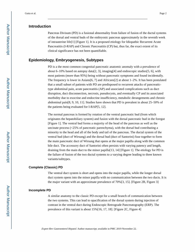
Pancreas Divisum (PD) is a fusional abnormality from failure of fusion of the ductal systems
of the dorsal and ventral buds of the embryonic pancreas approximately in the seventh week
of intrauterine life[1] [Figure 1]. It is a proposed etiology for Idiopathic Recurrent Acute
Pancreatitis (I-RAP) and Chronic Pancreatitis (CP) but, thus far, the exact extent of its
clinical significance has not been quantifiable.
Epidemiology, Embryogenesis, Subtypes
PD is a the most common congenital pancreatic anatomic anomaly with a prevalence of
about 6–10% based on autopsy data[2, 3], imaging[4] and endoscopic studies[5, 6], with
most patients (more than 95%) being without pancreatic symptoms and found incidentally.
The frequency is lower in Asians[6, 7] and Africans[2] at about 1–2%. It has been postulated
that a small subset of patients with PD are predisposed to recurrent attacks of pancreatic-
type abdominal pain, acute pancreatitis (AP) and associated complications such as duct
disruption, duct disconnection, necrosis, pseudocysts, and eventually CP and its associated
morbidity due to exocrine and endocrine insufficiency, metabolic derangements and chronic
abdominal pain[8, 9, 10, 11]. Studies have shown that PD is prevalent in about 25–50% of
the patients being evaluated for I-RAP[5, 12].
The normal pancreas is formed by rotation of the ventral pancreatic bud (from which
originates the hepatobiliary system) and fusion with the dorsal pancreatic bud in the foregut
[Figure 1]. The ventral bud forms a majority of the head of the pancreas as well as the
uncinate process (<25% of pancreatic parenchyma), with the dorsal bud contributing a
minority to the head and all of the body and tail of the pancreas. The ductal system of the
ventral bud (duct of Wirsung) and the dorsal bud (duct of Santorini) fuse together to form
the main pancreatic duct of Wirsung that opens at the major papilla along with the common
bile duct. The accessory duct of Santorini often persists with varying patency and length,
draining from the main duct to the minor papilla[13, 14] [Figure 1]. The etiology for PD is
the failure of fusion of the two ductal systems to a varying degree leading to three known
variants/subtypes.
Complete (Classic) PD
The ventral duct system is short and opens into the major papilla, while the longer dorsal
duct system opens into the minor papilla with no communication between the two ducts. It is
the major variant with an approximate prevalence of 70%[3, 15]. [Figure 2B, Figure 3]
Incomplete PD
A similar anatomy to the classic PD except for a small branch of communication between
the two systems. This can lead to opacification of the dorsal system during injection of
contrast in the ventral duct during Endoscopic Retrograde Pancreatography (ERP). The
prevalence of this variant is about 15%[16, 17, 18]. [Figure 2C, Figure 4]
Gutta et al. Page 2
Expert Rev Gastroenterol Hepatol. Author manuscript; available in PMC 2019 November 22.
Author M
anuscriptA
uthor Manuscript
Author M
anuscriptA
uthor Manuscript

Reverse PD
This is a rare variant wherein the accessory duct of Santorini does not communicate with the
main pancreatic duct, leading to a small isolated component of the dorsal pancreas. The
interruption in drainage system of the accessory duct in reverse PD can frequently be
interpreted as an obstruction by a malignant or a benign stricture. [Figure 2D]
The clinical behavior of complete and incomplete PD is similar except for the variation at
the time of ERP as noted [19, 20]. However, a reverse PD behaves more like a normal
pancreas wherein the severity of gallstone pancreatitis (from impaction of a gallstone at the
major papilla) can be much worse as a significant portion of the pancreas drains through the
major papilla unlike the preceding two variants where only a minority drains through the
major papilla.
Pseudo/False PD
An obstruction of the main pancreatic duct of Wirsung downstream to the origin of the
accessory duct of Santorini in a normal pancreas from injury due to acute pancreatitis, a
stricture due to chronic pancreatitis or a malignancy can give the appearance of true
pancreas divisum. This obstruction of the ventral duct in the head can lead to a decrease in
its size and compensatory increase in size of the accessory duct draining the body and tail
mimicking the anatomic findings of PD on imaging and ERP. It is important to recognize
this condition due to the possibility of malignancy.
Associated Pancreatic-Biliary Anomalies
PD has been found to be associated with other pancreatic and biliary anomalies. The most
frequent association is with annular pancreas where there is an abnormal rotation of the
ventral bud of the pancreas around the duodenum, leading to a ring or semi-ring of
pancreatic tissue around the second part of the duodenum[21]. Prior studies suggested that
there is about a 30–38% co-existence between the two conditions[21, 22, 23] but a recent
larger study suggested the prevalence is as high as 50%[24]. There have reports scattered in
the literature with PD co-existing with anomalous pancreatic-biliary junction (APBJ) and
choledochal cysts[25, 26, 27, 28, 29]. However, there have been no large-scale studies to
support any specific associations between these congenital anomalies. It has also been
reported recently that there is a higher prevalence of pancreatic and biliary tumors in patients
with PD than with a normal ductal system. (7.8% vs 3.5%)[30]. There have been reports of
partial agenesis of the dorsal pancreas with PD with some debate as to whether the agenesis
led to the PD or a consequence[14, 31].
Pathogenesis of Pancreatitis
The majority of patients with PD are without pancreas symptoms and therefore there is
considerable debate as to whether it is causally associated with pancreatitis or pancreatic-
type abdominal pain. It has been theorized that a larger and longer dominant dorsal duct
opening through a relatively smaller or stenotic minor papilla could lead to inadequate
drainage of pancreatic secretions with obstruction of flow. The resultant increase in
intraductal pressure and distention of the dorsal duct could then potentially lead to
Gutta et al. Page 3
Expert Rev Gastroenterol Hepatol. Author manuscript; available in PMC 2019 November 22.
Author M
anuscriptA
uthor Manuscript
Author M
anuscriptA
uthor Manuscript
abdominal pain and even pancreatitis. As a consequence of this imbalance, the term
“dominant duct syndrome” has been coined in the literature[32, 33]. Manometry studies
have shown an increased pressure in the dorsal duct and minor papilla when compared to the
ventral duct and major papilla in PD suggestive of dorsal duct and minor papilla
hypertension[34]. It has been suggested that this obstruction is transient and could be related
to intermittent plugging of the minor papilla by proteinaceous material in the pancreatic
secretions[9]. However, the large majority of patients with a dilated dorsal duct in PD are
asymptomatic, suggestive of a poor correlation between obstruction and symptoms[35].
Some observational studies of patients with I-RAP and CP have shown that the frequency of
pancreatitis was similar in patients with PD when compared to patients with normal duct
variants[8, 36, 37]. However, other studies have shown an increased prevalence of PD
(complete and incomplete) in adults[12, 38, 39, 40] and children[41] being evaluated for I-
RAP and CP, again supporting PD as a potentially pathologic entity. Similar support for
pancreas divisum as a potential pathologic entity is provided by studies that have shown
pathological changes of pancreatitis limited to the dorsal pancreas (i.e normal ventral
pancreas) in patients with PD[40, 42]. Studies have shown inconsistent and variable
response to minor papilla and dorsal duct therapy in patients who present with pancreatic-
type pain, with suboptimal results in the majority of patients[43, 44, 45, 46, 47, 48, 49, 50].
This heterogeneity suggests that PD may not be a primary driving force of pancreatic
pathology but rather a co-factor which in association with other factors leads to pancreatic
disease[51]. The basis for this line of thought comes from studies that show the prevalence
of I-RAP and CP is as high as 50% in adults and children with PD when associated with
certain genetic mutations of serine protease inhibitor Kazal type 1 gene (SPINK1), cystic
fibrosis transmembrane conductance regulator gene (CFTR), chymotrypsin C gene (CTRC)
[36, 41, 52, 53, 54]. These gene mutations or polymorphisms have been reported to have an
independent but not proven causal association with pancreatic disease[55, 56, 57].
Additionally, it has also been postulated that the presence of PD reduces the threshold for
pancreatic disease from other known primary factors of pancreatitis such as alcohol,
medications and trauma[34].
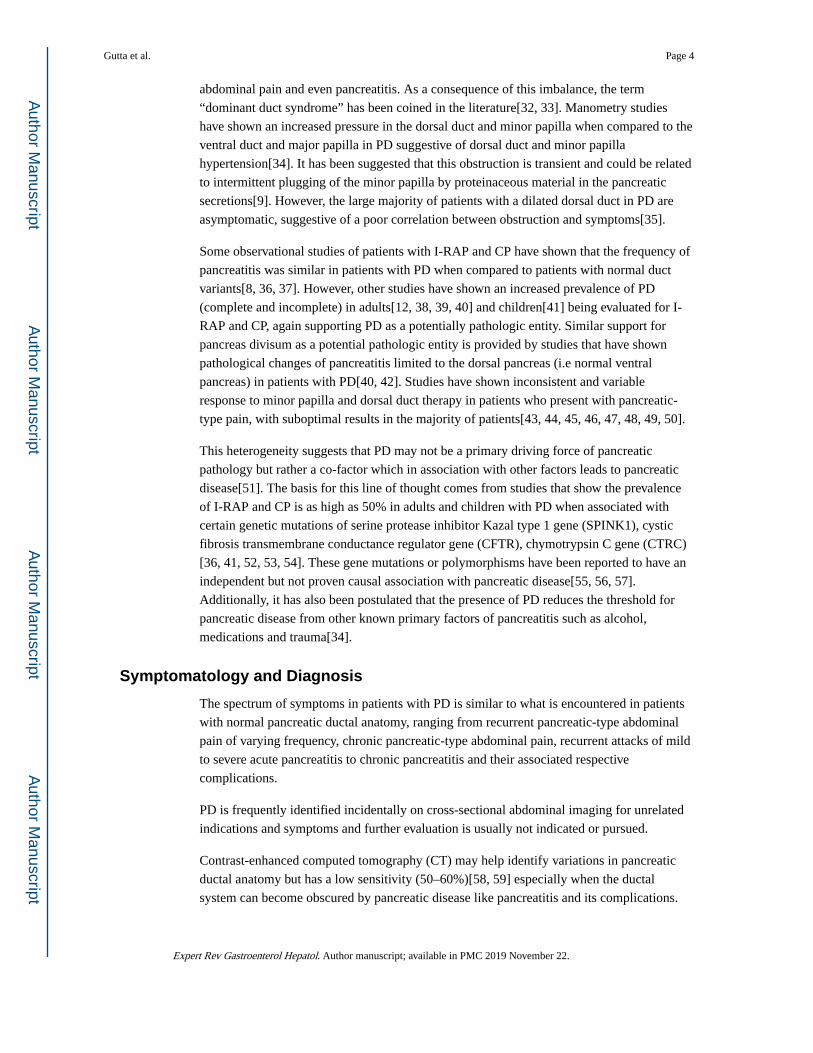
Symptomatology and Diagnosis
The spectrum of symptoms in patients with PD is similar to what is encountered in patients
with normal pancreatic ductal anatomy, ranging from recurrent pancreatic-type abdominal
pain of varying frequency, chronic pancreatic-type abdominal pain, recurrent attacks of mild
to severe acute pancreatitis to chronic pancreatitis and their associated respective
complications.
PD is frequently identified incidentally on cross-sectional abdominal imaging for unrelated
indications and symptoms and further evaluation is usually not indicated or pursued.
Contrast-enhanced computed tomography (CT) may help identify variations in pancreatic
ductal anatomy but has a low sensitivity (50–60%)[58, 59] especially when the ductal
system can become obscured by pancreatic disease like pancreatitis and its complications.
Gutta et al. Page 4
Expert Rev Gastroenterol Hepatol. Author manuscript; available in PMC 2019 November 22.
Author M
anuscriptA
uthor Manuscript
Author M
anuscriptA
uthor Manuscript

When the ducts are visualized, the sensitivity does increase to about 83% and the anatomy is
classically described as the dorsal duct crossing anteriorly and superiorly to the distal
common bile duct in the pancreatic head and opening separately into the minor papilla with
a small distinct ventral duct opening into the major papilla[60]. Dynamic CT with secretin
provocation (S-CT) can increase the visibility of the ducts when not visualized on initial
imaging with the sensitivity matching the previously described 83% with no immediate
complications[59].
Contrast-enhanced magnetic resonance cholangiopancreatography (MRCP) [Figure 5] is
reported to have sensitivity of about 50–70%[60, 61, 62, 63, 64, 65]. However, individual
studies have shown the sensitivity increases to up to 83–86% with a specificity of 97–99%
with secretin-enhanced MRCP (S-MRCP) [Figure 6][66, 67, 68, 69, 70, 71] with systematic
reviews and meta-analyses validating the results[64, 65, 72]. Secretin has also been shown to
be associated with minor side effects, but generally is well tolerated with no reported events
of exacerbating or precipitating pancreatic disease[71]. The description of the ductal
anatomy is similar to what is described on CT images[63]. Cystic dilation of the terminal
dorsal duct near the opening of the minor papilla described as a “Santorinicele” can also be
visualized on an S-MRCP[73, 74].
Endoscopic Ultrasound (EUS) has also been reported to have a high diagnostic accuracy for
PD with a sensitivity of 87%−95%[60, 75, 76] with secretin enhancement (S-EUS) offering
marginal benefit[77]. The absence of a “stack sign” (distal common bile duct, ventral
pancreatic duct and portal vein can be seen to run on a parallel axis in a normal pancreas),
the presence of a “crossed duct sign” (dorsal pancreatic duct will appear to cross over the
bile duct anteriorly and superiorly) are indicative of PD[78].
Systematic reviews suggest S-MRCP has a superior accuracy to EUS and is the preferred
diagnostic modality of choice[65, 72] with one study suggesting no significant difference
between S-MRCP and S-EUS[79].
Secretin-Enhanced Ultrasound (S-US) has been used in the past with studies showing
significant heterogeneity in regards to dilation of the dorsal duct in response to secretin and
the ability to predict the anatomy and response to treatment[80, 81, 82]. Factors such as
body habitus and overlying bowel gas have also led to variable accuracy and, therefore, S-
US has fallen out of favor.
Endoscopic retrograde cholangiopancreatography (ERCP) is considered the gold-standard
for diagnosis of PD[83, 84] but is seldom used currently when therapy is not planned given
the radiation exposure, procedure- and anesthesia-related complications and the high
accuracy of S-MRCP.
Secretin augments the visualization of the pancreatic ductal system by stimulating the
production of bicarbonate and water from the exocrine pancreas and thereby increasing the
volume of pancreatic secretions[85]. While the accuracy of all secretin augmented studies is
lower in patients with chronic pancreatitis and pancreatic exocrine insufficiency, secretin-
enhanced images may still yield more information than non-secretin studies.
Gutta et al. Page 5
Expert Rev Gastroenterol Hepatol. Author manuscript; available in PMC 2019 November 22.
Author M
anuscriptA
uthor Manuscript
Author M
anuscriptA
uthor Manuscript
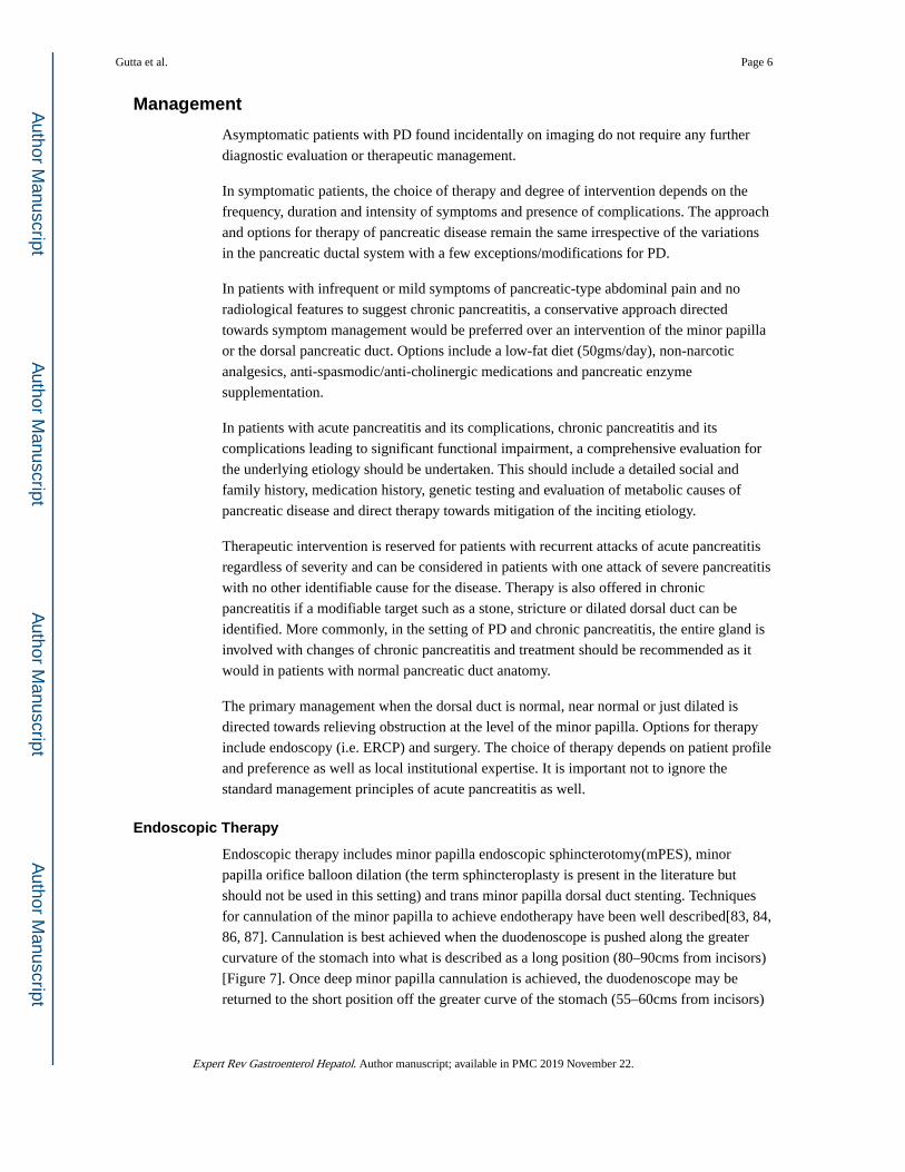
Management
Asymptomatic patients with PD found incidentally on imaging do not require any further
diagnostic evaluation or therapeutic management.
In symptomatic patients, the choice of therapy and degree of intervention depends on the
frequency, duration and intensity of symptoms and presence of complications. The approach
and options for therapy of pancreatic disease remain the same irrespective of the variations
in the pancreatic ductal system with a few exceptions/modifications for PD.
In patients with infrequent or mild symptoms of pancreatic-type abdominal pain and no
radiological features to suggest chronic pancreatitis, a conservative approach directed
towards symptom management would be preferred over an intervention of the minor papilla
or the dorsal pancreatic duct. Options include a low-fat diet (50gms/day), non-narcotic
analgesics, anti-spasmodic/anti-cholinergic medications and pancreatic enzyme
supplementation.
In patients with acute pancreatitis and its complications, chronic pancreatitis and its
complications leading to significant functional impairment, a comprehensive evaluation for
the underlying etiology should be undertaken. This should include a detailed social and
family history, medication history, genetic testing and evaluation of metabolic causes of
pancreatic disease and direct therapy towards mitigation of the inciting etiology.
Therapeutic intervention is reserved for patients with recurrent attacks of acute pancreatitis
regardless of severity and can be considered in patients with one attack of severe pancreatitis
with no other identifiable cause for the disease. Therapy is also offered in chronic
pancreatitis if a modifiable target such as a stone, stricture or dilated dorsal duct can be
identified. More commonly, in the setting of PD and chronic pancreatitis, the entire gland is
involved with changes of chronic pancreatitis and treatment should be recommended as it
would in patients with normal pancreatic duct anatomy.
The primary management when the dorsal duct is normal, near normal or just dilated is
directed towards relieving obstruction at the level of the minor papilla. Options for therapy
include endoscopy (i.e. ERCP) and surgery. The choice of therapy depends on patient profile
and preference as well as local institutional expertise. It is important not to ignore the
standard management principles of acute pancreatitis as well.
Endoscopic Therapy
Endoscopic therapy includes minor papilla endoscopic sphincterotomy(mPES), minor
papilla orifice balloon dilation (the term sphincteroplasty is present in the literature but
should not be used in this setting) and trans minor papilla dorsal duct stenting. Techniques
for cannulation of the minor papilla to achieve endotherapy have been well described[83, 84,
86, 87]. Cannulation is best achieved when the duodenoscope is pushed along the greater
curvature of the stomach into what is described as a long position (80–90cms from incisors)
[Figure 7]. Once deep minor papilla cannulation is achieved, the duodenoscope may be
returned to the short position off the greater curve of the stomach (55–60cms from incisors)
Gutta et al. Page 6
Expert Rev Gastroenterol Hepatol. Author manuscript; available in PMC 2019 November 22.
Author M
anuscriptA
uthor Manuscript
Author M
anuscriptA
uthor Manuscript
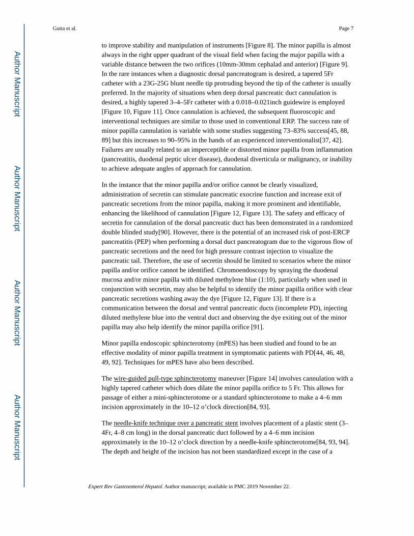
to improve stability and manipulation of instruments [Figure 8]. The minor papilla is almost
always in the right upper quadrant of the visual field when facing the major papilla with a
variable distance between the two orifices (10mm-30mm cephalad and anterior) [Figure 9].
In the rare instances when a diagnostic dorsal pancreatogram is desired, a tapered 5Fr
catheter with a 23G-25G blunt needle tip protruding beyond the tip of the catheter is usually
preferred. In the majority of situations when deep dorsal pancreatic duct cannulation is
desired, a highly tapered 3–4–5Fr catheter with a 0.018–0.021inch guidewire is employed
[Figure 10, Figure 11]. Once cannulation is achieved, the subsequent fluoroscopic and
interventional techniques are similar to those used in conventional ERP. The success rate of
minor papilla cannulation is variable with some studies suggesting 73–83% success[45, 88,
89] but this increases to 90–95% in the hands of an experienced interventionalist[37, 42].
Failures are usually related to an imperceptible or distorted minor papilla from inflammation
(pancreatitis, duodenal peptic ulcer disease), duodenal diverticula or malignancy, or inability
to achieve adequate angles of approach for cannulation.
In the instance that the minor papilla and/or orifice cannot be clearly visualized,
administration of secretin can stimulate pancreatic exocrine function and increase exit of
pancreatic secretions from the minor papilla, making it more prominent and identifiable,
enhancing the likelihood of cannulation [Figure 12, Figure 13]. The safety and efficacy of
secretin for cannulation of the dorsal pancreatic duct has been demonstrated in a randomized
double blinded study[90]. However, there is the potential of an increased risk of post-ERCP
pancreatitis (PEP) when performing a dorsal duct pancreatogram due to the vigorous flow of
pancreatic secretions and the need for high pressure contrast injection to visualize the
pancreatic tail. Therefore, the use of secretin should be limited to scenarios where the minor
papilla and/or orifice cannot be identified. Chromoendoscopy by spraying the duodenal
mucosa and/or minor papilla with diluted methylene blue (1:10), particularly when used in
conjunction with secretin, may also be helpful to identify the minor papilla orifice with clear
pancreatic secretions washing away the dye [Figure 12, Figure 13]. If there is a
communication between the dorsal and ventral pancreatic ducts (incomplete PD), injecting
diluted methylene blue into the ventral duct and observing the dye exiting out of the minor
papilla may also help identify the minor papilla orifice [91].
Minor papilla endoscopic sphincterotomy (mPES) has been studied and found to be an
effective modality of minor papilla treatment in symptomatic patients with PD[44, 46, 48,
49, 92]. Techniques for mPES have also been described.
The wire-guided pull-type sphincterotomy maneuver [Figure 14] involves cannulation with a
highly tapered catheter which does dilate the minor papilla orifice to 5 Fr. This allows for
passage of either a mini-sphincterotome or a standard sphincterotome to make a 4–6 mm
incision approximately in the 10–12 o’clock direction[84, 93].
The needle-knife technique over a pancreatic stent involves placement of a plastic stent (3–
4Fr, 4–8 cm long) in the dorsal pancreatic duct followed by a 4–6 mm incision
approximately in the 10–12 o’clock direction by a needle-knife sphincterotome[84, 93, 94].
The depth and height of the incision has not been standardized except in the case of a
Gutta et al. Page 7
Expert Rev Gastroenterol Hepatol. Author manuscript; available in PMC 2019 November 22.
Author M
anuscriptA
uthor Manuscript
Author M
anuscriptA
uthor Manuscript
Santorinicele where the extent of incision is modified to achieve the goal of unroofing the
dilated distal cystic segment of the dorsal duct[95].
Though it has been shown that the needle-knife technique is safer than the pull-type
sphincterotome technique when performing a major papilla pancreatic sphincterotomy (PES)
in high-risk patients[96], both techniques have been found to be safe and efficacious when
employed at the minor papilla, with similar complication rates[97, 98, 99]. The complication
profile of mPES is also similar to PES with the same traditional risk profile for PEP
(younger age, female gender, prior PEP, absence of chronic pancreatitis)[100, 101].
A modification of the needle-knife technique is a wire-assisted access sphincterotomy. This
involves deep dorsal duct cannulation with a guidewire, passing a needle-knife
sphincterotome alongside the wire and incising the minor papilla by cutting away from the
wire in the 10–12 o’clock direction. It has been shown to be equally efficacious as the other
techniques and can be used when the orifice is stenotic and does not allow the passage of
catheters and sphincterotomes.[102].
In situations described above where there is a failure of cannulation of the minor papilla, a
free-hand pre-cut needle-knife [Figure 15] maneuver may be helpful in cannulating the
minor papilla and gaining access to the dorsal duct to complete planned therapy. This
technique involves inserting a needle-knife sphincterotome 1–2 mm into the minor papilla
and making a 2–4 mm incision in the 10–12 o’clock direction. It has been shown to be
efficacious and safe in a small series of patients[103]. However, this technique should be
pursued with caution, as failed dorsal duct cannulation despite precut sphincterotomy may
significantly increase the risk of PEP.
Given the high risk of PEP in mPES (similar to high risk patients undergoing PES)[104], a
prophylactic temporary pancreatic stent can be placed after mPES to reduce the risk of PEP
[Figure 16], based on data extrapolated from studies related to PES[98, 105, 106, 107].
These temporary stents typically migrate out of the pancreatic duct spontaneously, usually
within 2 weeks[94, 108]. Migration can be confirmed on an upright abdominal X-ray (AXR/
KUB) and if the stent remains intraductal, it can be removed by performing an upper
endoscopy. On occasion, an urgent ERP may need to done to place a pancreatic stent or
replace a migrated stent in an attempt to modify the course of an evolving PEP and hasten
recovery[109]. Peri-procedure rectal indomethacin 100 mg[110, 111, 112, 113] and post
procedure intravenous lactated Ringer’s solution[114] have also been shown to reduce the
risk of PEP.
Minor papilla endoscopic orifice balloon dilation [Figure 17] has also been studied and
involves dilation of the minor papilla with comparable outcomes to mPES[115, 116]. As
with balloon dilation of the major papilla, it carries a higher risk of PEP and therefore almost
always is performed with placement of a protective pancreatic stent.
“Long-term” dorsal pancreatic duct stent placement either independently or in conjunction
with mPES or minor papilla orifice dilation has been shown to be an effective technique in
offering relief of symptoms related to PD[44, 45, 47, 50, 88, 92]. However it is associated
with complications such as occlusion, migration, ductal perforation, acute pancreatitis with
Gutta et al. Page 8
Expert Rev Gastroenterol Hepatol. Author manuscript; available in PMC 2019 November 22.
Author M
anuscriptA
uthor Manuscript
Author M
anuscriptA
uthor Manuscript

pseudocyst formation [117, 118], ductal and parenchyma changes indicating chronic
pancreatitis[119, 120, 121, 122].
The response and outcome to all pancreatic endotherapy from multiple studies over the years
has been found to be highly variable and primarily determined by clinical presentation.
Among them, the only randomized control trial by Lans et al [45] comparing endotherapy
(dorsal pancreatic duct stenting with stent changes every 3 months for one year) to sham in
19 patients with idiopathic recurrent acute pancreatitis showed 90% of the patients in the
intervention group reported >50% symptomatic improvement as compared to 11% in the
placebo. The mean follow-up times in the stent and control groups were 28.6 and 31.5
months respectively. There was a lower incidence of recurrent pancreatitis in the stent group
(1/10 vs 7/9), with 44% in the control group required stent placement due to uncontrolled
symptoms. The patients in the control group who underwent stenting were subsequently
followed up from 6 to 53 months and did not suffer from further episodes of pancreatitis or
require hospitalization. This study demonstrated significant objective and subjective clinical
improvement of symptoms with endotherapy compared with controls on short-term follow-
up. Four systematic reviews and meta-analyses of these studies[6, 123, 124, 125] reporting a
pooled response rate of 62–70%. The maximal improvement of symptoms was seen in
patients with recurrent acute pancreatitis (76–80%), with a lower response rate in chronic
pancreatitis (42–69%), and the least with chronic pancreatic-type abdominal pain (33–54%).
The incidence of PEP was reported to be at 10% with the rate of minor papilla stenosis
requiring re-intervention at 19%. The follow up period was between 14 to 64 months with
most studies reporting a period of 36 months. In an effort to identify if endotherapy therapy
is beneficial in PD, a sham-controlled, single blinded with a blinded outcome assessment,
multi-center, international, randomized clinical trial (SHARP) of ERP with mPES for the
treatment of RAP with PD is currently underway with planned total sample size of 234
subjects, and a planned maximum follow-up of 48 months (ClinicalTrials.gov Identifier: ).
Surgery
Surgical minor papilla sphincterotomy[48] and surgical minor papilla sphincteroplasty[33,
126] have both been described to provide comparable effective and durable relief of
symptoms with maximal efficacy noted in patients with recurrent acute pancreatitis (RAP).
The response to all surgical minor papilla therapy has also been variable with one systematic
review and meta-analysis[6] reporting a pooled response rate of 75%. Similar to
endotherapy, maximal symptomatic improvement was seen in patients with RAP (83%),
with a lower response rate in chronic pancreatitis (67%), and the least with chronic
pancreatic-type abdominal pain (52%). The follow up period was between 6 to 120 months
with most studies greater than 50 months.
The clinical presentation and response rates to endotherapy and surgery within the three
types of pancreas divisum were not significantly different[6, 89].
Given the comparable outcomes between endotherapy and surgery of minor papilla in
alleviating symptoms but with endotherapy having a favorable complication and mortality
rate, endoscopy (i.e. ERCP) is usually considered the first line treatment, with surgery
Gutta et al. Page 9
Expert Rev Gastroenterol Hepatol. Author manuscript; available in PMC 2019 November 22.
Author M
anuscriptA
uthor Manuscript
Author M
anuscriptA
uthor Manuscript
reserved for patients who fail minor papilla cannulation, endotherapy or have altered
anatomy (e.g., gastric bypass).
In patients with RAP who unfortunately fail minor papilla therapy or have progressed to
chronic pancreatitis, further therapeutic options are limited. These may include management
of symptoms related to chronic pancreatitis, including periods of pancreas rest with small
bowel nutrition, celiac plexus block, somatostatin analogues, and an intrathecal pain
pump[127]. Patients with significantly dilated dorsal pancreatic ducts may benefit from a
lateral pancreatojejunostomy (Puestow or Frey procedure)[128, 129]. Total dorsal
pancreatectomy has been proposed for benign or low grade malignant pancreatic tumors
limited to the dorsal pancreas as an alternative to total pancreatectomy and may be
extrapolated to patients with changes of chronic pancreatitis limited to the dorsal
pancreas[130]. Total pancreatectomy with auto-islet cell transplantation (TPAIT) is an
emerging option for patients with disease refractory to endotherapy. Reports of outcomes of
TPAIT data performed in patients with a normal anatomy suggest favorable outcomes and
potentially can be extrapolated to patients with PD[131, 132, 133, 134].
Expert Commentary
5-year review/Speculation
Pancreas divisum is the most common congenital pancreatic anomaly and has been reported
on autopsy, imaging and endoscopy for many years. As the majority of patients lack
pancreatic symptoms, its association with pancreatic disease is debatable. However, there
have been several studies that show a substantial association with pancreatic disease in the
background of other factors that predispose to pancreatic pathology. Over the past few
decades, several attempts have been made to understand the pathology of pancreatic disease
in relation to pancreas divisum and to prove a definite association but none have been
definitively successful. Numerous studies have been undertaken in an attempt to demonstrate
a therapeutic benefit with various modalities of minor papilla therapy but, thus far, the
results have been heterogenous and not definitive. Most of the data suggest a role for
endoscopic intervention in the setting of acute recurrent pancreatitis. However, most studies
have been of low quality, small patient sample, predominantly retrospective and without
long-term follow up. Surgical and endoscopic modalities have also not been directly
compared. With the current data available, it would not be prudent to routinely advise a
definitive line of management for pancreatic disease associated with pancreas divisum. This
should involve a comprehensive discussion with the individual patient to define expectations
before embarking on any medical and/or interventional therapy.
References
1. Kozu T, Suda K, Toki F. Pancreatic development and anatomical variation. Gastrointestinal endoscopy clinics of North America. 1995 1;5(1):1–30. [PubMed: 7728339]
2. Smanio T Proposed nomenclature and classification of the human pancreatic ducts and duodenal papillae. Study based on 200 post mortems. International surgery. 1969 8;52(2):125–41. [PubMed: 5793091]
Gutta et al. Page 10
Expert Rev Gastroenterol Hepatol. Author manuscript; available in PMC 2019 November 22.
Author M
anuscriptA
uthor Manuscript
Author M
anuscriptA
uthor Manuscript

3. Stimec B, Bulajic M, Korneti V, et al. Ductal morphometry of ventral pancreas in pancreas divisum. Comparison between clinical and anatomical results. The Italian journal of gastroenterology. 1996 Feb-Mar;28(2):76–80. [PubMed: 8781998]
4. Bulow R, Simon P, Thiel R, et al. Anatomic variants of the pancreatic duct and their clinical relevance: an MR-guided study in the general population. European radiology. 2014 Dec;24(12):3142–9. doi: 10.1007/s00330-014-3359-7.
5. Bernard JP, Sahel J, Giovannini M, et al. Pancreas divisum is a probable cause of acute pancreatitis: a report of 137 cases. Pancreas. 1990 5;5(3):248–54. [PubMed: 2343039]
6. Liao Z, Gao R, Wang W, et al. A systematic review on endoscopic detection rate, endotherapy, and surgery for pancreas divisum. Endoscopy. 2009 5;41(5):439–44. doi: 10.1055/s-0029-1214505. [PubMed: 19337962]
7. Suga T, Nakagawa T, Miyakawa H, et al. Clinical Features of Patients with Pancreas Divisum. 1994;6(1):80–86. doi: doi: 10.1111/j.1443-1661.1994.tb00668.x.
8. Delhaye M, Cremer M. Clinical significance of pancreas divisum. Acta gastro-enterologica Belgica. 1992 May-Jun;55(3):306–13. [PubMed: 1632145]
9. Cotton PB. Pancreas divisum--curiosity or culprit? Gastroenterology. 1985 12;89(6):1431–5. [PubMed: 4054536]
10. Brenner P, Duncombe V, Ham JM. Pancreatitis and pancreas divisum: aetiological and surgical considerations. The Australian and New Zealand journal of surgery. 1990 11;60(11):899–903. [PubMed: 2241651]
11. Richter JM, Schapiro RH, Mulley AG, et al. Association of pancreas divisum and pancreatitis, and its treatment by sphincteroplasty of the accessory ampulla. Gastroenterology. 1981 12;81(6):1104–10. [PubMed: 7286588]
12. Takuma K, Kamisawa T, Hara S, et al. Etiology of recurrent acute pancreatitis, with special emphasis on pancreaticobiliary malformation. Advances in medical sciences. 2012;57(2):244–50. doi: 10.2478/v10039-012-0041-7. [PubMed: 23183766]
13. Kamisawa T, Koike M, Okamoto A. Embryology of the pancreatic duct system. Digestion. 1999 Mar-Apr;60(2):161–5. doi: 10.1159/000007642. [PubMed: 10095158]
14. Kamisawa T, Takuma K, Egawa N, et al. A new embryological theory of the pancreatic duct system. Digestive surgery. 2010;27(2):132–6. doi: 10.1159/000286906. [PubMed: 20551658]
15. MacCarty RL, Stephens DH, Brown AL Jr., et al. Retrograde pancreatography in autopsy specimens. The American journal of roentgenology, radium therapy, and nuclear medicine. 1975 2;123(2):359–66.
16. Kamisawa T, Egawa N, Tu Y, et al. Pancreatographic investigation of embryology of complete and incomplete pancreas divisum. Pancreas. 2007 1;34(1):96–102. doi: 10.1097/01.mpa.0000240607.49183.7e. [PubMed: 17198190]
17. Moreira VF, Merono E, Ledo L, et al. Incomplete pancreas divisum. Gastrointestinal endoscopy. 1991 Jan-Feb;37(1):104–5.
18. Tulassay Z, Papp J, Farkas IE. Diagnostic aspects of incomplete pancreas divisum. Gastrointestinal endoscopy. 1986 12;32(6):428.
19. Kamisawa T, Tu Y, Egawa N, et al. Clinical implications of incomplete pancreas divisum. JOP : Journal of the pancreas. 2006 11 10;7(6):625–30. [PubMed: 17095842]
20. Kim MH, Lee SS, Kim CD, et al. Incomplete pancreas divisum: is it merely a normal anatomic variant without clinical implications? Endoscopy. 2001 9;33(9):778–85. doi: 10.1055/s-2001-16521. [PubMed: 11558032]
21. Zyromski NJ, Sandoval JA, Pitt HA, et al. Annular pancreas: dramatic differences between children and adults. Journal of the American College of Surgeons. 2008 5;206(5):1019–25; discussion 1025–7. doi: 10.1016/j.jamcollsurg.2007.12.009. [PubMed: 18471747]
22. Lehman GA, O’Connor KW. Coexistence of annular pancreas and pancreas divisum--ERCP diagnosis. Gastrointestinal endoscopy. 1985 2;31(1):25–8. [PubMed: 3979764]
23. Sandrasegaran K, Patel A, Fogel EL, et al. Annular pancreas in adults. AJR American journal of roentgenology. 2009 8;193(2):455–60. doi: 10.2214/ajr.08.1596. [PubMed: 19620443]
Gutta et al. Page 11
Expert Rev Gastroenterol Hepatol. Author manuscript; available in PMC 2019 November 22.
Author M
anuscriptA
uthor Manuscript
Author M
anuscriptA
uthor Manuscript
24. Gromski MA, Lehman GA, Zyromski NJ, et al. Annular pancreas: endoscopic and pancreatographic findings from a tertiary referral ERCP center. Gastrointestinal endoscopy. 2018 9 18. doi: 10.1016/j.gie.2018.09.008.
25. Cao J, Wang L, Zou X. Coexistence of Complete Pancreas Divisum and Anomalous Pancreaticobiliary Junction in a Patient With Type IA Choledochal Cyst. Clinical gastroenterology and hepatology : the official clinical practice journal of the American Gastroenterological Association. 2018 8 2. doi: 10.1016/j.cgh.2018.07.036.
26. Ng JW, Wong MK, Huang J, et al. Incomplete pancreas divisum associated with abnormal junction of pancreaticobiliary duct system. Gastrointestinal endoscopy. 1992 Jan-Feb;38(1):105–6. [PubMed: 1612369]
27. Patidar Y, Agarwal N, Gupta S, et al. Choledochocele with pancreas divisum: A rare co-occurrence diagnosed on magnetic resonance cholangiopancreatography. World journal of radiology. 2013 7 28;5(7):264–6. doi: 10.4329/wjr.v5.i7.264. [PubMed: 23908697]
28. Ransom-Rodriguez A, Blachman-Braun R, Sanchez-Garcia Ramos E, et al. A rare case of choledochal cyst with pancreas divisum: case presentation and literature review. Annals of hepato-biliary-pancreatic surgery. 2017 2;21(1):52–56. doi: 10.14701/ahbps.2017.21.1.52. [PubMed: 28317046]
29. Sanada Y, Sakuma Y, Sata N. Embryological etiology of pancreaticobiliary system predicted from pancreaticobiliary maljunction with incomplete pancreatic divisum: a case report. BMC surgery. 2018 8 2;18(1):50. doi: 10.1186/s12893-018-0385-4. [PubMed: 30068344]
30. Adibelli ZH, Adatepe M, Isayeva L, et al. Pancreas divisum: A risk factor for pancreaticobiliary tumors - an analysis of 1628 MR cholangiography examinations. Diagnostic and interventional imaging. 2017 2;98(2):141–147. doi: 10.1016/j.diii.2016.08.004. [PubMed: 27616039]
31. Lehman GA, Kopecky KK, Rogge JD. Partial pancreatic agenesis combined with pancreas divisum and duodenum reflexum. Gastrointestinal endoscopy. 1987 12;33(6):445–8. [PubMed: 3443265]
32. Madura JA. Pancreas divisum: stenosis of the dorsally dominant pancreatic duct. A surgically correctable lesion. American journal of surgery. 1986 6;151(6):742–5. [PubMed: 3717505]
33. Warshaw AL, Simeone JF, Schapiro RH, et al. Evaluation and treatment of the dominant dorsal duct syndrome (pancreas divisum redefined). American journal of surgery. 1990 1;159(1):59–64; [PubMed: 2403764]
34. Staritz M, Meyer zum Buschenfelde KH. Elevated pressure in the dorsal part of pancreas divisum: the cause of chronic pancreatitis? Pancreas. 1988;3(1):108–10. [PubMed: 3362837]
35. Cotton PB. Congenital anomaly of pancreas divisum as cause of obstructive pain and pancreatitis. 1980;21(2):105–114. doi: 10.1136/gut.21.2.105%JGut.
36. Bertin C, Pelletier AL, Vullierme MP, et al. Pancreas divisum is not a cause of pancreatitis by itself but acts as a partner of genetic mutations. The American journal of gastroenterology. 2012 2;107(2):311–7. doi: 10.1038/ajg.2011.424. [PubMed: 22158025]
37. Delhaye M, Engelholm L, Cremer M. Pancreas divisum: congenital anatomic variant or anomaly? Contribution of endoscopic retrograde dorsal pancreatography. Gastroenterology. 1985 11;89(5):951–8. [PubMed: 4043675]
38. Dhar A, Goenka MK, Kochhar R, et al. Pancrease divisum: five years’ experience in a teaching hospital. Indian journal of gastroenterology : official journal of the Indian Society of Gastroenterology. 1996 1;15(1):7–9. [PubMed: 8840617]
39. Morgan DE, Logan K, Baron TH, et al. Pancreas divisum: implications for diagnostic and therapeutic pancreatography. AJR American journal of roentgenology. 1999 7;173(1):193–8. doi: 10.2214/ajr.173.1.10397125. [PubMed: 10397125]
40. Wang DB, Yu J, Fulcher AS, et al. Pancreatitis in patients with pancreas divisum: imaging features at MRI and MRCP. World journal of gastroenterology. 2013 8 14;19(30):4907–16. doi: 10.3748/wjg.v19.i30.4907. [PubMed: 23946595]
41. Lin TK, Abu-El-Haija M, Nathan JD, et al. Pancreas Divisum in Pediatric Acute Recurrent and Chronic Pancreatitis: Report From INSPPIRE. Journal of clinical gastroenterology. 2018 6 2. doi: 10.1097/mcg.0000000000001063.
42. Benage D, McHenry R, Hawes RH, et al. Minor papilla cannulation and dorsal ductography in pancreas divisum. Gastrointestinal endoscopy. 1990 Nov-Dec;36(6):553–7. [PubMed: 2279641]
Gutta et al. Page 12
Expert Rev Gastroenterol Hepatol. Author manuscript; available in PMC 2019 November 22.
Author M
anuscriptA
uthor Manuscript
Author M
anuscriptA
uthor Manuscript
43. Borak GD, Romagnuolo J, Alsolaiman M, et al. Long-term clinical outcomes after endoscopic minor papilla therapy in symptomatic patients with pancreas divisum. Pancreas. 2009 11;38(8):903–6. doi: 10.1097/MPA.0b013e3181b2bc03. [PubMed: 19672208]
44. Chacko LN, Chen YK, Shah RJ. Clinical outcomes and nonendoscopic interventions after minor papilla endotherapy in patients with symptomatic pancreas divisum. Gastrointestinal endoscopy. 2008 10;68(4):667–73. doi: 10.1016/j.gie.2008.01.025. [PubMed: 18436218]
45. Lans JI, Geenen JE, Johanson JF, et al. Endoscopic therapy in patients with pancreas divisum and acute pancreatitis: a prospective, randomized, controlled clinical trial. Gastrointestinal endoscopy. 1992 Jul-Aug;38(4):430–4. [PubMed: 1511816]
46. Lehman GA, Sherman S, Nisi R, et al. Pancreas divisum: results of minor papilla sphincterotomy. Gastrointestinal endoscopy. 1993 Jan-Feb;39(1):1–8. [PubMed: 8454127]
47. Heyries L, Barthet M, Delvasto C, et al. Long-term results of endoscopic management of pancreas divisum with recurrent acute pancreatitis. Gastrointestinal endoscopy. 2002 3;55(3):376–81. doi: 10.1067/mge.2002.121602. [PubMed: 11868012]
48. Keith RG, Shapero TF, Saibil FG, et al. Dorsal duct sphincterotomy is effective long-term treatment of acute pancreatitis associated with pancreas divisum. Surgery. 1989 10;106(4):660–6; [PubMed: 2799640]
49. Kwan V, Loh SM, Walsh PR, et al. Minor papilla sphincterotomy for pancreatitis due to pancreas divisum. ANZ journal of surgery. 2008 4;78(4):257–61. doi: 10.1111/j.1445-2197.2008.04431.x. [PubMed: 18366396]
50. Siegel JH, Ben-Zvi JS, Pullano W, et al. Effectiveness of endoscopic drainage for pancreas divisum: endoscopic and surgical results in 31 patients. Endoscopy. 1990 5;22(3):129–33. doi: 10.1055/s-2007-1010727. [PubMed: 2103724]
51. Klein SD, Affronti JP. Pancreas divisum, an evidence-based review: part I, pathophysiology. Gastrointestinal endoscopy. 2004 9;60(3):419–25. [PubMed: 15332034]
52. Garg PK, Khajuria R, Kabra M, et al. Association of SPINK1 gene mutation and CFTR gene polymorphisms in patients with pancreas divisum presenting with idiopathic pancreatitis. Journal of clinical gastroenterology. 2009 10;43(9):848–52. doi: 10.1097/MCG.0b013e3181a4e772. [PubMed: 19593166]
53. Gelrud A, Sheth S, Banerjee S, et al. Analysis of cystic fibrosis gener product (CFTR) function in patients with pancreas divisum and recurrent acute pancreatitis. The American journal of gastroenterology. 2004 8;99(8):1557–62. doi: 10.1111/j.1572-0241.2004.30834.x. [PubMed: 15307877]
54. Choudari CP, Imperiale TF, Sherman S, et al. Risk of Pancreatitis with Mutation of the Cystic Fibrosis Gene [Original Contribution]. American Journal Of Gastroenterology. 2004 07/01/online;99:1358. doi: 10.1111/j.1572-0241.2004.30655.x. [PubMed: 15233679]
55. Bhatia E, Choudhuri G, Sikora SS, et al. Tropical calcific pancreatitis: strong association with SPINK1 trypsin inhibitor mutations. Gastroenterology. 2002 10;123(4):1020–5. [PubMed: 12360463]
56. Cohn JA, Friedman KJ, Noone PG, et al. Relation between mutations of the cystic fibrosis gene and idiopathic pancreatitis. The New England journal of medicine. 1998 9 3;339(10):653–8. doi: 10.1056/nejm199809033391002. [PubMed: 9725922]
57. Witt H, Luck W, Hennies HC, et al. Mutations in the gene encoding the serine protease inhibitor, Kazal type 1 are associated with chronic pancreatitis. Nature genetics. 2000 6;25(2):213–6. doi: 10.1038/76088. [PubMed: 10835640]
58. Asayama Y, Fang W, Stolpen A, et al. Detectability of pancreas divisum in patients with acute pancreatitis on multi-detector row computed tomography. Emergency radiology. 2012 4;19(2):121–5. doi: 10.1007/s10140-011-1008-x. [PubMed: 22167339]
59. Lindstrom E, Ihse I. Dynamic CT scanning of pancreatic duct after secretin provocation in pancreas divisum. Digestive diseases and sciences. 1990 11;35(11):1371–6. [PubMed: 1699712]
60. Kushnir VM, Wani SB, Fowler K, et al. Sensitivity of endoscopic ultrasound, multidetector computed tomography, and magnetic resonance cholangiopancreatography in the diagnosis of pancreas divisum: a tertiary center experience. Pancreas. 2013 4;42(3):436–41. doi: 10.1097/MPA.0b013e31826c711a. [PubMed: 23211370]
Gutta et al. Page 13
Expert Rev Gastroenterol Hepatol. Author manuscript; available in PMC 2019 November 22.
Author M
anuscriptA
uthor Manuscript
Author M
anuscriptA
uthor Manuscript
61. Carnes ML, Romagnuolo J, Cotton PB. Miss rate of pancreas divisum by magnetic resonance cholangiopancreatography in clinical practice. Pancreas. 2008 8;37(2):151–3. doi: 10.1097/MPA.0b013e318164cbaf. [PubMed: 18665075]
62. Chalazonitis NA, Lachanis BS, Laspas F, et al. Pancreas divisum: magnetic resonance cholangiopancreatography findings. Singapore medical journal. 2008 11;49(11):951–4; [PubMed: 19037565]
63. Kamisawa T, Tu Y, Egawa N, et al. MRCP of congenital pancreaticobiliary malformation. Abdominal imaging. 2007 Jan-Feb;32(1):129–33. doi: 10.1007/s00261-006-9005-3. [PubMed: 16680507]
64. Rustagi T, Njei B. Magnetic resonance cholangiopancreatography in the diagnosis of pancreas divisum: a systematic review and meta-analysis. Pancreas. 2014 8;43(6):823–8. doi: 10.1097/mpa.0000000000000143. [PubMed: 24743381]
65. Shen Z, Munker S, Zhou B, et al. The Accuracies of Diagnosing Pancreas Divisum by Magnetic Resonance Cholangiopancreatography and Endoscopic Ultrasound: A Systematic Review and Meta-analysis. Scientific reports. 2016 10 13;6:35389. doi: 10.1038/srep35389. [PubMed: 27734952]
66. Chamokova B, Bastati N, Poetter-Lang S, et al. The clinical value of secretin-enhanced MRCP in the functional and morphological assessment of pancreatic diseases. The British journal of radiology. 2018 4;91(1084):20170677. doi: 10.1259/bjr.20170677. [PubMed: 29206061]
67. Matos C, Metens T, Deviere J, et al. Pancreas divisum: evaluation with secretin-enhanced magnetic resonance cholangiopancreatography. Gastrointestinal endoscopy. 2001 6;53(7):728–33. doi: 10.1067/mge.2001.114784. [PubMed: 11375579]
68. Mosler P, Akisik F, Sandrasegaran K, et al. Accuracy of magnetic resonance cholangiopancreatography in the diagnosis of pancreas divisum. Digestive diseases and sciences. 2012 1;57(1):170–4. doi: 10.1007/s10620-011-1823-7. [PubMed: 21761168]
69. Sandrasegaran K, Cote GA, Tahir B, et al. The utility of secretin-enhanced MRCP in diagnosing congenital anomalies. Abdominal imaging. 2014 10;39(5):979–87. doi: 10.1007/s00261-014-0131-z. [PubMed: 24748158]
70. Sandrasegaran K, Tahir B, Barad U, et al. The Value of Secretin-Enhanced MRCP in Patients With Recurrent Acute Pancreatitis. AJR American journal of roentgenology. 2017 2;208(2):315–321. doi: 10.2214/ajr.16.16566. [PubMed: 27824485]
71. Sherman S, Freeman ML, Tarnasky PR, et al. Administration of secretin (RG1068) increases the sensitivity of detection of duct abnormalities by magnetic resonance cholangiopancreatography in patients with pancreatitis. Gastroenterology. 2014 9;147(3):646–654.e2. doi: 10.1053/j.gastro.2014.05.035. [PubMed: 24906040]
72. Wan J, Ouyang Y, Yu C, et al. Comparison of EUS with MRCP in idiopathic acute pancreatitis: a systematic review and meta-analysis. Gastrointestinal endoscopy. 2018 5;87(5):1180–1188.e9. doi: 10.1016/j.gie.2017.11.028. [PubMed: 29225082]
73. Boninsegna E, Manfredi R, Ventriglia A, et al. Santorinicele: secretin-enhanced magnetic resonance cholangiopancreatography findings before and after minor papilla sphincterotomy. European radiology. 2015 8;25(8):2437–44. doi: 10.1007/s00330-015-3644-0. [PubMed: 25693665]
74. Manfredi R, Brizi MG, Costamagna G, et al. Pancreas divisum and santorinicele: assessment by dynamic magnetic resonance cholangiopancreatography during secretin stimulation. La Radiologia medica. 2002 Jan-Feb;103(1–2):55–64. [PubMed: 11859301]
75. Lai R, Freeman ML, Cass OW, et al. Accurate diagnosis of pancreas divisum by linear-array endoscopic ultrasonography. Endoscopy. 2004 8;36(8):705–9. doi: 10.1055/s-2004-825663. [PubMed: 15280976]
76. Rana SS, Bhasin DK, Sharma V, et al. Role of endoscopic ultrasound in the diagnosis of pancreas divisum. Endoscopic ultrasound. 2013 Jan-Mar;2(1):7–10. doi: 10.7178/eus.04.003. [PubMed: 24949360]
77. Catalano MF RM, Geenen JE, et al. Pancreatic endotherapy of pancreas divisum (PDIV): Response based on clinical presentation and results of secretin stimulated endoscopic ultrasound: (S-EUS). Gastrointestinal endoscopy. 2001;53(5):AB133. doi: 10.1016/S0016-5107(01)80267-4.
Gutta et al. Page 14
Expert Rev Gastroenterol Hepatol. Author manuscript; available in PMC 2019 November 22.
Author M
anuscriptA
uthor Manuscript
Author M
anuscriptA
uthor Manuscript
78. Sharma M, Pathak A, Rameshbabu CS, et al. Imaging of pancreas divisum by linear-array endoscopic ultrasonography. Endoscopic ultrasound. 2016 Jan-Feb;5(1):21–9. doi: 10.4103/2303-9027.175878. [PubMed: 26879163]
79. Mariani A, Arcidiacono PG, Curioni S, et al. Diagnostic yield of ERCP and secretin-enhanced MRCP and EUS in patients with acute recurrent pancreatitis of unknown aetiology. Digestive and liver disease : official journal of the Italian Society of Gastroenterology and the Italian Association for the Study of the Liver. 2009 10;41(10):753–8. doi: 10.1016/j.dld.2009.01.009.
80. Glaser J, Stienecker K. Ultrasound secretin test in patients with pancreas divisum--an aid in the diagnosis of papillary or dorsal duct stenosis? Zeitschrift fur Gastroenterologie. 1999 7;37(7):585–8. [PubMed: 10458006]
81. Lowes JR, Lees WR, Cotton PB. Pancreatic duct dilatation after secretin stimulation in patients with pancreas divisum. Pancreas. 1989;4(3):371–4. [PubMed: 2660134]
82. Tulassay Z, Jakab Z, Vadasz A, et al. Secretin provocation ultrasonography in the diagnosis of papillary obstruction in pancreas divisum. Gastroenterologisches Journal : Organ der Gesellschaft fur Gastroenterologie der DDR. 1991;51(1):47–50. [PubMed: 1910496]
83. Lehman GA, Sherman S. Pancreas divisum. Diagnosis, clinical significance, and management alternatives. Gastrointestinal endoscopy clinics of North America. 1995 1;5(1):145–70. [PubMed: 7728342]
84. Lehman GA, Sherman S. Diagnosis and therapy of pancreas divisum. Gastrointestinal endoscopy clinics of North America. 1998 1;8(1):55–77. [PubMed: 9405751]
85. Chey WY, Chang TM. Secretin: historical perspective and current status. Pancreas. 2014 3;43(2):162–82. doi: 10.1097/01.mpa.0000437325.29728.d6. [PubMed: 24518494]
86. McCarthy J, Fumo D, Geenen JE. Pancreas divisum: a new method for cannulating the accessory papilla. Gastrointestinal endoscopy. 1987 12;33(6):440–2. [PubMed: 3443264]
87. O’Connor KW, Lehman GA. An improved technique for accessory papilla cannulation in pancreas divisum. Gastrointestinal endoscopy. 1985 2;31(1):13–7. [PubMed: 3979760]
88. Ertan A Long-term results after endoscopic pancreatic stent placement without pancreatic papillotomy in acute recurrent pancreatitis due to pancreas divisum. Gastrointestinal endoscopy. 2000 7;52(1):9–14. doi: 10.1067/mge.2000.106311. [PubMed: 10882955]
89. Jacob L, Geenen JE, Catalano MF, et al. Clinical presentation and short-term outcome of endoscopic therapy of patients with symptomatic incomplete pancreas divisum. Gastrointestinal endoscopy. 1999 1;49(1):53–7. [PubMed: 9869723]
90. Devereaux BM, Fein S, Purich E, et al. A new synthetic porcine secretin for facilitation of cannulation of the dorsal pancreatic duct at ERCP in patients with pancreas divisum: a multicenter, randomized, double-blind comparative study. Gastrointestinal endoscopy. 2003 5;57(6):643–7. doi: 10.1067/mge.2003.195. [PubMed: 12709690]
91. Park SH, de Bellis M, McHenry L, et al. Use of methylene blue to identify the minor papilla or its orifice in patients with pancreas divisum. Gastrointestinal endoscopy. 2003 3;57(3):358–63. doi: 10.1067/mge.2003.110. [PubMed: 12612516]
92. Gerke H, Byrne MF, Stiffler HL, et al. Outcome of endoscopic minor papillotomy in patients with symptomatic pancreas divisum. JOP : Journal of the pancreas. 2004 5;5(3):122–31. [PubMed: 15138333]
93. Rustagi T, Golioto M. Diagnosis and therapy of pancreas divisum by ERCP: a single center experience. Journal of digestive diseases. 2013 2;14(2):93–9. doi: 10.1111/1751-2980.12004. [PubMed: 23134252]
94. Siegel JH, Cohen SA, Kasmin FE, et al. Stent-guided sphincterotomy. Gastrointestinal endoscopy. 1994 Sep-Oct;40(5):567–72. [PubMed: 7988820]
95. Crinò SF, Bernardoni L, Conti Bellocchi MC, et al. Efficacy of Endoscopic Minor Papilla Sphincterotomy for Symptomatic Santorinicele. Clinical Gastroenterology and Hepatology. 2017;15(2):303–306. doi: 10.1016/j.cgh.2016.08.004. [PubMed: 27521510]
96. Varadarajulu S, Wilcox CM. Randomized trial comparing needle-knife and pull-sphincterotome techniques for pancreatic sphincterotomy in high-risk patients. Gastrointestinal endoscopy. 2006 11;64(5):716–22. doi: 10.1016/j.gie.2006.02.058. [PubMed: 17055863]
Gutta et al. Page 15
Expert Rev Gastroenterol Hepatol. Author manuscript; available in PMC 2019 November 22.
Author M
anuscriptA
uthor Manuscript
Author M
anuscriptA
uthor Manuscript
97. Attwell A, Borak G, Hawes R, et al. Endoscopic pancreatic sphincterotomy for pancreas divisum by using a needle-knife or standard pull-type technique: safety and reintervention rates. Gastrointestinal endoscopy. 2006 11;64(5):705–11. doi: 10.1016/j.gie.2006.02.057. [PubMed: 17055861]
98. Lawrence C, Romagnuolo J, Cotton PB, et al. Post-ERCP pancreatitis rates do not differ between needle-knife and pull-type pancreatic sphincterotomy techniques: a multiendoscopist 13-year experience. Gastrointestinal endoscopy. 2009 6;69(7):1271–5. doi: 10.1016/j.gie.2008.10.015. [PubMed: 19246037]
99. Lee JK, Park JK, Yoon WJ, et al. Risk for post-ERCP pancreatitis after needle knife precut sphincterotomy following repeated cannulation attempts. Journal of clinical gastroenterology. 2007 4;41(4):427–31. doi: 10.1097/01.mcg.0000225695.46874.b5. [PubMed: 17413615]
100. Moffatt DC, Cote GA, Avula H, et al. Risk factors for ERCP-related complications in patients with pancreas divisum: a retrospective study. Gastrointestinal endoscopy. 2011 5;73(5):963–70. doi: 10.1016/j.gie.2010.12.035. [PubMed: 21392753]
101. Freeman ML, DiSario JA, Nelson DB, et al. Risk factors for post-ERCP pancreatitis: a prospective, multicenter study. Gastrointestinal endoscopy. 2001 10;54(4):425–34. [PubMed: 11577302]
102. Maple JT, Keswani RN, Edmundowicz SA, et al. Wire-assisted access sphincterotomy of the minor papilla. Gastrointestinal endoscopy. 2009 1;69(1):47–54. doi: 10.1016/j.gie.2008.04.010. [PubMed: 18656861]
103. Sahin B, Parlak E, Cicek B, et al. Precutting of the minor papilla for pancreatic duct cannulation in pancreas divisum patients. Endoscopy. 2005 8;37(8):779. doi: 10.1055/s-2005-870133. [PubMed: 16032501]
104. Cheng CL, Sherman S, Watkins JL, et al. Risk factors for post-ERCP pancreatitis: a prospective multicenter study. The American journal of gastroenterology. 2006 1;101(1):139–47. doi: 10.1111/j.1572-0241.2006.00380.x. [PubMed: 16405547]
105. Sofuni A, Maguchi H, Itoi T, et al. Prophylaxis of post-endoscopic retrograde cholangiopancreatography pancreatitis by an endoscopic pancreatic spontaneous dislodgement stent. Clinical gastroenterology and hepatology : the official clinical practice journal of the American Gastroenterological Association. 2007 11;5(11):1339–46. doi: 10.1016/j.cgh.2007.07.008. [PubMed: 17981247]
106. Sherman S, Eversman D, Fogel E, et al. Sphincter of oddi dysfunction (SOD): Needle-knife pancreatobiliary sphincterotomy over pancreatic stent (NKOPS) has a lower post-procedure pancreatitis rate than pull-type biliary sphincterotomy (BES). Gastrointestinal endoscopy. 1997;45(4):AB148. doi: 10.1016/S0016-5107(97)80496-8.
107. Tarnasky PR. Mechanical prevention of post-ERCP pancreatitis by pancreatic stents: results, techniques, and indications. JOP : Journal of the pancreas. 2003 1;4(1):58–67. [PubMed: 12555017]
108. Rashdan A, Fogel EL, McHenry L Jr., et al. Improved stent characteristics for prophylaxis of post-ERCP pancreatitis. Clinical gastroenterology and hepatology : the official clinical practice journal of the American Gastroenterological Association. 2004 4;2(4):322–9. [PubMed: 15067627]
109. Kerdsirichairat T, Attam R, Arain M, et al. Urgent ERCP with pancreatic stent placement or replacement for salvage of post-ERCP pancreatitis. Endoscopy. 2014 12;46(12):1085–94. doi: 10.1055/s-0034-1377750. [PubMed: 25216326]
110. Elmunzer BJ, Scheiman JM, Lehman GA, et al. A randomized trial of rectal indomethacin to prevent post-ERCP pancreatitis. The New England journal of medicine. 2012 4 12;366(15):1414–22. doi: 10.1056/NEJMoa1111103. [PubMed: 22494121]
111. Garg R, Mohan BP, Krishnamoorthi R, et al. Pre-endoscopic retrograde cholangiopancreatography (ERCP) administration of rectal indomethacin in unselected patients to reduce post-ERCP pancreatitis: A systematic review and meta-analysis. Indian journal of gastroenterology : official journal of the Indian Society of Gastroenterology. 2018 3;37(2):120–126. doi: 10.1007/s12664-018-0841-1. [PubMed: 29619673]
112. Inamdar S, Han D, Passi M, et al. Rectal indomethacin is protective against post-ERCP pancreatitis in high-risk patients but not average-risk patients: a systematic review and meta-
Gutta et al. Page 16
Expert Rev Gastroenterol Hepatol. Author manuscript; available in PMC 2019 November 22.
Author M
anuscriptA
uthor Manuscript
Author M
anuscriptA
uthor Manuscript

analysis. Gastrointestinal endoscopy. 2017 1;85(1):67–75. doi: 10.1016/j.gie.2016.08.034. [PubMed: 27612923]
113. Lyu Y, Cheng Y, Wang B, et al. What is impact of nonsteroidal anti-inflammatory drugs in the prevention of post-endoscopic retrograde cholangiopancreatography pancreatitis: a meta-analysis of randomized controlled trials. BMC gastroenterology. 2018 7 4;18(1):106. doi: 10.1186/s12876-018-0837-4. [PubMed: 29973142]
114. Buxbaum J, Yan A, Yeh K, et al. Aggressive hydration with lactated Ringer’s solution reduces pancreatitis after endoscopic retrograde cholangiopancreatography. Clinical gastroenterology and hepatology : the official clinical practice journal of the American Gastroenterological Association. 2014 2;12(2):303–7.e1. doi: 10.1016/j.cgh.2013.07.026. [PubMed: 23920031]
115. Satterfield ST, McCarthy JH, Geenen JE, et al. Clinical experience in 82 patients with pancreas divisum: preliminary results of manometry and endoscopic therapy. Pancreas. 1988;3(3):248–53. [PubMed: 3387418]
116. Yamamoto N, Isayama H, Sasahira N, et al. Endoscopic minor papilla balloon dilation for the treatment of symptomatic pancreas divisum. Pancreas. 2014 8;43(6):927–30. doi: 10.1097/mpa.0000000000000148. [PubMed: 24826883]
117. Ikenberry SO, Sherman S, Hawes RH, et al. The occlusion rate of pancreatic stents. Gastrointestinal endoscopy. 1994 Sep-Oct;40(5):611–3. [PubMed: 7988829]
118. Johanson JF, Schmalz MJ, Geenen JE. Incidence and risk factors for biliary and pancreatic stent migration. Gastrointestinal endoscopy. 1992 May-Jun;38(3):341–6. [PubMed: 1607087]
119. Kozarek RA. Pancreatic stents can induce ductal changes consistent with chronic pancreatitis. Gastrointestinal endoscopy. 1990 Mar-Apr;36(2):93–5. [PubMed: 2335298]
120. Sherman S, Hawes RH, Savides TJ, et al. Stent-induced pancreatic ductal and parenchymal changes: correlation of endoscopic ultrasound with ERCP. Gastrointestinal endoscopy. 1996 9;44(3):276–82. [PubMed: 8885346]
121. Smith MT, Sherman S, Ikenberry SO, et al. Alterations in pancreatic ductal morphology following polyethylene pancreatic stent therapy. Gastrointestinal endoscopy. 1996 9;44(3):268–75. [PubMed: 8885345]
122. Mariani A, Di Leo M, Petrone MC, et al. Outcome of endotherapy for pancreas divisum in patients with acute recurrent pancreatitis. World journal of gastroenterology. 2014 12 14;20(46):17468–75. doi: 10.3748/wjg.v20.i46.17468. [PubMed: 25516660]
123. Kanth R, Samji NS, Inaganti A, et al. Endotherapy in symptomatic pancreas divisum: a systematic review. Pancreatology : official journal of the International Association of Pancreatology (IAP) [et al]. 2014 Jul-Aug;14(4):244–50. doi: 10.1016/j.pan.2014.05.796.
124. Michailidis L, Aslam B, Grigorian A, et al. The efficacy of endoscopic therapy for pancreas divisum: a meta-analysis. Annals of gastroenterology. 2017;30(5):550–558. doi: 10.20524/aog.2017.0159. [PubMed: 28845111]
125. Hafezi M, Mayschak B, Probst P, et al. A systematic review and quantitative analysis of different therapies for pancreas divisum. American journal of surgery. 2017 9;214(3):525–537. doi: 10.1016/j.amjsurg.2016.12.025. [PubMed: 28110914]
126. Bradley EL 3rd, Stephan RN. Accessory duct sphincteroplasty is preferred for long-term prevention of recurrent acute pancreatitis in patients with pancreas divisum. Journal of the American College of Surgeons. 1996 7;183(1):65–70. [PubMed: 8673310]
127. Anderson MA, Akshintala V, Albers KM, et al. Mechanism, assessment and management of pain in chronic pancreatitis: Recommendations of a multidisciplinary study group. Pancreatology : official journal of the International Association of Pancreatology (IAP) [et al]. 2016 Jan-Feb;16(1):83–94. doi: 10.1016/j.pan.2015.10.015.
128. Pappas SG, Pilgrim CH, Keim R, et al. The Frey procedure for chronic pancreatitis secondary to pancreas divisum. JAMA surgery. 2013 11;148(11):1057–62. doi: 10.1001/jamasurg.2013.3728. [PubMed: 24005579]
129. Skorzewska M, Romanowicz T, Mielko J, et al. Frey operation for chronic pancreatitis associated with pancreas divisum: case report and review of the literature. Przeglad gastroenterologiczny. 2014;9(3):175–8. doi: 10.5114/pg.2014.43581. [PubMed: 25097717]
Gutta et al. Page 17
Expert Rev Gastroenterol Hepatol. Author manuscript; available in PMC 2019 November 22.
Author M
anuscriptA
uthor Manuscript
Author M
anuscriptA
uthor Manuscript

130. Conci S, Ruzzenente A, Bertuzzo F, et al. Total Dorsal Pancreatectomy, an Alternative to Total Pancreatectomy: Report of a New Case and Literature Review. Digestive surgery. 2018 7 13:1–6. doi: 10.1159/000490198.
131. Bellin MD, Abu-El-Haija M, Morgan K, et al. A multicenter study of total pancreatectomy with islet autotransplantation (TPIAT): POST (Prospective Observational Study of TPIAT). Pancreatology : official journal of the International Association of Pancreatology (IAP) [et al]. 2018 4;18(3):286–290. doi: 10.1016/j.pan.2018.02.001.
132. Bellin MD, Kerdsirichairat T, Beilman GJ, et al. Total Pancreatectomy With Islet Autotransplantation Improves Quality of Life in Patients With Refractory Recurrent Acute Pancreatitis. Clinical gastroenterology and hepatology : the official clinical practice journal of the American Gastroenterological Association. 2016 9;14(9):1317–23. doi: 10.1016/j.cgh.2016.02.027. [PubMed: 26965843]
133. Wilson GC, Sutton JM, Salehi M, et al. Surgical outcomes after total pancreatectomy and islet cell autotransplantation in pediatric patients. Surgery. 2013 10;154(4):777–83; discussion 783–4. doi: 10.1016/j.surg.2013.07.003. [PubMed: 24074415]
134. Wilson GC, Sutton JM, Smith MT, et al. Completion pancreatectomy and islet cell autotransplantation as salvage therapy for patients failing previous operative interventions for chronic pancreatitis. Surgery. 2015 10;158(4):872–8; discussion 879–80. doi: 10.1016/j.surg.2015.04.045. [PubMed: 26173686]
Gutta et al. Page 18
Expert Rev Gastroenterol Hepatol. Author manuscript; available in PMC 2019 November 22.
Author M
anuscriptA
uthor Manuscript
Author M
anuscriptA
uthor Manuscript

Summary/Key-Issues
• Pancreas divisum is the most common congenital malformation of the
pancreas and occurs due to a failure of fusion of the ductal systems of the
dorsal and ventral pancreatic buds in the seventh week of intra-uterine life.
This leads to a dominant dorsal pancreatic duct draining through the minor
papilla and a small ventral pancreatic duct draining through the major papilla.
The prevalence in the western population is about 10% with a lower
frequency of 1–2% in Asians and Africans.
• More than 95% of patients with pancreas divisum lack pancreatic symptoms,
with the anomaly found incidentally on abdominal imaging (e.g.,
Computerized Tomography, Magnetic Resonance Cholangiopancreatography)
performed for unrelated symptoms and indications.
• The etiological role and clinical significance of pancreas divisum in
pancreatic disease such as pancreatic-type abdominal pain (intermittent or
chronic), acute pancreatitis or chronic pancreatitis has been suggested based
on its increased incidence in the IRAP population, pathologic studies
suggesting pancreatic disease limited to the dorsal pancreas and improvement
in outcome following minor papilla interventions. However, this remains
debatable with no clear causality identified.
• It has been demonstrated to predispose to pancreatic disease in association
with other factors such has genetic mutations of SPINK-1, CFTR, CTRC and
therefore should be checked in pertinent clinical situations. It has also been
shown to decrease the threshold for other known causes of pancreatic disease
such as alcohol, medications, toxins, trauma.
• Secretin augmented Magnetic Resonance Cholangiopancreatography is the
non-invasive imaging modality of choice for diagnosing pancreas divisum.
• The pathogenesis of pancreatic disease in pancreas divisum is not clearly
defined but is believed to be due to an imbalance between the drainage of a
larger and longer dominant dorsal pancreatic duct opening through a
relatively smaller or stenotic minor papilla, causing inadequate drainage of
pancreatic secretions and transient obstruction of flow. The resultant increase
in intraductal pressure and distention of the dorsal duct may then lead to
abdominal pain and pancreatitis.
• No further pancreatic evaluation or therapy is needed or recommended in
patients with asymptomatic pancreas divisum. In patients deemed to be
symptomatic from pancreas divisum, after a comprehensive evaluation for
causes of pancreatic disease is completed, therapy is offered for patients
suffering from recurrent attacks of acute pancreatitis and can be considered
for a severe attack of acute pancreatitis and its complications, chronic
pancreatitis and its associated complications and chronic pancreatic-type
abdominal pain causing significant functional impairment and debility. It is
Gutta et al. Page 19
Expert Rev Gastroenterol Hepatol. Author manuscript; available in PMC 2019 November 22.
Author M
anuscriptA
uthor Manuscript
Author M
anuscriptA
uthor Manuscript

important not to ignore the standard management principles of acute and
chronic pancreatitis.
• In patients with mild or infrequent symptoms of pancreatic-type abdominal
pain, conservative management is pursued and directed towards symptom
control.
• Endoscopic Retrograde Cholangiopancreatography with minor papilla
endotherapy is the current first line of therapy. Minor papilla sphincterotomy
is the predominant intervention due to the adverse event profile of balloon
dilation of the minor papilla and long-term pancreatic stents. The procedure-
related risk profile is similar to that of major papilla pancreatic
sphincterotomy in high-risk patients.
• Minor papilla surgical therapy has a comparable outcome with endotherapy
but due to a less favorable adverse event profile is currently reserved for
patients who fail endoscopic intervention. The choice of endoscopic or
surgical therapy also depends on patient profile and preference as well as
local institutional expertise.
• The response to either endoscopic or surgical therapy is variable with
maximal benefit seen in patients with recurrent acute pancreatitis, less with
chronic pancreatitis and least with chronic pancreatic-type abdominal pain.
• Data supporting either endoscopic or surgical therapy are of low quality as the
studies are predominantly retrospective with a relatively short follow-up
period.
• In patients who fail minor papilla therapy, management is directed towards
symptom control, with options including periods of pancreas rest with post-
ligament of Treitz enteral feeding, celiac plexus block, somatostatin
analogues and placement of an intrathecal pain pump.
• Additional surgical options including a pancreatojejunostomy (Puestow or
Frey procedure) or a total pancreatectomy with auto- islet cell transplantation
may be an option for a subset of patients.
Gutta et al. Page 20
Expert Rev Gastroenterol Hepatol. Author manuscript; available in PMC 2019 November 22.
Author M
anuscriptA
uthor Manuscript
Author M
anuscriptA
uthor Manuscript

Figure 1: Rotation of the ventral pancreatic bud/anlage (from which originates the hepatobiliary
system) and fusion with the dorsal pancreatic bud/anlage in the foregut. The failure of fusion
of the ductal systems of the dorsal and ventral anlage results in pancreas divisum with the
majority of the pancreas draining via the dorsal duct of Santorini through the minor papilla.
Gutta et al. Page 21
Expert Rev Gastroenterol Hepatol. Author manuscript; available in PMC 2019 November 22.
Author M
anuscriptA
uthor Manuscript
Author M
anuscriptA
uthor Manuscript

Figure 2: A - Normal Pancreas; B - Classic Pancreas Divisum; C – Incomplete Pancreas Divisum
(magnified image demonstrates the small communication between the dorsal and ventral
pancreatic ductal systems); D – Reverse Pancreas Divisum
Gutta et al. Page 22
Expert Rev Gastroenterol Hepatol. Author manuscript; available in PMC 2019 November 22.
Author M
anuscriptA
uthor Manuscript
Author M
anuscriptA
uthor Manuscript

Figure 3: The dorsal pancreatic duct (yellow arrow) crosses the bile duct (blue arrow) and empties at a
separate orifice/minor papilla (green arrow).
Gutta et al. Page 23
Expert Rev Gastroenterol Hepatol. Author manuscript; available in PMC 2019 November 22.
Author M
anuscriptA
uthor Manuscript
Author M
anuscriptA
uthor Manuscript
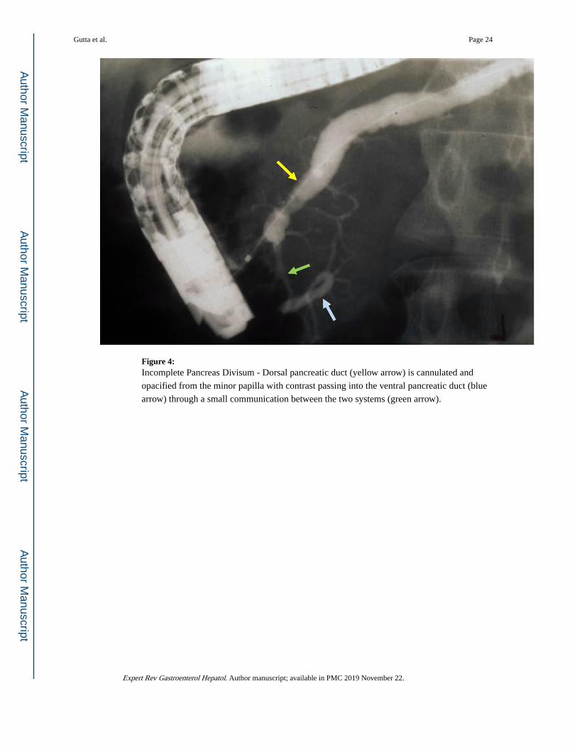
Figure 4: Incomplete Pancreas Divisum - Dorsal pancreatic duct (yellow arrow) is cannulated and
opacified from the minor papilla with contrast passing into the ventral pancreatic duct (blue
arrow) through a small communication between the two systems (green arrow).
Gutta et al. Page 24
Expert Rev Gastroenterol Hepatol. Author manuscript; available in PMC 2019 November 22.
Author M
anuscriptA
uthor Manuscript
Author M
anuscriptA
uthor Manuscript
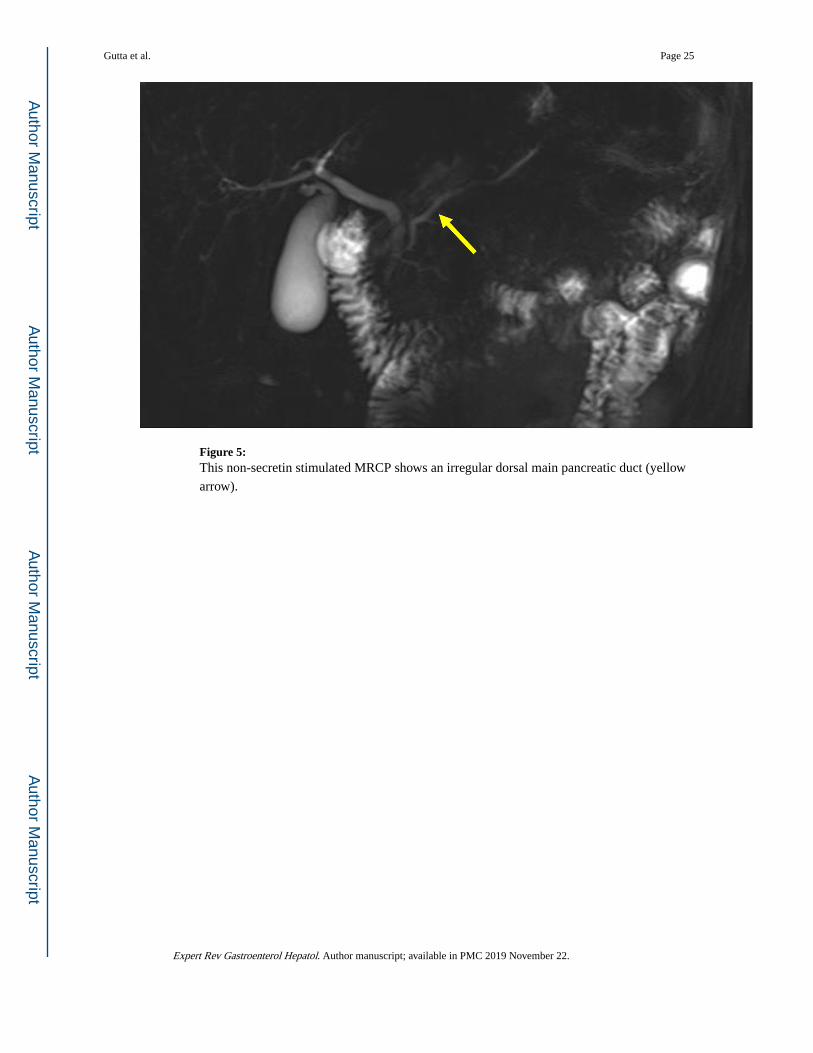
Figure 5: This non-secretin stimulated MRCP shows an irregular dorsal main pancreatic duct (yellow
arrow).
Gutta et al. Page 25
Expert Rev Gastroenterol Hepatol. Author manuscript; available in PMC 2019 November 22.
Author M
anuscriptA
uthor Manuscript
Author M
anuscriptA
uthor Manuscript

Figure 6: This secretin-stimulated MRCP shows a dilated and irregular dorsal pancreatic duct (yellow
arrow) crossing the bile duct and opening separately at the minor papilla (blue arrow).
Irregular side branches are also seen and are more prominent than the non-secretin enhanced
image (Figure 5).
Gutta et al. Page 26
Expert Rev Gastroenterol Hepatol. Author manuscript; available in PMC 2019 November 22.
Author M
anuscriptA
uthor Manuscript
Author M
anuscriptA
uthor Manuscript

Figure 7: Placing the duodenoscope in the long position, along the greater curvature of the stomach
(yellow arrow), provides the optimum angle for minor papilla cannulation and opacification
of the dorsal pancreatic duct (blue arrow).
Gutta et al. Page 27
Expert Rev Gastroenterol Hepatol. Author manuscript; available in PMC 2019 November 22.
Author M
anuscriptA
uthor Manuscript
Author M
anuscriptA
uthor Manuscript

Figure 8: Duodenoscope can be reduced to a short position along the lesser curvature of the stomach
(yellow arrow) once deep minor papilla cannulation is achieved to allow for improved
stability to interrogate and instrument the dorsal pancreatic duct (blue arrow).
Gutta et al. Page 28
Expert Rev Gastroenterol Hepatol. Author manuscript; available in PMC 2019 November 22.
Author M
anuscriptA
uthor Manuscript
Author M
anuscriptA
uthor Manuscript
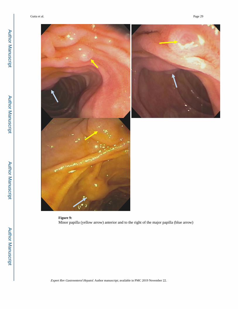
Figure 9: Minor papilla (yellow arrow) anterior and to the right of the major papilla (blue arrow)
Gutta et al. Page 29
Expert Rev Gastroenterol Hepatol. Author manuscript; available in PMC 2019 November 22.
Author M
anuscriptA
uthor Manuscript
Author M
anuscriptA
uthor Manuscript
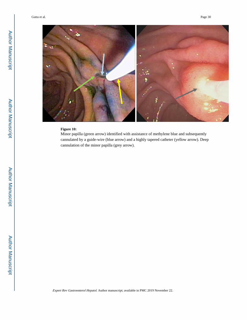
Figure 10: Minor papilla (green arrow) identified with assistance of methylene blue and subsequently
cannulated by a guide-wire (blue arrow) and a highly tapered catheter (yellow arrow). Deep
cannulation of the minor papilla (grey arrow).
Gutta et al. Page 30
Expert Rev Gastroenterol Hepatol. Author manuscript; available in PMC 2019 November 22.
Author M
anuscriptA
uthor Manuscript
Author M
anuscriptA
uthor Manuscript

Figure 11: Minor papilla (green arrow) cannulated by a guide-wire (blue arrow) and a highly tapered
catheter (yellow arrow).
Gutta et al. Page 31
Expert Rev Gastroenterol Hepatol. Author manuscript; available in PMC 2019 November 22.
Author M
anuscriptA
uthor Manuscript
Author M
anuscriptA
uthor Manuscript

Figure 12: Minor papilla (arrow) clearly identified after spraying duodenal mucosa with methylene blue
and administration of secretin to enhance pancreatic secretion.
Gutta et al. Page 32
Expert Rev Gastroenterol Hepatol. Author manuscript; available in PMC 2019 November 22.
Author M
anuscriptA
uthor Manuscript
Author M
anuscriptA
uthor Manuscript

Figure 13: The minor papilla (yellow arrow) is easily identified after spraying the duodenal mucosa
with methylene blue and administration of intravenous secretin. The pancreatic juice flow
results in a zone of central clearing (white arrow).
Gutta et al. Page 33
Expert Rev Gastroenterol Hepatol. Author manuscript; available in PMC 2019 November 22.
Author M
anuscriptA
uthor Manuscript
Author M
anuscriptA
uthor Manuscript

Figure 14: Minor papilla pull-type sphincterotomy.
Gutta et al. Page 34
Expert Rev Gastroenterol Hepatol. Author manuscript; available in PMC 2019 November 22.
Author M
anuscriptA
uthor Manuscript
Author M
anuscriptA
uthor Manuscript

Figure 15: Free hand needle-knife (blue arrow) sphincterotomy (yellow arrow) of minor papilla (green
arrow) followed by placement of a plastic stent (grey arrow) in the dorsal pancreatic duct.
Gutta et al. Page 35
Expert Rev Gastroenterol Hepatol. Author manuscript; available in PMC 2019 November 22.
Author M
anuscriptA
uthor Manuscript
Author M
anuscriptA
uthor Manuscript
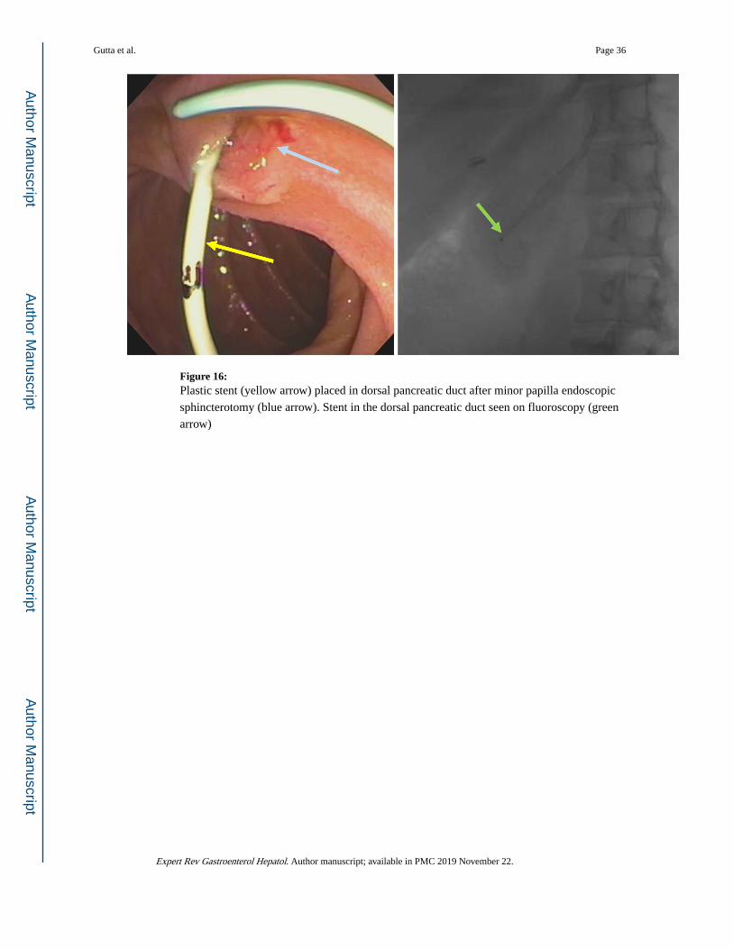
Figure 16: Plastic stent (yellow arrow) placed in dorsal pancreatic duct after minor papilla endoscopic
sphincterotomy (blue arrow). Stent in the dorsal pancreatic duct seen on fluoroscopy (green
arrow)
Gutta et al. Page 36
Expert Rev Gastroenterol Hepatol. Author manuscript; available in PMC 2019 November 22.
Author M
anuscriptA
uthor Manuscript
Author M
anuscriptA
uthor Manuscript
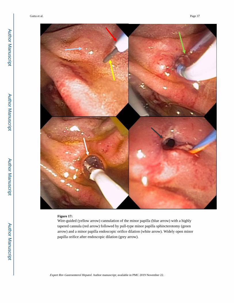
Figure 17: Wire-guided (yellow arrow) cannulation of the minor papilla (blue arrow) with a highly
tapered cannula (red arrow) followed by pull-type minor papilla sphincterotomy (green
arrow) and a minor papilla endoscopic orifice dilation (white arrow). Widely open minor
papilla orifice after endoscopic dilation (grey arrow).
Gutta et al. Page 37
Expert Rev Gastroenterol Hepatol. Author manuscript; available in PMC 2019 November 22.
Author M
anuscriptA
uthor Manuscript
Author M
anuscriptA
uthor Manuscript