adult onset sinonasal rhabdomyosarcoma - a rare … · adult onset sinonasal rhabdomyosarcoma - a...
TRANSCRIPT
169
Adult onset sinonasal rhabdomyosarcoma - a rare case report with cytohistological features
Neelam SOOD MD Pathology and Nisha SEHRAWAT MD Pathology
Department of Pathology and Laboratory Medicine, Deen Dayal Upadhyay Hospital, New Delhi
Abstract
Rhabdomyosarcoma (RMS) is a fast growing, malignant tumour arising from immature mesenchymal cells, committed to skeletal muscle differentiation. It is more often seen in the paediatric population and constitutes less than 1% of all malignancies and less than 3% of all soft tissue tumours. RMS of the paranasal sinuses constitutes 10-15 % of adult head and neck RMS, ethmoidal and maxillary sinuses being the most common. We report a 56-year-oldman presenting with left nasal obstruction, epistaxis on and off and left cheek swelling. Nasal endoscopy revealed a reddish friable mass, bleeding on touch, in the left nasal cavity. CECT scan showed a heterogeneous growth in the left maxillary sinus eroding the medial orbital wall and lateral nasal wall. FNAC of the left cheek swelling yielded highly cellular smears showing predominantly singly scattered round to ovoid neoplastic cells with scanty cytoplasm and indistinct nucleoli. Few of the cells had eccentric nuclei with moderate amount of eosinophilic cytoplasm. Attempted pseudorossette formation was seen. An impression of round cell tumour was given. A diagnosis of an adult onset sinonasal rhabdomyosarcoma was made on histopathological examination of the nasal biopsy, supported by immunohistochemistry (IHC) showing strong myogenin positivity, focal positivity for PAX8 and negativity for CK, LCA, S-100 and CD99. Parameningeal RMS is rare in adults especially the elderly. However, it needs to be considered whenever a poorly-differentiated neoplasm is seen in this age and IHC is a useful aid.
Key words: Rhabdomyosarcoma, cambium zone, parameningeal, small round cell tumour
Address for correspondence: Nisha Sehrawat, B-1/13, DLF Phase 1 Gurgaon, Haryana- 122001, New Delhi, India. Email: [email protected]
CASE REPORT
INTRODUCTION
Rhabdomyosarcoma (RMS) originates from immature mesenchymal cells that are committed to skeletal muscle differentiation. It was first described by Weber in 1854 as a fast growing, malignant tumour. It is more commonly found in children, constituting 6 % of all malignancies below 15 years of age.1 It accounts for less than 1% of all adult malignancies.2 Paranasal RMS comprises 10-15 % of head and neck RMS.3 We report a rare adult alveolar RMS with embryonal RMS-like areas presenting in the sinonasal tract (a parameningeal location) to share its histomorphological features.
CASE HISTORY
A 56-year-old male presented with complaints of left nasal obstruction, epistaxis on and off and left cheek swelling. On nasal endoscopic examination, a reddish friable mass, bleeding on touch was seen in the left nasal cavity. Contrast-
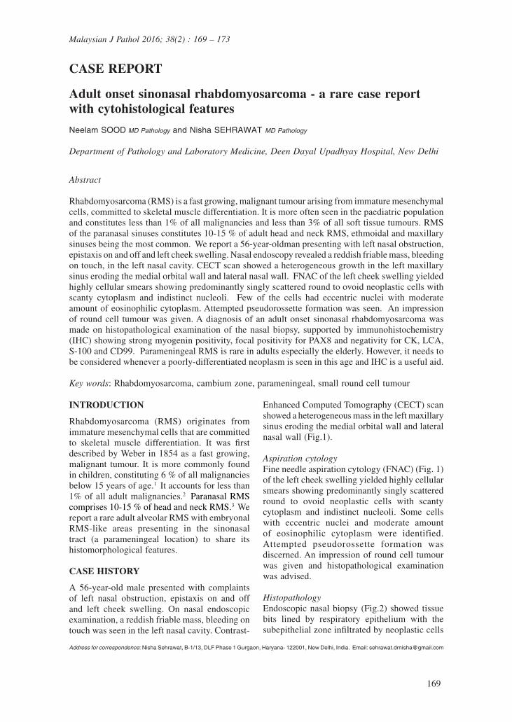
Enhanced Computed Tomography (CECT) scan showed a heterogeneous mass in the left maxillary sinus eroding the medial orbital wall and lateral nasal wall (Fig.1).
Aspiration cytologyFine needle aspiration cytology (FNAC) (Fig. 1)of the left cheek swelling yielded highly cellular smears showing predominantly singly scattered round to ovoid neoplastic cells with scanty cytoplasm and indistinct nucleoli. Some cells with eccentric nuclei and moderate amount of eosinophilic cytoplasm were identified. Attempted pseudorossette formation was discerned. An impression of round cell tumour was given and histopathological examination was advised.
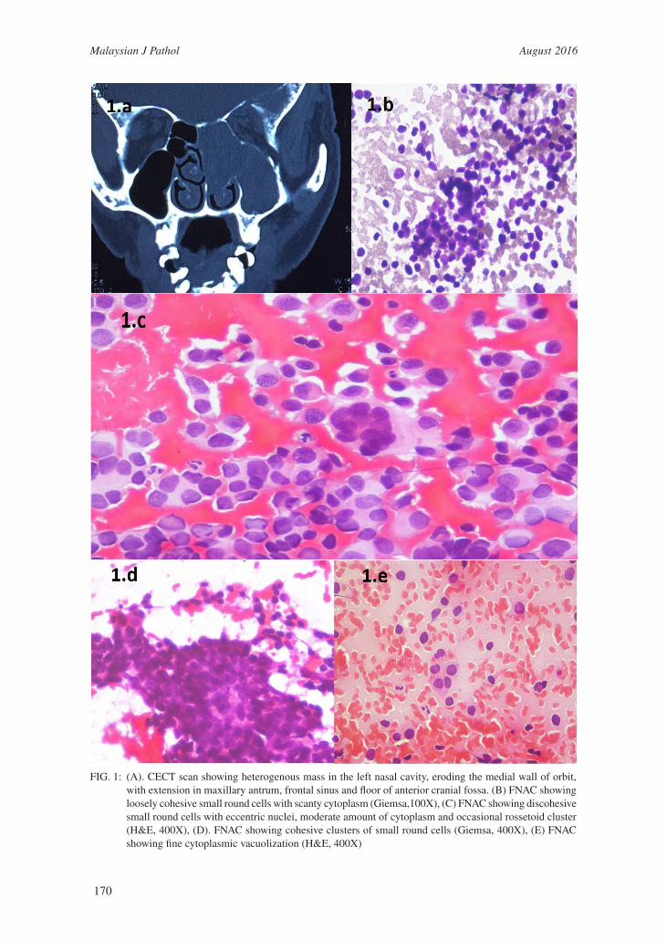
HistopathologyEndoscopic nasal biopsy (Fig.2) showed tissue bits lined by respiratory epithelium with the subepithelial zone infiltrated by neoplastic cells
Malaysian J Pathol 2016; 38(2) : 169 – 173
Malaysian J Pathol August 2016
170
FIG. 1: (A). CECT scan showing heterogenous mass in the left nasal cavity, eroding the medial wall of orbit, with extension in maxillary antrum, frontal sinus and floor of anterior cranial fossa. (B) FNAC showing loosely cohesive small round cells with scanty cytoplasm (Giemsa,100X), (C) FNAC showing discohesive small round cells with eccentric nuclei, moderate amount of cytoplasm and occasional rossetoid cluster (H&E, 400X), (D). FNAC showing cohesive clusters of small round cells (Giemsa, 400X), (E) FNAC showing fine cytoplasmic vacuolization (H&E, 400X)
171
ADULT SINONASAL RHABDOMYOSARCOMA
FIG. 2: (A) Respiratory mucosa (arrow head) with subepithelium densely infiltrated by small round cells (H&E40X). (B) neoplastic cells with abundant clear cytoplasm (H&E 400X), (C) hyperchromatic oval to spindle cells in loose myxoid stroma separated from overlying respiratory mucosa by cambium layer (arrow head). Rhabdomyoblasts also seen. (H&E 400X), (D) diffuse myogenic positivity in more than 70% of cells (red arrow) and respiratory mucosa (black arrow) (IHC for Myogenin FSD IgG1 Kappa RTU, 400X), (E). nuclear positivity for PAX8 in more than 70% cells (IHC for PAX8 BC12 Isotype: IgG1 RTU, 400X)
Malaysian J Pathol August 2016
172
entrapping mucosal glands. A clear cambium zone was identified focally. No characteristic alveolar histology was seen. The neoplastic cells were round to ovoid, medium-sized, mainly occurring in solid pattern with central nuclei, vesicular nuclear chromatin and scanty to moderate amounts of pale vacuolated cytoplasm. Mitosis was infrequent. Occasional multinucleated giant cells were also seen. The subepithelial zone focally showed discohesive ovoid cells with hyperchromatic nuclei, moderate anisonucleosis and focal rhabdomyoblastic differentiation in a loose stroma, with infrequent mitosis. On IHC, tumour cells showed 70% myogenin nuclear positivity and negativity for CK5, CK7, S-100 protein, CD99, GFAP and LCA. PAX 8 staining revealed focal nuclear positivity. A final diagnosis of solid alveolar RMS with areas simulating embryonal RMS was signed out. The patient was referred for further management to a more advanced center but was subsequently lost to follow-up.
DISCUSSION
RMS is a highly malignant sarcoma, seen mostly in the paediatric age group and less commonly in adults and has different presentation in the two age groups.4 Unlike the paediatric age group, adult RMS do not show any male preponderance. Adult RMS has a predilection for the extremities, while paediatric RMS is found predominantly in the head and neck region.4 In adults, RMS constitutes less than 1% of all malignancies and 3% of all soft tissue tumours.2 Only 15 % cases of adult RMS involve the head and neck, of which 10-15 % are seen in paranasal sinusesunlike a 40% involvement in children.3,4
Adult sinonasal RMS has been reported more commonly in those less than 35 years of age, whereas this case was 56 years old.5,6 The risk factors for sinonasal RMS are unknown. Exposure to radiation is one of the postulated factors, which was not noted in the present case.6
RMS is found in three sites in the head and neck region – orbit, parameninges (nasal cavity, paranasal sinuses, nasopharynx, infra-temporal fossa) and superficial (pharynx, scalp, buccal mucosa, parotid, external ear, tonsil and face).7 They commonly present with nasal congestion, rhinorrhea and nasal bleeding. Four histopathological patterns are seen in RMS - embryonal (70%), alveolar (20%), botyroid and pleomorphic (10%). In the sinonasal tract, however, RMS is predominantly
of alveolar type with solid alveolar subtype, mostly comprising undifferentiated tumour cells arranged in solid alveolar pattern surrounded by dense hyalinized stroma. Individual tumour cells are small, round with large hyperchromatic nuclei and scanty pale cytoplasm. Mitosis is common and multinucleated giant cells are prominent. Embryonal RMS (ERMS) shows variable cellularity with alternating hypercellular areas and loosely textured cells in myxoid matrix. Tumour cells are seen infiltrating the submucosa with overlying clear zone between mucosa and tumour cells, known as the cambium layer, as seen in this case. Individual tumour cells are round or spindle. These cells are immature with scanty cytoplasm and indistinct nucleoli. There is lack of cohesion.6
In cases where features of myogenesis are absent, immunohistochemistry (IHC) and RT-PCR are useful. RMS shows positivity for myogenin and desmin and negativity for cytokeratin, epithelial membrane antigen, CD45, CD 99 and S-100 protein.7,8 Alveolar RMS (ARMS) predominantly shows t(2;13) (q35;q14) translocation leading to formation of PAX3/FKHR fusion transcript or t(1;13)(p36;q14) translocation forming PAX7/FKHR fusion transcript, but could not be established in this case.6,7 Paired box gene antibody (PAX2, PAX5, PAX8) have been studied for differentiation of poorly-differentiated round cell tumour and it was found that PAX8 expression is noticed in 5/5 and 3/6 cases of ARMS and ERMS respectively.9 On the other hand Sullivan et al reported 67% immunoreactivity for PAX 5 in ARMS, whereas it was negative in all cases of ERMS.10 One antibody alone amongst the PAX group cannot be reliably used for differentiating between the two subtypes of RMS, which can be a limiting factor in laboratories in a resource poor setting. Ahmad and Tsokos reported 14 cases of sinonasal RMS with 1 case of ERMS and the rest being ARMS.7 Montone et al reported 13 cases of adult parameningeal RMS with 3 cases of ERMS, 9 ARMS and 1 unclassified.6 Our case is a rare ARMS with ERMS like areas presenting in the sinonasal tract. The presence of solid alveolar tumour cells beneath the sinonasal mucosa may give a false impression of acambium layer, posing diagnostic problems in differentiating it from embryonal RMS. In such cases, myogenin positivity is helpful, with ERMS having <50% positivity vs diffuse nuclear reactivity in ARMS.6
Differentiating embryonal RMS from
173
ADULT SINONASAL RHABDOMYOSARCOMA
alveolar RMS can be difficult on histology alone particularly in a biopsy, as in this case. The absence of an alveolar pattern, desmoplasia and presence of cambium layer, accompanied by low mitotic activity were the dilemmas but more diffuse myogenin positivity, a feature of alveolar RMS, was contributory in reaching the diagnosis of ARMS. However, FISH findings was not available for confirmation. The prognosis depends on the primary site, histological subtype and tumour size. A favorable prognosis is seen in tumours less than 5 cm, patient age < 20 years, lack of regional or distant metastasis and negative surgical margins. Adult RMS has a poor prognosis as it is usually advanced at presentation, with extensive local involvement making complete surgical excision difficult, as in our case. The 5-year survival rate is only 8 % in head and neck RMS.3 RMS in a parameningeal location such as the nasal cavity poses a significant risk of subarachnoid dissemination. Treatment of RMS can be radiation or chemotherapy.7
In conclusion, although parameningeal RMS is rare in adults especially in the elderly, it needs to be considered whenever a poorly-differentiated neoplasm is seen in this age group and IHC is a useful aid.
REFERENCES
1. Stuart A, Radhakrishnan J. Rhabdomyosarcoma. Indian J Pediatr. 2004; 71: 331-7.
2. Weiss SW, Goldblum JR. Rhabdomyosarcoma. In: Weiss SW, Goldblum JR, editors. Enzinger and Weiss’s soft tissue tumors. 4th ed. St. Louis: CV Mosby; 2001. p. 785-835.
3. Wu TH, Huang JS, Wang HM, Wang CH, Yeh KY. Long-term survivors of adult rhabdomyosarcoma of maxillary sinus following multimodal therapy: case reports and literature reviews. Chang Gung Med J. 2010; 33: 466-71.
4. Erkul E, Pinar D, Yilmaz I, Cincik H, Cekin E, Salihoglu M. Rare adult sinonasal embryonal rhabdomyosarcoma with optic involvement. Otolaryngology. 2012; 2: 118.
5. Manucha V, Castellani R, Sun CC. Alveolar rhabdomyosarcoma of the paranasal sinuses in a 57-year-old women with 1:16 translocation. Int J Surg Pathol. 2006; 14: 238-42.
6. Montone KT, Barr FG, Zhang PJ, Feldman MD, LiVolsi VA. Embryonal and alveolar rhabdomyosarcoma of parameningeal sites in adults: a report of 13 cases. Int J Surg Pathol. 2009; 17: 22-30.
7. Ahmad AA, Tsokos M. Sinonasal rhabdomyosarcoma in children and young adults. Int J Surg Pathol. 2007; 15: 160-5.
8. Kumar S, Perlman E, Harris CA, Raffeld M,
Tsokos M. Myogenin is a specific marker for rhabdomyosarcoma: an immunohistochemical study in paraffin-embedded tissues. Mod Pathol. 2000; 13: 988-93.
9. Fan R. PAX immunoreactivity in poorly differentiated small round cell tumors of childhood. Fetal Pediatr Pathol. 2014; 33: 244-52.
10. Sullivan LM, Atkins KA, LeGallo RD. PAX immunoreactivity identifies alveolar rhabdomyosarcoma. Am J Surg Pathol. 2009; 33: 775-80.