advance dental simulation module on crown …...step by step procedure in completing full crown...
TRANSCRIPT
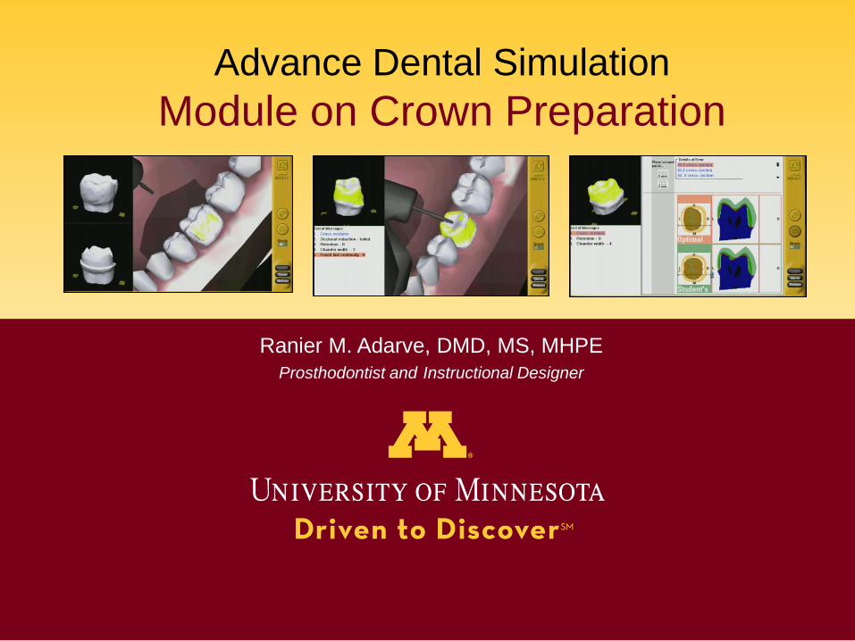
Advance Dental Simulation
Module on Crown Preparation
Ranier M. Adarve, DMD, MS, MHPE
Prosthodontist and Instructional Designer

Introduction
Welcome! This module is developed to guide dental
students in preparing Full Crown restoration using the
Advance Dental Simulation Technology-DentSim®.
The second part of the module covers the suggested
step by step procedure in completing full crown
preparation.
The first part of the module covers the Principles of
Preparation for Gold Crown Restoration for Tooth #19
(36) as prescribed by DentSim ®.
At the end of the module, students are required to
complete the test to evaluate performance.

Objectives
Upon completion of this module, you will be able to:
a. Understand the Principles of Preparation
b. Identify the specific dimensions, forms and
measurements of tooth reductions.
c. Prepare accurately tooth #19 to receive
Gold Crown restoration.

Part 1
Principles of Preparing
Tooth #19
for Gold Crown Restoration
Tips
• There are 16 Principles of Tooth Preparation for Gold
Crown Restoration as prescribed by the DentSim®.
• Each of the principle will be listed in chronological order.
• More information will be provided by clicking the info
button
• Use the forward arrow to navigate through the course
and backward arrow if you need to review the
previous slide.
• You may need to take down notes to remember all the
dimensions and angulations for tooth reduction. Good
luck!
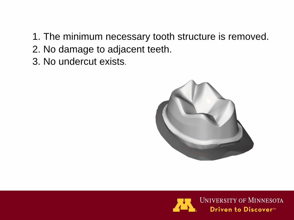
1. The minimum necessary tooth structure is removed.
2. No damage to adjacent teeth.
3. No undercut exists.

4. In the occlusal surface appear 5 cusps:
ML, DL, MB, B, and DB.
These cusps follow the original contour of the
tooth cusps and preserve the occlusal anatomy.

5. The tips of the cusps are located under
the tips of the original cusps of the tooth.

6. Occlusal reduction:
B cusps - 1.5 mm
L cusps – 1.0 mm

1.0
7. Functional cusps beveling is located on
the buccal cusps, its width 1.0mm.

8. Taper of opposing walls:
The M and D walls have taper of 4-6°.
The B and L walls have taper of 4-6°.

9. Height of contour of the preparation walls
is located parallel to the original height of
contour of the enamel surface.
10.The buccal walls have 3 surfaces:
from the margin to the HOC, from the HOC
to the Functional Bevel, and the Functional Bevel.
Height of Contour
(HOC)
Height of Contour
(HOC)
1
2
3

Height of Contour
(HOC)
1
2
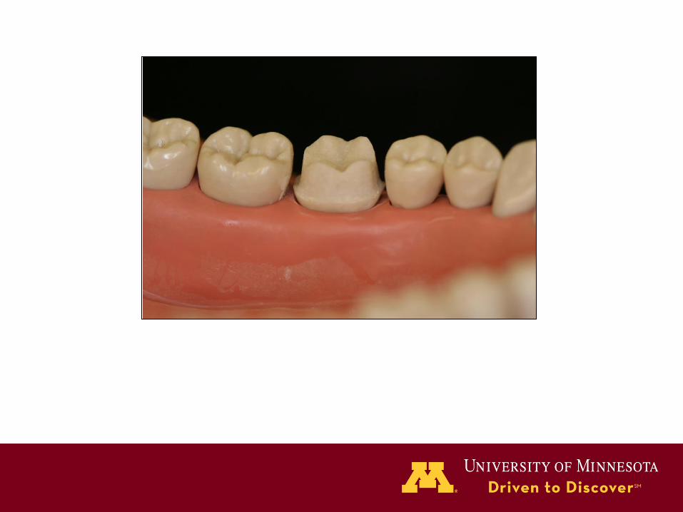
11. The lingual cusps have 2 surfaces:
from the margin to the HOC,
and from the HOC to the tip of the cusp.
12. The gradual change in the surfaces begins
between the transitional line angles.
13. All the line angles of the walls and the cusps
are rounded. All walls are smooth.

14. Marginal geometry: chamfer configuration.
The anatomy of the chamfer was created
according to the shape of a round-end taper
diamond bur, 0.8mm at its tip.
15. Axial reduction / chamfer width:
Buccal chamfer width – 0.5 mm
Lingual chamfer width - 0.5 mm.

16. Margins are located 1.0mm occlusal
to the free gingival line.
As a result of the margin location,
clearance is left from the adjacent teeth.

Procedure Evaluation
Test Grade
Damage to adjacent
tooth
Point reduced only in run-
time
Pulp exposure Point reduced only in run-
time
Undercut 10
Occlusal reduction 12
Wall incline 16
Retention 10
Resistance (hinge
movement)
10
Wall smoothness 10
Margin location 6
Margin width 10
Finish line continuity 10
Interproximal
clearance
6
Passing grade 60

Part 2
Step by Step Procedure

Tips
• There are 9 suggested step by step procedures.
• For beginners, it is best to follow the recommended
sequence. Otherwise, you may proceed in the order that
you are most comfortable with.
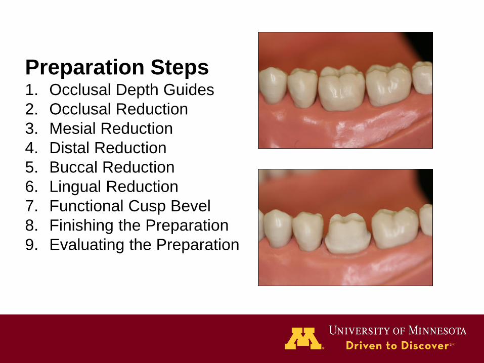
Preparation Steps 1. Occlusal Depth Guides
2. Occlusal Reduction
3. Mesial Reduction
4. Distal Reduction
5. Buccal Reduction
6. Lingual Reduction
7. Functional Cusp Bevel
8. Finishing the Preparation
9. Evaluating the Preparation

Step 1
Occlusal Depth Guides


Step 2
Occlusal Reduction


Step 3
Mesial Reduction


Step 4
Distal Reduction


Step 5
Buccal Reduction


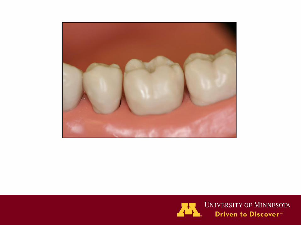
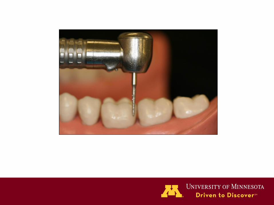
Step 6
Lingual Reduction


Step 7
Functional Cusp Bevel


Step 8
Finishing the Preparation


Step 9
Evaluating the Preparation


