advanced image processing methods applied to digital ... · transfer of knowledge the aidpath...
TRANSCRIPT

Advanced Image Processing
Methods Applied to Digital
Pathology. AIDPATH
Gloria Bueno
E.T.S.I. Industriales - UCLM
http://visilab.etsii.uclm.es

2

Digital PathologyEuroTelepath
COST Action Project

Partners Sector Country
1. UNIVERSIDAD DE CASTILLA-LA
MANCHA
Public SPAIN
2. ASTRAZENECA UK LIMITED Commercial UK
3. BARCO NV Commercial BELGIUM
4. TISSUEGNOSTICS GMBH Commercial AUSTRIA
5. LEICA MICROSYSTEMS CMS
GMBH
Commercial IRELAND
GERMANY
6. UNIVERSITA DEGLI STUDI DI
UDINE
Public ITALY
7. UNIVERSITATEA DE MEDICINA SI
FARMACIE GR.T.POPA IASI
Public ROMANIA
8. THE UNIVERSITY OF
NOTTINGHAM
Public UK
9. SERVICIO DE SALUD DE CASTILLA
LA MANCHA & ANDALUZ
Public SPAIN
10. VIESOJI ISTAIGA VILNIAUS
UNIVERSITETO LIGONINES
SANTARISKIU KLINIKOS
Public LITHUANIA
11. LOUGHBOROUGH UNIVERSITY Public UK
Partners
4

Transfer of Knowledge
The AIDPATH Supervisory Board takes overall charge of the researcher’s personalcareer development (PCD), knowledge transfer and training needs and activities.
Inter-host Secondments & Training Programs

Secondments Newly Recruited Researchers
ERS ER(<10 years)
ER( >10 years)
ER(<10 years)
ER(>10 years)
Part
icip
an
t N
am
e
Tota
l R
esea
rch
er-
mo
nth
s
Res
earc
her
s
Tota
l R
esea
rch
er-
mo
nth
s
Res
earc
her
s
Tota
l R
esea
rch
er-
mo
nth
s
Res
earc
her
s
Tota
l R
esea
rch
er-
mo
nth
s
Res
earc
her
s
Tota
l R
esea
rch
er-
mo
nth
s
Res
earc
her
s
UCLM 0 0 8 3 4 2 12 1 0 0
AZ 20 5 12 4 15 5 24 1 0 0
BAR 18 4 4 2 8 4 24 1 0 0
TG 18 4 9 4 8 4 24 1 0 0
LEICA 4 2 0 0 6 3 12 1 0 0
UNIUD 0 0 2 1 3 1 12 1 0 0
UMF 0 0 2 1 2 1 12 1 0 0
UNOTT 0 0 8 3 2 1 12 1 0 0
SESCAM 0 0 2 1 6 3 12 1 0 0
VUHSK 0 0 2 1 6 3 12 1 0 0
LU 0 0 6 3 0 0 12 1 0 0
Total 60 15 55 23 60 27 168 11 0 0
76 researchers.

7
• 4 years
• 3.000.000 €
• 11 partners
• 76 researchers

Introduction to Barco Healthcare
• Image Processing & Analysis
• Standards
• Image Quality Control
• Biomarker Analysis
8

Introduction to Barco Healthcare
• Clinical Validation
• Quality Control
• Database Annotation
Pathologist
9

Tools already Implemented
•Goals: To research and develop novel medical image displaytechnology, processing and standards for digital pathology.
•Tools Developed:• Web viewer• Metrics for image quality• Tools for Image Processing• Detection and Classification Tools

Web Viewer TelePath Viewer - VISILAB (c)
A Web Viewer to explore microscopic images has beenimplemented by VISILAB Group. The viewer is able to upload alltype of images including .svs format, visualize the image atdifferent magnifications, create annotations and select regions ofinterest. The size of microscopic images obtained from wholeslide tissue samples are usually larger than 1GB, therefore it isimpossible to visualize them with a common software. TelePathViewer is able to work with this images, providing a useful tool tobe used in Digital Pathology.

Web Viewer TelePath Viewer - VISILAB (c)
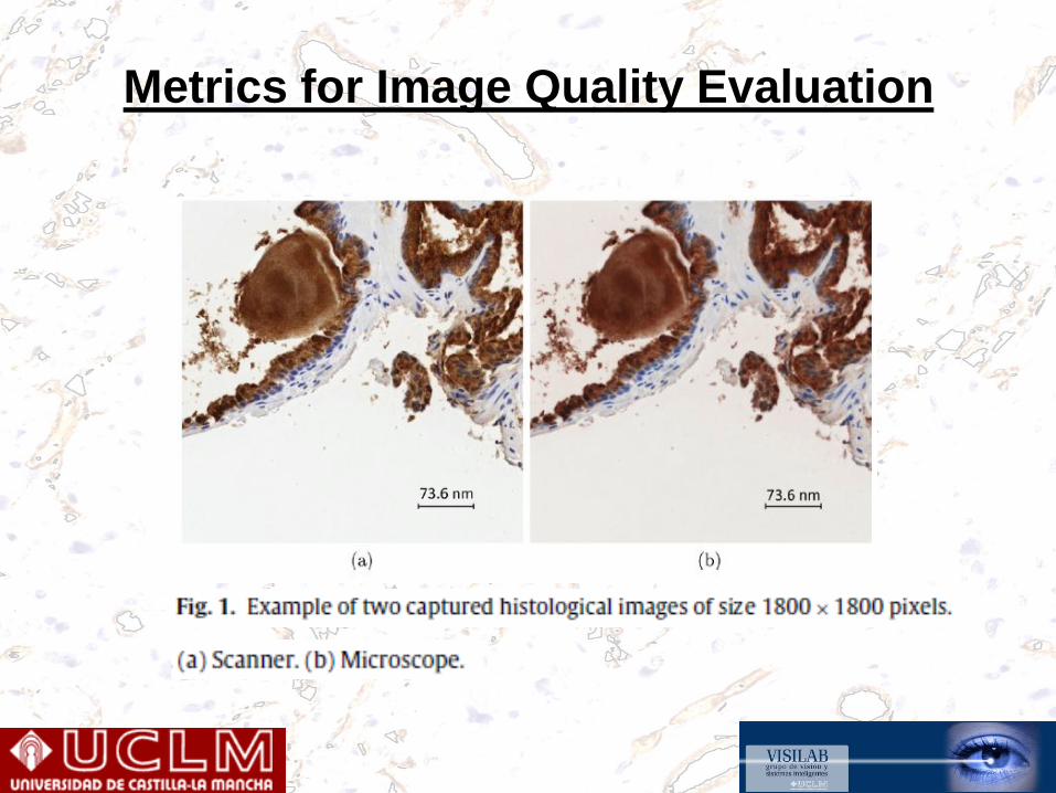
Metrics for Image Quality Evaluation

Digital Pathology

∆x?, ∆y?
• Stitching
Original scanned images
Advanced Image Processing

Advanced Image Processing
Serial section alignment

• Registration result: HE+ER
Advanced Image Processing

• Registration result: HER2 + ER
Advanced Image Processing

ANGIOPATH – VISILAB (c)
ANGIOPATH is a morphometric tool allowing us to measure differentaspects of the shape and size of vascular vessels in a complete andaccurate way. The developed tool detect and close all vesselsproviding useful parameter for angiogenesis research. Therefore,ANGIOPATH is based on vessel closing which is an essential propertyto properly characterize the vascular and lymphatic vessels.http://dx.doi.org/10.1155/2013/263190

ANGIOPATH – VISILAB (c)

CORE DETECTION AND EXTRACTION
This tool allows automatic segmentation and archiving of tissuemicroarray (TMA) cores in microscopy images at differentmagnifications. A crucial step to improve the speed and quality ofTMA analysis is the correct localization of each tissue core in thearray. However, usually the tissue cores are not aligned in themicroarray, the TMA cores are incomplete and the images are noisyand with distorted colors. Need for a robust tool able to workunder these conditionshttp://dx.doi.org/10.1109/JBHI.2013.2282816

Advanced Image Processing

Advanced Image Processing

Advanced Image Processing
CORE CLASSIFICATION
The TMA system core viewer
Final TMA core image

Advanced Image Processing

• The challenge is to exploit the new and emerging digital pathology technologies effectively in order to process and model all the heterogeneous tissue-derived data.
• This requires joint research projects and collaborative programmes between academia and industry. Thus, biomedical scientists will be equipped with broad knowledge and tools of modern imaging and data processing, whereas engineers with have an understanding of the complex disease processes and the clinical needs.
• This will help developing efficient and innovative products to fulfil the needs of digital pathology. The AIDPATH project addresses this challenge
Conclusion

THANKS
http://visilab.etsii.uclm.es
http://aidpath.eu