advanced review systems analysis of biological networks in...
TRANSCRIPT
Advanced Review
Systems analysis of biologicalnetworks in skeletal musclefunctionLucas R. Smith,1 Gretchen Meyer1 and Richard L. Lieber2,3∗
Skeletal muscle function depends on the efficient coordination among subcellularsystems. These systems are composed of proteins encoded by a subset of genes, allof which are tightly regulated. In the cases where regulation is altered because ofdisease or injury, dysfunction occurs. To enable objective analysis of muscle geneexpression profiles, we have defined nine biological networks whose coordinationis critical to muscle function. We begin by describing the expression of proteinsnecessary for optimal neuromuscular junction function that results in the musclecell action potential. That action potential is transmitted to proteins involvedin excitation–contraction coupling enabling Ca2+ release. Ca2+ then activatescontractile proteins supporting actin and myosin cross-bridge cycling. Forcegenerated by cross-bridges is transmitted via cytoskeletal proteins through thesarcolemma and out to critical proteins that support the muscle extracellularmatrix. Muscle contraction is fueled through many proteins that regulate energymetabolism. Inflammation is a common response to injury that can result inalteration of many pathways within muscle. Muscle also has multiple pathwaysthat regulate size through atrophy or hypertrophy. Finally, the isoforms associatedwith fast muscle fibers and their corresponding isoforms in slow muscle fibersare delineated. These nine networks represent important biological systems thataffect skeletal muscle function. Combining high-throughput systems analysis withadvanced networking software will allow researchers to use these networks toobjectively study skeletal muscle systems. © 2012 Wiley Periodicals, Inc.
How to cite this article:WIREs Syst Biol Med 2013, 5:55–71. doi: 10.1002/wsbm.1197
INTRODUCTION
Skeletal muscle’s primary function is to generate forceand produce movement. This requires coordinationamong many physiological pathways and their asso-ciated components. Loss of skeletal muscle functionbecause of disease results from altered transcriptionalpathways that have responded to mechanical, bio-logical, and chemical stimuli. Thus, understanding
∗Correspondence to: [email protected] of Bioengineering, University of California, San Diego,La Jolla, CA, USA2Department of Orthopaedic Surgery, University of California, SanDiego, La Jolla, CA, USA3Department of Orthopaedic Surgery, V.A. San Diego HealthcareSystem, San Diego, CA, USA
components that regulate muscle function is a pre-requisite to understanding mechanisms of musclepathology. This review highlights the components thatare most critical to muscle function and places themin a context of muscle physiology as a whole usingcurrent reviews in muscle physiology and muscle geneontology.1 We do not provide an exhaustive list ofgenes and proteins that regulate muscle function, butrather explore how various pathways are distorted ina variety of muscle pathologies and the downstreamconsequences of altered gene expression. The net-works created here provide a foundation from whichto build more detailed and specific networks. The net-works have been created in a Cytoscape (Cytoscape2.8)2 for use in the interpretation of current high-throughput and system level technologies such asmicroarrays3 and protein arrays.4 This work will
Volume 5, January/February 2013 © 2012 Wiley Per iodica ls, Inc. 55

Advanced Review wires.wiley.com/sysbio
also be useful to provide a general reference forstudying the interaction between transcription andmuscle function.
SYSTEMS OVERVIEW
Proper muscle function requires coordination of manyintegrated biological networks. Muscle contraction(MC) is initiated at the specialized neuromuscularjunction (NMJ) where acetylcholine (ACh) releasefrom the nerve ending triggers an action potential. Theaction potential propagates across the sarcolemmaand into the transverse tubules to initiate calciumrelease from the sarcoplasmic reticulum (SR) in theprocess known as excitation–contraction coupling(ECC). Calcium binding to regulatory proteins onthe thin filament triggers the myosin cross-bridgecycle that creates MC. MC force is transmittedthrough specialized networks of proteins within thecell cytoskeleton (CYSK) to the costameres and outto the extracellular matrix (ECM). Myosin cross-bridge cycling requires ATP, and thus skeletal musclefunction also requires metabolism (MET) and storageof carbohydrates and fatty acids. A variety of damageparadigms may cause an inflammatory (INF) responsein muscle. With chronically altered use, muscleadapts by coordinating a change in muscle mass viasynchronized muscle hypertrophy or atrophy (HA).Most aspects of these muscle networks are slightlytuned in the different kinds of muscle cells known asfiber types (FTs).
Understanding muscle diseases requires knowl-edge of the protein used for force production.Duchenne muscular dystrophy (DMD) is the mostfrequently studied muscle disease, and although itresults from the loss of a single gene product, dys-trophin, many muscle functions are compromised.5
Dystrophin is part of the costamere complex that linksMC to the ECM and, when disrupted, allows mechan-ically induced membrane damage.6 This allows cal-cium influx that contributes ECC alterations andmuscle degradation and damage7 and is associatedwith a large INF response8,9 as well as cycles ofregeneration.10 As the HA pathway is exhausted, themuscle undergoes an increase in ECM fibrosis.11 Thisresponse illustrates the interconnectivity among mus-cle subsystems and demonstrates how understandinga single protein’s role is ultimately critical to under-standing muscle pathology. This review provides aframework for those investigating muscle diseaseand adaptation to efficiently inspect muscle functionas a system of related proteins, especially to takeadvantage of the high-throughput technologiescurrently available.
Neuromuscular JunctionAt the NMJ, the motor neuron provides the initia-tion signal to the muscle and does so rapidly andtransiently, necessitating an off switch. NMJ functionrequires coordination of many transcripts that areexpressed primarily or exclusively local to the NMJ(Figure 1). MC is initiated by ACh release from themotor neuron, which crosses the synaptic basal laminato bind to the nicotinic ACh receptor (CHRN), whichconsists of five subunits.12 The γ subunit (CHRNG)is expressed in immature muscle and replaced by theε subunit upon maturity.13 CHRN is a ligand-gatedchannel that allows sodium influx upon binding tocreate an endplate potential. When sufficient AChbinding allows the endplate potential to reach thresh-old, muscle sodium channels (SCN4A) are activatedto create an action potential, which propagates acrossthe sarcolemma and throughout the muscle.14
Neurons communicate their ‘connection’ to mus-cle cells and maintain proper clustering of the proteinsrequired for transmission in the postsynaptic muscle.This is performed with motor neuron release of agrin(AGRN). AGRN binds to a receptor on the muscletransmembrane receptor MUSK along with its extra-cellular coreceptor LRP4.12 MUSK binding eventuallyresults in the activation of RAPSN, which acts as anintracellular scaffold for CHRN. MUSK also inter-acts with 14-3-3γ (YWHAG), a signal transductionprotein involved in synaptic gene expression. Synapticgene expression is also mediated through neuregulin(NRG1), a glycoprotein that binds to a family epider-mal growth factor receptor (ERBB). ERBB binding toNRG1 results in the activation of GABP transcriptionfactors for synaptic genes.13 This provides a methodfor nuclei in close proximity to the NMJ to act incoordination and turn on the genes required for theNMJ within a multinucleated muscle fiber.
The synaptic basal lamina plays an importantrole in organizing the NMJ by maintaining a shortdistance for ACh to diffuse across and to be quicklyprocessed by maintaining a unique subset of proteinsthat are expressed preferentially in the region. Thebasal lamina is primarily composed of COL4 with theα3–6 subunits, instead of the α1–2 subunits foundaround the rest of muscle, and laminin subunitsLAMA5 and LAMB2.12 Collagen and lamininnetworks are linked by the glycoprotein NID1.Perlecan (HSPG2) is the major proteoglycan presentat the synapse. The synaptic basal lamina also includesCOLQ, which, along with HSPG2, binds and anchorsacetylcholine esterase (ACHE) to the NMJ. Thefunction of ACHE is to hydrolyze ACh that terminatesmuscle fiber activation.14 ACHE begins hydrolyzingACh almost immediately upon release so as to limit
56 © 2012 Wiley Per iodica ls, Inc. Volume 5, January/February 2013

WIREs Systems Biology and Medicine Biological networks in skeletal muscle function
MotorNeuron
Motor Neuron
NRG1 AGRN
LRP4
ACh ACHE
COLQ NID1
Action potentialCHRNMUSK
GABP YWHAG RAPSN Immaturemuscle Cytoskeleton
ERBBSarcolemma
SynapticNucleus
Synaptic geneexpression
UTRNITGB1CHRNG
COL4Synaptic basallamina
HSPG2 LAMA5 LAMB2
FIGURE 1 | Neuromuscular junction (NMJ). Motor neurons release the neurotransmitter acetylcholine, which triggers an action potential. NMJformation is also induced by motor neuron factors that signal muscle proteins. For all figures, we use the following node conventions: (bluecircle) entrez gene symbols, (blue circle within grayed box) complexes, (blue triangle) non-protein molecules, (blue square with roundededges) modules or functions. There are the following interactions: (plus in white circle) positive, (minus in red circle) negative, (black circle) binding,(double back slash) intermediate, and (question mark with circle) unknown. There are the following lines: (solid thick line) basic, (solid thick line witharrow) A proceeds to B, (horizontal thin line with vertical right end thick line) A does not proceed to B, and (curved arrow) translocation of A. Geneswithin complexes are listed in Table S1, Supporting Information. Some complexes are interacting proteins; however, others are multiple isoforms of aprotein that have the same function.
transmission time and allow the neuron to take up theproducts necessary to resynthesize ACh efficiently.
Excitation–Contraction CouplingThe endplate potential generated at the NMJ activatesmuscle voltage-gated sodium channels (SCN4A)to transform the focal endplate potential into aglobal muscle cell action potential, which propagatesthroughout the muscle and to the interior of themuscle via the transverse-tubule (T-tubules) system(Figure 2). Translation of the action potential intoan intracellular Ca2+ signal for contraction takesplace at the junction between T-tubules and theSR where the terminal cisternae permit ECC.14 TheSR is held in close approximation to the T-tubulesystem by the linking protein junctophilin (JPH1).15
The action potential activates the voltage-gated Ca2+channel DHPR, which consists of five subunits. Thecoupling of DHPR on T-tubules and the ryanodinereceptor RYR1 on the SR ensures that Ca2+ enteringthe cell triggers opening of RYR1 to release Ca2+from the SR store.16,17 A unique ryanodine receptor(RYR3) is expressed in immature muscle and isreplaced by RYR1 during development.18 Ca2+ serves
as the intracellular trigger for MC, but must betightly regulated for efficient force production. Toaccomplish this RYR1 has multiple proteins thatmodulate Ca2+ release. FKBP1A is an SR proteinthat directly interacts with RYR1 and is required forfull conductance16; S100A1 also binds to RYR1 toincrease open probability,19 and SYLP2 acts fromthe T-tubules to increase open probability withoutincreasing current amplitude.18 Aberrant regulation ofRYR1 can occur as a reaction to common anestheticsproducing life-threatening malignant hyperthermia inwhich muscle activity overwhelms the body. Thestandard treatment for malignant hyperthermia isdantrolene sodium, which inhibits RYR1 activity.20
Coordinated movement requires controlled mus-cle relaxation as well, which can be the rate-limitingstep and which requires a surprisingly large portionof the cellular energy supply. For muscle relaxation tooccur, Ca2+ is pumped back into the SR via theATP-dependent SERCA pumps (ATP2A), whichhave different isoforms for fast and slow muscle18
(Figure 2). Fast muscle fibers have faster relaxationtimes to permit higher frequency contraction. Addi-tionally, fast fibers contain more abundant PVALB,
Volume 5, January/February 2013 © 2012 Wiley Per iodica ls, Inc. 57

Advanced Review wires.wiley.com/sysbio
T-T
ubul
e
SCN4A
Action potential
FKBP1A S100A1
Sarcolemma
Sarcoplasmicreticulum
DHPR RYR1 Ca2+ATP2A
SYPL2 RYR3
TRDN
JPH1
ASPH
CASQPLNSLN
PVALB
Ca2+
Ca2+
TRPC
CAPN CALM1
Musclecontraction
CAMK2 PPP3CA
Sarcoplasmicreticulum
Immaturemuscle
T-Tubule
FIGURE 2 | Excitation–contraction coupling. Action potentials travel into the T-tubule system and induce Ca2+ release from the sarcoplasmicreticulum (SR) through the ryanodine receptor. Intracellular Ca2+ triggers muscle contraction and is then pumped back into the Sr. Ca2+ also plays arole in several critical intracellular signaling pathways that regulate muscle mass and muscle fiber type.
which acts as an intracellular Ca2+ buffer by bindingfree Ca2+.21 Conversely, muscle also has the capabil-ity to limit relaxation times through SR proteins SLNand PLN, which, when dephosphorylated, slow relax-ation by inhibiting ATP2A reuptake of Ca2+.18,22 TheSERCA pumps must work against a higher concentra-tion gradient of calcium, but are facilitated by Ca2+-binding proteins within the SR, CASQ, which also hasdifferent isoforms in fast and slow muscle (Figure 9).CASQ is held within the SR and near RYR1 by luminalproteins triadin (TRDN) and junctin (ASPH).
Aside from initiating contraction, Ca2+ may alsoplay a signaling role in muscle. In some disease states,Ca2+ activates CAPN, a family of proteolytic enzymesimportant in muscle.23 Through the activation ofcalmodulin (CALM1) Ca2+ can also activate manygrowth and metabolic responses that are discussed insubsequent sections through CAMK2 or calcineurin(PPP3CA) activation.21 Sarcolemmal channels such asTRPCs, which allow Ca2+ into the cell, also contributeto activation of these Ca2+ pathways.24 These path-ways can link muscle activity patterns to intracellular
signaling mechanisms that control cell fate. The broadactions of Ca2+ as both contraction initiator andadaptation controller highlight the critical importanceof maintaining proper Ca2+ levels and localizationwithin a normal muscle cell.
Sarcomere ContractionThe basic functional unit of MC is the sarcomere,where force generation is produced by interactionbetween thick and thin filaments25,26 (Figure 3). Thethick filament is made up primarily of type 2 myosin,which contains two myosin heavy chain (MYH) andfour myosin light chain (MYL) subunits.27 The myosinhead interacts with the thin filament by binding toactin (ACTA) in a force-generating mechanism andis termed as the cross-bridge cycle. To control thebinding of MYH to ACTA the thin filament has aset of regulatory proteins. Tropomyosin (TPM) wrapsaround the thin filament and obscures the MYH-binding pocket of ACTA filaments. The position ofTPM is regulated by the troponin (TN) complex,
58 © 2012 Wiley Per iodica ls, Inc. Volume 5, January/February 2013

WIREs Systems Biology and Medicine Biological networks in skeletal muscle function
TNNC
TNNIThin filament Thin filament
Thick filament
Thick filament
Half sarcomere
TNNT
ACTA
TMOD NEB MYPN
CAPZ
ACTN
TCAPTTNMYOM
MYH
MYBPC
MYL5
MYL4MYH8 MYH3
Immaturemuscle
MYL
Z-disc
MYOT
TPM
ATPMuscle
contraction
Ca2++
+
+
+
+
−
−
FIGURE 3 | Muscle contraction (MC). Myosin binds to actin and undergoes cross-bridge cycling to produce contractile force. Myosin (thick) andactin (thin) filaments slide past each other during contraction. Sarcomeres are separated by Z-discs. The force generated by the myosin cross-bridgepowers active MC.
which consists of three subunits.28 Regulation ismediated by Ca2+ binding to TNNC, which removesthe inhibitory subunit TNNI from its position withTPM and both subunits are anchored to the thinfilament by the third TNNT.29 MYH is an ATPasethat requires ATP for the release phase of the cross-bridge cycle. Deficits of ATP cause the muscle to stiffeninto a state of rigor, where cross-bridges attach but donot cycle, such as occurs after death. Myosin-bindingproteins (MYBPC) are important for thick filamentformation and structural maintenance.
The functional components of the sarcomererequire dynamic coordination. Thus, the distinctproperties of muscle FTs require separate proteinisoforms for optimal function. MYH is the majordeterminant of FTs with slow fibers characterized bymyosin heavy chain I (MYH7) and is more oxidativeand used for repetitive contractions. Fast fibersare generally larger, more glycolytic, and requiredfor brief high force contractions.30 Fast fibers inhumans have two myosin heavy chain isoformsexpressed in mature muscle, myosin heavy chain IIa(MYH2), from fast fibers with oxidative capacity, andmyosin heavy chain IIx (MYH1), from the fastestmost glycolytic fibers. Both MYL and MYBPC havedifferent isoforms corresponding to FT that alsoeffect cross-bridge cycling rates and force production.Differential regulation of MYH binding by the thinfilament regulatory proteins also requires distinct
isoforms of TPM and TN. MYH and MYL alsohave particular isoforms (MYH3 and MYH8) and(MYL4 and MYL5), respectively, that are expressedin immature muscle and also serve as a marker ofmuscle that undergoing regeneration.27
Sarcomere structure is maintained by a varietyof proteins. The largest protein in the body is titin(TTN), which spans a half sarcomere from myosinin the middle of the sarcomere near the m-line andinteracts with myomesin (MYOM) at the Z-disc endof the sarcomere. Because it spans the thick and thinfilaments, TTN plays an important role in conferringpassive stiffness to muscle.31 The Z-disc is made upprimarily of α-actinin (ACTN) with many interactingproteins that anchor muscle within the cytoskeleton.One of those is TCAP, which localizes titin to theZ-disc. It also interacts with CAPZ, which caps thebarbed end of the actin thin filament. The large pro-tein nebulin (NEB), which extends most of the lengthof the thin filament and contains many repeated actin-binding sites, maintains thin filament structure.27 Onthe pointed end, NEB interacts with the actin-cappingprotein TMOD, which has different isoforms corre-sponding to muscle type.32 NEB is anchored to theZ-disc by MYPN, and MYOT also plays a role inthin filament stability.27 The dependence of muscleon force production on sarcomere length is directlyrelated to thin filament length.33 Nemaline myopathiesare congenital muscle disorders resulting in muscle
Volume 5, January/February 2013 © 2012 Wiley Per iodica ls, Inc. 59
Advanced Review wires.wiley.com/sysbio
Extracellularmatrix
ITGB1
OBSCN ANK SPT
FLNC
SYNC SYNM
NOS1 VIM
Immaturemuscle
Nucleus
Nucleus
LMNA
EMD
Mitochondria
MitochondriaZ-disc
Z-discACTNACTA
TTN
Sarcoplasmicreticulum
Sarcoplasmicreticulum CSRP3
MYOZ2 LDB3
TLN1DES
VCL
DTN SNT
ITGA7 DYSF SGC DMD ?
DAG1
UTRN LARGE FKTN SarcolemmaMembranestability
+
+
+
+
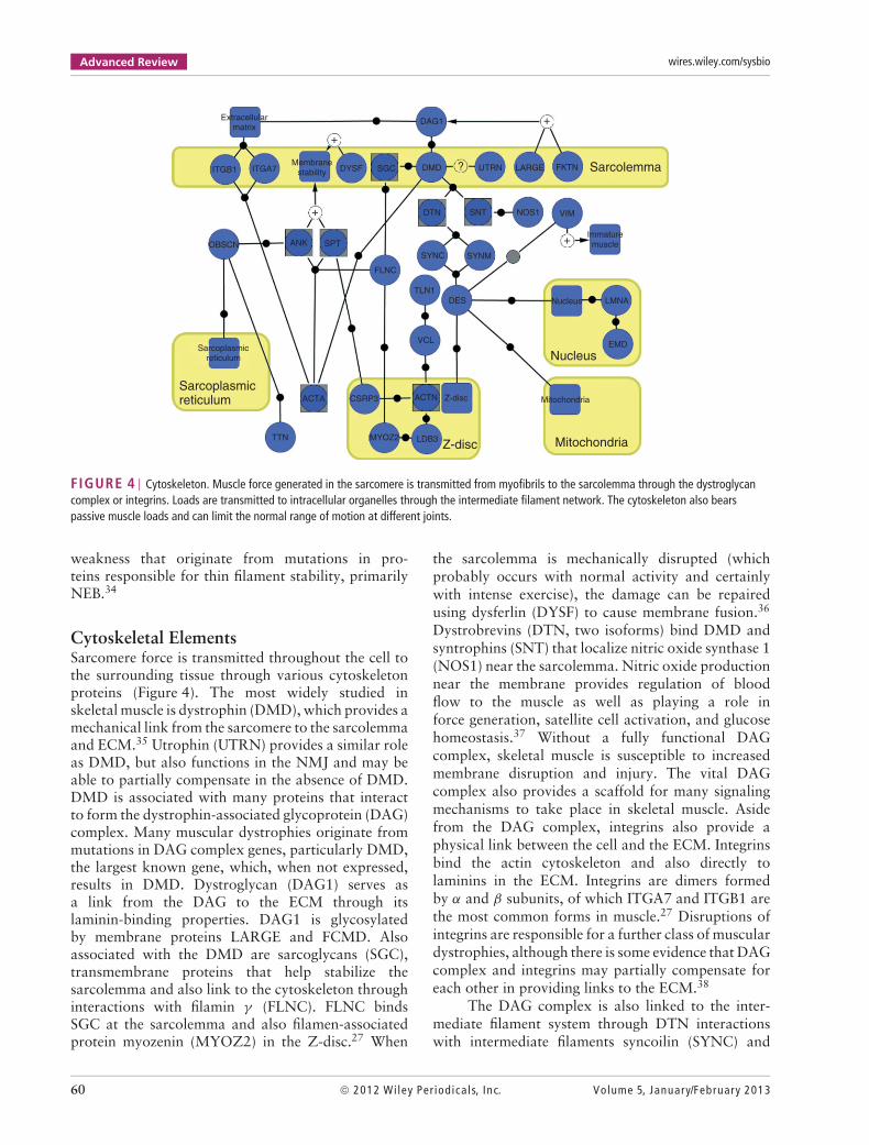
FIGURE 4 | Cytoskeleton. Muscle force generated in the sarcomere is transmitted from myofibrils to the sarcolemma through the dystroglycancomplex or integrins. Loads are transmitted to intracellular organelles through the intermediate filament network. The cytoskeleton also bearspassive muscle loads and can limit the normal range of motion at different joints.
weakness that originate from mutations in pro-teins responsible for thin filament stability, primarilyNEB.34
Cytoskeletal ElementsSarcomere force is transmitted throughout the cell tothe surrounding tissue through various cytoskeletonproteins (Figure 4). The most widely studied inskeletal muscle is dystrophin (DMD), which provides amechanical link from the sarcomere to the sarcolemmaand ECM.35 Utrophin (UTRN) provides a similar roleas DMD, but also functions in the NMJ and may beable to partially compensate in the absence of DMD.DMD is associated with many proteins that interactto form the dystrophin-associated glycoprotein (DAG)complex. Many muscular dystrophies originate frommutations in DAG complex genes, particularly DMD,the largest known gene, which, when not expressed,results in DMD. Dystroglycan (DAG1) serves asa link from the DAG to the ECM through itslaminin-binding properties. DAG1 is glycosylatedby membrane proteins LARGE and FCMD. Alsoassociated with the DMD are sarcoglycans (SGC),transmembrane proteins that help stabilize thesarcolemma and also link to the cytoskeleton throughinteractions with filamin γ (FLNC). FLNC bindsSGC at the sarcolemma and also filamen-associatedprotein myozenin (MYOZ2) in the Z-disc.27 When
the sarcolemma is mechanically disrupted (whichprobably occurs with normal activity and certainlywith intense exercise), the damage can be repairedusing dysferlin (DYSF) to cause membrane fusion.36
Dystrobrevins (DTN, two isoforms) bind DMD andsyntrophins (SNT) that localize nitric oxide synthase 1(NOS1) near the sarcolemma. Nitric oxide productionnear the membrane provides regulation of bloodflow to the muscle as well as playing a role inforce generation, satellite cell activation, and glucosehomeostasis.37 Without a fully functional DAGcomplex, skeletal muscle is susceptible to increasedmembrane disruption and injury. The vital DAGcomplex also provides a scaffold for many signalingmechanisms to take place in skeletal muscle. Asidefrom the DAG complex, integrins also provide aphysical link between the cell and the ECM. Integrinsbind the actin cytoskeleton and also directly tolaminins in the ECM. Integrins are dimers formedby α and β subunits, of which ITGA7 and ITGB1 arethe most common forms in muscle.27 Disruptions ofintegrins are responsible for a further class of musculardystrophies, although there is some evidence that DAGcomplex and integrins may partially compensate foreach other in providing links to the ECM.38
The DAG complex is also linked to the inter-mediate filament system through DTN interactionswith intermediate filaments syncoilin (SYNC) and
60 © 2012 Wiley Per iodica ls, Inc. Volume 5, January/February 2013

WIREs Systems Biology and Medicine Biological networks in skeletal muscle function
synemin (SYNM). These intermediate filaments con-nect to desmin (DES), the primary intermediate ofskeletal muscle. However, vimentin (VIM) predomi-nates expression during muscle development, but isthen lost at maturity. DES also links mitochondriaand the nucleus across the cell. The nuclear membraneanchorage to the cytoskeleton is mediated by emerin(EMD), which also binds nuclear lamin (LMNA) withits role in nuclear stability and gene expression. Thecritical role of EMD and LMNA regulating nuclearenvelope structure in muscle is emphasized by the factthat mutations in each cause Emery–Dreifuss mus-cular dystrophy. DES binds these organelles to themuscle structure at the Z-disc anchor. The Z-discitself consists of the overlapping barbed-end actin fil-aments from adjacent sarcomeres with its principlecomponent ACTN connecting actin filaments. TheZ-disc also contains ACTN-binding protein cypher(LDB3), for the linkage of filaments through MYOZ2,and muscle LIM protein (CSRP3), which links to theankryn (ANK) and spectrin (SPT) network within themuscle. ANK and SPT interact with actin and supportmembrane stability in prevention of muscle injury.They also interact with OBSCN, which plays a rolein localizing the SR through interactions with boththe SR and TTN in the sarcomere.27 The numerouscytoskeletal proteins present within and without themuscle cell and their role in various skeletal musclemyopathies highlight the critical importance of thestructured coordination of expression and functionamong these genes (Box 1).
BOX 1
DESMIN IN SKELETAL MUSCLE
Desmin represents the muscle-specific intermedi-ate filament protein that interconnects sarcom-eres at their Z-discs and permits efficient forcetransmission throughout the sarcomere.39 Inter-estingly, mice lacking desmin generate lowerstress because of this ‘impaired’ mechanicalforce transmission, but also show less overtsigns of injury.40 Recent studies showed thatthe loss of desmin also results in a chronicinflammatory effect that ultimately leads toskeletal muscle fibrosis of the ECM.41 As thedesmin knockout muscle fibers are more com-pliant compared to wild type,42 but the ECMof knockouts is stiffer compared to wild type,this suggests that the knockout may mount a‘compensatory’ response to loss of desmin andincreased fiber compliance. The precise mech-anism for this compensation is not known.
Extracellular MatrixThe cytoskeleton provides a means to transmit forcefrom the sarcomere to the ECM, which surroundseach muscle fiber with multiple layers of organization(Figure 5). The basal lamina ensheathes each fiber witha mesh-like network consisting primarily of COL4(COL4A1 and COL4A2) and laminin (LAM; mostcommonly LAMA2, LAMB1/LAMB2, and LAMC1in muscle).43 HSPG2 is a proteoglycan in thebasal lamina that binds both COL4 and LAM.Other basal laminar proteoglycans include syndecans(SDC), which play an important role in satellite celldifferentiation, and biglycan (BGN), an importantbinding partner for UTRN.35,44 The glycoproteinfibronectin (FN1) is a multimeric protein that servesas a link to many proteins within the basal lamina.45
The components of the basal lamina contain site ofbinding directly to the DCG complex or integrinswithin the muscle cell.
As opposed to the basal lamina, the fibrillarECM has strong load-bearing capabilities that canlimit the strain imposed on muscles. The fibrillarECM is made up primarily of collagen I (COL1)and collagen III (COL3A1) and is contiguous withinlayers of ECM in muscle and to the tendon beyondthe musculotendinous junction. Collagen VI (COL6)serves as an important link between the fibrillarand laminar ECM.43 Decorin (DCN) is the majorfibrillar proteoglycan in muscle.44 FN1 also playsan important link in fibrillar ECM. It binds toglycoprotein tenascin C (TNC) and provides strengthand elasticity to the ECM and is highly expressedin regenerating fibers and at the muscle–tendonjunction.45 Lysyl oxidase (LOX) is a copper enzymeprimarily responsible for cross-linking collagen, whichcontributes to ECM stiffness.46 Increased collagencross-linking has been proposed as a mechanism ofmuscle stiffening with age.47
The ECM also has a program for degradationand regulation through zinc-dependent matrixmetalloproteinases (MMPs). MMP2 and MMP9 areabundant gelatinases in muscle that breakdownCOL4 and are found in the basal lamina andfurther the breakdown of degraded fibrillar collagens.MMP2 is activated by membrane type MMP14 atthe sarcolemma. MMP1 functions as a proteasein the fibrillar ECM breaking down both COL1and COL3A.48 MMPs are expressed highly inperiods of ECM remodeling such as during muscleregeneration.49 To prevent excessive ECM breakdownand control MMP activity, muscles express tissueinhibitors of metalloproteinases (TIMPs), of whichTIMP1 and TIMP2 and present in skeletal muscle.48
The precise manner in which MMPs and TIMPs
Volume 5, January/February 2013 © 2012 Wiley Per iodica ls, Inc. 61

Advanced Review wires.wiley.com/sysbio
MMP1
COL3A1COL1
COL6
COL4 MMP9
MMP2
MMP14
TIMP1 TIMP2CTGF
LTBP4
TGFB1ECM
strength
PLOD3
Collagenexpression
Collagenprocessing P4H
Dystroglycancomplex
+
+
+ +
+
+
+
−
−
−
−
−
−
LOX
TNC FN1
BGN SDC HSPG2
LAM
DCN
Fibrillar ECM
Basal lamina
Sarcolemma
FIGURE 5 | Extracellular matrix (ECM). Provides the network for intracellular loads to be transmitted extracellularly. The basal lamina is amesh-like network, while the fibrillar ECM is made up of larger collagen fibrils and associated proteins. Several important growth factors are involvedin ECM formation and several enzyme systems regulate its state.
interact to regulate muscle ECM properties is notknown.
Production of ECM proteins is controlled byspecific growth factors. The most well-studied inducerof ECM in skeletal muscle for its role in fibrosis istransforming growth factor, β1 (TGFB1).50 TGFB1binds BGN in skeletal muscle and its activity is alsoinhibited through binding to LTBP4.51,52 Connec-tive tissue growth factor (CTGF) is another criticalcomponent in ECM signaling that leads to expres-sion of collagen genes.50 After expression, collagenmust be processed before becoming functional. Pro-lyl 4-hydroxylase (P4) catalyzes the formation ofhydroxyproline, and PLOD3 catalyzes posttransla-tional modification and both proteins reflect the rateof collagen biosynthesis.46 High levels of TGFB1 andCTGF are markers of muscle fibrosis, the build-up ofexcessive connective tissue that is present in most mus-cle diseases.53 As a mechanical tissue, skeletal muscleis adversely affected by stiff fibrous tissue in additionto the barrier it presents from typical cell interactionswith the ECM. Once fibrosis is prevalent within skele-tal muscle it prevents muscle regeneration making itpredominately irreversible.53
Energy MetabolismAs the motor for the body, muscle requires an exten-sive energy supply in the form of ATP. As mentioned
above, ATP is used to power the cross-bridge cycle andmaintain appropriate ion gradients by calcium trans-port in relaxation (Figure 6). Much of the metabolicmachinery is similar to that found in other cells; how-ever, many enzymes involved have muscle-specificisoforms expressed. Muscle can burn energy fasterthan can be produced within the cell necessitatinga buffer system that uses creatine kinase (CKM) totransfer a high-energy phosphate from phosphocrea-tine stores to ADP to form ATP.54 For short bursts ofactivity, skeletal muscle relies upon glycolysis for ATPproduction. This is the primary form of energy pro-duction within fast type II fibers because of the rapidATP production relative to oxidative metabolism. Tofacilitate glycolysis, glucose is transported across thesarcolemma by GLUT4 (SLC2A4). SLC2A4 translo-cation to the sarcolemma is controlled by AMPK.AMPK is an energy-sensing enzyme that becomes acti-vated in response to low energy levels.55 Through anindependent mechanism SLC2A4 also provides insulinsensitivity by increasing glucose uptake in response toelevated insulin levels.56 Intracellular glucose can bestored in the form of glycogen via the enzyme glyco-gen synthase 1 (GYS1), which can then be brokendown back into glucose via the enzyme glycogen phos-phorylase (PYGM) when energy demands are high.57
Glucose is broken down into pyruvate during glycol-ysis, which nets the required energy in the form of
62 © 2012 Wiley Per iodica ls, Inc. Volume 5, January/February 2013

WIREs Systems Biology and Medicine Biological networks in skeletal muscle function
Sarcolemma
VEGFA Glucose
GlucoseGlucogen
SLC2A4
PYGM
Fatty acid
Fatty acid
Fatty acidCPT1B
PNPLA2LPL
LIPE
FABP3
CD36
Ca2+
CAMK2
CAMK4PPP3CA
CREB1
NFATC1
MB O2
NFE2L2
TFAM
TFB
TP53 LDHA
PFKM
HK2
GYS1
Glycolysis
Pyruvate
GPAM
Triglyceride
PDH
NADH
Mitochondrialtranscription
ATP5 COX CYC SDH NDUF
CS
TCA cycle
MLYCD HADH
β oxidation
Acetyl–CoA
Oxidativephosphorylation
PPARGC1A
Nucleus
Mitochondria
NRF1
MEF2 ATF2
ATP
MAPK14
P–Creatine CKM
AMPK
+
+ +
+
+
+
+ + + +
+++
+
+
+
+
+
++
+ +
+
+
+
+ +
+
+
+
+
+
+
+++
+
+−
+
FIGURE 6 | Energy metabolism. Muscles use ATP as their energy source for contraction and much of relaxation. ATP is generated bothglycolytically and oxidatively in the muscle from glucose or fatty acids, respectively. Muscle has energy-sensing mechanisms that permit adaptation ofmetabolic systems to changes in energy demand.
ATP. Glucose is prepared for glycolysis by the phos-phorylating enzyme hexokinase (HK1); glycolysis ismaintained by lactate dehydrogenase A (LDHA), andthe rate-limiting step of glycolysis is controlled by theenzyme phosphofructokinase (PFKM).58 Disruptionsof the typical pathway from glycogen storage throughglycolysis often have pathologic consequences in skele-tal muscle characterized by the glycogen storage dis-eases. Glycogen storage diseases are often associatedwith exercise intolerance from limited energy supplyand excessive build-up of glycogen within the cell.59
When energy levels must be sustained overlonger periods muscle must utilize the more efficientoxidative phosphorylation process. This is more activein slow type I fibers that experience repetitive lowforce contractions and to a lesser extent in type IIafibers. Pyruvate conversion to acetyl-CoA by pyruvatedehydrogenase (PDH) allows progression throughthe tricarboxylic acid (TCA) cycle and subsequentoxidative phosphorylation.58 Muscles also utilizemore energy dense fatty acids during sustained activityin order to produce acetyl-CoA. Fatty acid uptake
into the cell is also regulated through an AMPK-mediated transporter CD36.55 Shuttling proteins fattyacid-binding protein 3 (FABP) and lipoprotein lipase(LPL) also mediate fatty acid transport.60 Intracellularfatty acids may be stored as triglycerides for which theenzyme GPAM catalyzes the initial and committingstep.55 Hormone-sensitive lipase (LIPE) is responsiblefor triglyceride breakdown to free fatty acids in musclealong with ATGL (PNPLA2) for the initial step andalso assisted by LPL for triglyceride hydrolysis.60 Tobe used in energy metabolism fatty acids must betransported into the mitochondria via CPT1B.58 Here,fatty acids can undergo β-oxidation to produce acetyl-CoA and NADH, which is catalyzed by enzymesMYLCD and HADH.60 Efficient processing of fattyacids is critical in skeletal muscle as a build-up ofmuscle lipid intermediates has been shown to result indecreased insulin sensitivity.61
Type I and type IIa muscle fibers have a highvolume fraction of mitochondria, which are requiredto meet their oxidative metabolism demands. Withinthe mitochondria, acetyl-CoA enters the TCA cycle
Volume 5, January/February 2013 © 2012 Wiley Per iodica ls, Inc. 63

Advanced Review wires.wiley.com/sysbio
to produce NADH. Citrate synthase (CS) catalyzesthe rate-limiting step within the TCA cycle, whichalso requires the enzyme succinate dehydrogenase(SDH).62 NADH is then used as an electron carrierin oxidative phosphorylation that uses oxygen asan oxidizing agent.63 Oxygen is provided to themuscle through the vasculature, which is stimulatedby VEGF, and transported within the muscle byMB.64,65 Oxidative phosphorylation is catalyzed bya set of four complexes in series (NDUF, SDH, CYC,and COX). The energy from the electron gradientproduced is then converted to ATP by ATP synthase(ATP5).63 Mutations in mitochondrial genes can leadto a set of severe mitochondrial myopathies in whichcompromised mitochondria signal for apoptosis andmuscle necrosis.66
Skeletal muscle is capable of adjusting thequantity of metabolic machinery present in order tomeet changes in energy demands. The productionof metabolic transcripts is largely controlled byPGC-1α (PPARGC1A) within skeletal muscle inconjunction with many other transcription factors.67
AMPK activates PPARGC1A when energy levelsare low.55 Ca2+ also plays a role through the
activation of PPP3CA, CAMK4, and CAMK2.PPP3CA activates the transcription factor NFATC1that produces muscle metabolic genes includingmyoglobin (MB).68 PPP3CA and CAMK4 bothactivate CREB1, a transcription factor that is integralto PPARGC1A-mediated expression.62 CAMK2activates p38 (MAPK14) that activates transcriptionfactors MEF2 and ATF2, which participate inmetabolic transcription through PPARGC1A.62,63
NRF1 together with PPARGC1A plays a large role inmitochondrial expression through transcription factorA, mitochondrial (TFAM). TFAM works in concertwith transcription factor B1 and B2, mitochondrial(TFB), and nuclear respiratory factor 2 (NFE2L2)in the mitochondrial transcription complex, which isalso maintained by TP53.69 These proteins make upthe refinery that allows muscle to convert a variety ofenergy sources into ATP used in force production.
InflammationInjury to skeletal muscle initiates a coordinated inflam-matory response that is localized to the damage site(Figure 7). This is a critical step in the process of
IL8 + PTGS2
Neutrophil Macrophage 1Macrophage 2ROS/NO
+
IFNG
IGF1 IL1B TNF
–
–
–+
IL10 TGFB1
+
Fibrosis
+ + + + + + +
–
IKBKE MAPK14 Ca2+SOCS3
–+++++
+
+
++
+ +NFKB1 HSF2
HSF1
FBXO32 TRIM63
–
–
–
–
Muscleregeneration
+
Nucleus
SMADPathway
CASP1
JAK-STATPathway
Sarcolemma
IL6
FIGURE 7 | Inflammation. Early macrophages and neutrophils enter damaged muscle to clear debris and produce an inflammatory signal. Ifpresent chronically, inflammation can lead to secondary tissue degradation. Secondary macrophages enter to limit inflammatory signals and repairmuscle.
64 © 2012 Wiley Per iodica ls, Inc. Volume 5, January/February 2013

WIREs Systems Biology and Medicine Biological networks in skeletal muscle function
muscle repair and, if not properly regulated, leads todeterioration and fibrosis.50,70,71 In the early stages ofthe inflammatory process, proinflammatory cytokinessuch as interleukin 8 (IL8), interferon γ , (IFNG), andCOX-2 (PTGS2) are released at the injury site attract-ing circulating neutrophils and classically activatedmacrophages, which then act to clear myofiber debrisand promote myoblast proliferation.70,71 These mono-cytes secrete other proinflammatory cytokines suchas tumor necrosis factor-α (TNF) and interleukin-1β (IL1B), which stimulate phagocytosis.50,72
The primary pathway for inflammation-mediated protein degradation is the nuclear factorκB (NFKB)-dependent pathway. Activation of NFKBis controlled by the Iκβ kinase (IKBKE) complex,which phosphorylates Iκβ targeting it for degrada-tion and enabling the translocation of NFKB tothe nucleus.73,74 NFKB affects protein turnover byincreasing the expression of the ubiquitin ligaseMuRF1 (TRIM63) and by binding to and activat-ing interleukin-6 (IL6). IL6 is thought to act in ahormone-like manner to regulate glucose homeostasispossibly via AMPK and can also be produced by themuscle itself.74–77 IL6 may also be activated by heatshock factors 1 and 2 and by calcium via its activa-tion of NFKB.70,77 NFKB is also activated by reactiveoxygen and nitrogen species as well as SOCS378,79;however, inside the nucleus, reactive species inhibitNFKB activity.78 Once activated, NFKB and IL6inhibit muscle regeneration. Evidence suggests thatthe mitogen-activated protein kinase p38 (MAPK14)is also activated in response to TNF and IL1B. It thenupregulates atrogin-1 (FBXO32), a transcript involvedin muscle atrophy. Additionally, p38 has been shownto activate IL6.76 TNF is also shown to increase circu-lating levels of interferon-γ (IFNG), which activatesthe JAK-STAT pathway and inhibits cell growth andproliferation.80 TNF and IL1B inhibit the expressionof IGF1, a key muscle growth factor.81
After initial invasion of neutrophils and clas-sically activated macrophages, a second populationof macrophages secrete cytokines such as interleukin10 (IL10) and TGFB1 and reduce the inflammatoryresponse.50 IL10 and TGFB1 negatively regulate IFNGproduction and IL10 inhibits the proteolytic effects ofIL1B.70,80 TGFB1 plays a role in both the initiationof fibrosis in skeletal muscle by stimulating fibroblastproliferation and inducing myogenic cells to differ-entiate into myofibroblastic cells. TGFB1 has beenshown to inhibit regeneration via activation of Smadproteins.50 While this inflammatory response is criti-cal to muscle repair, persistent inflammation as seen inmany myopathies leads to detrimental muscle fibro-sis and an inability of muscle to regenerate.53 It is
thus probably not surprising that there are severalreports in the literature that short-term inhibition ofinflammation after injury leads to long-term muscledysfunction.
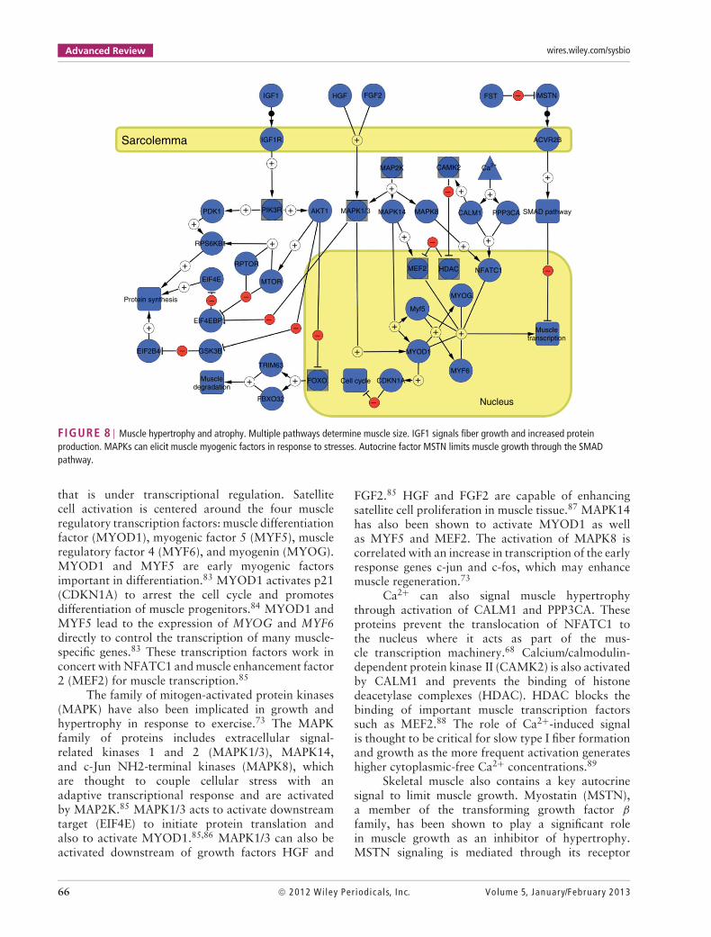
Muscle Hypertrophy and AtrophySkeletal muscle hypertrophy and atrophy are requiredto maintain the appropriate skeletal muscle massin response to altered levels of use and to recoverfrom injury. These processes can be triggered atthe cellular level by a variety of cues includinggrowth factors, nutritional signals, and mechanicalstress74,75 (Figure 8). Insulin and insulin-like growthfactor 1 (IGF1) are potent inducers of hypertrophy viaIGF1 receptor (IGF1R) and the PI3K/Akt pathway.75
Activated phosphatidylinositol 3 kinase (PI3KR)creates a lipid-binding site on the cell membrane forAKT1, a serine/threonine kinase. AKT1 then results inthe activation of the mammalian target of rapamycin(MTOR). Activation of MTOR then activatesRPS6KB1, which activates proteins responsible forprotein synthesis. In addition, PDK1 has alsobeen shown to phosphorylate RPS6KB1 directly.75
MTOR also participates in the growth process byphosphorylating EIF4EBP1, which results in thedissociation of the EIF4EBP1/EIF4E complex allowingEIF4E to initiate protein translation. A regulatoryassociated protein of MTOR (RPTOR) facilitates thephosphorylation of RPS6KB1 and 4EBP1 by MTORand has been shown to bind both proteins. AKT1 mayalso influence translation by inhibiting the activityof glycogen synthase kinase 3β (GSK3B) as GSK3Binhibits EIF2B, thereby blocking its promotion ofprotein translation.74,75 Along with the increasedprotein synthesis, IGF1 signaling is also able tolimit active protein degradation. AKT1 prevents theforkhead box proteins (FOXO) from entering thenucleus via phosphorylation.74,76 FOXO transcriptionfactors are known to promote transcription ofTRIM63 and FBXO32, which are ubiquitin ligasesinvolved in muscle degradation.82 These muscleatrophy proteins are critical for the active processof muscle atrophy that occurs during unloading orimmobilization where muscle mass is removed.
Muscle hypertrophy requires increased protein,but multinucleated muscle fibers also require morenuclear content with growth that is provided bythe local satellite cell population. Satellite cells areregenerating myogenic progenitor cells that residenext to muscle fiber beneath the basal laminaand are also activated by IGF1. Satellite cellsundergo processes of proliferation, differentiation,migration, and fusion with muscle fibers in a process
Volume 5, January/February 2013 © 2012 Wiley Per iodica ls, Inc. 65
Advanced Review wires.wiley.com/sysbio
IGF1 HGF FGF2 FST MSTN
IGF1R
PDK1
RPS6KB1
ElF4E
ElF4EBP
GSK3B
TRIM63
FBXO32
EIF2B4
FOXO Cell cycle CDKN1A
MYOD1
MYF6
Nucleus
Myf5
MYOG
MEF2 HDAC NFATC1
Muscletranscription
Muscledegradation
MTOR
RPTOR
Protein synthesis
ACVR2B
PIK3R AKT1 MAPK1/3 MAPK14 MAPK8 CALM1 PPP3CA SMAD pathway
MAP2K CAMK2 Ca2+
Sarcolemma
+
+
+
+
++
+
++
+
+
++
++
+
+
+
+
+
+
+
+ −
−
−
−
−
− −
−−
−
−
+
FIGURE 8 | Muscle hypertrophy and atrophy. Multiple pathways determine muscle size. IGF1 signals fiber growth and increased proteinproduction. MAPKs can elicit muscle myogenic factors in response to stresses. Autocrine factor MSTN limits muscle growth through the SMADpathway.
that is under transcriptional regulation. Satellitecell activation is centered around the four muscleregulatory transcription factors: muscle differentiationfactor (MYOD1), myogenic factor 5 (MYF5), muscleregulatory factor 4 (MYF6), and myogenin (MYOG).MYOD1 and MYF5 are early myogenic factorsimportant in differentiation.83 MYOD1 activates p21(CDKN1A) to arrest the cell cycle and promotesdifferentiation of muscle progenitors.84 MYOD1 andMYF5 lead to the expression of MYOG and MYF6directly to control the transcription of many muscle-specific genes.83 These transcription factors work inconcert with NFATC1 and muscle enhancement factor2 (MEF2) for muscle transcription.85
The family of mitogen-activated protein kinases(MAPK) have also been implicated in growth andhypertrophy in response to exercise.73 The MAPKfamily of proteins includes extracellular signal-related kinases 1 and 2 (MAPK1/3), MAPK14,and c-Jun NH2-terminal kinases (MAPK8), whichare thought to couple cellular stress with anadaptive transcriptional response and are activatedby MAP2K.85 MAPK1/3 acts to activate downstreamtarget (EIF4E) to initiate protein translation andalso to activate MYOD1.85,86 MAPK1/3 can also beactivated downstream of growth factors HGF and
FGF2.85 HGF and FGF2 are capable of enhancingsatellite cell proliferation in muscle tissue.87 MAPK14has also been shown to activate MYOD1 as wellas MYF5 and MEF2. The activation of MAPK8 iscorrelated with an increase in transcription of the earlyresponse genes c-jun and c-fos, which may enhancemuscle regeneration.73
Ca2+ can also signal muscle hypertrophythrough activation of CALM1 and PPP3CA. Theseproteins prevent the translocation of NFATC1 tothe nucleus where it acts as part of the mus-cle transcription machinery.68 Calcium/calmodulin-dependent protein kinase II (CAMK2) is also activatedby CALM1 and prevents the binding of histonedeacetylase complexes (HDAC). HDAC blocks thebinding of important muscle transcription factorssuch as MEF2.88 The role of Ca2+-induced signalis thought to be critical for slow type I fiber formationand growth as the more frequent activation generateshigher cytoplasmic-free Ca2+ concentrations.89
Skeletal muscle also contains a key autocrinesignal to limit muscle growth. Myostatin (MSTN),a member of the transforming growth factor β
family, has been shown to play a significant rolein muscle growth as an inhibitor of hypertrophy.MSTN signaling is mediated through its receptor
66 © 2012 Wiley Per iodica ls, Inc. Volume 5, January/February 2013

WIREs Systems Biology and Medicine Biological networks in skeletal muscle function
MYH7 MYH1MYH2MYL2
MYL3
MYBL2
TNNT1
TNNI1TNNC1
TPM3
TMOD1
ATP2A2
CASQ2
Slowfibers
Fastfibers
MYL1
MYBPS2
TNNT3
TNNI2TNNC2
TPM1
TMOD4
ATP2A1
CASQ1
FIGURE 9 | Muscle fiber type (FT). Genes that have similar function but are expressed specifically in either a fast or slow muscle FT are listedhere. MYH is the major determinant of FT, but numerous genes exist that are coregulated in the various FTs.
activin IIB (ACVR2B), which conducts a signal to thenucleus through the SMAD pathway. Expression offollistatin (FSTN) has been shown to increase musclemass through action as a MSTN antagonist.50,74
This presents MSTN as a potent and muscle-specificpotential drug target for therapies aimed at increasingmuscle mass. Indeed, various methods of therapy usingFSTN, soluble ACVR2B, and anti-MSTN antibodieshave been attempted in order to inhibit MSTN actionand are currently under research.90
Muscle Fiber TypesSkeletal muscles have different FTs that play a role inmuscle function (Figure 9) with myosin heavy chainas the major differentiator among FT in skeletalmuscle.30 In fact, muscle FTs are often referred toin terms of their dominant myosin heavy chain astype I, type IIa, type IIx, and type IIb (not expressedin humans). Rather than define the details of eachmuscle FT in this section, we have integrated thisdiscussion into the processes described above. It isclear that many transcripts related to muscle functionare differentially regulated according to FT. A fulldescription of the proteins related to muscle FTdetermination has been provided elsewhere.91–95 Inaddition to the various MYH genes expressed, severalof the complexes listed in the sections above havedistinct isoforms that are activated specifically in eitherfast or slow muscle fibers. Figure 9 helps in identifyingthe individual genes for each FT-specific protein. Thisprovides a useful description of muscle FT beyondjust MYH and highlights the fact that ‘FT’ simplyrefers to a general physiological function (e.g., fastor slow contracting, high or low endurance) that iscreated by coordinated expression of scores of genes(Box 2).
BOX 2
FIXED MUSCLE CONTRACTURES
Skeletal muscle is an extremely adaptable tissuewith motor neuron firing being a major deter-minant of muscle characteristics. Upper motorneuron lesions like those seen in stroke or cere-bral palsy are known to create spastic muscles.This spasticity and altered use often causes mus-cles to adapt into a pathologic state known as a‘fixed muscle contracture’.96 When muscle stiff-ness, without any active force generation, limitsthe functional range of motion of a joint, thepatient is considered to have a fixed contrac-ture. This pathologic state is not the result ofany genetic abnormalities of muscle genes them-selves, but represents a maladaptive state.97
The mechanism that leads to restricted mus-cle in contracture is largely unknown. Recentstudies on children with cerebral palsy havesuggested that many of the functional mus-cle networks described here are also altered incerebral palsy.98 Many proteins related to ECCare upregulated, such as parvalbumin, a proteininvolved that assists in relaxing the muscle. TheECM also showed many transcripts that wereupregulated including the major component ofthe basal lamina collagen IV. The muscle fromchildren with cerebral palsy also had conflict-ing growth signals with both IGF1, a proteinthat signals hypertrophy, and MSTN, a proteinexpressed to limit growth, being upregulated.98
These results demonstrate how the networkapproach described here may be used to inves-tigate muscle adaptations and lead to thera-peutic interventions to reverse these maladies.a
Volume 5, January/February 2013 © 2012 Wiley Per iodica ls, Inc. 67
Advanced Review wires.wiley.com/sysbio
CONCLUSION
For skeletal muscle to function properly, coordinationamong multiple biological networks is required. Eachnetwork has critical components responsible for itsfunction, most of which have been established in theliterature. We delineated the critical components ofthese nine networks and their relationship to musclefunction.
A systems approach such as that described hereis critical to define the appropriate regulatory systemsthat enable normal muscle function and to understandmuscle pathological states. Importantly, these regula-tory networks are likely to involve multiple genes sothat a systematic, quantitative analytical approach,using these nine networks, is critical to dissecting thedisease process. For example, if the typical networkincludes 20 genes and 3 genes interact to produce adisease state, it would require 120 combinations of3 gene sets to sort out this network. The systemsapproach defined here produces topological networkmodels which in turn can be cast into equation-drivenmathematical models. These models lend themselvesto quantitative studies of normal and diseased func-tion. For instance, reduced concentration or absenceof a species can be incorporated into the model toassess alterations in the phenotypes.
The models can also serve the function of assess-ing the effect of pharmacological intervention. In fact,a large-scale screening of therapeutic compounds canbe assessed quantitatively. Muscle cells grow wellin culture and can be used as a screening tool when
combined with a systems approach to analyzing exper-imental results. Drugs can be titrated with regard tobeneficial and potential detrimental side effects, andcomplete cocktails of drugs can be developed by linearcombination of experimental results.
One specific application of the use of musclemicroarray data is in patients with DMD. One canimagine that expression data can be plugged intothe pathways to quickly investigate the transcrip-tional effects on skeletal muscle. These muscle-specificpathways would allow rapid analysis of how associ-ated cytoskeletal transcripts are expressed. Beyond theexpected network adaptations in glucose metabolism,inflammation, or cytoskeletal remodeling, unexpectedadaptations of muscle systems could also be defined.Certainly, some of these details are already availablescattered across the literature; however, this reviewprovides a framework to simplify analysis of high-throughput technologies by placing gene networks ina physiological context.
NOTEaThis systems approach to understanding complexmuscle adaptation in disease was used in the case ofchildren’s cerebral palsy. In this approach, the geneexpression pattern present in human muscle biop-sies was first determined,98 and then the biochemical,biomechanical, and structural properties of the tissueitself were also determined.99 Using these data, wehope that therapeutic strategies can be developed tocombat this devastating muscle adaptation.
ACKNOWLEDGMENTS
This work was supported by grants from the National Institute of Health (AR057393 and R24HD050837) andthe Department of Veterans Affairs.
REFERENCES1. Feltrin E, Campanaro S, Diehl AD, Ehler E, Faulkner G,
Fordham J, Gardin C, Harris M, Hill D, Knoell R, et al.Muscle research and gene ontology: new standards forimproved data integration. BMC Med Genomics 2009,2:6.
2. Cline MS, Smoot M, Cerami E, Kuchinsky A, Landys N,Workman C, Christmas R, Avila-Campilo I, Creech M,Gross B, et al. Integration of biological networks andgene expression data using Cytoscape. Nat Protoc 2007,2:2366–2382.
3. Lockhart DJ, Dong H, Byrne MC, Follettie MT, GalloMV, Chee MS, Mittmann M, Wang C, Kobayashi M,
Horton H, et al. Expression monitoring by hybridiza-tion to high-density oligonucleotide arrays. NatBiotechnol 1996, 14:1675–1680.
4. Hu S, Xie Z, Qian J, Blackshaw S, Zhu H. Functionalprotein microarray technology. Wiley Interdiscip RevSyst Biol Med 2010, 3:255–268.
5. Deconinck N, Dan B. Pathophysiology of Duchennemuscular dystrophy: current hypotheses. Pediatr Neurol2007, 36:1–7.
6. Petrof BJ, Shrager JB, Stedman HH, Kelly AM, SweeneyHL. Dystrophin protects the sarcolemma from stresses
68 © 2012 Wiley Per iodica ls, Inc. Volume 5, January/February 2013
WIREs Systems Biology and Medicine Biological networks in skeletal muscle function
developed during muscle contraction. Proc Natl AcadSci U S A 1993, 90:3710–3714.
7. Hopf FW, Turner PR, Denetclaw WF Jr, Reddy P, Stein-hardt RA. A critical evaluation of resting intracellularfree calcium regulation in dystrophic mdx muscle. AmJ Physiol 1996, 271:C1325–C1339.
8. Haslett JN, Sanoudou D, Kho AT, Bennett RR, Green-berg SA, Kohane IS, Beggs AH, Kunkel LM. Geneexpression comparison of biopsies from Duchenne mus-cular dystrophy (DMD) and normal skeletal muscle.Proc Natl Acad Sci U S A 2002, 99:15000–15005.
9. Spencer MJ, Montecino-Rodriguez E, Dorshkind K,Tidball JG. Helper (CD4(+)) and cytotoxic (CD8(+))T cells promote the pathology of dystrophin-deficientmuscle. Clin Immunol 2001, 98:235–243.
10. Schmalbruch H. Regenerated muscle fibers in Duchennemuscular dystrophy: a serial section study. Neurology1984, 34:60–65.
11. Duance VC, Stephens HR, Dunn M, Bailey AJ,Dubowitz V. A role for collagen in the pathogenesisof muscular dystrophy? Nature 1980, 284:470–472.
12. Wu H, Xiong WC, Mei L. To build a synapse: sig-naling pathways in neuromuscular junction assembly.Development 2010, 137:1017–1033.
13. Schaeffer L, de Kerchove d’Exaerde A, Changeux JP.Targeting transcription to the neuromuscular synapse.Neuron 2001, 31:15–22.
14. Patton BL. Basal lamina and the organization of neuro-muscular synapses. J Neurocytol 2003, 32:883–903.
15. Beard NA, Wei L, Dulhunty AF. Ca(2+) signaling instriated muscle: the elusive roles of triadin, junctin, andcalsequestrin. Eur Biophys J 2009, 39:27–36.
16. Dulhunty AF. Excitation-contraction coupling from the1950s into the new millennium. Clin Exp PharmacolPhysiol 2006, 33:763–772.
17. Flucher BE, Obermair GJ, Tuluc P, Schredelseker J,Kern G, Grabner M. The role of auxiliary dihydropy-ridine receptor subunits in muscle. J Muscle Res CellMotil 2005, 26:1–6.
18. Treves S, Vukcevic M, Maj M, Thurnheer R, Mosca B,Zorzato F. Minor sarcoplasmic reticulum membranecomponents that modulate excitation-contraction cou-pling in striated muscles. J Physiol 2009, 587:3071–3079.
19. Volkers M, Rohde D, Goodman C, Most P. S100A1:a regulator of striated muscle sarcoplasmic reticulumCa2+ handling, sarcomeric, and mitochondrial function.J Biomed Biotechnol 2010, 2010:178614.
20. Rosenberg H, Davis M, James D, Pollock N, Stowell K.Malignant hyperthermia. Orphanet J Rare Dis 2007,2:21.
21. Berchtold MW, Brinkmeier H, Muntener M. Calciumion in skeletal muscle: its crucial role for musclefunction, plasticity, and disease. Physiol Rev 2000,80:1215–1265.
22. Traaseth NJ, Ha KN, Verardi R, Shi L, Buffy JJ, Mas-terson LR, Veglia G. Structural and dynamic basis ofphospholamban and sarcolipin inhibition of Ca(2+)-ATPase. Biochemistry 2008, 47:3–13.
23. Allen DG, Whitehead NP, Yeung EW. Mechanisms ofstretch-induced muscle damage in normal and dys-trophic muscle: role of ionic changes. J Physiol 2005,567:723–735.
24. Patel A, Sharif-Naeini R, Folgering JR, Bichet D, DupratF, Honore E. Canonical TRP channels and mechan-otransduction: from physiology to disease states.Pflugers Arch 2010, 460:571–581.
25. Huxley AF, Niedergerke R. Structural changes in mus-cle during contraction; interference microscopy of livingmuscle fibres. Nature 1954, 173:971–973.
26. Huxley H, Hanson J. Changes in the cross-striations ofmuscle during contraction and stretch and their struc-tural interpretation. Nature 1954, 173:973–976.
27. Clark KA, McElhinny AS, Beckerle MC, Gregorio CC.Striated muscle cytoarchitecture: an intricate web ofform and function. Annu Rev Cell Dev Biol 2002,18:637–706.
28. Huxley HE. The cross-bridge mechanism of muscularcontraction and its implications. J Exp Biol 1985, 115:17–30.
29. Gordon AM, Homsher E, Regnier M. Regulation ofcontraction in striated muscle. Physiol Rev 2000, 80:853–924.
30. Zierath JR, Hawley JA. Skeletal muscle fiber type: influ-ence on contractile and metabolic properties. PLoS Biol2004, 2:e348.
31. Maruyama K. Connectin/titin, giant elastic protein ofmuscle. FASEB J 1997, 11:341–345.
32. McElhinny AS, Kolmerer B, Fowler VM, Labeit S, Gre-gorio CC. The N-terminal end of nebulin interacts withtropomodulin at the pointed ends of the thin filaments.J Biol Chem 2001, 276:583–592.
33. Gordon AM, Huxley AF, Julian FJ. The variation inisometric tension with sarcomere length in vertebratemuscle fibres. J Physiol 1966, 184:170–192.
34. Ochala J. Thin filament proteins mutations associatedwith skeletal myopathies: defective regulation of musclecontraction. J Mol Med (Berl) 2008, 86:1197–1204.
35. Allikian MJ, McNally EM. Processing and assemblyof the dystrophin glycoprotein complex. Traffic 2007,8:177–183.
36. Han R, Campbell KP. Dysferlin and muscle membranerepair. Curr Opin Cell Biol 2007, 19:409–416.
37. Stamler JS, Meissner G. Physiology of nitric oxide inskeletal muscle. Physiol Rev 2001, 81:209–237.
38. Mayer U. Integrins: redundant or important players inskeletal muscle? J Biol Chem 2003, 278:14587–14590.
39. Lazarides E. Intermediate filaments as mechanical inte-grators of cellular space. Nature 1980, 283:249–256.
Volume 5, January/February 2013 © 2012 Wiley Per iodica ls, Inc. 69

Advanced Review wires.wiley.com/sysbio
40. Sam M, Shah S, Friden J, Milner DJ, CapetanakiY, Lieber RL. Desmin knockout muscles generate lowerstress and are less vulnerable to injury compared to wild-type muscles. Am J Physiol 2000, 279:C1116–C1122.
41. Li Z, Mericskay M, Agbulut O, Butler-Browne G,Carlsson L, Thornell LE, Babinet C, Paulin D. Desminis essential for the tensile strength and integrity ofmyofibrils but not for myogenic commitment, differen-tiation, and fusion of skeletal muscle. J Cell Biol 1997,139:129–144.
42. Shah SB, Davis J, Weisleder N, Kostavassili I, McCul-loch AD, Ralston E, Capetanaki Y, Lieber RL. Struc-tural and functional roles of desmin in mouse skeletalmuscle during passive deformation. Biophys J 2004,86:2993–3008.
43. Grounds MD, Sorokin L, White J. Strength at the extra-cellular matrix-muscle interface. Scand J Med Sci Sports2005, 15:381–391.
44. Jenniskens GJ, Veerkamp JH, van Kuppevelt TH. Hep-aran sulfates in skeletal muscle development and physi-ology. J Cell Physiol 2006, 206:283–294.
45. Jarvinen TA, Kannus P, Jarvinen TL, Jozsa L, Kalimo H,Jarvinen M. Tenascin-C in the pathobiology and healingprocess of musculoskeletal tissue injury. Scand J MedSci Sports 2000, 10:376–382.
46. Takala TE, Virtanen P. Biochemical composition ofmuscle extracellular matrix: the effect of loading. ScandJ Med Sci Sports 2000, 10:321–325.
47. Palokangas H, Kovanen V, Duncan A, Robins SP. Age-related changes in the concentration of hydroxypyri-dinium crosslinks in functionally different skeletal mus-cles. Matrix 1992, 12:291–296.
48. Carmeli E, Moas M, Reznick AZ, Coleman R. Matrixmetalloproteinases and skeletal muscle: a brief review.Muscle Nerve 2004, 29:191–197.
49. Kherif S, Lafuma C, Dehaupas M, Lachkar S, FournierJG, Verdiere-Sahuque M, Fardeau M, Alameddine HS.Expression of matrix metalloproteinases 2 and 9 inregenerating skeletal muscle: a study in experimen-tally injured and mdx muscles. Dev Biol 1999, 205:158–170.
50. Serrano AL, Munoz-Canoves P. Regulation and dysreg-ulation of fibrosis in skeletal muscle. Exp Cell Res 2010,316:3050–3058.
51. Young MF, Bi Y, Ameye L, Chen XD. Biglycan knock-out mice: new models for musculoskeletal diseases.Glycoconj J 2002, 19:257–262.
52. Goldstein JA, McNally EM. Mechanisms of muscleweakness in muscular dystrophy. J Gen Physiol 2010,136:29–34.
53. Serrano AL, Mann CJ, Vidal B, Ardite E, Perdiguero E,Munoz-Canoves P. Cellular and molecular mechanismsregulating fibrosis in skeletal muscle repair and disease.Curr Top Dev Biol 2011, 96:167–201.
54. Bessman SP, Geiger PJ. Transport of energy in mus-cle: the phosphorylcreatine shuttle. Science 1981, 211:448–452.
55. Ruderman NB, Park H, Kaushik VK, Dean D, Con-stant S, Prentki M, Saha AK. AMPK as a metabolicswitch in rat muscle, liver and adipose tissue afterexercise. Acta Physiol Scand 2003, 178:435–442.
56. Lauritzen HP, Schertzer JD. Measuring GLUT4 translo-cation in mature muscle fibers. Am J Physiol EndocrinolMetab 2010, 299:E169–E179.
57. Westerblad H, Bruton JD, Katz A. Skeletal muscle:energy metabolism, fiber types, fatigue and adaptability.Exp Cell Res 2010, 316:3093–3099.
58. Jeukendrup AE. Regulation of fat metabolism in skele-tal muscle. Ann N Y Acad Sci 2002, 967:217–235.
59. Hicks J, Wartchow E, Mierau G. Glycogen storage dis-eases: a brief review and update on clinical features,genetic abnormalities, pathologic features, and treat-ment. Ultrastruct Pathol 2011, 35:183–196.
60. Holloway GP, Luiken JJ, Glatz JF, Spriet LL, Bonen A.Contribution of FAT/CD36 to the regulation of skeletalmuscle fatty acid oxidation: an overview. Acta Physiol(Oxf) 2008, 194:293–309.
61. Kraegen EW, Cooney GJ. Free fatty acids and skeletalmuscle insulin resistance. Curr Opin Lipidol 2008, 19:235–241.
62. Hood DA, Irrcher I, Ljubicic V, Joseph AM. Coordina-tion of metabolic plasticity in skeletal muscle. J ExpBiol 2006, 209:2265–2275.
63. Holloway GP, Bonen A, Spriet LL. Regulation of skele-tal muscle mitochondrial fatty acid metabolism inlean and obese individuals. Am J Clin Nutr 2009, 89:455S–462S.
64. Kanatous SB, Mammen PP. Regulation of myoglobinexpression. J Exp Biol 2010, 213:2741–2747.
65. Hudlicka O, Brown MD. Adaptation of skeletal musclemicrovasculature to increased or decreased blood flow:role of shear stress, nitric oxide and vascular endothelialgrowth factor. J Vasc Res 2009, 46:504–512.
66. DiMauro S. Pathogenesis and treatment of mitochon-drial myopathies: recent advances. Acta Myol 2010, 29:333–338.
67. Olesen J, Kiilerich K, Pilegaard H. PGC-1α-mediatedadaptations in skeletal muscle. Pflugers Arch 2010, 460:153–162.
68. Schiaffino S. Fibre types in skeletal muscle: a personalaccount. Acta Physiol (Oxf) 2010, 199:451–463.
69. Ljubicic V, Joseph AM, Saleem A, Uguccioni G, Collu-Marchese M, Lai RY, Nguyen LM, Hood DA. Tran-scriptional and post-transcriptional regulation of mito-chondrial biogenesis in skeletal muscle: effects ofexercise and aging. Biochim Biophys Acta 2010, 1800:223–234.
70. Peake J, Della Gatta P, Cameron-Smith D. Aging andits effects on inflammation in skeletal muscle at rest and
70 © 2012 Wiley Per iodica ls, Inc. Volume 5, January/February 2013

WIREs Systems Biology and Medicine Biological networks in skeletal muscle function
following exercise-induced muscle injury. Am J PhysiolRegul Integr Comp Physiol 2010, 298:R1485–R1495.
71. Tidball JG. Inflammatory processes in muscle injuryand repair. Am J Physiol Regul Integr Comp Physiol2005, 288:R345–R353.
72. Smith C, Kruger MJ, Smith RM, Myburgh KH. Theinflammatory response to skeletal muscle injury: illu-minating complexities. Sports Med 2008, 38:947–969.
73. Kramer HF, Goodyear LJ. Exercise, MAPK, and NF-κBsignaling in skeletal muscle. J Appl Physiol 2007, 103:388–395.
74. Zhang P, Chen X, Fan M. Signaling mechanismsinvolved in disuse muscle atrophy. Med Hypotheses2007, 69:310–321.
75. Glass DJ. Skeletal muscle hypertrophy and atrophy sig-naling pathways. Int J Biochem Cell Biol 2005, 37:1974–1984.
76. Febbraio MA, Pedersen BK. Muscle-derived interleukin-6: mechanisms for activation and possible biologicalroles. FASEB J 2002, 16:1335–1347.
77. Pedersen BK, Fischer CP. Physiological roles of muscle-derived interleukin-6 in response to exercise. Curr OpinClin Nutr Metab Care 2007, 10:265–271.
78. Morgan MJ, Liu ZG. Crosstalk of reactive oxygenspecies and NF-κB signaling. Cell Res 2011, 21:103–115.
79. Spangenburg EE, Brown DA, Johnson MS, Moore RL.Exercise increases SOCS-3 expression in rat skele-tal muscle: potential relationship to IL-6 expression.J Physiol 2006, 572:839–848.
80. Schroder K, Hertzog PJ, Ravasi T, Hume DA.Interferon-γ : an overview of signals, mechanisms andfunctions. J Leukoc Biol 2004, 75:163–189.
81. Li H, Malhotra S, Kumar A. Nuclear factor-κB signal-ing in skeletal muscle atrophy. J Mol Med 2008, 86:1113–1126.
82. Attaix D, Ventadour S, Codran A, Bechet D, Tail-landier D, Combaret L. The ubiquitin-proteasome sys-tem and skeletal muscle wasting. Essays Biochem 2005,41:173–186.
83. Ten Broek RW, Grefte S, Von den Hoff JW. Regulatoryfactors and cell populations involved in skeletal muscleregeneration. J Cell Physiol 2010, 224:7–16.
84. Kitzmann M, Fernandez A. Crosstalk between cell cycleregulators and the myogenic factor MyoD in skeletalmyoblasts. Cell Mol Life Sci 2001, 58:571–579.
85. Keren A, Tamir Y, Bengal E. The p38 MAPK signalingpathway: a major regulator of skeletal muscle develop-ment. Mol Cell Endocrinol 2006, 252:224–230.
86. Williamson D, Gallagher P, Harber M, Hollon C,Trappe S. Mitogen-activated protein kinase (MAPK)pathway activation: effects of age and acute exercise onhuman skeletal muscle. J Physiol 2003, 547:977–987.
87. Yablonka-Reuveni Z, Day K, Vine A, Shefer G. Defin-ing the transcriptional signature of skeletal muscle stemcells. J Anim Sci 2008, 86:E207–E216.
88. McGee SL. Exercise and MEF2-HDAC interactions.Appl Physiol Nutr Metab 2007, 32:852–856.
89. Schiaffino S, Serrano A. Calcineurin signaling and neu-ral control of skeletal muscle fiber type and size. TrendsPharmacol Sci 2002, 23:569–575.
90. Tsuchida K. Targeting myostatin for therapies againstmuscle-wasting disorders. Curr Opin Drug DiscovDevel 2008, 11:487–494.
91. Peter JB, Barnard RJ, Edgerton VR, Gillespie CA, Stem-pel KE. Metabolic profiles of three fiber types of skeletalmuscle in guinea pigs and rabbits. Biochemistry 1972,11:2627–2633.
92. Gauthier GF. On the relationship of ultrastructural andcytochemical features of color in mammalian skeletalmuscle. Z Zellforsch Mikrosk Anat 1969, 95:462–482.
93. Brooke MH, Kaiser KK. Muscle fiber types: how manyand what kind? Arch Neurol 1970, 23:369–379.
94. Schiaffino S, Reggiani C. Molecular diversity of myofib-rillar proteins: gene regulation and functional signifi-cance. Physiol Rev 1996, 76:371–423.
95. Gillespie CA, Simpson DR, Edgerton VR. High glyco-gen content of red as opposed to white skeletal musclefibers of guinea pigs. J Histochem Cytochem 1970, 18:552–558.
96. Farmer SE, James M. Contractures in orthopaedic andneurological conditions: a review of causes and treat-ment. Disabil Rehabil 2001, 23:549–558.
97. O’Dwyer NJ, Ada L. Reflex hyperexcitability and mus-cle contracture in relation to spastic hypertonia. CurrOpin Neurol 1996, 9:451–455.
98. Smith LR, Ponten E, Hedstrom Y, Ward SR, Cham-bers HG, Subramaniam S, Lieber RL. Novel transcrip-tional profile in wrist muscles from cerebral palsypatients. BMC Med Genomics 2009, 2:44.
99. Smith LR, Lee KS, Ward SR, Chambers HG, Lieber RL.Hamstring contractures in children with spastic cerebralpalsy result from a stiffer ECM and increased in vivo sar-comere length. J Physiol (Lond) 2011, 589:2625–2639.
FURTHER READINGThe networks described in this article are available for analysis and manipulation using the tools of Cytoscape software.Available at www.cytoscape.org.
Volume 5, January/February 2013 © 2012 Wiley Per iodica ls, Inc. 71