advanced techniques in biology & medicine · phyllanthus niruiri. and . ... niruri and...
TRANSCRIPT
Research Article Open Access
Nelson et al., Adv Tech Biol Med 2013, 1:1http://dx.doi.org/10.4172/atbm.1000101
Research Article Open Access
Advanced Techniques in Biology & Medicine
Volume 1 • Issue 1 • 1000101Adv Tech Biol MedISSN: ATBM, an open access journal
Keywords: Adhatoda vasica; Leptospira interrogans; Rat fever; Inclusion body
IntroductionLeptospirosis is one of the worlds’ most widespread zoonotic diseases
[1], causing extensive vacuites of micro vessels in multiple organs [2]. Faine [3] reported that Leptospiral lesion disrupt the integrity of the cell membrane of endothelial cells lining small blood vessels in all parts of the body, and leading to capillary leakage and hemorrhage. Damage of blood vessels in the renal cortex leads to renal failure [4], and in the liver, cause haemorrhagic jaundices. In Tamilnadu state of India, several outbreaks of Leptospirosis had been reported [5]. Jeyakumar et al. [6] had reported the seroprevalence of Leptospirosis among the people involved in agricultural, livestock and veterinary activities in the city. Leptospires are corkscrew shaped bacteria (Spirochaetes), with many species divergence. The pathogenic Leptospira interrogans has several serovars [7], and reported to cause Leptospirosis in many places in South India. Debnath et al. [8] had reported a high incidence of Leptospirosis in rural people, particularly in people associated with agricultural activities. Hence, an investigation into herbal remedy for Leptospirosis could help villagers to take immediate remedial measures for this infection.
Hence, an attempt has been made to study the efficacy of the extracts of Andrographis paniculata, Azadirachta indica, Phyllanthus niruiri and Adhatoda vasica, to inhibit the activity of Leptospira interrogans. Based on the antiLeptospiral activity, the extracts of the plant, Adhatoda vasica, was chosen for further study. Electron microscopical observation of Adhatoda vasica treated Leptospira interrogans. Also, the motility of Leptospira interrogans, an indication for its living was recorded in Adhatoda vasica treated and normal Leptospira interrogans. To find out the efficacy of plant extract to inhibit Leptospirosis was the prime objective of the study.
Materials and MethodsFor the present study, four plants, Andrographis paniculata,
Azadirachta indica, Phyllanthus niruiri and Adhatoda vasica were used, and these materials collected from Western Ghats, Tamilnadu, India. Voucher specimens were deposited in the Dept. of Biology, Sri Paramakalyani College, Alwarkurichi. The leaves of the four plants were shade dried and powdered. Using solvents like water, benzene, ethanol and methanol, extracts were taken in soxhelt apparatus. The extracts were concentrated separately and vacuum dried. For antiLeptospiral activity, the extracts were tested separately using micro dilution techniques. Before doing the micro dilution technique in 96 wells ‘U’ bottom titre plate, the extracts were diluted to five different concentrations viz 0.1, 1.0, 10, 100, 1000 mg/ml using phosphate buffered saline (PBS, pH 7.2). In to each well of the titre plate, 90 µl of 7 days old culture of Leptospira interrogans serover Louisiana (KIT, Royal Tropical Tropical Research Centre, and The Netherlands) were added. The Leptospiral concentration was adjusted to 1×10⁸ cells/ml. 10 µl of the diluted plant extracts were introduced into each well using micropipette. Control well was maintained using standard and antibiotic, Penicillin. Triplicate wells were maintained for each concentration and experiments were conducted.
After incubation for 2 hrs, the presence of living cells in the wells were tested. For the test, 5 µl of the sample from each well of the
*Corresponding author: AJA Ranjit Singh, Department of Advanced Zoology and Biotechnology, Sri Paramakalyani College, Alwarkurichi, India, E-mail: [email protected]
Received December 10, 2012; Accepted February 12, 2013; Published February 15, 2013
Citation: Nelson J, Chairman K, Ranjit Singh AJA, Padmalatha C, Hepsibah B (2013) Cytomorphological Changes and Inhibition of Inclusion Body Formation in Leptospira interrogans on Treatment with the Extracts of Adhatoda vasica. Adv Tech Biol Med 1: 101. doi:10.4172/atbm.1000101
Copyright: © 2013 Nelson J, et al. This is an open-access article distributed under the terms of the Creative Commons Attribution License, which permits unrestricted use, distribution, and reproduction in any medium, provided the original author and source are credited.
AbstractLeptospirosis is most common bacterial zoonosis worldwide, caused by spirochetes of the genus Leptospira
that are transmitted from animals to humans. To treat leptospirosis, traditional healers use phytoremedy. In the present study, a scientific study was made on the plants used by the traditional healers to cure liver problems with symptoms of leptospirosis. Methanolic and ethanolic extracts of four plants were screened for antiLeptospiral activity. Of the four plants, Adhatoda vasica extracts exhibited a good anti-Leptospiral activity. On treatment of Leptospira interrogans culture with leaf extracts of the plant Adhatoda vasica, the cellular architechure of lengthy spirochaete L. interrogans was severely damaged. An electron microscopic observation showed breaks at different locations in the lengthy cell, and inhibition of the development of inclusion body responsible for virulence. The minimum inhibitory concentration of extracts was observed to be 5 mg/ml. The antiLeptospiral effect of A.vasica was compared with that of penicillin, and it was found that the medicine used by traditional healers to cure liver disorders has very effective inhibition activity over the microbe responsible for Leptospirosis. Hence, an alternate phytotheraphy can be recommended and the bioactive compound present in leaves of A.vasica has to be identified.
Cytomorphological Changes and Inhibition of Inclusion Body Formation in Leptospira interrogans on Treatment with the Extracts of Adhatoda vasicaJeyakumar Nelson1, K Chairman2, AJA Ranjit Singh2*, C Padmalatha3 and Beaulla Hepsibah4
1AIMST University, Malaysia2Department of Advanced Zoology and Biotechnology, Sri Paramakalyani College, Alwarkurichi, India3M.V.M. Govt. Art and Science College, Dindugal, Dindugal District, Tamilnadu, India4Rani Anna Govt. College for Women, Gandhinagar, Tirunelvli, Manonmaniam Sundaranar University, Tamil Nadu, India
Citation: Nelson J, Chairman K, Ranjit Singh AJA, Padmalatha C, Hepsibah B (2013) Cytomorphological Changes and Inhibition of Inclusion Body Formation in Leptospira interrogans on Treatment with the Extracts of Adhatoda vasica. Adv Tech Biol Med 1: 101. doi:10.4172/atbm.1000101
Page 2 of 4
Volume 1 • Issue 1 • 1000101Adv Tech Biol MedISSN: ATBM, an open access journal
microtitre plate was taken and kept on clean micro slide, and covered with cover slip. Slides were examined under a dark field microscope with low and high power magnification. The motility of the organisms was observed, and based on motility, the inhibitory activity was recorded. The results were compared with control.
Based on the efficacy of plant extracts to inhibit the motility of Leptospira for Minimum Inhibitory Concentration (MIC), the MIC of the extracts was recorded (Table 1). Of the four plants, Adhatoda vasica was found to show high anti-Leptospiral activity in the microtitre plate assay. Based on the intensity of antiLeptospiral activity, the extracts of Adhatoda vasica was screened for minimum inhibitory concentration (MIC) assay. Minimum inhibition concentration was tested by using dilution method [9]. For all the experiments, triplicates were maintained.
Electron microscopic observation
The morphological changes and toxicity of the plant extracts to the cells of Leptospira interrogans serovar. Louisiana was further tested by taking a sample from the minimum inhibitory dose of Adhatoda vasica Linn. Electron microscopic study was used to find out the cellular architechire of Leptospira, that were cells treated with plant extracts using the negative staining method. Leptospiral culture was grown in Ellinghausen-Mc Cullough-Johnson Harris broth (EMJH), with 5 mg/ml of Adhatoda vasica extracts. After 7 days, the cells were harvested and centrifuged at 4000 rpm for 20 mins to exclude extraneous matter, and then at 17,500 rpm for 30 mins at 4°C to concentrate the organism. The pellets were washed once with 0.01 M phosphate buffered saline (pH 7.2).
The sample was placed in 300 mesh carbon coated copper grids. Excess fluid was removed with filter paper and a drop of 1% phosphotungstic acid was added, and the excess was removed after 30 seconds using filter paper strips, the preparations were air dried before examination. The gird was examined at 80 KV in JEOL 100 C Transmission Electron Microscope (TEM) in a magnification of ×8500, ×15000 and ×35000 simultaneously, to observe any change in the morphology of the organism. The results for the control and plant extracts treated Leptospiras were statistically tested.
Results and DiscussionIn the present investigation, four types of plants that are used
by the tribal people to cure “rat disease” like infection was tested for antileplospiral activity (Table 2). For the testing of anti-Leptospiral activity, Leptospira interrogans serovar Louisiana (reference strain) was
used. Of the four types of plant extracts, Adhatoda vasica, Phyllanlhus niruri and Andrographis paniculala were found to have anti-leptospiral activity. Of these three plants, Adhatoda vasica was able to inhibit the motility of Leptospira in all the concentrations tested. Phyllanlhus niruri and Andrographis paniculala were able to reduce the activity of the Leptospiral strain, only at higher concentrations (100 mg/ml and 1000 mg/ml).
Of the four types of solvents used to for plant extraction, aqueous methanol and ethanol extracts were found to have antiLeptospiral role. Methanol and aqueous extracts were able to inhibit the motility of Leptospira interrogans at higher concentrations (10 to 1000 mg/ml). But ethanol extracts of Adhatoda vasica was able to control Leptospiral activity in all the concentrations tested (0.1 to 1000 mg/ml). This clearly indicates that Adhatoda vasica is an effective plant remedy to low inhibit Leptospiral activity. Adhatoda vasica has been used by traditional medicinal practitioners to cure respiratory infections and jaundice. Adhatoda vasica had been reported to contain the active principle, vasicine and vasicinone [10]. Adhatoda leaf extracts is also prescribed for curing bleeding due to peptic ulcer, piles, etc. [10]. Rajani and Pundarikakshuda [11] and Thappa et al. [12] had reported that the extracts of Adhatoda vasica to control diphtheria, intermittent fever, tuberculosis and typhoid. So far, there is no report on the antiLeptospiral activity of any medicinal plants. Hence, the result of the present study on antiLeptospiral activity of Adhatoda vasica will be of much use to develop a safe antiLeptospiral drug.
The leaf of the Adhatoda vasica had been reported to contain Quinazonlinc Alkaloids (vasicine, N-oxides of vasicine, vasicinone, deoxyvasieine. oxyvasicinine malontone), and essential oil [12]. The compounds showed antimicrobial activity. Hence, a further study was made to observe the cellular changes in the lengthy bacteria, Leptospira interrogans serovar Louisiana, after Adhatoda vasica treatment. The minimum inhibitory concentration of ethanolic extracts of Adhatoda vasica on Leptospira interrogans was compared with the standard
Extract concentration (mg/ml) Leptospira interrogans serovar Louisiana O.D. value at 420 nm n=3
Ethanol 5 0.853 ± 0.00410 0.807 ± 0.00515 0.763 ± 0.00420 0.732 ± 0.00725 0.712 ± 0.006
Standard penicillin concentration (µg/ml) 2 0.651 ± 0.003
4 0.604 ± 0.0046 0.556 ± 0.0068 0.55 ± 0.002110 0.407 ± 0.012
Culture control - 0.754 ± 0.008
Table 1: Minimum Inhibitory Concentration (MIC) of the ethanolic extracts of Adhatoda vasica for Leptospira interrogans (n=3).
Extract concentration (mg/
ml)
Azadirachta indica
Phyllanthus niruri
Andrographis paniculata
Adhatoda vasica
Ethanol 0.1 - - + + 1.0 - - + +
10.0 - - + +100.0 - + + +1000.0 - + - +
Methanol 0.1 - - + -1.0 - - + -
10.0 - - - +100.0 - - - +1000.0 - - - +
Benzene 0.1 - - - -1.0 - - - -10.0 - - - -100.0 - - - -1000.0 - - - -
Water 0.1 - - - -1.0 - - - -10.0 - - - +
100.0 - + - +1000.0 - + + +
+=Positive-=Negative
Table 2: Preliminary screening of medicinal plants for anti- leptospiral activity.

Citation: Nelson J, Chairman K, Ranjit Singh AJA, Padmalatha C, Hepsibah B (2013) Cytomorphological Changes and Inhibition of Inclusion Body Formation in Leptospira interrogans on Treatment with the Extracts of Adhatoda vasica. Adv Tech Biol Med 1: 101. doi:10.4172/atbm.1000101
Page 3 of 4
Volume 1 • Issue 1 • 1000101Adv Tech Biol MedISSN: ATBM, an open access journal
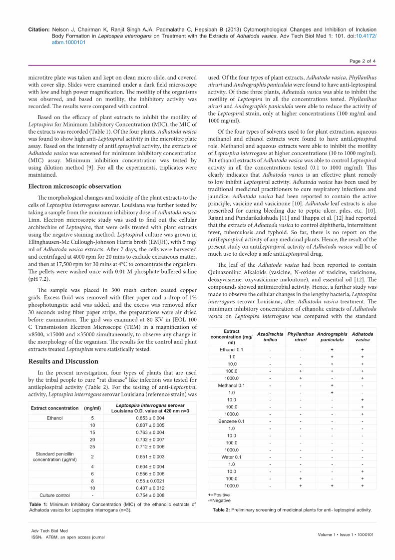
antibiotic, penicillin. At a concentration of 5 mg/ml. the OD value for Leptospiral culture was 0.853, but at a concentration 25 mg/ml, OD value was 0.712. This indicates a decline in the concentration of Leptospiral culture, when the dose is increased. The observed inhibition of Leptospira interrogans in the ethanolic extracts of Adhatoda vasica is close to the impact of penicillin. In the control, the cells of the Leptospira interrogans showed normal architecture (Length 5-10 µ, without any break and with the presence of inclusion body). An electron microscopic observation showed the nature as a lengthy spirochete, Leptospira interrogans. The same strain L.interogans serovar. Lousiana showed break in the cellular architecture on treatment with the extracts of A.vasica. Almost all the cells (200) observed (100%) after Adhatoda vasica treatment showed the absence of inclusion body (Tables 3 and 4). The lengthy spirochaete cells showed breaks at different locations due to Adhatoda vasica treatment. In 20.4% of the total cells observed, a single break was noticed in their lengthy structure. In 50.5% of the cells, double breaks were observed. In 29% of the cells, more than 2 breaks were noticed. The cellular damage by Adhatoda vasica extracts confirmed their efficiency to control Leptospira interrogans, and to inhibit their motility. This breakage in the cell confirmed the action of the bioactive compound present in Adhatoda vasica, on the lipopolysaccharide content of the cell wall of the Leptospira interrogans.
Inclusion body in Leptospira interrogan is normally associated with virulence of this microorganism [13], and it is absent in old cultures. In the present study, inclusion body (surface vacuole) on the outer envelope of the fresh isolate of L.interrogans was not observed in Adhatoda treated sample. This clearly confirmed the inhibition of virulence in this microorganism due to the phytochemicals present in Adhatoda vasica. Ellis et al. [13] and Alex et al. [14], had reported that the inclusion body was absent in the non-virulent culture of Leptospira interrogans. Penicillin had been reported to cause damage to the cell wall of bacteria during antibiotic therapy [15].
Electron microscopic analysis
Alex et al. [14] had made an electron microscopic observation of the Leptospira isolates for the first time in Indian subcontinent. In the
electron microscopy of the present study, the detail of normal and Adhatoda vasica treated Leptospira interrogans serovar Louisiana are recorded. The Leptospiral cell has an outer envelope consisting of a surface layer and an outer membrane (Figure 1a). Underneath the outer envelope is the peptidoglycon layer, which gives the cell its rigidity and the cytoplasmic membrane which envelope the cell cytoplasm.
At both the ends, the organism showed a bend but hook like appearance, which is prominent on one side. Surface vacuoles or inclusion body was seen on the outer envelope in different places (Figure 1b). The presence of inclusion body is attributed to the virulence of the organism [13,14].
On treatment of fresh isolates of Leptospira interrogans serovar Louisiana with the ethanol extracts of Adhatoda vasica (25 mg/ml), showed a significant damage to the cells of the spirochete. The normal cells are 5-12 µl long, but the Adhatoda vasica treated Leptospira interrogans serovar Louisiana cells showed break in more than 4 places in the cell (Figure 1c). Such cellular break in Leptospira interrogans serovar Louisiana indicates the inhibitory activity of the extracts. Besides the cellular break, inclusion bodies were also absent in the Adhatoda vasica treated Leptospira interrogans serovar Louisiana cells.
Although the cells of the Leptospira interrogans serovar Louisiana were fresh isolates, the loss of inclusion body after Adhatoda vasica treatment indicated the action of bioactive compound present in the plant Adhatoda vasica.
ConclusionIn the present study, the reduction of inclusion body in the
S. No Character observed
Percentage of damage observed in the cells of Leptospira interrogans
Adhatoda extracts (25 mg/ml)
Penicillin (6 µg/ml)
1. Single break in a cell 20.4 15.42. Double break in a cell 50.5 30.43. More than two breaks in a cell 29.0 56.41
Table 3: Changes in the cytomorphology of Leptospira interrogans treated with antibiotic penicillin and Adhatoda vasica extract.
TreatmentNumber of inclusion bodies observed in a
cell
Number of breaks observed in a cell of
Leptospira interrogans1.Normal 15 ± 2.0 -
2. Adhatoda vasica (mg/ml) 5 6 ± 0.5 -
10 4 ± 0.5 4± 115 - 6 ± 0.520 - 6 ± 0.525 - 7 ±0.5
3.Penicillin treatment (6 µg/ml) - 5 ± 1.0
Table 4: Effect of Adhatoda vasica on the formation of inclusion body in the cells of Leptospira interrogans serovar Louisiana-TEM observation of plant drug and Penicillin treated Leptospires (n=3).
Figure 1: (a) Electron microscopic figure showing the normal appearance of Leptospira interrogans serovar Louisiana. The spiral cell shows folding at scrotal places (F). Both ends of the spirochaete are hooked (H). Inclusion bodies appear (IB) throughout the length of the cell (Cax500). (b) A normal Leptospira interrogans serovar Louisiana cell under further magnification (Cax15,000). In the normal cell, Inclusion bodies (IB) are prominent. Spiral structure is clear and continuous throughout the cell. Hook (H) is very prominent. A big size inclusion body (IB) is present in the hooked end. The cell is highly intact. (c) An electron photomicrograph of a damaged cell of the spirochete, Leptospira interrogans serovar Louisiana. The cell is hypertrophied (Hy). The hooked end shows disintegration (D). Breaks appeared at several sites (B). The cell wall had been damaged. The big size inclusion body present in the hooked region is isolated. The inner region of the cell has low electron dense part (Cax35,000).
(a)
IB
IBH
F
H
Hy
B D(c)(b)
Leptospira treated with Adhatoda vasica extracts

Citation: Nelson J, Chairman K, Ranjit Singh AJA, Padmalatha C, Hepsibah B (2013) Cytomorphological Changes and Inhibition of Inclusion Body Formation in Leptospira interrogans on Treatment with the Extracts of Adhatoda vasica. Adv Tech Biol Med 1: 101. doi:10.4172/atbm.1000101
Page 4 of 4
Volume 1 • Issue 1 • 1000101Adv Tech Biol MedISSN: ATBM, an open access journal
cells of Leptospira interrogans serovar Louisiana vividly explains the antiLeptospiral activity of the plant drug. The loss of inclusion body indicates the loss of virulence of the cell [13]. Hence, the ability of the Adhatoda extracts to damage the inclusion body and motility confirmed the loss of virulence in the Leptospira interrogans serovar Louisiana cells.
Acknowledgement
Authors are thankful to Dr. K. Nataraja Sreenivasan, Professor, Bharathidhasan University, Trichy, Tamilnadu. Authors are thankful to Veterinary College, Chennai for TEM Study. Dr. N. Jeyakumar thanks the management to AIMST University, Malaysia.
References
1. Greig DJ, Gulland FMD, Kreuder C (2005) A decade of live California sea lion (Zalophus californianus) strandings along the central California coast: Causes and trends, 1991–2000. Aquatic Mammals 31: 11-22.
2. Vachvanichsanong P, Dissaneewate P, Mitarnun W (1999) Tubulointerstitial renal failure in childhood Leptospirosis. Pediatr Emerg Care 15: 332-334.
3. Faine S (1994) Leptospira and Leptospirosis, CRC Press, Boca Raton, Florida.
4. Feigin RD, Anderson DC (1975) Human Leptospirosis. CRC Crit Rev Clin Lab Sci 5: 413-467.
5. Natarajaseenivasan K, Boopalan M, Selvanayaki K, Suresh SR, Ratnam S (2002) Leptospirosis among rice mill workers of Salem, South India. Jpn J Infect Dis 55: 170-173.
Submit your next manuscript and get advantages of OMICS Group submissionsUnique features:
• Userfriendly/feasiblewebsite-translationofyourpaperto50world’sleadinglanguages• AudioVersionofpublishedpaper• Digitalarticlestoshareandexplore
Special features:
• 250OpenAccessJournals• 20,000editorialteam• 21daysrapidreviewprocess• Qualityandquickeditorial,reviewandpublicationprocessing• IndexingatPubMed(partial),Scopus,DOAJ,EBSCO,IndexCopernicusandGoogleScholaretc• SharingOption:SocialNetworkingEnabled• Authors,ReviewersandEditorsrewardedwithonlineScientificCredits• Betterdiscountforyoursubsequentarticles
Submityourmanuscriptat:http://www.omicsonline.org/submission/
Citation: Nelson J, Chairman K, Ranjit Singh AJA, Padmalatha C, Hepsibah B (2013) Cytomorphological Changes and Inhibition of Inclusion Body Formation in Leptospira interrogans on Treatment with the Extracts of Adhatoda vasica. Adv Tech Biol Med 1: 101. doi:10.4172/atbm.1000101
6. Jeyakumar N, Khan AA, Dhasarathan P, Singh AJ (2004) Seroprevelence of Leptospirosis in clinically suspected cases in Chennai city. Indian J Med Microbiol 22: 67.
7. Ramadass P, Jarvis BD, Corner RJ, Penny D, Marshall RB (1992) Genetic characterization of pathogenic Leptospira species by DNA hybridization. Int J Syst Bacteriol 42: 215-219.
8. Debnath C, Pal NK, Pramanik AK, Biswas M (2005) A serological study of Leptospirosis among hospitalized jaundice patients in and around Kolkata. Indian J Med Microbiol 23: 68-69.
9. Baker FJ, Breach MR (1980) Medical microbiological techniques. (1st Edn), Butterworth-Heinemann Ltd, London, UK.
10. Atal CK (1980) Chemistry and pharmacology of vasicine: a new oxytocic and abortifacient. Regional Research Laboratory, Jammu, India.
11. Rajani, M, Pundarikakshudu K (1996) A note on the seasonal variation of alkaloids in Adhatoda vasica Nees. Int J Pharma 34: 308-309.
12. Thappa RK, Agarwal SG, Dhar KL, Gupta VK, Goswami KN (1996) Two pyrroloquinazolines from Adhatoda vasica. Phytochemistry 42: 1485-1488.
13. Ellis WA, Hovind-Hougen K, Moller S, Birch-Andresen A (1983) Morphological changes upon subculturing of freshly isolated strains of Leptospira interrogans. Zentralbl Bakteriol Mikrobiol Hyg 255: 323-335.
14. Alex C, Ratnam S, Chandramohan A, Padmanaban VD (1995) Electron microscopy of a Leptospiral isolate. Indian Vet J 72: 1094-1095.
15. Voet D, Voet JG (2004) Biochemistry. (3rd edn), John Wiley and Sons, Hoboken, USA.