advances in the use of biotinylated diaminopyridine (bap...
TRANSCRIPT
Glycobiology vol. 4 no. 5 pp. 653-663, 1994
Advances in the use of biotinylated diaminopyridine (BAP) as aversatile fluorescent tag for oligosaccharides
Derek K.Toomre and Ajit Varki1
Glycobiology Program, UCSD Cancer Center, and Division of Cellular andMolecular Medicine, University of California, San Diego, La Jolla, CA92093, USA
'To whom correspondence should be addressed
We recently described a novel fluorescent compound, 2-amino,6-amidobiotinyl-pyridine (BAP), that allows thetagging of oligosaccharides, their fractionation by reversed-phase HPLC with picomole scale detection, and the forma-tion of functional neoglycoprotein equivalents with (strept)avidin for the detection of receptors and the generation ofmonospedflc antibodies (Rothenberg etaL, Proc. Natl Acad.Set USA, 90,11939-11943,1993). Here, we describe the en-hancement of this approach by the following, (i) A simpleone-step purification of BAP from its synthetic precursorsand other reactants. (ii) Development of HPLC sizing col-umn methods to quickly purify BAP-coupled oligosac-charides away from free BAP and other reactants. (iii)Development of anion-exchange and amine-adsorptionHPLC procedures for the fractionation of BAP-oligo-saccharide adducts by charge and size, respectively, (iv)Investigation of the affinity of BAP-oligosaccharides for(strept)avidin, confirming the formation of stable comp-lexes, (v) The use of BAP for sensitive monosaccharidecompositional analysis of glycoproteins. (vi) Formation ofstable BAP adducts without reduction and its implicationsfor the mechanism of adduct formation. These advancesmake available a multitude of techniques for the fractiona-tion of BAP-coupled oligosaccharides based on severaldifferent physical parameters. Distinct species of BAP-coupled oligosacharides can be isolated and subjected todetailed structural analysis. Such defined molecules formstable complexes with streptavidin that are effectively neo-glycoproteins, which can be used in a variety of biologicalapplications. Notably, all of these approaches require rel-atively inexpensive materials and conventional equipmentavailable In most laboratories.
Key words: analysis/biotin/fluorescence/fractionation/neoglyco-conjugates
Introduction
Oligosaccharides are involved in many diverse biological roles,such as intracellular trafficking of lysosomal enzymes (Dahmset al, 1989), receptor-mediated endocytosis of asialoglyco-proteins (Lodish, 1991; Lee, 1992) and cell-cell interactionsmediated by the selectins (Cummings and Smith, 1992).The determination of the structure of a given oligosaccharide
and probing of its potential biological functions can be quitechallenging. Frequent hurdles in structural analyses includelow quantities of material, linkage and branching hetero-geneity, and additional modifications (e.g. acetylation, sulpha-tion and phosphorylation) (Baenziger and Green, 1988; Dahmset al, 1989; Varki, 1993; Parekh, 1994). The purification ofoligosaccharides from complex mixtures requires a varietyof techniques, as well as sensitive and specific detection.Classically, the reducing end of oligosaccharides has beenlabelled either radioactively (Kobata, 1994) or by chromo-phores (Lee et al, 1991; Hase, 1994) to aid in sensitivedetection. The hydrophobic or anionic character of thechromophoric groups can be exploited to improve oligo-saccharide fractionation by either high-performance liquidchromatography (HPLC) or electrophoresis techniques(Jackson, 1991; Lee et al, 1991; Stack and Sullivan, 1992;Hase, 1994). Detection of unlabelled sugars by pulsedamperometric detection (Townsend et al., 1989) is alsopossible, but the technique requires expensive equipment andsubjects the sugars to strongly basic conditions which cancause de-O-acetylation and/or epimerization. Often, two ormore chromatographic techniques, based on different physicalproperties, are required to provide resolution of complexmixtures. An example of this approach is the recently described'two-dimensional mapping' of pyridylamino (PA)-coupledoligosaccharides by size and hydrophobicity (Tomiya et al,1991; Hase, 1994).
A fundamental limitation with the above techniques is thatonce the oligosaccharides of interest have been isolated andcharacterized, they are of relatively limited use for functionalor biological studies. A recently described option is to convertfluorescent PA sugars to biotinylated sugars by reductivehydrogenation, followed by hydrazinolysis and subsequent re-derivatization with biotin (Hase, 1992). By replacing the PAgroups with biotin, (strept)avidin neoglycoproteins could beprepared for functional studies. However, this techniquerequires separate rederivatization and repurification of eachPA-sugar of interest. Furthermore, the hydrazinolysis maycause the degradation of functionally important groups such asO-acetyl or sulphate esters (Patel and Parekh, 1994).
Previously (Rothenberg et al, 1993), we described a novelfluorescent compound, 2-amino,6-amidobiotinyl-pyridine(BAP), that allows tagging of oligosaccharides and theirfractionation by reversed-phase HPLC with picomole-leveldetection [see Rothenberg et al (1993) for the structure of BAPand the oligosaccharide-BAP adduct]. Since biotin is alreadyattached to the fluorophore, numerous applications couldsubsequently exploit the multivalent biotin-(strept)avidininteraction (Wilchek and Bayer, 1988; Shao et al., 1990; Shao,1992). The biotin moiety permits the formation of neoglyco-protein equivalents with (strept)avidin for the detection ofreceptors and the generation of monospecific antibodies
© Oxford University Press 653

D.K.Toomre and A.Varki
(Rothenberg et al., 1993). However, the versatility andeffectiveness of BAP as an oligosaccharide tag requires muchfurther investigation.
Here, we present improved methods for the preparation andpurification of both BAP and BAP-oligosaccharides, andtechniques for fractionating BAP adducts by size and charge. Amethod for using BAP for accurate and sensitive monosac-charide composition analysis of glycoproteins is also presen-ted. Further, we address the stability of BAP adducts to (strept)avidin under various conditions. Unlike other recently de-veloped high-technology approaches, the systems describedhere employ conventional and relatively inexpensive materialsand instrumentation.
Results and discussion
One-step purification of BAP from its synthetic precursorsand other reactants
In our previous study (Rothenberg et al., 1993), the reportedpurification method for preparing BAP was: (i) technicallydifficult and time consuming, involving many steps and lastingmore than a week; (ii) expensive because large volumes ofsolvents were required for flash chromatography; and (iii)impractical for small-scale preparations of BAP. Here, wedescribe an improved and simplified method for purifying BAPfrom reactants and side products after synthesis.
BAP was synthesized using minor modifications of the pre-viously described method (Rothenberg et al., 1993). Asindicated under Materials and methods, we have now foundthat the entire reaction mixture of substrates, side products andBAP can be directly loaded onto a 'Spice C18 tube' afterdilution in water, allowing a one-step purification. The onlylimiting factor is the capacity of the Spice tube for BAP (-50mg) and any remaining unreacted biotin. During the waterwash, a yellowish band (also fluorescent) adheres to the topof the Cig tube, a blue band migrates slowly, while thestrongly fluorescent excess of 2,6 diaminopyridine (DAP)is slowly eluted. As seen in lane 1 of Figure 1, A/-hydroxy-sulphosuccinimide (NHS) and fluorescent DAP run through theCig tube during the water wash. A second wash with 10%acetonitrile is used to elute remaining traces of DAP. The paleyellow and UV-fluorescent BAP is eluted with 50%acetonitrile. This material has an identical mobility to authenticBAP, as detected by both UV and iodine visualization (seeFigure 1, lane 3). Reversed-phase (RP)-HPLC and fast atombombardment mass spectrometry (FAB-MS) confirmed theidentity of the material as BAP (mol. wt 336, data not shown).No additional material was eluted with 100% acetonitrile.A blue compound that co-migrated with l-ethyl-3-(3-dimethyl-aminopropyl) carbodiimide hydrochoride (EDC) was elutedduring a subsequent 100 ml column regeneration wash (seeFigure 1, lane 5). Smaller capacity C18 Spice cartridges werealso effective in the purification of BAP, although BAP elutedwith lower concentrations of acetonitrile. The final yield ofBAP from the 50% acetonitrile eluate material was 31% (byweight, based on original biotin input). The yield increased to38% when a 10-fold molar excess of DAP to biotin was used inthe initial reaction, instead of the previously described 3-foldmolar excess. The moderate yields of 31-38% may reflect thelimited stability of activated EDC complexes under aqueousconditions and are currently under investigation. The purity of
- -S.F.
BAP
| NHS, Biotin
DAP
EDC
lane 1 2 3 4 5
Fig. 1. HPTLC of BAP purification on a C,8 tube. Equal aliquols of variousC|j washes were spotted on a silica gel TLC plate, developed in 85% ethanoland stained with iodine vapour. Lanes 1-5 are the sequential elution of theC|S tube with water, 10% acetonitrile, 50% acetonitrile, 100% acetonitrile and2:1 chloroform/methanol in 1% glacial acetic acid, respectively. Migrationpositions of standards and the solvent front (S.F.) are indicated on the right.The TLC was scanned and processed using an Applescan scanner.
BAP was slightly improved from 95% to >99% (as judged byRP-HPLC fluorescence) by a second passage over a C18 tube(data not shown). The purity of the final preparation iscomparable to that obtained by the previously describedmethod (Rothenberg et al., 1993).
The new method is: (i) quick, the purification takes ~1 day;(ii) inexpensive, only minimal amounts of organics and Ci8
tubes or cartridges are required; (iii) adaptable, it can be scaledup or down as desired. Thus, reasonable quantities of BAP cannow be quickly prepared at low cost.
General properties of BAP
The biotin moiety causes BAP to be slightly hydrophobiaHowever, the solubility of BAP in water at room temperature is~1 mg/ml (or 3000 nmol BAP/ml). This can be increased (> 10-fold) by the presence of very small quantities of organicmodifiers [(e.g. <1% dimethyl sulphoxide (DMSO)]. More-over, BAP-oligosaccharide adducts contain hydrophillic sugarswhich should also increase solubility. In practice, organicmodifiers (e.g. acetonitrile) facilitate the fractionation of BAPadducts on 'toyopearl' (Tosohass) or silica-based supports.However, we have noticed some smearing on Sepharose andSephadex resins due to non-specific interactions with BAP. Aspreviously reported, the optimum pH for sensitive detection is<5 (Rothenberg et al, 1993). The adducts show good stabilityat room temperature (1 day), and for over a year at - 20°C (datanot shown). Finally, it should be noted, that as with otherpreviously described oligosaccharide-tagging methods involv-ing reductive amination, the coupling reactions are not stoich-iometric, and depend to some degree on the nature of theoligosaccharide being coupled. Using BAP, we have typicallynoted coupling efficiencies ranging from 40 to 90%, with thelarger, more charged chains tending to be less efficient.
654
Use of BAP as a fluorescent tag
LUOZ111owLUCCO=5
1I
-BA
P
I
vl£
___-=
=»
BA
P
V10 20
TIME (min.)
30
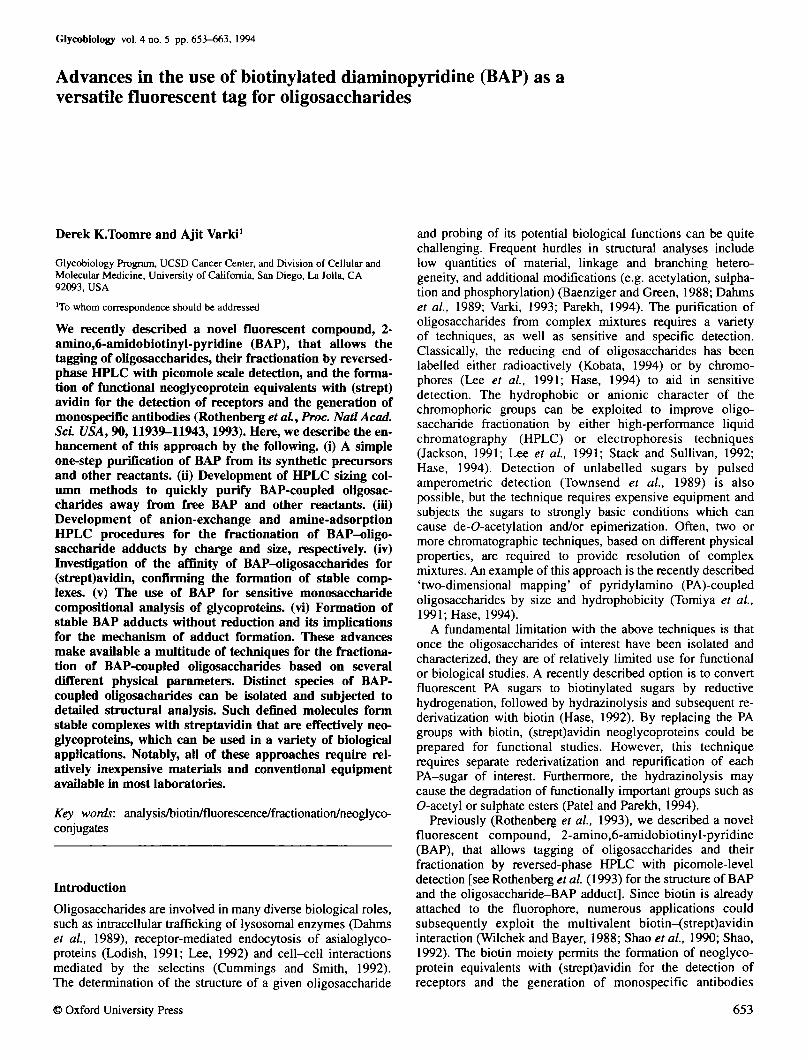
Fig. 2. Removal of BAP and fractionation of small BAP adducts by size-exclusion chromatography (SEC-HPLC). A mixture of sialylactose-BAP,chitobiose-BAP, fucose-BAP and BAP was injected onto a TSK-G3000PWxl HPLC column operating isocratically and the fluorescencewas monitored. The void volume (where large BAP adducts elute) ismarked by i v
Development of HPLC sizing column methods to fractionatedistinct species of BAP-coupled oligosaccharides away fromfree BAP, and from one another
Historically, after oligosaccharide derivatization, excess coup-ling reagents and contaminants have been removed by gelfiltration, evaporation or extraction with benzene (Hase et al,1984; Iwase et al, 1990; Kuraya and Hase, 1992; Linhardt,1994). Evaporation techniques are usually only partiallyeffective in removing excess reagents, and must be followed bygel-filtration chromatography. Slow column flow rates cannecessitate lengthy run times, sometimes exceeding 24 h(Rothenberg et al., 1993; Hase, 1994). To avoid these time-consuming or chemically toxic processes, an HPLC frac-tionation of BAP-oligosaccharides away from free BAP wasperformed by size exclusion on a TSK-G3000PWxl column.
As shown in Figure 2, this system gave an excellentseparation of oligosaccharide adducts (mono-, di- and tri-saccharides), based on size, with high reproducibility. LargerBAP-oligosaccharides eluted close together near the voidvolume (data not shown). Since all coupled sugars are wellseparated from free BAP, the column is routinely usedpreparatively to remove excess BAP and other reagents fromderivatized samples. The short run time, coupled with the
H-1 H-2
8-1 8-N
P-1
"l-AddGtycopfotein
FBxtnogen
bovine LautWzngHofmone
Pentaman nosyiP t l
I10
I20
TIME (min.)
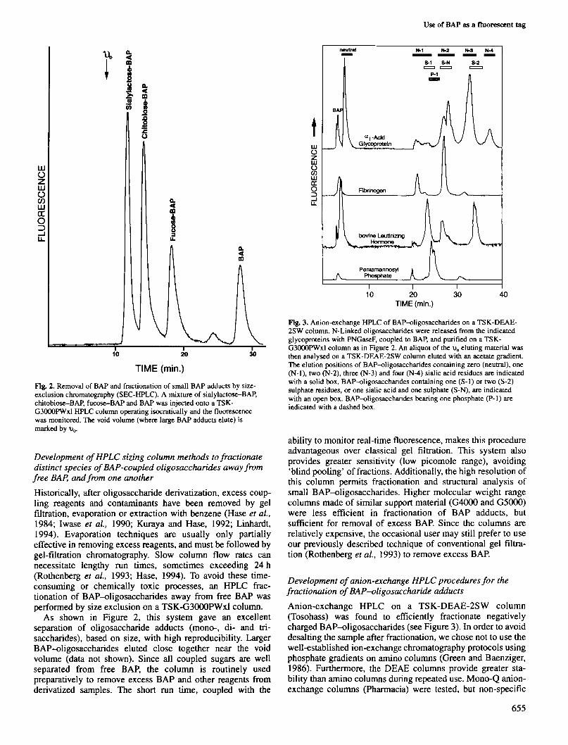
Fig. 3. Anion-exchange HPLC of BAP-oligosaccharides on a TSK-DEAE-2SW column. N-Linked oligosaccharides were released from the indicatedglycoproteins with PNGaseF, coupled to BAP, and purified on a TSK-G3000PWxl column as in Figure 2. An aliquot of the v0 eluting material wasthen analysed on a TSK-DEAE-2SW column eluted with an acetate gradient.The elution positions of BAP-oligosaccharides containing zero (neutral), one(N-l), two (N-2), three (N-3) and four (N-4) sialic acid residues are indicatedwith a solid box. BAP-oligosacchandes containing one (S-1) or two (S-2)sulphate residues, or one sialic acid and one sulphate (S-N), are indicatedwith an open box. BAP-oligosacchandes bearing one phosphate (P-1) areindicated with a dashed box.
ability to monitor real-time fluorescence, makes this procedureadvantageous over classical gel filtration. This system alsoprovides greater sensitivity (l°w picomole range), avoiding'blind pooling' of fractions. Additionally, the high resolution ofthis column permits fractionation and structural analysis ofsmall BAP-oligosaccharides. Higher molecular weight rangecolumns made of similar support material (G4000 and G5000)were less efficient in fractionation of BAP adducts, butsufficient for removal of excess BAP. Since the columns arerelatively expensive, the occasional user may still prefer to useour previously described technique of conventional gel filtra-tion (Rothenberg et al, 1993) to remove excess BAP.
Development of anion-exchange HPLC procedures for thefractionation of BAP—oligosaccharide adducts
Anion-exchange HPLC on a TSK-DEAE-2SW column(Tosohass) was found to efficiently fractionate negativelycharged BAP-oligosaccharides (see Figure 3). In order to avoiddesalting the sample after fractionation, we chose not to use thewell-established ion-exchange chromatography protocols usingphosphate gradients on amino columns (Green and Baenziger,1986). Furthermore, the DEAE columns provide greater sta-bility than amino columns during repeated use. Mono-Q anion-exchange columns (Pharmacia) were tested, but non-specific
655

D.K.Toomre and A.Varkl
interactions caused excessive band broadening. Owing to thehydrophobic nature of BAP, the DEAE system was operatedin the presence of 25% acetonitrile. Under the stated con-ditions, oligosaccharides containing all commonly foundanionic charges (sialic acid, sulphate, phosphate or uronic acid)bound to the column, and were eluted by an increasing gradientof acetate. Separation of different oligosaccharides into theindicated classes was based primarly on the net negativecharge. As seen in Figure 3, N-linked oligosaccharides froma r ac id glycoprotein, where the negative charge is dueexclusively to sialic acid, were separated into five discreteregions containing molecules with 0-4 sialic acids. Moreover,structural heterogeneity within a given charge class can beseen, and most likely represents a partial separation ofoligosaccharides that differ in molecular size, as has beenreported previously for aj-acid glycoprotein (Ohta et al.,1990). For unknown samples, this additional level offractionation could potentially be utilized in structuralanalyses.
Since the column is run at pH - 4, the pK,s of differentanionic groups influence the net negative charge and hence thebinding to the column. A single negative charge from sulphateshows increased retention times relative to sialic acid, althoughvariations in sugar linkage positions can also effect elution(Green and Baenziger, 1986; Ohta et al, 1990). Increasedretention of P-Man^BAP relative to sialic acid (Figure 3) mostlikely results from a stronger net negative charge, due to partialdeprotonation of phosphate hydroxyl groups (pK,, = 2.1 and5.5) at pH 4.
Sulphated oligosaccharides from bovine luteinizing hormone(bLH) fall into separate, although partially overlapping, regionswith sialylated oligosaccharides. Green and Baenziger (1986)previously reported separation of sialylated and sulphatedoligosaccharides on an amine column at pH 1.7, but not at pH4. The partial resolution of these oligosaccharides even at pH 4may reflect the difference in column stationary phase or thepresence of organic modifiers. By further lowering the pH, theseparation of sialic acid and sulphated regions could possiblybe further enhanced.
This system provides efficient and reliable separation ofBAP-oligosaccharide adducts by negative charge. Each run canbe completed in an hour and no column regeneration is re-quired. Treatment of the BAP-oligosaccharides either chem-ically or enzymatically provides a simple means of confirmingthe nature of the negatively charged moiety. Unlike mostsystems which typically use sodium or phosphate salts, thismetiiod uses a completely volatile buffer system, avoids desalt-ing and facilitates subsequent chromatographic or enzymaticsteps.
After anion-exchange HPLC, the pooled fractions may befurther purified or structurally characterized by other HPLCtechniques. As a model study, structural sequencing of pre-viously well-known biantennary BAP-oligosaccharides fromfibrinogen was carried out Analysis of these oligosaccharidesisolated from the N-2 region of the anion-exchange column(Figure 3) was performed by sequential exoglycosidasedigestions and RP-HPLC analysis (Figure 4). Treatment withglycosidases caused the product to become more hydrophobicand elute with increased acetonitrile concentrations, resultingin distinct shifts after each digestion.
Although very useful for such analysis, we have foundthat RP-HPLC has the following general limitations: (i)failure to resolve oligosaccharide species of similar relative
AUS P-Oal P-Hex a-Man
34 40 min.
Fig. 4. Analysis of BAP adducts on RP-HPLC by sequential exoglycosidasedigestions. BAP adducts of fibrinogen oligosaccharides were collected fromthe N-2 region on a TSK-DEAE-2SW column (see Figure 3) and treatedwith A.ureafaciens sialidase (AUS), jack bean f}-galactosidase (|J-Gal),S.pneumoniae P-AZ-acetylhexosaminidase (jJ-Hex) and a-mannosidase(a-Man) as indicated, and a fraction was analysed on a Microsorb C,jcolumn. Owing to differences in the pH optima of the enzymes the majorpeak, after treatment with AUS, was collected and sequentially treated withthe remaining enzymes. Sham treatments without enzyme showed no effects.
hydrophicity; (ii) difficulty in estimating the number of sugarresidues of an unknown sample based solely on hydro-phobicity, since factors such as branching can play importanteffects (Oku et al., 1990); (iii) poor resolution of oligo-saccharides with >12 monosaccharide units. Therefore, separa-tion according to another physical parameter, such as sizefractionation by amine-adsorption chromatography, wasexplored.
Development of an amine-adsorption HPLC system for sizefractionation of neutral BAP-oligosaccharides
An amine-adsorption HPLC system provided separation ofneutral BAP adducts based on the number of hydroxyl groups.Using an inexpensive NH2 column (Microsorb, Rainin) and anincreasing water gradient, excellent fractionation of a BAP-oligosaccharide ladder from partially hydrolysed dextran wasobtained (see Figure 5a). Baseline separation of (Glc)n-BAPadducts containing 1-35 glucose residues was achieved in <50min. The minor peaks in between the major ones are probablydue to isomers. Although the upper limits of this system werenot explored, these results compare very favourably withamine-adsorption methods of size fractionation for non-derivatized oligosaccharides (Mellis and Baenziger, 1981). Inthe present system, BAP-oligosaccharides from RNase Byielded a pattern very similar to published reports (Rudd et al.,1992). The elution position of the first major peak, cor-responding to Man5-GlcNAc2-BAP, near the Glc8 standardmay result from increased retention of the mannose oligo-saccharide containing a 1,6-linkage (Blanken et aL, 1985).Oligosaccharide adducts from ovalbumin contained a cluster of
656

Use of BAP as a fluorescent tag
LLJO
zuowLLJrr
hLU
U6 7 8 9 10 11 12 13
JUUUUJULA
< 3 5 7 9t
1
1
t
\JvV
ttpi
•13
t i S 17
III I r t •?1?7|31|3B
I Dextran -BAP
A ' RNAase-BAP
Ovalbumin-BAP
25TIME (min.)
50
Fig. 5. Amine-adsorption HPLC fractionation of neutral BAP-oligosacchandes. Aliquots of various neutral BAP-oligosaccharides wereanalysed on a Microsorb NH2 column. A dextran-BAP ladder was preparedby partial acid hydrolysis and coupling to BAP. Coupled sugars were purifiedon a C|g cartridge. The number of glucose residues is indicated above theHPLC peaks. RNase B and ovalbumin N-linked oligosaccharides werereleased by PNGase F, coupled to BAP and initially purified on aTSK-G3000PWxl column. The inset shows an expanded view of theregion including the ovalbumin and RNase B adducts.
over a dozen different neutral species which were partiallyresolved consistent with known structural heterogenity. Sinceoligosaccharides show a relatively predictable behaviour onamine columns, it is possible to determine the relativeoligosaccharide size by comparison with glucose standards.Based on these findings, it is likely that an amine-adsorption:ion-suppression system (Green and Baenziger,1986) could also be developed as an alternative method for thefractionation of negatively charged BAP-oligosaccharides.Together, the combination of amine-adsorption HPLC withanion-exchange HPLC and RP-HPLC represent a powerfulapproach for the fractionation and analysis of heterogeneousoligosaccharide mixtures.
100
80
60
40
20
10 min.
2 hours36 hours
3 weeks
STREPTAVIDIN
(a)H20 6MUrea 6M G-HCI 10mM Biotin
Solvent
100
H20
(b)
6M Urea 6M G-HCI
Solvent
10mM Biotin
Fig. 6. Stability of BAP adduct attachment to streptavidin and avidin.[3H]Gal-chitobiose-BAP was bound to either streptavidin (a) or avidin (b).Centncon-10 ultrafiltration tubes were used to remove unbound adducts.The release of oligosaccharide adducts was monitored after incubation withwater, 6 M urea, 6 M guanidine-HCl (G-HCI) or 10 mM biotin for varioustimes, and was expressed as a percentage of released radioactive materialfrom remaining bound material at each step.
Determination of the stability of BAP—oligosaccharideadducts attached to avidin and streptavidin
The original purpose of including biotin in the fluorophore wasto have an oligosaccharide tag that permitted the formation ofneoglycoproteins for functional studies. In order for BAPcompounds to be practical in such functional studies, it isimportant that the affinity for (strept)avidin be high, as seenwith unmodified biotin (Kd= 10-12-10-13) (Livnah et al, 1993).Prior literature has shown that, in some cases, the derivitizationof biotin without an additional spacer arm may stericallyinterfere with binding to (strept)avidin (Green, 1990). Also, the(strept)avidin-biotin complex could possibly be destabilized bythe proximity of the positively charged pyridinium ring.
[3H]Galactose-chitobiose-BAP was chosen as a modelcompound to investigate the stability of BAP adducts tostreptavidin and avidin (see Figure 6). The stability of bindingto either denaturation or competition with cold biotin wasexamined. After incubation in water for 3 weeks at roomtemperature, <3% of the BAP adduct was released in each case.Even in the presence of 6 M urea, relatively little material wasreleased. However, 6 M guanidine-HCl caused release of 18and 67% of BAP adduct from streptavidin and avidin after 3weeks, respectively. The differences between the two proteinsmay reflect the fact that in 6 M guanidine-HCl streptavidin istetrameric, while avidin is known to dissociate into a lower-affinity monomeric form (Green, 1990). For avidin, the binding
657

D.K.Toomre and A.Varki
of BAP adducts versus [3H]biotin was directly examined bycompetition with 10 mM biotin, corresponding to a 50 000 Mexcess of unlabelled biotin. After 36 h, only 3% of the free [3H]biotin was displaced (data not shown), whereas 37% of theBAP adduct was displaced. Although BAP adducts show aslightly reduced affinity, by a factor of -10, these valuesindicate very stable complexes of BAP adducts withstreptavidin or avidin. Thus, the pyridinium ring does notmarkedly reduce the affinity of BAP adducts for (strept)avidinand the interaction can be considered practically covalent, evenin rather severe conditions. To release bound BAP adducts, it isnecessary to either denature the protein into the monomericform or use a large excess of free biotin at room temperature forlong durations of time. The realistic implication is that thebinding of BAP adducts to streptavidin or avidin is practicallynon-reversible in all but the harshest conditions, thus formingstable neoglycoproteins for functional and biological studies.Note, however, that binding of BAP adducts to columns ofmonomeric avidin (Pierce) is reversible with mild acid(Kohanski and Lane, 1990), allowing the use of such columnsto retrieve BAP-oligosaccharide adducts from reactionmixtures if necessary.
Use of BAP for sensitive monosaccharide compositionalanalysis of glycoproteins
The procedures described below provide a convenient andsensitive means of analysing both neutral and amino mono-saccharides from glycoproteins by RP-HPLC. Although thereare many methods for monosaccharide analysis by pre-columnderivatization, several require additional chromatographic stepsor specialized equipment to remove excess reagents (Suzuki,1991; Spiro and Spiro, 1992; Hase, 1994). Furthermore, themajority require specialized columns as well as heating orcooling jackets (Muramoto et al., 1987; Suzuki, 1991; Spiroand Spiro, 1992; Kwon and Kim, 1993).
In our studies, inexpensive Microsorb-MV C18 columns pro-vided good peak resolution of different BAP-monosaccharidesat ambient temperatures under isocratic conditions. The mono-saccharides frequently found in glycoproteins are wellresolved: Gal, Man, Xyl, Fuc, GlcNAc and GalNAc (seeFigure 7). Glucose, a common environmental contaminant,eluted as a shoulder behind galactose. Other C18 columns aswell as C4 columns (authors' unpublished observations) alsowork well, although optimal acetonitrile concentrations varybetween columns. This simple method requires no additionalchromatography or workup of BAP adducts prior to HPLCanalysis, and is sensitive from the low picomole range (-20pmol) up to 10 nmol of reducing sugar.
To minimize sample loss and maintain good reproducibility,it is preferable not to remove excess BAP before HPLCanalysis. Thus, to avoid column overload, only 100 nmol ofBAP were used in each reaction. This corresponds to a ^5-foldmolar excess of BAP over the total monosaccharides analysed.When 25% of such a sample was injected, there was no peakoverlap between BAP-monosaccharides and free BAP. Thestandard curves for different monosaccharides show a linearresponse from <100 pmol to ~3 nmol of monosaccharidemixtures (see Figure 8). If greater quantities of sugars arepresent, they will be underrepresented. However, this can easilybe avoided by serial dilutions prior to derivatization. Thevariations in peak area for different monosaccharides,particularly the lower reactivity of the 2-amino-acetyl sugars,
TIME (min.)Fig. 7. RP-HPLC analysis of reduced BAP-monosaccharide adducts. Amonosaccharide mixture containing equal molar amounts of galactose (Gal),mannose (Man), xylose (Xyl), fucose (Fuc), W-acetylglucosamine (GlcNAc)and N-acetylgalactosamine (GalNAc) was coupled to BAP and subsequentlyreduced. Samples corresponding to 1 nmol (a) or 20 pmol (b) were injectedonto a Microsorb Cla HPLC under isocratic conditions of 74% Buffer A and26% Buffer B (resulting in a final acetonitrile concentration of 13%) at aflow rate of 0.5 ml/min, and the fluorescence was monitored. TheBAP-monosaccharide peaks are marked, while unmarked peaks arebyproducts of the coupling reaction. The sensitivity of the range wasincreased for trace (b).
Fig. 8. Standard curve of coupling monosaccharides to BAP. A mature ofmonosaccharides (100 pmol to 3 nmol) was coupled to BAP and reduced;10% of each aliquot was analysed by RP-HPLC, as described in Figure 7.
658

Use of BAP as a fluorescent tag
Table I. Monosaccharide compositional analysis of glycoprotcins
Glycoprotein*
Ovalbumin
Fetuin
Mucin(bovine submaxillary]
Monosaccharide Qig/mg)
Gal
6.1(0-15)
28.4(35-46)
9.61 (10-36)
Man
24(20-28)
26.6(23-30)
5(0-5.4)
Xyl
0(0)
0(0)
0.7(0-1.1)
Fuc
0(0)
0(0)
4.2(3-18)
GlcNAc
22.9(12-27)
30.8(26-56)
11.2(13-69)
GalNAc
0(0)
10.2(5.4-7)
73.6(30-168)
'1 mg each of ovalbumin, fetuin and bovine submaxillary mucin were acid hydrolysed, the monosacchandes re-A'-acetylated (Kwon and Kim, 1993), and afraction (1%) was coupled to BAP and subsequently reduced. Aliquots (10%) of each were analysed by RP-HPLC as described under Materials and methods.The results (average of two experiments) are expressed as Hg/mg relative to known monosaccharide standards. The values in parentheses correspond to the rangeof published values from prior literature [summarized in Kwon and Kim (1993)].
reflects variable coupling efficiency under these conditions oflimited BAP excess. This is not due to a decreased detectorresponse, since 25 pmol of purified Xyl and GlcNAc adducts(measured by biotin content) had similar RP-HPLC fluorescentpeak areas (data not shown). Thus, under these conditions oflow BAP concentrations there is a linear but incomplete coup-ling to some monosacchandes. It should be noted that underconditions where higher concentrations of BAP are used (i.e. incoupling to oligosaccharides), the efficiency of coupling ofGlcNAc as a reducing terminal monosaccharide can be as highas 90%.
To test the applicability of this system to monosaccharideanalysis of intact glycoproteins, we acid hydrolysed severalpreviously well-characterized glycoproteins, re-N-acetylatedand BAP-coupled the monosaccharides. The values of mono-saccharide composition obtained (by comparison to mono-saccharide standards) from the various glycoproteins correlatedwell with published values (Table I). The amount of materialinjected per HPLC run typically represented ~ 1 (ig of glyco-protein. Thus, the method is useful for monosaccharide ana-lysis of glycoproteins.
Monosaccharide analysis of samples containing glucose
In the samples studied above, glucose contamination was not aproblem. Since most glycoproteins do not contain muchglucose (Kornfeld and Kornfeld, 1985; Kwon and Kim, 1993),analysis of such molecules can be accomplished in a single run.However, in some cases the fact that the glucose adductpartially overlaps in elution with the galactose adducts couldpose a potential problem. Likewise, glucose can arise fromhydrolysis of contaminating polysaccharides from theenvironment. In order to separate galactose and glucosederivatives (a problem frequently encountered in RP-HPLCmonosaccharide analysis) (Eggert and Jones, 1985; Muramotoet ai, 1987; Kwon and Kim, 1993), monosaccharides werecoupled to BAP and the subsequent reduction step was omitted.Under these conditions, the elution order and resolution of themonosaccharide adducts was altered such that galactose andglucose adducts are readily resolved (see Figure 9). Althoughnon-reductive coupling standard curves of the linear range arestill needed, samples with glucose contamination couldpresumably be analysed in two runs, with and without adductreduction.
10TIME (min.)
Fig. 9. RP-HPLC resolution of Glc from Gal and Man monosacchandescoupled to BAP under non-reducing conditions. A mixture containing Gal,Glc, Man, Xyl, Fuc, GlcNAc and GalNAc was coupled to BAP withoutsubsequent reduction. An aliquot was injected onto a Microsorb C,8 columnunder isocratic conditions of 72% Buffer A and 28% Buffer B (resulting in afinal acetonitrile concentration of 14%) at a flow rate of 1 ml/min. Peakscorresponding to Gal, Glc, Man and Xyl adducts are marked, while the othermonosaccharides co-elute with BAP. Peaks marked a and b correspond toreaction byproducts and monosaccharide anomers, respectively.
This procedure for monosaccharide analysis has the advantagethat the process can be easily performed in a single reactionvial. Furthermore, for laboratories conducting only occasionalmonosaccharide analysis, BAP provides an alternative routewithout requiring the acquisition of additional equipment, otherthan a fluorescence HPLC detector.
BAP-oligosaccharide adducts can be stable withoutsubsequent reduction
Traditionally, the tagging of sugars by reductive amination isaccomplished in two steps: the formation of a reversible Schiffbase, and subsequent reduction to obtain an irreversible cova-lent adduct. In the section above, we noted the somewhat
659

D.K.Toomre and A.Varkl
100
O
oo
0.085 42 60 85 425 1060 2125 8500
mM REDUCING AGENT
Fig. 10. BAP forms stable, but acid-labile adducts without reduction. [3H]Gal-chitobiose was coupled to BAP and subsequently reduced with varing amounts ofBDA. Half of the sample was loaded onto a C|g cartridge, washed with water, eluted with 50% acetonitnle and fractions were counted. The percentage of couplingis shown with dashed bars and refers to the amount of bound radioactive material relative to the total. The error bars show the SD of the mean of threeexperiments. The other half was dried, treated with 2 M acetic acid for 3 h at 80°C (to break glycosylamine adducts), and similarly passed over a C|8 cartridge andcounted and shown with solid bars.
surprising finding that monosaccharide-BAP adducts appearedto be relatively stable, even without reduction. [3H]Galpl-4GlcNAcPl-4GlcNAc ([3H]Gal-chitobiose) was used as amodel compound to study the effects of increasing amounts ofreducing agent on coupling efficiency. As shown in Figure 10,a maximum coupling efficiency of 92% was observed with theaddition of -1 M borane dimethyl amine (BDA). Even in theabsence of added reducing agent, most of the radioactivematerial remained bound to the C|g cartridge, a characteristic ofa hydrophobic BAP-oligosaccharide complex. Furthermore,the non-reduced adduct was stable in water for >10 min at100°C (data not shown), suggesting that it may not simply bean unreduced Schiff base. In the absence of reducing agents,the formation of stable cyclic glycosylamines complexed with2-aminopyridine has been reported (Li and Her, 1993). Todetermine if the non-reduced material showed the acid labilitytypical of glycosylamines (Her et al., 1987), half of thevariously reduced samples were heated in 2 M acetic acid at80°C for 3 h, and then analysed. As seen in Figure 10, the acidtreatment greatly diminished the C|8-bound material in thepartially reduced or non-reduced samples. Additional acidtreatment of the non-reduced sample caused a further 50%decrease in binding (data not shown). The stability of the non-reduced BAP adduct in aqueous conditions, and its degradation
under acidic conditions (Amadori rearrangement products arestable to acid hydrolysis; Her et al., 1987), would suggest thatit is not a Schiff base nor an Amadori rearrangement product,respectively. This suggests that the adduct is most likely aglycosylamine. Further work is needed to verify this proposal.
One advantage of coupling oligosaccharides to BAP undernon-reducing conditions is that the closed ring of a glycosyl-amine can impart different physical properties on the adduct.For instance, others (Her et al., 1987) reported improved HPLCresolution of PA-glycosylamines compared to open ringreduced Schiff bases. We have shown above that non-reducedBAP monosaccharides display different elution profilescompared to their reduced counterparts. This can be advan-tageous for separating monosaccharides containing galactose,glucose and mannose, a problem sometimes encounteredin monosaccharide analysis by pre-column derivatizationmethods (Eggert and Jones, 1985; Kwon and Kim, 1993). Forthe most part, glycosylamines were purposely avoided in thepast, due to the additional complexity of the anomeric linkageto the sugar (a or p); however, there are instances whereinglycosylamines can be a useful alternative for extractingstructural information. Furthermore, the oligosaccharides canbe released reversibly with mild acid for the recovery of freeoligosaccharides that have an available reducing end. Finally,
660
Use of BAP as a fluorescent tag
RELEASEDOLJGOSACCHARIDES
MONOSACCHARIDEANALYSIS BY RP-HPLC
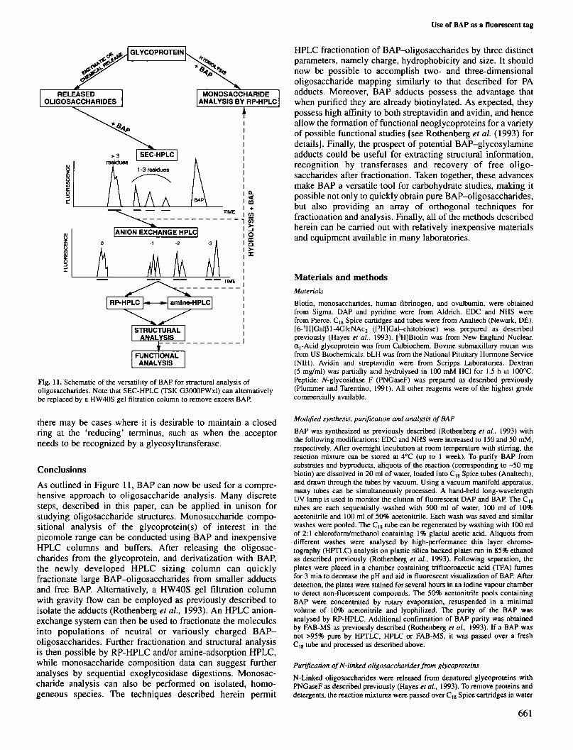
Fig. 11. Schematic of the versatility of BAP for structural analysis ofoligosaccharides. Note that SEC-HPLC (TSK G3000PWxl) can alternativelybe replaced by a HW40S gel filtration column to remove excess BAP.
there may be cases where it is desirable to maintain a closedring at the 'reducing' terminus, such as when the acceptorneeds to be recognized by a glycosyltransferase.
Conclusions
As outlined in Figure 11, BAP can now be used for a compre-hensive approach to oligosaccharide analysis. Many discretesteps, described in this paper, can be applied in unison forstudying oligosaccharide structures. Monosaccharide compo-sitional analysis of the glycoprotein(s) of interest in thepicomole range can be conducted using BAP and inexpensiveHPLC columns and buffers. After releasing the oligosac-charides from the glycoprotein, and derivatization with BAP,the newly developed HPLC sizing column can quicklyfractionate large BAP-oligosaccharides from smaller adductsand free BAP. Alternatively, a HW40S gel filtration columnwith gravity flow can be employed as previously described toisolate the adducts (Rothenberg et al., 1993). An HPLC anion-exchange system can then be used to fractionate the moleculesinto populations of neutral or variously charged BAP-oligosaccharides. Further fractionation and structural analysisis then possible by RP-HPLC and/or amine-adsorption HPLC,while monosaccharide composition data can suggest furtheranalyses by sequential exoglycosidase digestions. Monosac-charide analysis can also be performed on isolated, homo-geneous species. The techniques described herein permit
HPLC fractionation of BAP-oligosaccharides by three distinctparameters, namely charge, hydrophobicity and size. It shouldnow be possible to accomplish two- and three-dimensionaloligosaccharide mapping similarly to that described for PAadducts. Moreover, BAP adducts possess the advantage thatwhen purified they are already biotinylated. As expected, theypossess high affinity to both streptavidin and avidin, and henceallow the formation of functional neoglycoproteins for a varietyof possible functional studies [see Rothenberg et al. (1993) fordetails]. Finally, the prospect of potential BAP-glycosylamineadducts could be useful for extracting structural information,recognition by transferases and recovery of free oligo-saccharides after fractionation. Taken together, these advancesmake BAP a versatile tool for carbohydrate studies, making itpossible not only to quickly obtain pure BAP-oligosaccharides,but also providing an array of orthogonal techniques forfractionation and analysis. Finally, all of the methods describedherein can be carried out with relatively inexpensive materialsand equipment available in many laboratories.
Materials and methodsMaterials
Biotin, monosaccharides, human fibrinogen, and ovalbumin, were obtainedfrom Sigma. DAP and pyridine were from Aldrich. EDC and NHS werefrom Pierce. C|8 Spice cartidges and tubes were from Analtech (Newark, DE).[6-3H]Galpl-4GlcNAc2 ([3H]Gal-chitobiose) was prepared as describedpreviously (Hayes et al, 1993). [3H]Biotin was from New England Nuclear.0Ci-Acid glycoprotein was from Calbiochem. Bovine submaxillary mucin wasfrom US Biochemicals. bLH was from the National Pituitary Hormone Service(NIH). Avidin and streptavidin were from Scripps Laboratories. Dextran(5 mg/ml) was partially acid hydrolysed in 100 mM HC1 for 1.5 h at 100°C.Peptide: A/-glycosidase F (PNGaseF) was prepared as described previously(Plummer and Tarentino, 1991). All other reagents were of the highest gradecommercially available.
Modified synthesis, purification and analysis of BAP
BAP was synthesized as previously described (Rothenberg et al., 1993) withthe following modifications: EDC and NHS were increased to 150 and 50 mM,respectively. After overnight incubation at room temperature with stirring, thereaction mixture can be stored at 4°C (up to 1 week). To purify BAP fromsubstrates and byproducts, aliquots of the reaction (corresponding to -50 mgbiotin) are dissolved in 20 ml of water, loaded into C,8 Spice tubes (Analtech),and drawn through the tubes by vacuum. Using a vacuum manifold apparatus,many tubes can be simultaneously processed. A hand-held long-wavelengthUV lamp is used to monitor the elution of fluorescent DAP and BAP. The Cjgtubes are each sequentially washed with 500 ml of water, 100 ml of 10%acetonitrile and 100 ml of 50% acetonitrile. Each wash was saved and similarwashes were pooled. The CI8 tube can be regenerated by washing with 100 mlof 2:1 chloroform/methanol containing 1% glacial acetic acid. Aliquots fromdifferent washes were analysed by high-performance thin layer chromo-tography (HPTLC) analysis on plastic silica backed plates run in 85% ethanolas described previously (Rothenberg et al., 1993). Following separation, theplates were placed in a chamber containing trifluoroacetic acid (TFA) fumesfor 3 min to decrease the pH and aid in fluorescent visualization of BAP. Afterdetection, the plates were stained for several hours in an iodine vapour chamberto detect non-fluorescent compounds. The 50% acetonitrile pools containingBAP were concentrated by rotary evaporation, resuspended in a minimalvolume of 10% acetonitrile and lyophilized. The purity of the BAP wasanalysed by RP-HPLC. Additional confirmation of BAP purity was obtainedby FAB-MS as previously described (Rothenberg et al, 1993). If a BAP wasnot >95% pure by HPTLC, HPLC or FAB-MS, it was passed over a freshCu tube and processed as described above.
Purification of N-linked oligosaccharides from glycoproteins
N-Linked oligosaccharides were released from denatured glycoproteins withPNGaseF as described previously (Hayes et al, 1993). To remove proteins anddetergents, the reaction mixtures were passed over Clt Spice cartridges in water
661

D.K.Toomre and A.Varki
and washed with an additional 10 ml of water. The pooled run-through materialwas concentrated by shaker evaporation, extensively dialysed (molecularweight cut-off = 500) against water and lyophilized.
Coupling of oligosaccharides to BAP
Dry N-linked oligosaccharides or [3H]Gal-chitobiose were coupled for 1 h at80°C with 1 mg of BAP in 10 nl of 2:1 pyridine/dry glacial acetic acid (tracesof water were removed from the glacial acetic acid by placing on ice untilfrozen and pouring off any liquid; this process was repeated twice) aspreviously described (Rothenberg et al., 1993). Oligosaccharide adducts werethen reduced for 1 h at 80°C with 10 |il of 2.1 M BDA in 2:1 pyridine/dryglacial acetic acid. In some experiments, various concentrations of BDA weretried for [3H]Gal-chitobiose samples. Elimination of water in samples andother reagents was found to be important in order to obtain high couplingefficiencies. Thus, prior drying of samples was performed as indicated undermonosaccharide compositional analysis. If solubility of sugars is a problem(e.g. dextran), minimal amounts of dry DMSO can be added.
Analysis of BAP coupling efficiency using [3H]Gal-chitobiose
After coupling [3H]Gal-chitobiose to BAP using various amounts of reducingagent, 50% of each sample was transferred to a tube containing 5 ml of water.The samples were loaded with a syringe onto pre-washed Spice C,8 cartridges(Analtech). The run-through was reloaded onto the cartridge once. Thecartridge was washed three times with 5 ml of water to elute unbound material,and four times with 5 ml of 50% acetonitnle to elute BAP adducts. Aliquots of0.5 ml were removed from the fractions, added to 4.5 ml of scintillationcocktail (Ecoscint) and radioactivity monitored. For acid treatment, the other50% of the coupled [3H]Gal-chitobiose samples were taken to dryness,resuspended in 2 M acetic acid and heated for 3 h at 80°C. They weresubsequently taken to dryness again, dissolved in 5 ml of water and analysedwith C,5 cartridges as described above.
Size-fractionation HPLC analysis
A TSK-G3000PWxl (Tosohass; 7.8 x 300 mm) column was used isocraticallywith Solvent A (10 mM ammonium formate, pH 4.0) and Solvent B (10 mMammonium formate, pH 4.0 in 50% acetonitrile) in a ratio of A:B of 50:50(v/v%) at a flow rate of 0.8 ml/min.
Anion-exchange HPLC analysis
For fractionation by anion exchange, a TSK-DEAE-2SW (Tosohass; 4.6 x 250mm) column was used with Solvent A (0.5 mM pyridine acetate, pH 4.0 in 25%acetonitrile) and Solvent B (2 M pyridine acetate, pH 4.0 in 25% acetonitrile).After injection, Solvent A was washed through at a flow rate of 0.8 ml/min for10 min, after which a 30 min linear gradient up to a 50:50 ratio of A:B wasapplied.
Amine-adsorption HPLC
Fractionation of BAP adducts by amine adsorption was performed on aMicrosorb NH2 column (Rainin; 4.6 x 250 mm). A biphasic aqueous gradientwas performed over 60 min with various ratios of solvent A (10 mMammonium formate, pH 3.0 in 95% acetonitrile) and solvent B (10 mMammonium formate, pH 3.0 in water) at a flow rate of 1 ml/min. After injection,the ratio of A:B was linearly changed from 95:5 (v/v%) to 60:40 over the first20 min, and then to 40:60 over the remaining 40 min.
RP-HPLC analysis
RP-HPLC was conducted on a Microsorb-MV d column (Rainin; 4.6 x250 mm). Elution was performed isocratically with various ratios of Solvent A(10 mM ammonium formate, pH 4.0) and Solvent B (10 mM ammoniumformate, pH 4.0 in 50% acetonitrile) at flow rates of 0.5—1 ml/min. For reducedmonosaccharide adduct analysis after reduction, a ratio of A:B of 74:26 (v/v%)at a flow rate of 0.5 ml/min was typically used, while analysis of non-reducedmonosaccharide adducts used a ratio of A:B of 72:28 (v/v%) at a flow rate of1 ml/min. RP-HPLC analysis of BAP adducts of fibnnogen was performedusing a 50 min linear gradient from 100% A to a ratio of A:B of 50:50 (v/v/%)at a flow rate of 1 ml/min.
HPLC detection of BAP adducts
Detection of BAP adducts on all HPLC systems was performed using aSpectronics fluorescence HPLC detector with ex = 345 nm and em = 400 nm.
Monosaccharide compositional analysis
Glycoproteins were hydrolysed in 2 M TFA for 4 h and dried. Re-W-acetylalionwas performed by the addition of equal volumes of water, saturated sodiumbicarbonate and 10% acetic acid, and incubation for 30 min at roomtemperature (RT) as previously described (Kwon and Kim, 1993). Aliquots ofhydrolysed re-W-acetylated or pure monosaccharide standards were placed in200 |il tapered glass reactivials (Pierce), lyophilized and dried further forseveral hours in a P2O3 desiccator. Then 10 ^1 of a 10 mM stock BAP solutionin 2:1 pyridine/dry glacial acetic acid were added to each sample. The vialswere capped with Teflon 'tuff-bond' septae (Pierce), vortexed and heated for1 h at 80°C with an additional vortexing after the first 5 min of heating. Somesamples were removed, cooled to RT, dissolved in water and analysed by RP-HPLC. Other samples (to be reduced) were cooled to RT, and 10 nl of a fresh2.1 M BDA solution in 2:1 pyridine/dry glacial acetic acid added. The sampleswere capped, vortexed and heated for one additional hour at 80°C. Sampleswere subsequently stored at 4°C and were dissolved in 80 |il of water beforeHPLC analysis. Typically, 10-20% of each sample was injected onto aRP-HPLC column for analysis.
Enzymatic digestions of BAP adducts
Sequential enzymatic digestions were performed on purified bisialylatedbiantennary fibrinogen-BAP oligosaccharides. -100 pmol of fibrinogen-BAPwere treated overnight with 1 mU of Arthrobacter ureafaciens sialidase (AUN,Calbiochem) in 50 mM cacodylate buffer (pH 5.8) containing 0.1% bovineserum albumin (BSA). The sample was then boiled for 3 min, centrifuged in amicrocentrifuge, and the supernatant removed for analysis. The desialylatedproduct was isolated by RP-HPLC (see the section on RP-HPLC analysisabove for details), dried and similarly treated sequentially with jack beanpVgalactosidase (Oxford Glycosystems), Streptococcus pneumoniae $-N-acetylglucosaminidase (Oxford Glycosystems) and a-D-mannosidase (V-labs)as per the manufacturers' instructions. RP-HPLC was used to study the productafter each digestion
Determination of relative off-rates of [3H]Gal-chitobiose-BAP from avidinand streptavidin
One hundred picomoles of either [3H]Gal-chitobiose-BAP (1600 c.p.m.) or[3H]biotin (NEN; 3700 c.p.m.) were incubated for 10 min at 4°C with 1 nmolof either avidin or streptavidin in Centricon-10 ultrafiltration tubes (Amicon) ina total volume of 0.5 ml. The samples were centrifuged for 30 min at 4°C in aSorval SS-34 rotor at 5000 g. Unbound molecules were completely removed byrepeated ultrafiltration (four times) in water (residual volume of boundcomplexes -50 uJ). A 0.5 ml volume of solvent containing either water, 6 Murea, 6 M guanidine hydrochloride or 10 mM biotin was added, the samplesincubated for 10 min at RT, centrifuged again and the filtrate monitored forradioactivity. An additional 0.5 ml of water was added to the samples, and theywere centrifuged again to recover all released counts. Following the addition of0.5 ml of the various solutions above, the remaining complexes were incubatedat RT for 2 h, upon which released radioactivity was determined as describedabove. Two successive rounds of incubation and analysis were carried out for36 h and 3 weeks, respectively. The percentage of released radioactive materialfrom the remaining bound material at each step was determined.
AcknowledgementsWe thank Andre1 Klein and Leland Powell for helpful discussions and help inthe preparation of this manuscript.
AbbreviationsBAP, 2-amino,6-amidobiotinyl-pyridine; BDA, borane dimethyl amine;bLH, bovine luteinizing hormone; BSA, bovine serum albumin; DAP,2,6 diaminopyridine; DMSO, dimethyl sulphoxide; EDC, l-ethyl-3-(3-dimethylaminopropyl) carbodiimide hydrochoride; FAB-MS, fast atombombardment mass spectrometry; Gal-chitobiose, Galp>l-4GlcNAcp"l-4GlcNAc; HPLC, high performance liquid chromatography; HPTLC, high-performance thin layer chromatography; NHS, yV-hydroxysulphosuccinimide;PA, pyridylamino; PNGaseF, peptide: W-glycosidase F; RP, reversed phase; RT,room temperature; SEC, size-exclusion chromatography; TFA, trifluoroaceticacid.
662

Use of BAP as a fluorescent tag
References
BaenzigerJ.U. and Green.E.D (1988) Pituitary glycoprotein hormoneoligosaccharides: structure, synthesis and function of the asparagine-linkedoligosaccharides on lutropin, follitropin and thyrotropin. Biochim. Biophys.Acta, 947, 287-306.
Blanken,W.M., BerghJvl.L., Koppen.P.L. and van den Eijnden.D.H. (1985)High-pressure liquid chromatography of neutral oligosaccharides: effects ofstructural parameters. Anal. Biochem., 145, 322-330.
Cummings.R.D. and Smith.D.F. (1992) The selectin family of carbohydrate-binding proteins: Structure and importance of carbohydrate ligands for celladhesion. BioEssays, 14, 849-856.
Dahms^N.M., LobelJ". and Kornfeld,S. (1989) Mannose 6-phosphate receptorsand lysosomal enzyme targeting. /. Biol. Chem., 264, 12115-12118.
Eggert.F.M. and Jones,M. (1985) Measurement of neutral sugars in glyco-proteins as dansyl derivatives by automated high-performance liquidchromatography./. Chromatogr.,333, 123-131.
Green,E.D. and BaenzigerJ.U. (1986) Separation of anionic oligosaccharidesby high-performance liquid chromatography. Anal. Biochem., 158, 42—49.
Green,N.M. (1990) Avidin and streptavidin. Methods Enzymol, 184, 51-67.Hase,S. (1992) Conversion of pyridylamino sugar chains to 1-amino-1-deoxy
derivatives, intermediates for tagging with fluorescein and biotin.J. Biochem. (Tokyo), 112, 266-268.
Hase.S. (1994) High-performance liquid chromatography of pyridylaminatedsaccharides. Methods Enzymol, 230, 225-237.
Hase.S., Ibuki.T. and Ikenaka,T. (1984) Reexamination of the pyridylaminationused for fluorescence labeling of oligosaccharides and its application toglycoproteins. /. Biochem. (Tokyo), 95, 197-203.
Hayes.B.K., Freeze.H.H. and Varki, A. (1993) Biosynthesis of oligosac-charides in intact Golgi preparations from rat liver. Analysis of AMinkedglycans labeled by UDP-[6-3H]A'-acetylglucosamine. J. Biol. Chem., 268,16139-16154.
Her.G.R., Santikam.S., Reinhold.V.N. and Williams, J.C. (1987) Simplifiedapproach to HPLC precolumn fluorescent labeling of carbohydrates: N-(2-pyridinyl)-glycosylamines. J. Carbohydr. Chem., 6, 129-139.
Iwase,H., Ishii-Karakasa.I., Urata.T., Saito.T. and Hotta,K. (1990) Extractionmethod for preparing pyridylamino sugar derivatives and application toporcine gastric mucus glycoprotein analysis. Anal. Biochem., 188, 200-202.
Jackson.P. (1991) Polyacrylamide gel electrophoresis of reducing saccharideslabeled with the fluorophore 2-aminoacridone: Subpicomolar detectionusing an imaging system based on a cooled charge-coupled device. Anal.Biochem., 196, 238-244.
KobataA (1994) Size fractionation of oligosaccharides. Methods Enzymol.,230, 200-208.
Kohanski.R.A. and LaneJvl.D. (1990) Monovalent avidin affinity columns.Methods Enzymol., 184, 194-200.
Komfeld.R. and Komfeld.S. (1985) Assembly of asparagine-linked oligo-saccharides. Annu. Rev. Biochem., 54, 631-664.
KurayaJ>J. and Hase.S. (1992) Release of O-linked sugar chains fromglycoproteins with anhydrous hydrazine and pyridylamination of the sugarchains with improved reaction conditions. J. Biochem. (Tokyo), 112,122-126.
Kwon.H. and KimJ. (1993) Determination of monosaccharides in glyco-proteins by reverse-phase high-performance liquid chromatography. Anal.Biochem., 215,243-252.
Lee,K.-B., Al-Hakim,A., Loganathan.D. and Linhardt,R.J. (1991) A newmethod for sequencing linear oligosaccharides on gels using charged,fluorescent conjugates. Carbohydr. Res., 214, 155-168.
Lee.Y.C. (1992) Biochemistry of carbohydrate-protein interaction. FASEB J.,6, 3193-3200.
Li.D.T. and Her.G.R. (1993) Linkage analysis of chromophorc-labeleddisaccharides and linear oligosaccharides by negative ion fast atombombardment ionization and collisional-induced dissociation with B/Escanning. Anal. Biochem., 211, 250-257.
Linhardt.RJ. (1994) Capillary electrophoresis of oligosaccharides. MethodsEnzymol., 230, 265-280.
Livnah.O., Bayer.E.A., Wilchek.M. and SussmanJ.L. (1993) Three-dimensional structures of avidin and the avidin-biotin complex. Proc. NatlAcad. Sci. USA, 90, 5076-5080.
Lodish.H.F. (1991) Recognition of complex oligosaccharides by the multi-subunit asialoglycoprotein receptor. Trends Biochem. Sci., 16, 374-377.
Mellis.S J. and BaenzigerJ.U. (1981) Separation of neutral oligosaccharides byhigh-performance liquid chromatography. Anal. Biochem., 114, 276-280.
Muramoto.K., Goto,R. and Kamiya,H. (1987) Analysis of reducing sugars astheir chromophoric hydrazones by high-performance liquid chromatography.Anal Biochem., 162, 435-442.
Ohtajvl., Kobatakejvl., Matsumura,A. and Matsuura,F. (1990) Separation ofAsn-linked sialylohgosaccharides labeled with />-aminobenzoic acid ethylester by high-performance liquid chromatography. Agric. Biol. Chem., 54,1045-1047.
Oku,H., Hase.S. and Ikenaka,T. (1990) Separation of oligomannose-type sugarchains having one to five mannose residues by high-performance liquidchromatography as their pyridylamino derivatives. Anal Biochem., 185,331-334.
Parekh.R.B. (1994) Glycoform analysis of glycoproteins. Methods Enzymol.,230,340-348.
Patel.T.P. and ParekhJtB. (1994) Release of oligosaccharides from glyco-proteins by hydrazinolysis. Methods Enzymol, 230, 57-66.
Plummer.T.H. Jr and Tarentino^A.L. (1991) Purification of the oligosaccharide-cleaving enzymes of Flavobacterium meningosepticum. Clycobiology, 1,257-263.
Rothenberg3.E., Hayes3-K., Toomre.D., Manzi^A.E. and VarkiA (1993)Biotinylated diaminopyridine: An approach to tagging oligosaccharides andexploring their biology. Proc. Natl Acad. Sci.USA, 90, 11939-11943.
Rudd,P.M., ScraggJ.G., Coghill.E and Dwek,R.A. (1992) Separation andanalysis of the glycoform populations of ribonuclease B using capillaryelectrophoresis. Glycoconjugate J., 9, 86-91.
Shao,M -C. (1992) The use of streptavidin-biotinylglycans as a tool forcharacterization of oligosacchande-binding specificity of lectin. Anal.Biochem., 205, 77-82.
Shao,M.-C., Chen.L.-M. and Wold.F. (1990) Complex neoglycoproteins.Methods Enzymol, 184, 653-659.
Spiro.M.J. and Spiro.R.G. (1992) Monosaccharide determination ofglycoconjugates by reverse-phase high-performance liquid chromatographyof their phenylthiocarbamyl derivatives. Anal Biochem., 204, 152-157.
Stack.R.J. and Sullivan.M.T. (1992) Electrophoretic resolution andfluorescence detection of N-linked glycoprotein oligosaccharides afterreductive animation with 8-aminonaphthalene-l,3,6-trisulphonic acid.Glycobiology, 2, 85-92.
SuzukiJ. (1991) Method for analysis of component sugars by fluorescentlabeling. Trends Glycosci. GlycotechnoL, 3, 48-50.
Tomiya,N., Lee.Y.C , Yoshida.T, Wada.Y, AwayaJ., Kurono.M. andTakahashijN. (1991) Calculated two-dimensional sugar map of pyridylami-nated oligosaccharides: Elucidation of the jack bean a-mannosidasedigestion pathway of Mar^GlcNAc2. Anal. Biochem., 193, 90-100.
Townsend.R.R., Hardy.M.R. and Lee.Y.C. (1989) Separation of oligo-saccharides using high-performance anion-exchange chromatography withpulsed amperometric detection. Methods Enzymol, 179, 65—75.
VarkiA (1993) Biological roles of oligosaccharides: All of the theories arecorrect. Glycobiology, 3, 97-130.
Wilchek,M. and Bayer.E.A. (1988) The avidin-biotin complex in bioanalyticalapplications. Anal. Biochem., 171, 1-32.
Received on April 28, 1994; revised on June 6, 1994; accepted on June 12,1994
663