affinity of human erythrocyte transglutaminase for a 42-kda gelatin
TRANSCRIPT

Proc. Natl. Acad. Sci. USAVol. 90, pp. 3152-3156, April 1993Biochemistry
Affinity of human erythrocyte transglutaminase for a 42-kDagelatin-binding fragment of human plasma fibronectin
(collagen-binding domain/ELISA/fluorescence polarization/thermolytic fragment)
JAMES T. RADEK*, JONG-MOON JEONG*, S. N. PRASANNA MURTHY*, KENNETH C. INGHAMt,AND LASZLO LORAND*t*Department of Biochemistry, Molecular Biology and Cell Biology, Northwestern University, Evanston, IL 60208; and the tHolland Laboratory, AmericanRed Cross, Rockville, MD 20855
Contributed by Laszlo Lorand, December 29, 1992
ABSTRACT Complex formation between the humanerythrocyte transglutaminase (protein-glutamine:amine -glu-tamyltransferase, EC 2.3.2.13) and fibronectin or its fragmentswas examined by immunoanalytical procedures and by fluo-rescence polarization. A 42-kDa gelatin-binding structure,obtained from human plasma fibronectin by thermolytic di-gestion, showed as high an affinity for the cytosolic enzyme asthe parent fibronectin chains themselves. A 21-kDa fragmentcomprising type I modules 8 and 9, the last two modules in the42-kDa fragment, bound with an affinity 100-fold less than the42-kDa fragment. Binding was remarkably specific and couldbe exploited for the affinity purification of transglutaminasedirectly from the hemoglobin-depleted erythrocyte lysate. Inspite of the high affinity, it was possible to elute active enzymefrom the 42-kDa fragment column with 0.25% monochloro-acetic acid. This solvent might have general applicability inother systems involving separation of tightly bound ligands.
Plasma fibronectin is thought to play an important homeo-static role by acting as a scavenger for cytosolic transglu-taminases (TGs; protein-glutamine:amine -glutamyltrans-ferase, EC 2.3.2.13). Such enzymes occur in many differentcell types and could pose the danger of polymerizing proteinsif discharged freely into the circulation (1-3). We havefocused on the interaction of human plasma fibronectin withthe human erythrocyte (RBC) enzyme (4). Binding betweenthe two proteins is instantaneous and very tight and occurseven in the absence of Ca2 , which indicates that it does notdepend on the unmasking of the active center of TG. Studieswith chymotryptic fragments of fibronectin showed that thegelatin (collagen)-binding domain of the molecule was in-volved also in the binding ofTG (5). However, the two sitesseem to act independently because attachment to TG andgelatin can take place simultaneously in a ternary complex.Electron microscopic examination demonstrates that, alongthe contour lengths of the constituent chains of fibronectin,human RBC TG was bound at a distance of 5-10 nm from theN termini, frequently forming ring-like structures (6).By using well-characterized thermolytic fragments of hu-
man fibronectin (7), it is now possible to further define theTG-associating domains of the plasma protein. It will beshown in the present paper that an -42-kDa gelatin-bindingstructure, which is sequentially composed of a type I, fol-lowed by two type II and three type I motifs (16-111-II2-17-18-19), displays a full strength of binding for the human RBCenzyme. The high specificity for binding was exploited for theaffinity chromatographic purification ofTG directly from thehemoglobin-depleted RBC lysate.
MATERIALS AND METHODSProtein Preparations. The 42-kDa and 30-kDa gelatin-
binding fragments of human plasma fibronectin were ob-tained by digestion with thermolysin as described by Borsi etal. (8). Further treatment of the 42-kDa fragment with pepsinwas employed for producing a yet smaller 21-kDa gelatin-binding fragment (6). For use in immunoassays, fibronectinwas purified as a by-product during the course of isolatingfactor XIII from outdated human CPDA-1 (1.9 mM citricacid/11 mM sodium citrate/20 mM dextrose/2 mM sodiumphosphate/0.025 mM adenine) plasma (9). The protein wasstored at -20°C in 50 mM Tris-HCl, pH 7.5/1 mM EDTA/Trasylol (FBA Pharmaceuticals, West Haven, CT; 10 units/ml). The 42-kDa thermolytic product (6.7 mg/ml) was takenup in 20 mM Tris-HCl, pH 7.4/0.02% NaN3; the 30-kDa (5mg/ml) and 21-kDa (2.3 mg/ml) fragments were dissolved in20 mM Tris-HCI, pH 7.4/150 mM NaCl/1 mM EDTA/0.02%NaN3. All products were stored at 4°C. Protein concentra-tions were estimated on the basis ofthe followingA% values:12.8 for fibronectin (10); 16.1, 16.7, and 15.3 for the 42-kDa,30-kDa, and 21-kDa fragments, respectively (11).
Labeling of the 42-kDa (0.2 mg), 30-kDa (0.3 mg), and21-kDa (0.3 mg) gelatin-binding fragments of fibronectin withfluorescein isothiocyanate (FITC; Sigma) was carried out asdescribed by Freyssinet et al. (12). A 6-fold molar excess ofFITC was mixed with the proteins in 0.1 M Tris-HCl/1 mMEDTA, pH 9.0 (room temperature, 1 hr, dark). UnreactedFITC was removed by gel filtration on a Sephadex G-50(Sigma) column (1 x 16 cm) equilibrated in 100 mMTris-HCl/1 mM EDTA, pH 7.5. The fluorescein-labeledproteins were concentrated on a PM10 membrane (Amicon)and dialyzed overnight against 75 mM Tris HCl/1 mMEDTA, pH 7.5, containing 2% (wt/vol) activated charcoal(Norit-A; MCB Chemical) at 4°C. By using a molar extinctioncoefficient of 3.4 x 10⁴ (13), the extent of labeling was foundto be 1.5, 1.4, and 0.37 mol/mol of 42-, 30-, and 21-kDafragments, respectively. Concentrations of the labeled deriv-atives were determined by the method of Lowry et al. (14)using bovine serum albumin (BSA; Pierce) as standard.Human RBC TG was isolated either by a published pro-
cedure (15) or by the affinity chromatographic method ofpurification presented in Results and Discussion. For thelatter purpose, blood (collected by Life Source, Glenview,IL, in CPDA-1 anticoagulant 2 weeks earlier) was centrifugedat 3000 x g for 10 min. The plasma and buffy coat wereremoved by aspiration, the RBCs were mixed with 2 vol ofbuffer K (0.1 M KC1/0.06 M NaCl/5 mM Tris-HCl, pH7.4/0.01 M glucose), and the suspension was passed througha column of sterile absorbent cotton wool (3.5 x 7 cm,
The publication costs of this article were defrayed in part by page chargepayment. This article must therefore be hereby marked "advertisement"in accordance with 18 U.S.C. §1734 solely to indicate this fact.
3152
Abbreviations: RBC, erythrocyte; TG, transglutaminase; FITC,fluorescein isothiocyanate; BSA, bovine serum albumin.*To whom reprint requests should be addressed.

Proc. Natl. Acad. Sci. USA 90 (1993) 3153
A B C 0.195
97.4-66.2-
45- i
31-- :
21.5-
1 4.4-a b a b a b
FIG. 1. Overlay assay demonstrates binding of human RBC TGto the 42-kDa fragment of human plasma fibronectin. (A) Proteinstain. (B) Immunoblot for detecting the binding of TG with amonospecific antibody to the enzyme. (C) Control blot withoutoverlaying of TG. Lanes: a, 42-kDa fragment; b, 30-kDa fragment.Molecular mass markers are shown on left in kDa.
Johnson & Johnson, New Brunswick, NJ). RBCs wereseparated by centrifugation (3000 x g for 10 min) and werewashed three times with buffer K. Approximately 110 ml ofpacked RBCs was lysed by adding 10 vol of 5 mM imidazolehydrochloride, pH 7.5/1 mM EDTA/1 mM dithiothreitol/protease inhibitors [phenylmethylsulfonyl fluoride (20 ,ug/ml)/leupeptin (2 ,ug/ml)/pepstatin (2 ,ug/ml)/1 mM benza-midine/Trasylol (10 units/ml)]. Membranes were discardedafter centrifugation (47,000 x g for 20 min) and 1045 ml ofsupernatant was mixed with DEAE-Sephadex A-50 [Sigma;6 g, preswollen in 300 ml of 0.2 M imidazole hydrochloride(pH 7.5) and then equilibrated in 20 mM imidazole hydro-chloride pH 7.5/1 mM EDTA] and stirred for 16 hr at 4°C.The suspension was poured into a column (2.5 x 40 cm) andwashed with 300 ml of 0.2 M NaCl/1 mM EDTA/20 mMimidazole hydrochloride, pH 7.5, to ensure complete removalof hemoglobin. During subsequent elution with 0.5 M NaClin 20 mM imidazole hydrochloride, pH 7.5/1 mM EDTA/10% (vol/vol) glycerol, protein was collected between 85 and
100
80 [
0-o60
5, 400
0
10 30 100 - 3,000 10,000 30,000 100,000HFN or fragment in fluid phase, nM
FIG. 2. Inhibition of binding of RBC TG to solid-phase humanfibronectin (HFN) by fluid-phase HFN or its gelatin-binding frag-ments. Mixtures of fixed amounts of TG (5 pmol) were prepared inthe fluid phase with various concentrations of fibronectin (e) or itsfragments [i.e., 42 kDa (o), 21 kDa (o), or 30 kDa (o)], and the freeTG remaining was allowed to bind to microtiter plates coated withfibronectin (1 ,ug). Binding was detected with a monospecific anti-body to the enzyme. The ordinate represents the calculated percentof TG left unbound in the fluid-phase mixtures of the enzyme withfibronectin (given as a single-chain equivalent) or its fragments.
0.185
0.175
0.165
4
3
cx0
0 200 400 600 800Time, sec
0.0 0.6 1.2TG, M x 107
1000 1200
1.8 2.4
FIG. 3. Binding of RBC TG to fluorescein-labeled fragments offibronectin as examined by the technique of fluorescence polariza-tion. (A) Anisotropy values (r) observed in a solution of 41 nMfluorescein-labeled 42-kDa fragment (42KF) upon injection ofTG toincreasing mole proportions ofTG to 42KF (0.5:1, 1:1, 1.5:1, and 2:1).(Inset) Changes in anisotropy (Ar) as a function of the [TG]/[42KF1ratio are plotted. (B) Changes in fluorescence anisotropy of 30 nMsolutions of fluorescein-labeled 21-kDa (e) and 30-kDa (v) fragmentsof fibronectin with various concentrations of TG. (Inset) Double-reciprocal plot of the data for the 21-kDa fluorescent fragment.
138 ml of effluent and was pooled for an affinity purificationstep employing the 42-kDa fragment of fibronectin. Theaffinity chromatographic column (1 x 2 cm; 2.6 mg of the42-kDa fragment coupled to 2 ml of Affi-Gel-15 according toinstructions of Bio-Rad) was equilibrated with 20 mM imid-azole hydrochloride, pH 7.5/1 mM EDTA/10% glycerol. Theprotein (representing an input of -144 A280 units) was passedthrough the column (flow rate, 8 ml/hr). The bulk of theunbound proteins (- 130 A280 units) was removed by washingwith 80 ml of 0.5 M NaCl/1 mM EDTA/20 mM imidazolehydrochloride, pH 7.5/10% glycerol. When the A280 of theeffluent dropped to near zero (20 ml of additional washbuffer), a solution of 0.25% monochloroacetic acid in 0.2 MNaCl and 10% glycerol was applied. In the course of collect-ing, the pH of each fraction (1 ml) was raised to pH =6.3 byaddition of 0.1 ml of 0.75 M imidazole hydrochloride, pH7.2/10% glycerol/5 mM EDTA. TG activity was measured(on 2-,ul aliquots of fractions in a total reaction volume of 40,u) by the incorporation (37°C for 60 min) of [14C]putrescine
20 -
Biochemistry: Radek et al.

Proc. Natl. Acad. Sci. USA 90 (1993)
0.20
0.19
0.18
0.17
0.20
0.19
0.18
0.17
0
0
200 400
200 400Time, sec
600
600
FIG. 4. Competition of unlabeled fragments offibronectin againstthe fluorescein-labeled 42-kDa fragment for binding to TG. (A) TGwas added (50 nM; 120 sec, arrow) to solutions of the 42-kDafluorescent fragment (41 nM) premixed with various concentrationsof unlabeled 42-kDa fragment (0-168 nM). Fluorescence anisotropyvalues (r) were measured (ordinate). (B) Results for similar experi-ments of adding TG to 41 nM 42-kDa fluorescent fragment inmixtures with unlabeled 30-kDa fragment (3200 nM, line 2) orunlabeled 21-kDa fragment (400 nM, line 3; 800 nM, line 4; and 1600nM, line 5). Line 1 is a control with the 42-kDa fluorescent fragment(41 nM) alone.
(0.5 mM, 4.8 ,Ci/,umol; 1 Ci = 37 GBq) into N,N-dimethylcasein (2 mg/ml) in the presence of 10 mM dithio-threitol and 5 mM CaC12, by using the filter paper assaydescribed by Lorand et al. (16). Ten active fractions werepooled and concentrated on a Centricon-30 microconcentra-tor (Amicon) by centrifuging at 3000 x g. The concentratedenzyme (=3 A280 units in 0.45 ml) was stored at -80°C. Purityof the enzyme preparation was also assayed with one- andtwo-dimensional polyacrylamide gel electrophoresis (17, 18)and by activity staining with dansylcadaverine after nonde-naturing electrophoresis in agarose (19).
Immunoanalytical procedures. Dot-blot assay. One micro-liter of fibronectin or its proteolytic fragments (42, 30, and21 kDa) was applied in various dilutions, containing from 4 to1000 ng of protein, to nitroceilulose [BA83, 0.2 Am (poresize); Schleicher & Schuell], which was then washed in0.05% Tween 20/10 mM sodium phosphate/0.145 M NaCl,pH 7.4 (TPBS; three 10-min 50-ml washes), and immersed ina solution of purified human RBC TG (120 ,ug in 50 ml ofTPBS for 1 hr at room temperature). The control wasincubated in TPBS alone. After further washing with three
10-min 50-ml changes ofTPBS, the nitrocellulose sheets wereincubated overnight with a rabbit antibody to human RBC TG(1:2000 dilution in 50 ml ofTPBS). This was followed by three10-min 50-ml TPBS washes. Alkaline phosphatase-conju-gated goat IgG to rabbit IgG (Promega; diluted 1:5000 in 50ml ofTPBS) was used as secondary antibody during a 90-mintreatment, followed by three 10-min 50-ml TPBS washes anda final rinse with alkaline phosphatase buffer (100 mMTris HCI, pH 9.5/100 mM NaCl/5 mM MgCl2). Color wasdeveloped by immersion in 0.37 mM 5-bromo-4-chloro-3-indolyl phosphate/0.39 mM nitro blue tetrazolium (Sigma) inthe above phosphatase buffer. The nitrocellulose sheets werewashed with water after full color development.SDS/PAGE overlay assay. Solutions of fibronectin or its
fragments were diluted 4:1 with 25% glycerol/5% (wt/vol)SDS/5 mM EDTA/50 mM Tris HCl, pH 6.8, and electro-phoresed (3 ug of proteins per lane) by the procedure ofLaemmli (17) on 12.5% polyacrylamide gels. Transfer tonitrocellulose (20) was accomplished in an LKB transferelectroblotting unit (Bromma, Sweden) in 25 mM Tris/192mM glycine/20%o methanol, at pH 8.3 (2 hr, 0.6 A, 4°C).Overlaying with human TG and probing of the attachedenzyme by antibody were carried out as described for thedot-blot assay.ELISA competition assay. Microtiter wells (Dynatech)
were coated with a solution of human plasma fibronectin (1,ug in 100 ,l of PBS per well) for a period of 2 hr at roomtemperature on an orbit shaker (Lab-Line, Melrose Park, IL)and were washed for three 5-min periods with 0.5% BSA(Sigma) in 200 ,u of PBS per well. Parallel to this blockingprocedure, 50 Al ofRBC TG (5 pmol in PBS) was mixed with50 ul of fibronectin or the 42-kDa (0-10 pmol), 30-kDa, or21-kDa fragments (0-4 nmol) in various dilutions with PBS;after a 30-min incubation, 900 Al of PBS with 0.5% BSA wasadmixed, and 100 ul was pipetted into the wells to allow forthe free TG remaining in the solutions to bind to the fibronec-tin-coated wells. This step was followed by three 5-minwashings with 0.5% BSA in 200 ,ul of PBS per well and thenby the addition of the rabbit antibody to human RBC TG (200,l4 per well, 1:2000 dilution with 0.5% BSA in PBS; 1 hr) andby three 5-min washings with 200 ,ul of 0.5% BSA in PBS.Alkaline phosphatase-conjugated goat IgG to rabbit IgG(Promega) was used as secondary antibody (200 Al per well,1:5000 dilution with 0.5% BSA in PBS; 1 hr). The plates werewashed for two 5-min periods with 0.5% BSA in PBS and,finally, with the alkaline phosphatase buffer (5 min) prior toinitiating color development by adding p-nitrophenyl phos-phate (Sigma; 1 mg/ml in the alkaline phosphatase buffer) at100 IlI per well. A410 values were read after 30 min with aDynatech plate reader (Chantilly, VA). A calibration curvewas obtained for human RBC TG (0-6 pmol per well) on afibronectin-coated plate and was employed for computing theamounts of TG that were left unbound in the fluid-phasemixtures of the enzyme with fibronectin or its fragments.
Fluorescence Polarization. Experiments were performed ona SLM 8000C double-emission spectrofluorometer (SLMAminco, Urbana, IL) with Glan-Thompson calcite prismpolarizers in the excitation and emission channels (21).Protein solutions (=2 ml) of 0.6-3.0 ,ug/ml in 75 mMTris-HCl/150 mM NaCl/0.5 mM EDTA, pH 7.5, were placedin a 1 x 1 cm quartz cuvette with continuous stirring (37°C).Rapid additions were made with a microsyringe (Hamilton)through the port of the sample chamber. Excitation andemission wavelengths were set to 490 and 530 nm, respec-tively. Polarization was calculated as
P = [(Rver/Rhorz) - l]/[(Rvert/Rhoriz) + 1]
= (Rcor - 1)/(Rcorr + 1),
3154 Biochemistry: Radek et al.
Proc. Natl. Acad. Sci. USA 90 (1993) 3155
0.6
0.4()
coN
l:~
0.2
0.0
0 5 10 15 20Fraction
8000
E
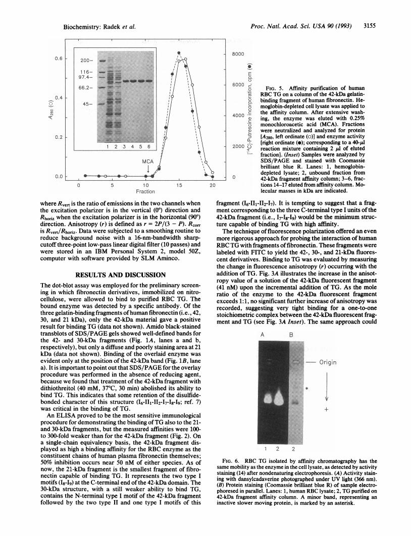
6000 go FIG. 5. Affinity purification of humanE RBC TG on a column of the 42-kDa gelatin-° binding fragment of human fibronectin. He-o moglobin-depleted cell lysate was applied to
4000 *C the affinity column. After extensive wash-40 ing, the enzyme was eluted with 0.25%5 monochloroacetic acid (MCA). Fractionsa were neutralized and analyzed for proteina [A280, left ordinate (o)] and enzyme activity
2000 [right ordinate (o); corresponding to a 40,ul2 reaction mixture containing 2 ,ul of eluted
fraction]. (Inset) Samples were analyzed bySDS/PAGE and stained with Coomassiebrilliant blue R. Lanes: 1, hemoglobin-depleted lysate; 2, unbound fraction from
0 42-kDa fragment affinity column; 3-6, frac-tions 14-17 eluted from affinity column. Mo-lecular masses in kDa are indicated.
where Rvert is the ratio of emissions in the two channels whenthe excitation polarizer is in the vertical (00) direction andRhonz when the excitation polarizer is in the horizontal (90°)direction. Anisotropy (r) is defined as r = 2P/(3 - P). RCOIis Rvert/Rhoriz. Data were subjected to a smoothing routine toreduce background noise with a 16-nm-bandwidth sharp-cutoff three-point low-pass linear digital filter (10 passes) andwere stored in an IBM Personal System 2, model 50Z,computer with software provided by SLM Aminco.
RESULTS AND DISCUSSIONThe dot-blot assay was employed for the preliminary screen-ing in which fibronectin derivatives, immobilized on nitro-cellulose, were allowed to bind to purified RBC TG. Thebound enzyme was detected by a specific antibody. Of thethree gelatin-binding fragments ofhuman fibronectin (i.e., 42,30, and 21 kDa), only the 42-kDa material gave a positiveresult for binding TG (data not shown). Amido black-stainedtransblots of SDS/PAGE gels showed well-defined bands forthe 42- and 30-kDa fragments (Fig. 1A, lanes a and b,respectively), but only a diffuse and poorly staining area at 21kDa (data not shown). Binding of the overlaid enzyme wasevident only at the position of the 42-kDa band (Fig. 1B, lanea). It is important to point out that SDS/PAGE for the overlayprocedure was performed in the absence of reducing agent,because we found that treatment of the 42-kDa fragment withdithiothreitol (40 mM, 37°C, 30 min) abolished its ability tobind TG. This indicates that some retention of the disulfide-bonded character of this structure (16-II1-112-47-18-19; ref. 7)was critical in the binding of TG.An ELISA proved to be the most sensitive immunological
procedure for demonstrating the binding ofTG also to the 21-and 30-kDa fragments, but the measured affinities were 100-to 300-fold weaker than for the 42-kDa fragment (Fig. 2). Ona single-chain equivalency basis, the 42-kDa fragment dis-played as high a binding affinity for the RBC enzyme as theconstituent chains of human plasma fibronectin themselves;50% inhibition occurs near 50 nM of either species. As ofnow, the 21-kDa fragment is the smallest fragment of fibro-nectin capable of binding TG. It represents the two type Imotifs (I8-49) at the C-terminal end of the 42-kDa domain. The30-kDa structure, with a still weaker ability to bind TG,contains the N-terminal type I motif of the 42-kDa fragmentfollowed by the two type II and one type I motifs of this
fragment (16-II1-412-17). It is tempting to suggest that a frag-ment corresponding to the three C-terminal type I units ofthe42-kDa fragment (i.e., I7-18-19) would be the minimum struc-ture capable of binding TG with high affinity.The technique offluorescence polarization offered an even
more rigorous approach for probing the interaction of humanRBC TG with fragments offibronectin. These fragments werelabeled with FITC to yield the 42-, 30-, and 21-kDa fluores-cent derivatives. Binding to TG was evaluated by measuringthe change in fluorescence anisotropy (r) occurring with theaddition of TG. Fig. 3A illustrates the increase in the anisot-ropy value of a solution of the 42-kDa fluorescent fragment(41 nM) upon the incremental addition of TG. As the moleratio of the enzyme to the 42-kDa fluorescent fragmentexceeds 1:1, no significant further increase of anisotropy wasrecorded, suggesting very tight binding for a one-to-onestoichiometric complex between the 42-kDa fluorescent frag-ment and TG (see Fig. 3A Inset). The same approach could
A B
- Origin
FIG. 6. RBC TG isolated by affinity chromatography has thesame mobility as the enzyme in the cell lysate, as detected by activitystaining (14) after nondenaturing electrophoresis. (A) Activity stain-ing with dansylcadaverine photographed under UV light (366 nm).(B) Protein staining (Coomassie brilliant blue R) of sample electro-phoresed in parallel. Lanes: 1, human RBC lysate; 2, TG purified on42-kDa fragment affinity column. A minor band, representing aninactive slower moving protein, is marked by an asterisk.
Biochemistry: Radek et al.

Proc. Natl. Acad. Sci. USA 90 (1993)
be used to demonstrate a fairly tight binding of TG to the21-kDa fluorescent fragment (Kd.app = 1.8 x 10-v; Fig. 3B,upper curve and Inset). Binding of the enzyme to the 30-kDafluorescent fragment (Fig. 3B, lower curve) was marginal.
Fluorescence polarization could also be employed as a testfor competition of unlabeled fibronectin fragments (42, 30,and 21 kDa) against the binding of the 42-kDa fluorescentfragment to TG. In the experiment presented in Fig. 4A,increasing amounts (0-168 nM) of the unlabeled 42-kDafragment were mixed with the 42-kDa fluorescent fragment(41 nM) prior to the injection of TG (50 nM). The unlabeledfragment was found to be an equal competitor in the sensethat the anisotropy value of the system was depressed by50%o when near equal amounts of the labeled and unlabeled
fragments were present. Under similar experimental condi-tions, no significant competition could be demonstrated withthe unlabeled 30-kDa fragment, even when it was added in an=76-fold excess over the 42-kDa fluorescent fragment (line 2in Fig. 4B). The unlabeled 21-kDa fragment was capable ofcompeting with the 42-kDa fluorescent fragment for bindingto TG (lines 3-5 in Fig. 4B), albeit competition by theunlabeled 21-kDa fragment was nearly 40-fold weaker thanwith the unlabeled 42-kDa fragment.
Binding of the RBC TG to the 42-kDa fragment of humanplasma fibronectin was also found to be of remarkably highspecificity. A single passage ofthe hemoglobin-depleted RBClysate (110 ml) through an affinity column with the covalentlycoupled 42-kDa fragment (2.6 mg) as ligand caused a near-total removal of TG from among the many proteins of thelysate. The most obvious difference between the proteinprofiles of the material applied to the column and the pass-through fraction was the absence of an =80-kDa band cor-responding to TG (Fig. 5 Inset, compare lanes 1 and 2).Desorbing the enzyme with fair retention of activity provedto be difficult because, possibly on account of the tightbinding, conventional methods of elution [e.g., with 10%o(vol/vol) acetic acid, 50 mM Tris'HCl, pH 7.5/1 M NaCl, 0.5M MgCl2, or 4 M urea; 4°C] were ineffective. However,satisfactory results were obtained in a single step of purifi-cation with 0.25% monochloroacetic acid (Fig. 5 Inset, lanes3-6), allowing an =18% recovery of enzyme activity, mea-sured by the [14C]putrescine incorporation assay (16).
Mobility of the purified TG in nondenaturing electropho-resis, as observed by activity staining with dansylcadaverine(19), was indistinguishable from that of the enzyme in the celllysate prior to hemoglobin depletion (Fig. 6). However, eventhough reduced SDS/PAGE showed a single protein of 80kDa (Fig. 5 Inset, lanes 3-6), nondenaturing electrophoresisin agarose revealed a minor slower-migrating band (Fig. 6,
asterisk) without TG activity. The small displacement ofpeaks observed for the activity and protein eluted from theaffinity column (Fig. 5) also suggests the presence of someinactive protein in the purified product. Two-dimensionalelectrophoresis (18) indicated the presence of isoforms sim-ilar to those found in TG preparations (data not shown)isolated by another procedure (15).
At Northwestern University, this work was aided by a U.S. PublicHealth Service Research Career Award (HL-03512) and by grantsfrom the National Institutes of Health (HL-45168 and HL-02212); atthe American Red Cross, this work was aided by a grant from theNational Institutes of Health (HL-21791).
1. Murthy, S. N. P. & Lorand, L. (1990) Proc. Natl. Acad. Sci.USA 87, 9679-9682.
2. Murthy, S. N. P., Wilson, J., Guy, S. L. & Lorand, L. (1991)Proc. Natl. Acad. Sci. USA 88, 10601-10604.
3. Shainoff, J. R., Urbanic, D. A. & DiBello, P. M. (1991) J. Biol.Chem. 266, 6429-6437.
4. Lorand, L., Dailey, J. E. & Turner, P. M. (1988) Proc. Natl.Acad. Sci. USA 85, 1057-1059.
5. Turner, P. M. & Lorand, L. (1989) Biochemistry 28, 628-635.6. LeMosy, E. K., Enrckson, H. P., Beyer, W. F., Jr., Radek,
J. T., Jeong, J. M., Murthy, S. N. P. & Lorand, L. (1992) J.Biol. Chem. 267, 7880-7885.
7. Ingham, I. C., Brew, S. A. & Migliorin, M. M. (1989) J. Biol.Chem. 264, 16977-16980.
8. Borsi, L., Castallani, P., Balza, E., Sini, A., Pellecchia, C.,DeScalzi, F. & Zardi, L. (1986) Anal. Biochem. 155, 335-345.
9. Lorand, L., Credo, R. B. & Janus, T. J. (1981) MethodsEnzymol. 80, 333-341.
10. Mosesson, M. W. & Umfleet, R. A. (1970) J. Biol. Chem. 245,5728-5736.
11. Litvinovich, S. V., Strickland, D. K., Medved, L. V. & Ing-ham, K. C. (1991) J. Mol. Biol. 217, 563-575.
12. Freyssinet, J.-M., Lewis, B. A., Holbrook, J. J. & Shore, J. D.(1978) Biochem. J. 169, 403-410.
13. Churchich, J. E. (1967) Biochim. Biophys. Acta 147, 511-517.14. Lowry, 0. H., Rosebrough, N. J., Farr, A. L. & Randall, R. J.
(1951) J. Biol. Chem. 193, 265-275.15. Brenner, S. C. & Wold, F. (1978) Biochim. Biophys. Acta 522,
74-83.16. Lorand, L., Campbell-Wilkes, L. K. & Cooperstein, L. (1972)
Anal. Biochem. 50, 623-631.17. Laemmli, U. K. (1970) Nature (London) 227, 680-685.18. O'Farrell, P. H. (1975) J. Biol. Chem. 250, 4007-4021.19. Lorand, L., Siefring, G. E., Jr., Tong, Y. S., Bruner-Lorand,
J. & Gray, J. A., Jr. (1979) Anal. Biochem. 93, 453-458.20. Towbin, H., Staehelin, T. & Gordon, J. (1979) Proc. Natl.
Acad. Sci. USA 76, 4350-4354.21. Lakowicz, J. R. (1983) Principles of Fluorescence Spectros-
copy (Plenum, New York), pp. 111-153.
3156 Biochemistry: Radek et al.