after mapping of the axilla: radiotherapy or surgery? · eortc 10981-22023 amaros version 5.0 3/63...
TRANSCRIPT

International Association under Belgian Law
INTERGROUP STUDY EORTC 10981-22023
AMAROS
After Mapping of the Axilla: Radiotherapy Or Surger y?
Coordinating group: EORTC BREAST CANCER GROUP PROTOCOL 10981 (E. Rutgers)
Collaborating groups: EORTC RADIOTHERAPY GROUP PROTOCOL 22023 (G. Van Tienhoven) BOOG, Borstkanker Onderzoeksgroup Nederland (C.J.H. Van de Velde) ALMANAC (axillary lymphatic mapping against nodal axillary clearance) Trialists Group (R E Mansel)
Study coordinator: E.J.Th. Rutgers (EORTC Breast Cancer Group) Phone: +31.20.5122552 Fax: +31.20.5122554 e-mail: [email protected]
Pharmacovigilance Unit:
Phone: +32 2 774 16 76 Fax: +32 2 772 80 27
e-mail: [email protected]
November 5, 1998 NTC-PRC outline approval July 20, 2000 PRC full protocol approval Version 1.0 May 22, 2001 First amendment Version 2.0 February 7, 2003 Second amendment Version 3.0 February 15, 2005 Third amendment Version 4.0 February 22, 2008 Fourth amendment Version 5.0
Version 5.0 Copyright EORTC 2008

EORTC 10981-22023 AMAROS
Version 5.0 2/63 22 February 2008
THE AMAROS TRIAL IN OPERABLE BREAST CANCER
EORTC BCG Study Coordinators Rutgers EJT
Van de Velde CJH
Borger J
EORTC RA Study Coordinator
Van Tienhoven G
BOOG Study Coordinator
Van de Velde CJH
ALMANAC Study Coordinator
Mansel R E
Writing Committee
1. Surgery
Cataliotti L
Christiaens MR
Nieweg OE
Jaskiewicz J
Ruers T
Rutgers EJT
Snoj M.
Van de Velde CJH
2. Radiotherapy
Jassem J
Fourquet A
Leer JW
3. Pathology
Peterse JL
4. Nuclear Medicine
Pijpers R
5. Quality of life assessment
Bottomley A
6. Statistician
Bogaerts Jan
7. Medical advisor
Demonty G
8.Cost-Benefit Evaluation
Neymark N
9. Data Management
Duez N
10. Monitors
Straver ME

EORTC 10981-22023 AMAROS
Version 5.0 3/63 22 February 2008
Table of Contents
1. BACKGROUND AND INTRODUCTION 7
2. OBJECTIVES OF THE TRIAL 11
3. PATIENT SELECTION CRITERIA 11
4. TRIAL DESIGN 12
5. THERAPEUTIC REGIMENS, SN PROCEDURE, PATHOLOGY, SURGERY, RADIOTHERAPY 13
5.1 Definition Sentinel node 13
5.2 Lymphoscintigraphy 13
5.3 Imaging and reporting 14
5.4 Surgical procedure of sentinel node biopsy 15
5.5 Pathology 16
5.6 Radiation hazard and safety precautions 16
5.7 Learning phase 17
5.8 Axillary lymph node dissection 17
5.9 Mastectomy 18
5.10 Radiation of the axilla 18
5.11 Physical training programme 19
5.12 Adjuvant systemic therapy guidelines 19
5.13 Treatment of recurrence 20
6. CLINICAL EVALUATION, LABORATORY TESTS AND FOLLOW-UP 20
6.1 Before treatment 20
6.2 Treatment 20
6.3 After the end of treatment 21
6.4 Summary 21
7. CRITERIA OF EVALUATION 21

EORTC 10981-22023 AMAROS
Version 5.0 4/63 22 February 2008
8. PATIENT RANDOMIZATION/REGISTRATION PROCEDURE 23
8.1. RANDOMIZATION 23
8.2 REGISTRATION AFTER THE SENTINEL NODE PROCEDURE 23
9. FORMS AND PROCEDURES FOR COLLECTING DATA 24
9.1 Case report forms and schedule for completion 24
9.2 DATA FLOW 25 9.2.1 Paper CRFs 26 9.2.2 Using the electronic forms system (RDC) 26
10. REPORTING ADVERSE EVENTS 27
10.1 Definitions 27
10.2 REPORTING PROCEDURES OF SERIOUS ADVERSE EVENTS 27
11. STATISTICAL CONSIDERATIONS 29
12. QUALITY OF LIFE ASSESSMENT 30
13. ECONOMIC EVALUATION 32
14. INDEPENDENT DATA MONITORING COMMITTEE 32
15. QUALITY ASSURANCE 32
15.1 Control of data consistency 32
15.2 On-site quality control 32
16. ETHICAL CONSIDERATIONS 32
16.1 Patient protection 32
16.2 Subject identification 33
16.3 INFORMED CONSENT 33
17. INVESTIGATOR COMMITMENT STATEMENT 33
18. ADMINISTRATIVE RESPONSIBILITIES 34
19. TRIAL SPONSORSHIP/FINANCING 36

EORTC 10981-22023 AMAROS
Version 5.0 5/63 22 February 2008
20. TRIAL INSURANCE 37
21. PUBLICATION POLICY 37
22. REFERENCES 38
23. APPENDICES 43
Appendix I: results of sentinel lymph node biopsy obtained by various investigators 43
Appendix II: technique and results of lymphoscintigraphy by various investigators 44
Appendix III: radiation activity related to time interva l after tracer injection 45
Appendix IV: review form when visiting candidate center for the AMAROS trial 46
Appendix V: Informed Consent AMAROS trial 49
Appendix VI: EORTC Quality of life questionnaire-C30 57
Appendix VII: Commitment statement / Study acknowledgment 61
Appendix VIII: ALMANAC Trialists Group Specific Appe ndix to the EORTC protocol 10981- 22023 62

EORTC 10981-22023 AMAROS
Version 5.0 6/63 22 February 2008
Protocol Summary
After mapping of the axillary: radiotherapy or surgery, is a phase III study comparing a complete axillary lymph node dissection with radiotherapy to the axilla in sentinel biopsy positive patients, where-as sentinel node negative patients are followed for the end-points of the study as well. The main objective of the trial is to prove equivalent local/regional control for patients with proven axillary lymph node metastasis by sentinel node biopsy with reduced morbidity if treated with axillary radiotherapy instead of axillary lymph node dissection. A second objective is to investigate whether adequate axillary control can be obtained by not subjecting patients with a negative sentinel lymph node to axillary lymph node dissection.
The involved patients will have an operable invasive breast cancer of over 5 mm and less than 5 centimetres, without clinical suspect regional lymph nodes. Patients will have FNA or core biopsy proven invasive breast cancer and should be fit to undergo either treatment. Patients will be stratified by institution and will be randomized between complete axillary lymph node dissection and radiotherapy of the axilla. Sentinel node biopsies will be performed by the combined technique using preoperative lymphoscintigraphy by intra- or peritumoural injection of 99Tc Nanocolloid, immediate pre-operative injection of Patent Blue Dye and SN-retrieval by both discoloration and intra-operative use of a detection probe. Per-operative frozen section is allowed. All patients will undergo a wide excision or segmentectomy of the primary tumour or a mastectomy. Adjuvant systemic therapy and radiotherapy is advised according to the institutional guidelines provided that: 1. dose or schedule is related to tumour characteristics and nodal positivity, 2. treatment policies, which should be maintained during the study period, are send to the study
coordinator.

EORTC 10981-22023 AMAROS
Version 5.0 7/63 22 February 2008
1. BACKGROUND AND INTRODUCTION
Knowledge of the presence or absence of dissemination to the axillary lymph nodes represents important information for prognosis and staging of patients with breast cancer. Clearing the axilla also assures regional tumour control and might in some cases improve survival 1,2. Metastatic cancer is found in these nodes in 26- 34% of the patients 1-3. In the remaining 70%, no therapeutic benefit is
derived from axillary node dissection Yet these patients are exposed to the considerable morbidity associated with the procedure 5-7. It is understandable that less invasive approaches are sought for staging the axilla. Palpation is unreliable for this purpose 8,9. The merit of methods like lymphoscintigraphy with a radiolabeled colloid 10,11, lymphangiography 12, CT-scanning 13, ultra-sound 14, scintigraphy with 99mTechnetium-Sestamibi (99mTc-Sestamibi) 15 and positron emission tomography 16,17 has not yet been clearly established. Characteristics of the primary lesion such as tumour type, tumour size, site of the primary lesion, nuclear grade, hormone receptor status, ploidy, S-phase fraction and HER2/NEU expression have been extensively studied and cannot replace the axillary lymph node status 18,19. Lymphatic mapping with sentinel node biopsy is emerging as a new technique to determine the lymph node status 20. This novel approach involves lymphoscintigraphy and a minimally invasive surgical technique, and appears to allow the same information for staging and prognosis to be gathered with a limited morbidity. The concept was proved to be correct in melanoma and it was shown that the technique lends itself to widespread application 21. Several investigators were quick to presume that this procedure could also be of value in breast cancer 22-25. IN VIVO LYMPHATIC MAPPING AND SENTINEL LYMPH NODE BIOPSY.
A literature search was performed to identify and analyse all publications on this subject (Appendix I). Looking at the various publications, it becomes clear that no standardised technique exists. There are substantial differences in various aspects among the various published papers. There are differences in the patient populations enrolled in the various studies. A little over a third of the investigators use preoperative lymphoscintigraphy. A number of different radiopharmaceuticals are used for this purpose. Different surgical techniques are used. Some surgeons use a radioactive tracer and a gamma detection probe, others prefer a vital dye. Still others use both intraoperative detection techniques. Only six groups employ all three detection techniques 29, 56,32-35. This chapter is based on the review paper of Nieweg et al. (Nieweg et al.Eur J Nucl Medicine. Vol. 26 (suppl), April 1999: S11-S16). LYMPHOSCINTIGRAPHY
The purpose of lymphoscintigraphy for lymphatic mapping is to demonstrate the lymphatic drainage pathway of the neoplasm. To be more precise: to determine the number of lymph nodes on a direct drainage pathway, to differentiate these first-tier nodes from subsequent nodes and to locate these sentinel nodes. It should be emphasised that the radiopharmaceuticals used for this purpose are not tumour seeking agents, but rather lymph node seeking agents. They are accumulated in lymph nodes whether these contain metastatic disease or not. Uptake is non-specific and does not infer nodal metastasis per se. In fact, heavily invaded nodes may not accumulate the tracer and can remain undetected 29. Furthermore, the gamma cameras that are used for imaging have a limited resolution. This implies that the lymphoscintigraphy images usually do not allow the visualisation of sufficient anatomic detail to distinguish a tumour-containing node either through abnormal shape or structure.

EORTC 10981-22023 AMAROS
Version 5.0 8/63 22 February 2008
Macrophages have a great avidity for colloidal radiopharmaceuticals, although this does not ensure that all of the tracer that reaches the sentinel node is retained there. Some of it may pass through to efferent lymphatics to be absorbed by subsequent nodes. Eleven studies have been published describing results from 739 lymphoscintigrams (Appendix II). These studies show that a sentinel node is visualised in 75 - 98% of the patients. There are marked differences in the technique as used by various investigators, but the published results are too scarce to tell which technique is to be preferred. A number of issues are unresolved as yet. For instance, what is the optimum size of the colloid particles? The behaviour of colloids injected interstitially depends on their particle size. Very large particles fail to migrate and tend to remain in the interstitium at the injection site. Very small particles travel so quickly that only a fraction is retained in the first lymph node and then secondary nodes light up as well. The very small particles also tend to penetrate the capillaries and enter the blood stream. There is a trade off. A smaller particle size agent is preferred when quick accumulation and nice flow images are considered to be important to visualise the lymphatic duct(s) in order to distinguish first-echelon nodes from second-echelon nodes. The down side of a small particle size is depiction of secondary nodes that may be difficult to distinguish from the first-echelon node when the duct is not visible after all. A larger particle size tracer, on the other hand, will limit the number of "hot" non-sentinel nodes depicted on the images but not visualise the lymphatic duct and probably also not visualise some sentinel nodes. Other issues that need to be clarified are the dose and volume of the radiopharmaceutical. The doses that are used by various investigators range from seven to 370 MBq (Appendix II). The volumes that are injected range from 0.2 to 4 ml, a difference by a factor of 20. Investigators who use a small volume prefer not to disturb the physiology of lymph flow and avoid the risk of visualising non-sentinel nodes. Those who use the larger volumes argue that they do want to change the physiology and thereby increase the chance of visualising a lymph node. It is unknown what the best place is to inject the tracer (or the vital dye). Since we want to visualise drainage from the tumour, it makes sense to inject the tracer into or closely around the primary lesion. Injecting in the overlying skin increased the likelihood of depicting a lymphatic duct and a lymph node because drainage from the skin is far richer than drainage from the breast parenchyma 29,36. But injecting further away from the lesion carries the risk that a watershed is crossed and a node is visualised that drains another area of the breast and not the area with the tumour. It is a telltale sign that the skin of the breast in our melanoma patients never drains to internal mammary nodes. The question has been raised whether scintigraphy contributes anything to lymphatic mapping and should be done at all 37. We would like to argue in favour of lymphoscintigraphy for several reasons. A sentinel node sometimes contains so little radioactivity that it cannot be identified with a probe through the intact skin. Sometimes the sentinel node cannot be picked up with the probe because of a location so close to the primary lesion site - where the bulk of the radioactivity stays behind - that its counts are overwhelmed by shine-through from the injection site. Another reason to advocate preoperative lymphoscintigraphy is that sentinel nodes are located outside the axilla in a substantial number of patients 38. Lymphoscintigraphy will point out such sentinel nodes. Based on the available evidence, one can conclude that a number of technical issues need to be resolved but it is clear that preoperative lymphoscintigraphy increases the likelihood of finding (all) sentinel nodes. The nuclear medicine physician provides the road map that guides the surgeon.

EORTC 10981-22023 AMAROS
Version 5.0 9/63 22 February 2008
SURGERY
There are two techniques to find the sentinel node during the operation: instrument-guided mapping and visually guided mapping 39. One may use a gamma detection probe as a guide to the sentinel node after administration of a radioactive tracer. The radioactivity that remains in the node can be exploited to this end when the operation is done within a day after scintigraphy. Otherwise, the tracer can be administered shortly before the operation. With the probe, the location of the node can be determined through the intact skin. Preoperative knowledge of the node's exact location helps to minimise the extent of the dissection. The site and the direction of the incision are chosen based on this knowledge, keeping in mind that formal axillary node dissection possibly needs to follow. An incision length of a few centimetres is sufficient. One then proceeds with the dissection in the direction of the highest count rate. After a while, the probe is inserted into the wound so that the direction that one is moving into can be adjusted if necessary. The gamma probe signal intensifies each time this sequence is repeated until the sentinel lymph node is found. The other technique to find a sentinel node is with the aid of a vital dye, (isosulfan blue, patent blue) which is administered immediately prior to the operation. The area is massaged for several minutes to increase the lymph flow. Once the dye is taken up by the lymphatic system, it stains the lymphatic channel. The channel is identified where it enters the axilla and it is dissected until it enters and stains a first-echelon node. Appendix I shows how various investigators perform the procedure and shows their results. Eleven of the 27 groups (41%) use preoperative lymphoscintigraphy. Eight investigators (30%) rely on the blue dye technique. Six investigators (22%) use only a radioactive tracer and a gamma detection probe. Twelve investigators (44%) use various combinations of detection techniques. The success rates in identifying sentinel nodes range from 41% to 98%. The false negative rates also show wide variations: 0-40%. A few things are becoming clear from the work that has been done so far. For instance, there is a learning phase. In their initial study, Giuliano and co-workers identified a sentinel node in 66% of their patients 40. In a subsequent study, they identified the sentinel node in 93% of the patients 41. The identification rate improved as the investigator became familiar with the nuances of the technique. The false negative rate was 11% in the first series and improved to 0% in the second. This latter study shows that excellent results can be obtained with blue dye alone, without preoperative scintigraphy and without a gamma detection probe. One wonders how is it possible that these results are so good when the groups of Cox and of De Vries, who use blue dye as well as a probe, found that 30-40% of the sentinel nodes are not blue but only radioactive 32,34. The added value of a probe is easily pointed out. With the probe one can find a sentinel node in odd locations like the breast parenchyma or in the subclavicular fossa. Such nodes are bound to be overlooked when only blue dye is used. With the probe, one can identify the sentinel node when the blue lymphatic duct is accidentally damaged and one loses the guiding track to the sentinel node. This is likely to happen when one starts doing this, during the learning phase. In addition to showing that sentinel nodes may be only radioactive, De Vries also found the reverse: 16% of the sentinel nodes in their study were only blue but not radioactive 34. In a larger series by Cox and co-workers this was even twice as much: 32% 32. So, relying on a probe and omitting a blue dye also leads to the situation where relevant nodes are left behind.

EORTC 10981-22023 AMAROS
Version 5.0 10/63 22 February 2008
There is another reason for using a vital dye. The dye can help differentiate between first-echelon nodes and nodes with secondary drainage. This is particularly useful when scintigraphy shows accumulation of the radiopharmaceutical in a multitude of nodes without indicating the drainage sequence. Blue dye does not only visualise the lymph nodes but also the lymphatic ducts: the order of drainage is mapped out. And it is the lymphatic duct that comes from the direction of the primary tumour that proves that the node that it goes to is a first-echelon node. The probe may enable identification of lymph nodes but does not visualise the ducts that are important when faced with this situation. COMPLETE AXILLARY DISSECTION
At least level I-II axillary lymph node dissection (ALND) with a minimum of 10-lymph nodes removed and examined, provides for a good axillary control with recurrence rates of less than 5% at 10 years 58. Morbidity of axillary treatment consists of lymph edema (3-17%) and shoulder function impairment (5-19%) 6,7,59,60. Regional recurrence rates increases with increased involvement of axillary lymph nodes. However, regional control is directly related to the extent and completeness of ALND. Much debate exists on the indication of adjuvant radiotherapy after complete ALND of the axilla and tumour positive nodes for this study. RADIATION THERAPY TO THE AXILLA
The search for alternatives for ALND resulted in a series of randomized clinical trials in the sixties and seventies 61-67. The original objective was to test the hypothesis of improving survival by maintaining an immunological barrier in the axillary lymph nodes. By leaving these nodes in situ and treating the axilla with irradiation the process of dissemination was supposed to be influenced in a positive way. Simple mastectomy was the primary surgical procedure in most of these trials and patients were randomly assigned to ALND or axillary radiotherapy (ART). With respect to the endpoint of survival no difference was found between the two approaches to the axilla except for the Guy’s trial which showed worse survival due to lower axillary control rates 63. Axillary recurrence rates varied between 3 and 19% for all trials and between 3 and 12 % for those using megavoltage radiation equipment 61,64,65,67. Of these latter studies the NSABP study 67 was the only trial for which clinically node negative patients only were eligible. Apart from randomized studies many clinical series report on the effectiveness of ART as a less invasive alternative for ALND in the treatment of early breast cancer in the context of breast conserving therapy (BCT) 68-75. The axillary recurrence rates in these series with mainly megavoltage techniques and more uniform dose levels (50 Gy/5 weeks) vary between 0.6 and 3.6 % with one exception reporting a recurrence rate of 16% 74,76,77. It should be mentioned that these studies were performed in populations of breast cancer patients with an average incidence rate of pathologically confirmed axillary lymph node metastases of approximately 40% 67. About 60% of the axillae were node negative which is comparable with the contemporary sentinel node setting in which the positive sentinel node appears to be the only positive node in 50% of the cases. In terms of local control, therefore, ART in positive sentinel node cases is expected to result in an at least as favourable outcome as in the above-mentioned experience. Armedema (0-2%), shoulder malfunction (1%), radiopneumonitis (3-6%) and brachial plexus neuropathy (< 1%) are observed as possible side effects of ART 78, the latter probably as a result of inappropriate field junction techniques. Modern field matching techniques have become available for safe administration of combinations of breast tangentials and regional node portals 76,79-81. The incidence of radiopneumonitis may be different nowadays because of the combination of shoulder radiotherapy with chemotherapy, especially doxorubicin. Because of limited clinical experience with this aspect, patients in the ART arm of AMAROS should have strict surveillance

EORTC 10981-22023 AMAROS
Version 5.0 11/63 22 February 2008
with regard to the occurrence of radiopneumonitis when (anthracyclin containing) chemotherapy is combined with radiotherapy, irrespective the sequence. Considering the design and endpoints of this study radiotherapy should be given directly after surgery and not be postponed in favour of the early administration of chemotherapy. ADJUVANT SYSTEMIC TREATMENTS
A recent re-evaluation of the indications for chemotherapy gave recommendation on T rather than N-stage, since the proportionally reduction in events was similar in node negative and node positive breast cancer. However, in the structure of this trial, indications for systemic treatment should be based on tumour characteristics and nodal positivity. Different schemes on the basis of the number of positive nodes are applicable for this study, but not recommended. However, patients with clinically or pathologically involved lymph nodes, are excluded from this study.
2. OBJECTIVES OF THE TRIAL
This is a phase III randomised non-inferiority trial. The aim of this trial is to prove equivalence in local control between the two treatment modalities of the axilla with reduced the morbidity. The primary end point is axillary recurrence rate after 5 years. Secondary endpoints in the sentinel node positive group are shoulder function analysis, quality of life assessment as are the axillary recurrence free survival rate, disease free survival and the survival. Furthermore axillary recurrence free rate in the sentinel node negative group will be observed and compared to historical controls. Quality control of the involved disciplines is mandatory for the trial and the infrastructure of the Cooperative Groups (Pathology Group, Surgical Quality Control Group) is very important for the success of this study. The Group will organise a number of training workshops for new centres before starting participation. With this study we hope to proof benefits for all involved parties. Patients will benefit because of the well-controlled use of the sentinel lymph node mapping and the avoidance of unnecessary axillary dissection. Surgeons meet together with the team of nuclear physicists and pathologists, to assure quality control of this new technology. Radiation oncologists have to control the quality of radiotherapy directed to the axilla in order to compare morbidity of the two procedures together with perhaps greater patient satisfaction if all arms reveal similar loco regional and distant control of the disease. Approximately 70% of all axillary staging operations could be made redundant 3,4. Consequently, it is expected that the registration arm will be at least twice as large as the arm for which the evaluation will take place. This study will yield important information on local control, morbidity, quality of life, and quality of treatment by comparing the different treatment groups. For quality of life assessment the EORTC QLQ-C30 and QLQ-BR23 quality of life questionnaires will be used.
3. PATIENT SELECTION CRITERIA
ELIGIBILITY CRITERIA
♦ Invasive breast cancer proven by core biopsy or ‘triple diagnosis’: clinical palpation concordant with malignancy, imaging (mammography, ultrasound or MRI) and tumour positive FNA cytology; diagnosis by excisional tumourectomy is allowed. Clinically occult invasive cancer should be proven by histology,

EORTC 10981-22023 AMAROS
Version 5.0 12/63 22 February 2008
♦ tumour larger than 5 and smaller than 50 mm in its largest diameter, measured by mammography, ultrasound or MRI
♦ Multifocal (within one quadrant and sharing the same histological characteristics) breast cancer is allowed, but multicentric (in different quadrants) breast cancer is not allowed
♦ bilateral invasive breast cancer is allowed, (bilateral mammogram is mandatory)
♦ clinically negative axillary lymph nodes,
♦ patient has to be fit to undergo any of the following treatments: SN-biopsy, axillary clearance, breast surgery, radiation therapy of the axilla,
♦ before patient registration/randomization, informed consent must be obtained according to ICH/EU GCP, and national/local regulations,
♦ female gender,
♦ absence of any psychological, familial, sociological or geographical condition potentially hampering compliance with the study protocol and follow-up schedule; those conditions should be discussed with the patient before registration in the trial,
♦ no metastatic disease (routine investigations are not required: symptoms should be investigated on indication),
♦ no previous treatment of the axilla by surgery or radiotherapy,
♦ no previous treatment of the primary breast tumour by neoadjuvant hormonal or other systemic treatment
♦ no previous treatment of cancer, except Basal Cell Carcinoma of the skin and in situ carcinoma of the cervix,
♦ no pregnancy.
Patients eligible for the AMAROS trial and the EORTC MINDACT trial (EORTC 10041), can be entered in both trials simultaneously since the primary endpoints of these trials differ.
4. TRIAL DESIGN
After diagnosis of invasive breast cancer, informed consent and eligibility check, patients are randomized. Randomization will take place before the sentinel node procedure. The patient will know before surgery whether she will have a complete axillary dissection or radiotherapy if the sentinel node(s) is (are) tumour positive on frozen section or definitive histology. Patients with negative sentinel node(s) will have no further treatment of the axilla and will be followed. Patients with only submicrometastasis/isolated tumour cells (< 0.02) could be considered as sentinel node negative. No further axillary treatment is allowed. The decision to omit further axillary treatment will be made per institution.
EORTC 10981-22023 AMAROS
Version 5.0 13/63 22 February 2008
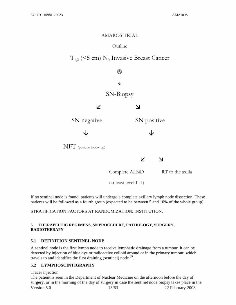
AMAROS-TRIAL
Outline
T1,2 (<5 cm) N0 Invasive Breast Cancer
�
SN-Biopsy
� �
SN negative SN positive
� �
NFT (positive follow up)
� �
Complete ALND RT to the axilla
(at least level I-II)
If no sentinel node is found, patients will undergo a complete axillary lymph node dissection. These patients will be followed as a fourth group (expected to be between 5 and 10% of the whole group).
STRATIFICATION FACTORS AT RANDOMIZATION: INSTITUTION.
5. THERAPEUTIC REGIMENS, SN PROCEDURE, PATHOLOGY, SURGERY, RADIOTHERAPY
5.1 DEFINITION SENTINEL NODE
A sentinel node is the first lymph node to receive lymphatic drainage from a tumour. It can be detected by injection of blue dye or radioactive colloid around or in the primary tumour, which travels to and identifies the first draining (sentinel) node 39.
5.2 LYMPHOSCINTIGRAPHY
Tracer injection The patient is seen in the Department of Nuclear Medicine on the afternoon before the day of surgery, or in the morning of the day of surgery in case the sentinel node biopsy takes place in the

EORTC 10981-22023 AMAROS
Version 5.0 14/63 22 February 2008
afternoon. 99 mTC Colloid albumin (Nanocoll tracer dose, see Appendix III), dissolved in 0.2 to 1 ml 0,09% NaCl (Saline), is generally used as tracer and injected by the nuclear physician or surgeon. The tracer solution is divided in equal aliquots and injected in two to four depots into the breast tissue surrounding the primary tumour using a 0.6 mm needle. This may be subdermally if the tumour is superficially located. In case the tracer is injected in the tumour, aliquots of tracer volume should be small 39. To promote drainage of tracer the patient can be asked to massage the breast. The time interval between injection of tracer and surgery should at least be two hours in order to allow the tracer to reach the draining lymph node(s). The tracer dose that needs to be injected to obtain adequate radioactive signals at time of operation is dependent on the time interval between tracer injection and surgery and is mainly determined by the physical half life time of 99m Technetium. The amount of tracer activity after different time intervals is given in Appendix III.
5.3 IMAGING AND REPORTING
Lymphoscintigraphy (128x128 matrix) is performed 2-3 hours after injection of the tracer and may be repeated 1-2 hours before operation. Frequently, dynamic images starting shortly after injection during 30 minutes are helpful to depict the SN 82,83. Therefore, dynamic lymfoscintigraphy is advised. As this is a more time consuming procedure, logistics in the nuclear medicine department may preclude dynamic images in every patient. Consequently, at least static images should be made as described. Manipulation of images on computer can be useful to identify all focal accumulations. In case of high tracer dose and a short time interval between injection and scanning, imaging can be improved by using a medium energy collimator (lower septum penetration). The following views are advised:
♦ anterior view with the patient in supine position,
♦ the “hanging” breast technique with patient in prone position, the breast hanging in an opening of the mattress provides excellent images of the axilla,
♦ lateral view with the patient on her (non-affected) side and the gamma-camera above the axilla,
♦ in addition an anterior oblique view can be obtained with manual medial displacement of the breast and the arm on the affected site abducted at an angle of 90º. This position corresponds with the position during surgery,
♦ the skin overlying the sentinel node is marked with indelible ink,
♦ the use of a flood source or other technique to delineate the body contour are helpful and recommended.
The report by the nuclear physician should include the following details:
♦ site of injection (in tumour/around tumour),
♦ number of hot spots in the ipsilateral axilla,
♦ presence of hot spots outside the ipsilateral axilla (internal mammary chain, supraclavicular, lateral extension breast etc).
EORTC 10981-22023 AMAROS
Version 5.0 15/63 22 February 2008
The images should be discussed with the surgeon pre-operatively and should be available during surgery.
5.4 SURGICAL PROCEDURE OF SENTINEL NODE BIOPSY
Surgery is to be performed within 24 hours after injection of the tracer. After induction of anaesthesia, proper positioning of the patient, desinfection and sterile draping, the hot spots marked on the skin are verified using a gamma probe. The blue dye is preferably injected after cleaning and sterile draping 0.5– 2ml Patent Blue V dye is injected in or around the tumour or alternatively sub- or intradermally in the skin overlying the tumour or near the areola 36,41. Massage of the breast will increase lymphatic drainage: after five to ten minutes sufficient blue dye will be transported to visualise lymphatic and the blue sentinel lymph node. In contrast to the Nanocoll, the blue dye is not retained in the lymph node by macrophages and will soon flow to additional lymph nodes in the axilla. Generally, there will be concordance between the blue nodes and the radioactive (hot) nodes. In this case the sentinel node is the first blue lymph node with radioactivity that drains the afferent lymphatic vessel from the tumour. Under certain circumstances the first hot node is not blue, for example because of a discrepancy in injection site of the blue dye and the radioactive tracer. In this case the first hot node (not blue) is classified as the sentinel node. On the other hand it may occur that the first node that seems to drain the lymph vessel from the tumour is blue but shows no radioactivity, whereas the first hot node is situated more distally from the tumour. In this case it is less clear which node should be identified as the sentinel node and both nodes are removed. Blue nodes situated more distally from the tumour than the first blue node or the first hot node are considered as second echelon nodes and therefore can be left in situ. Hot nodes situated more distally than the first hot node however, should be removed, as it is difficult to determine which hot node is the real sentinel node. An appropriate incision is made in the axilla that can be used also in case of further axillary dissection or mastectomy. The sentinel node(s) is identified by placing the gamma probe (sterile packed) in the wound and moving it slowly to locate any hot spots. The direction of dissection is determined by aiming the gamma probe toward the site of maximal radioactivity emitted from the sentinel node(s). Careful dissection is used to identify also the blue-stained afferent lymphatic vessels that can guide the dissection toward the sentinel node. The sentinel node(s) is removed and background radiation in the bed of the sentinel-node(s) resection site is determined. In case of significant background activity, dissection should be continued in search for additional sentinel node(s) 32. Knowledge of the lymphoscintigraphy may contribute to find additional nodes. After removal of sentinel nodes, care should be taken to secure lymphatic vessels by ligation. Meticulous closure of the SN biopsy site is advocated to prevent dead space, lymphatic leakage and seroma formation. As this study concerns morbidity related to axillary treatment and subsequent morbidity, only SN’s from the axilla (including those located in level III or interpectoral) have to be retrieved. However, removal of SN outside the axilla i.e. internal mammary chain nodes is at the discretion of the treating physician. Registration of these extra procedures is requested. Wide local excision of the tumour or simple mastectomy will follow. Depending on the preferred procedure, if after a tumour positive SN an ALND is allotted, this can be performed immediately. However not always feasible, a complete removal of the sentinel biopsy site in the axilla is aimed at. In case of tumours in the lateral outer quadrant of the breast, localisation of the sentinel node may be difficult due to interfering radiation from the primary tumour. Initial removal of the tumour before the sentinel node procedure or a lead shield between probe and tumour may prevent this problem. For proper use of the gamma probe it is advised to check regularly the energy window of the instrument.

EORTC 10981-22023 AMAROS
Version 5.0 16/63 22 February 2008
If no sentinel node can be found a complete ALND is carried out, independent of the allotted treatment after randomization. If during the search for the SN the surgeon encounters a clinically suspect non-sentinel node, this node should be taken out for frozen section or paraffin histology. If such a non-sentinel node contains metastasis, this is considered as a failure of the procedure and a complete ALND is carried out.
5.5 PATHOLOGY
For pathological examination, each sentinel node is processed separately. FROZEN SECTIONS
Lymph nodes > 4 mm are bisected and frozen sections are performed from both edges. After frozen section, the sentinel node tissue is processed routinely for permanent sections. Each node is blocked individually. PARAFFIN SECTIONS
As a minimal requirement three histological levels (500 micron distance) for each sentinel node are examined. On each level two parallel sections are performed, one for immunohistochemistry and one for hematoxylin and eosin staining. Immunohistochemical staining is performed for markers containing at least cytokeratin 8 and 18 (e.g. CAM 5.2, NCL5D3). Cytokeratin immuno-histochemical (IHC) staining is done only when H&E staining is negative. Lymph nodes submitted for pathological examination which are marked by the surgeon as non-sentinel nodes are examined with H&E and cytokeratin IHC staining. PATHOLOGY REPORT
The pathology report should mention the results of IHC staining and H&E for each sentinel node. In case of additional axillary lymphadenectomy the conclusion should consist: the total amount of lymph nodes examined, the number of sentinel nodes subjected, staining results of each sentinel node, staining results of non-sentinel nodes. Whether the lymph node metastases is considered to be a micrometastases (< 2 mm) or a submicrometastasis/isolated tumour cells (< 0.2 mm), should also be mentioned in the pathological report. It should be noted that incidentally benign cytokeratin positive cells are encountered in normal lymph nodes. SUBMICROMETASTASIS/ISOLATED TUMOUR CELLS Submicrometastasis or isolated tumour cells (<0.2mm) may be considered as sentinel node negative. It is allowed to give no further axillary treatment in case of submicrometastasis/isolated tumour cells (<0.2 mm) solely.
5.6 RADIATION HAZARD AND SAFETY PRECAUTIONS
Based on a standard tracer dose of 60 MBq the maximum total body amount of radiation absorbed by the surgeon will be 4,7 microSv/hr. The maximum amount of radiation allowed per year for the hands, that are most exposed during the procedure, is 15 milliSv. Most of the radiation dose is coming from the injection site, only a few percent originates from the sentinel node. Exposure of the other operating staff and pathology staff will be lower as the distance to the radiation source is further and the exposure time is shorter.

EORTC 10981-22023 AMAROS
Version 5.0 17/63 22 February 2008
For transportation within the hospital a leakproof bag or box will suffice. For transportation outside the hospital e.g. to the pathology department, different country regulations may apply. It is advised that the person responsible for safety regulation within the hospital is informed about the procedure and is asked for advice concerning local regulations.
5.7 LEARNING PHASE
For this section, ALMANAC investigators must refer to the Group Specific Appendix. Each surgeon and nuclear physician in a potential participating centre involved in the sentinel node program for breast cancer should have followed one of the training courses. There is a certain learning phase. Before the participating centre is allowed to enter patients, the team has to perform at least thirty sentinel node procedures followed by a complete ALND 84-86. Histopathology of separately retrieved sentinel nodes and the nodes of the remaining axilla has to be compared carefully. All steps of the procedure (scintigraphy, surgery, probe findings, blue nodes, hot nodes, histopathology) should be documented carefully. At least in 27 patients the sentinel node has to be retrieved. Not more than one false negative should be encountered. After the 30 cases, the participating centre will be site visited by the study coordinator or the international study monitor and all the cases will be reviewed. If quality criteria are fulfilled, the participating team can enter patients in the trial. If not, the learning phase will be extended by steps of 10 patients until the last 30 patients have met the above mentioned criteria. However, if the team has performed 30 SNB procedures without ALND under the guidance of a surgeon who has performed at least 30 SNB procedures followed by ALND in accordance to the criteria mentioned in before, this surgeon is also allowed to enter patients in the trial if in at least 27 patients the sentinel node has been retrieved. Forms to be used in this quality control are listed in Appendix IV. THE TECHNIQUE OF SN-BIOPSY
All starting centres should employ the combined technique, including lymphoscintigraphy, pre-operative patent blue dye and perspective use of the gamma probe as described in paragraph 5.2-4. However, if a centre can present a long standing experience with the lymphoscintigraphy technique and the intraoperative use of the probe, (without using patent blue dye), resulting in an identification rate of > 90% in the last 60 patients (55 or more patients the SN has to be retrieved) with a false negative rate of less than 5% (missing a positive axillary node found in a complete level I, II ALND after the retrieval of a false negative -SN), this centre is allowed to enter patients in the AMAROS trial.
5.8 AXILLARY LYMPH NODE DISSECTION
♦ the complete ALND should be performed within 12 weeks after retrieval of the tumour positive SN,
♦ all axillary fat from at least levels I and II and preferably III should be excised in one specimen,
♦ the medial border is formed by a curved plain ranging from the muscles to the vessels and nerves going to the pectoral muscles (in the interpectoral area, the interpectoral fat, which might contain nodes, should be dissected carefully from this neurovascular bundles). The medial line is further indicated by a sagittal plain through the medial border of the m. pectoralis minor (patient in supine position),
♦ the cranial border is formed by plexus and axillary vein,
♦ the lateral border is from the white tendon downward to the latissimus dorsi,

EORTC 10981-22023 AMAROS
Version 5.0 18/63 22 February 2008
♦ the dorsal border is the fascia of subscapular muscles; the n. thoracodorsalis and vessels should be spared,
♦ caudally the upper-outer quadrant of the breast. Level III axillary fat (apical, subclavicular) may be resected, en bloc with the specimen or separately for staging procedures.
5.9 MASTECTOMY
1 Total (simple mastectomy) Removal of the entire breast, including the nipple areola complex, with the pectoralis major muscle fascia, but without axillary node dissection.
2 Modified radical mastectomy (conservative radical mastectomy) Type Madden: en bloc removal of the complete breast and the axilla; for the extent of axillary clearance see paragraph 5.8.
5.10 RADIATION OF THE AXILLA
AXILLARY RADIOTHERAPY (ART) VS AXILLARY LYMPH NODE DISSECTION (ALND)
To prevent large differences in timing of axillary treatment in SN positive patients in both arms of the study, both treatments should be given within comparable time limits. Therefore considering the design and endpoints of this study, radiotherapy should be given not later than12 weeks after surgery and should not be postponed (ALMANAC investigators must also refer to the Group Specific Appendix). Considering the design and endpoints of this study radiotherapy should be given directly after surgery and should not be postponed in favour of the early administration of chemotherapy. Concomitant RT/CT schemes are not recommended because of more early mucosal and skin reactions. POSTOPERATIVE IRRADIATION OF THE AXILLA
Patients undergoing axillary dissection may turn out to have extensive nodal involvement necessitating postoperative irradiation. The probability of that occurring in this trial population is expected to be small and the possible bias resulting from such an intervention in terms of outcome (survival) may be neglected. Postoperative axillary irradiation, therefore, is allowed in patients with 4 or more nodes positive provided that more than one axillary level is involved. Patients with incomplete resections at the primary tumour site or in the axilla should receive treatment according the individual institute’s guidelines. Treatment policies in this regard should be provided to the trial coordinators by the participating centres. IRRADIATION OF THE AXILLA
Target volume The contents of all 3 levels of the axilla together with the medial part of the supraclavicular fossa are considered as target area for radiotherapy of the axilla. The levels are defined according to their relation with the minor pectoral muscle, level I being lateral, level II directly beneath and level III medial to this muscle. As a radiological landmark its origin, the coracoid process, will be used. Treatment of full patient thickness is necessary for level II and I (lateral to the coracoid process). The target volume of level III can be defined at a depth of 3 cm from the anterior skin surface. The cranial border is determined by the sternoclavicular joint which should be included with a margin of 3 cm, the medial border by the midline of the sternum and the lateral border by the insertion of the major pectoral muscle at the humerus. Special attention should be paid in sparing at least 1 cm skin

EORTC 10981-22023 AMAROS
Version 5.0 19/63 22 February 2008
in the cranial part of the field (m. trapezius) i. e. to avoid glancing of the field at this site. The caudal border may be the cranial end of chest wall or breast fields but should be at least at the level of the sternal insertion of the second rib (angulus sterni), medially and at the level of the fourth rib, laterally. Treatment techniques The patient is treated in a supine position with the arm in 90º abduction. An inclined position on the treatment couch by use of a 10-250 board support is recommended. An anterior photon field is given with the above mentioned field borders; a smaller PA photon field is given with identical field borders except for the medial border, which is situated at the coracoid process. Dose homogeneity can be obtained by transmission blocks in the lateral part of the AP field matching the PA field and by selection of the appropriate photon beam energy according to the diameter of the target volume. Special techniques are required for field matching with chest wall or breast beams. Internal mammary fields are allowed only when matched appropriately and caudally to the axillary field. Parts of the shoulder joint should be protected by blocks with the limitation that blocks do not extend to more than half the humerus shaft thickness towards the axilla and no further than the acromio-clavicular joint, medially. Dose Specification The dose to the axilla should be given at full patient thickness at level II and I (lateral to coracoid process) and at 3 cm depth at level III. The dose should therefore be specified both medially at 3 cm depth from the anterior skin surface (3 cm from medial and cranial field borders) and laterally at half patient’s thickness along the beam axis of the smaller posterior axillary field. The dose should be specified and identical in both of the above mentioned points and not vary more than plus or minus 5% of the specified dose across the target volume. Dose Prescription The prescribed dose to the axilla as a whole is 50 Gy in 25 fractions of 2 Gy treated on a daily basis, five days a week. A biologically equivalent dose may be used calculated according to the LQ model using an a/ß ratio of 2 Gy. A maximum fraction dose of 2 Gy is allowed. Field matching and different axillary irradiation techniques Techniques for axillary irradiation different from the one described above and field matching techniques between the axillary field and the breast/chest wall as well as internal mammary field should be documented in advance by the participating centres and will be subject to on site inspection by the trial coordinators if necessary.
5.11 PHYSICAL TRAINING PROGRAMME
A professionally guided active physical training program for improvement and maintenance of shoulder mobility as part of the follow-up of all patients is optional. The indication hitherto is at the decision of the patient and the physician. Evaluation of shoulder mobility should be performed at each follow-up visit and appropriate action be taken in case of impairment.
5.12 ADJUVANT SYSTEMIC THERAPY GUIDELINES
USE OF CHEMOTHERAPY WITHOUT KNOWLEDGE OF NODAL STATUS
In a previous EORTC Breast Cancer Cooperative Group Protocol 10902 for tumours larger than 1 centimetre chemotherapy was used without knowledge of the nodal status. Recent overviews by Peto and meetings in St. Gallen have given substantiation for the use of systemic treatment on the basis of

EORTC 10981-22023 AMAROS
Version 5.0 20/63 22 February 2008
T rather than N-stage. It is mandatory that a policy statement is made per centre, so that stratification per centre is used to allow any difference as a result of the use of systemic treatments.
5.13 TREATMENT OF RECURRENCE
Axillary recurrences are preferably treated by surgery. After sentinel node procedure a complete axillary lymph node dissection can be performed. Adjuvant radiation therapy depends on the extent of the lymph node involvement. Also lymph node recurrences after axillary dissection should be considered for surgical treatment. If no initial radiotherapy is been given, radiation therapy after surgical treatment of axillary lymph node recurrence after axillary lymph node dissection is advised. Breast recurrence, superclavicular-, mammary chain- and distant-relapses are treated according to the institutional guidelines and the best insight and possibility judged by the attending physicians.
6. CLINICAL EVALUATION, LABORATORY TESTS AND FOLLOW -UP
6.1 BEFORE TREATMENT
1. Patient history, physical examination (including arm circumference and shoulder function) and description of axilla and breast abnormalities
2. Mammography (bilateral) 3. Ultrasound or MRI of mamma and axilla (on indication) 4. FNA or core biopsy tumour positive 5. Quality of life questionnaire 6. Sentinel Node Procedure
6.2 TREATMENT
1. Lymphoscintigraphy:
♦ injection site
♦ location of hot spots
♦ number of hot spots
2. Surgical procedure:
♦ type of breast surgery
♦ use of probe and blue dye
♦ number of sentinel nodes
♦ type of axillary dissection (immediate/delayed, level)
♦ reconstruction
3. Pathology:
♦ H&E and IHC staining
♦ report sentinel nodes and primary tumour
♦ Presence of micrometastasis (2-0.2 mm) and submicrometastasis (<0.2mm)
4. Radiotherapy:
♦ total dose and fractions of axillary and other irradiation
♦ schedule

EORTC 10981-22023 AMAROS
Version 5.0 21/63 22 February 2008
5. Adjuvant systemic therapy:
♦ chemotherapy: schedule and given dose
♦ hormonal therapy: type
6.3 AFTER THE END OF TREATMENT
Patients will be followed at least annually, according to the institutional guidelines. The minimal follow up requirements for this study are:
♦ annual physical examination,
♦ annual mammography,
♦ quality of life examination at 1, 2, 3 and 5 and 10 years after surgery for SN positive patients,
♦ arm circumference and shoulder function at 1,3,5 and 10 years after surgery for SN positive patients,
♦ imaging techniques for detecting possible recurrence/metastasis on indication,
♦ FNA/core biopsy on indication,
♦ economic evaluation for SN positive patients.
6.4 SUMMARY
Baseline 1 year
2 years
3 years
4 years
5 years
4-9 years
10 years
PH/PE/breast/axilla X x x x x x x x MG X x x x x x x x Sentinel Node Procedure X US/MRI ----------------------------------on indication------------------------- FNA/Core X ------------------on indication-------------- Imaging to detect recurrence/metastasis
-------------------------------on indication---------------------
Quality of life (* ) X x x x - x - x Arm function test(* ) X x - x - x - x Arm circumference(* ) X x - x - x - x
PH= patient history, PE= physical examination, MG= mammography, US= ultrasound of the breast or axilla.
(*) ONLY FOR SN POSTIVE PATIENTS
7. CRITERIA OF EVALUATION
Main end point is axillary recurrence. If regional recurrence is suspected every measure should be undertaken, necessary to establish or reject the diagnosis. Quality of life assessments, arm function test and edema will be measured at indicated intervals for all patients. Differences between study groups will be analysed. Definition of tumour positive sentinel node: tumour deposit ≥ 0.2 mm in the node, and in the afferent or efferent lymph vessels is considered as tumour positive sentinel node. Tumour deposits may be
EORTC 10981-22023 AMAROS
Version 5.0 22/63 22 February 2008
recognised in H&E sections and/or IHC stained sections. Tumour deposits between 0.2 and 2 mm are micrometastases and belong to this category. Definition of submicrometastasis: Cell clusters or isolated tumour cells smaller than 0.2 mm Diagnosis of recurrence outside the axilla and infraclavicular fossa: one or more of the following must be positive for the diagnosis of tumour recurrence to be accepted, even if the symptoms have necessitated a change in management:
1. histology or cytology. 2. autopsy examination.
Definition of axillary recurrence: tumour recurrence in lymph nodes draining the primary tumour site namely nodes in the ipsilateral axilla, infraclavicular fossa, and interpectoral area by FNA, core biopsy or surgical biopsy. Definition of date of axillary recurrence: date on which a clinically suspicious lesion is first recorded in the patient file provided action is taken as result of which the diagnosis axillary recurrence is confirmed. Definition of time to axillary recurrence: the time between randomization and the date of first suspicion of axillary recurrence, measured in days. Definition of axillary recurrence free survival: the time interval between the date of randomization and date of first suspicion of axillary recurrence or date of death, whichever comes first, measured in days. Patients whom did not experience axillary recurrence but are still alive are censored at the date of last follow up. Definition of local recurrence: this includes recurrence after mastectomy in the skin or soft tissue of the chest wall within the anatomical area bounded by the mid-sternal line, the clavicle, the posterior axillary line and the costal margin or any type of breast carcinoma in the breast after conservation therapy. Definition of distant spread: all other sites of recurrence are included under this heading and are classified as: soft-tissue category, visceral category and skeletal spread. Definition of disease free survival: time interval between the date of randomization and disease progression or death, whichever comes first, measured in days. If neither has been observed, then the patient is censored at the date of last follow up. Definition of date of disease progression: date on which a clinically suspicious lesion is first recorded in the patient file provided action is taken as result of which the diagnosis of any type of recurrence is confirmed. Definition of survival: the time interval between the date of randomization and the date of death. Patients whom are still alive are censored at the date of last follow up. Shoulder function Six different type of function of the shoulder will be assessed objectively in both arms by measuring the amplitud of the movement in degrees: abduction, adduction , external rotation , internal rotation, anteversion and retroversion.

EORTC 10981-22023 AMAROS
Version 5.0 23/63 22 February 2008
8. PATIENT RANDOMIZATION/REGISTRATION PROCEDURE
For all collaborative groups the following procedures will apply. All patients will be randomized before knowing the results of the sentinel node biopsy, to allow some institutions to follow their practice of applying breast surgery and axillary surgery simultaneously for sentinel node positive patients randomized to ALND. The institution will be used as stratification factor. Patient randomization will only be accepted from authorised investigators. A patient can be randomized after verification of eligibility directly on the EORTC Headquarters computer, 24 hours a day, 7 days a week, through the INTERNET network. To access the interactive randomization program, the investigator needs a username and a password (that can be interactively requested: http://www.eortc.be/random). Alternatively, randomization can be done by telephone to the EORTC Headquarters from 9.00 am to 5.00 p.m. (Belgian local time) Monday through Friday. This must be done before the start of the protocol treatment.
Telephone: +32 2 77416 00 Internet: http://www.eortc.be/random
8.1. RANDOMIZATION
A list of questions to be answered during the randomization procedure is included in the randomization checklist Form no1, which is part of the case report forms. The responsible investigator should complete this checklist before the patient is randomized. The following questions will be asked: ♦ institution number?
♦ protocol number?
♦ step number? (1)
♦ name of the responsible investigator?
♦ patient's initials (maximum 4 letters)?
♦ patient's chart number (if available)?
♦ patient's birth date (day/month/year)?
♦ eligibility criteria?
all eligibility criteria will be checked;
actual values of the eligibility parameters will be requested when applicable At the end of the procedure, the treatment will be randomly allocated to the patients, as well as a patient sequential identification number. This number and the allocated treatment have to be recorded on the randomization checklist, along with the date of randomization. The completed checklist must be signed y the responsible investigator and returned to the Headquarters with the initial data of the patient. The sequential identification number attributed to the patient at the end of the randomization procedure identifies the patient and must be reported on all case report forms.
8.2 REGISTRATION AFTER THE SENTINEL NODE PROCEDURE
A registration must be done by Internet or by phone within 6 weeks after randomization to report the SN-biopsy result. The responsible investigator should complete the Registration Form no 3 before the patient is registered. The following questions will be asked:

EORTC 10981-22023 AMAROS
Version 5.0 24/63 22 February 2008
♦ institution number?
♦ protocol number?
♦ step number? (2)
♦ name of the responsible investigator?
♦ patient's initials (maximum 4 letters)?
♦ patient's chart number (if available)?
♦ patient's birth date (day/month/year)?
♦ SN-biopsy results?
♦ Was the patient taken off protocol treatment?
If SN-biopsy results is positive treatment allocation will be confirmed as it was in step 1. The completed registration form must be signed by the responsible investigator and returned to the Headquarters with the baseline data of the patient. 9. FORMS AND PROCEDURES FOR COLLECTING DATA This section applies to all investigators. However, ALMANAC investigators will have to send CRFs to the address mentioned in the Group Specific Appendix. In this trial data will be collected by using two different systems: paper CRFs (randomization checklist, second registration form, QoL questionnaires and SAE form can only be completed in paper CRFs) AND Electronic Remote Data Capture (RDC) (all other forms). 9.1 CASE REPORT FORMS AND SCHEDULE FOR COMPLETION Data reported on paper CRFs will be reported on the EORTC forms and sent to:
Nicole Duez, EORTC Headquarters Avenue Emmanuel Mounier, 83, bte 11 B-1200 Brussels Belgium
Case report forms must be completed according to the following schedule: Initial work-up period: for all patients Forms that have to be completed before randomization and sent to the DC within 6 weeks after randomization: ♦ Randomization Checklist Form no1 (paper)
♦ On Study Form no2 (RDC)
♦ Baseline Shoulder Function Form no 6 (RDC)
♦ Baseline Quality of life Questionnaire QLQ-C30 (paper)
After Sentinel Node Procedure:

EORTC 10981-22023 AMAROS
Version 5.0 25/63 22 February 2008
♦ Registration Form no 31 (paper): must be completed for all patients at second registration time i.e. just after the sentinel node biopsy and sent to the DC within six weeks of randomization.
♦ Sentinel Node Procedure Form no 42 (RDC): must be completed for SN-positive and SN-negative patients and sent to the DC within 3 months after randomization.
♦ Do not complete Form no 42 when sentinel nodes are not identified or when SN-biopsy is not done.
♦ Pathology Form no 52 (RDC): must be completed for SN-positive and SN-negative patients and sent to the DC within 5 months after randomization.
♦ Do not complete Form no 52 when sentinel nodes are not identified or when SN-biopsy is not done.
♦ The investigator should send a copy of the original sentinel lymph node pathology report. Personal data of the patient (name, hospital chart number or other personal data) must not appear in the copy.
Treatment Period: forms to be completed for SN-positive patients only ♦ Therapy Form no 7 (RDC): must be completed at the end of treatment period and sent to the DC
within 5 months after randomization.
♦ Adjuvant Treatment Form no 81 (RDC): must be completed at the end of adjuvant treatment period and sent to the DC within 9 months after breast surgery.
Follow-up Period ♦ Follow-up Form no 9 (RDC): must be completed yearly for all patients and upon date of
progression/recurrence (according to the schedule specified in the protocol, i.e. at least annually according the institutional guidelines).
♦ Shoulder Function form no 6 (RDC): must be completed only for SN-positive patients at 1, 3, 5 and 10 years after surgery
♦ Quality of life questionnaire QLQ-C30 (paper): must be completed only for SN-positive patients at 1, 2, 3, 5 and 10 years after surgery
9.2 DATA FLOW Most of the forms should be electronically completed according to the schedule defined in the CRF guidelines through the EORTC web based Remote Data Capture (RDC) system that can be accessed at http://rdc.eortc.be/. The Randomization checklist, the second registration form, the SAE form and the Quality of Life form can NOT be filled out electronically (see section 9.2.1 “Paper CRFs”). Paper copies will be provided for these forms to all centers. All other forms should be filled out electronically (see section 9.2.2 “Using the electronic forms system”).
EORTC 10981-22023 AMAROS
Version 5.0 26/63 22 February 2008
The list of staff members authorized to sign paper Case Report Forms and to enter electronic forms through the RDC system (with a sample of their signature) must be identified on the signature log and sent to the EORTC Headquarters by the responsible investigator. 9.2.1 Paper CRFs The case report forms must be completed and signed by the investigator or one of his/her authorized staff members as soon as the requested information is available, according to the above described schedule. In all cases, it remains the responsibility of the investigator to check that original case report forms are sent to the Headquarters and that they are completely and correctly filled out. The original copy must be immediately returned to the EORTC Headquarters and the investigator must keep a copy. The EORTC Headquarters will perform extensive consistency checks on the received data and issue Query Forms in case of inconsistent data. These Query Forms will be sent by email (PDF) or regular mail, and must be filled out on the printed paper. The original form must be returned to the EORTC Headquarters by regular mail and a copy must be stored by the investigator. The EORTC data manager will subsequently apply the corrections into the database. When satellite institutions are involved all contacts are done exclusively with the primary institution, for purposes of data collection and all other study related issues. The EORTC Headquarters will perform extensive consistency checks on the CRFs and issue Query Forms in case of inconsistent data. Those Query Forms must be immediately answered and signed by the investigator. If an investigator (or an authorized staff member) needs to modify a CRF after the original form has been returned to the allocated data center, he/she should notify the EORTC Headquarters by using the Data Correction Form. The original Data Correction Form should be sent to the EORTC Headquarters and a copy should be kept with the other CRF copies. The investigator's copy of the CRFs may not be modified unless modifications are reported on a Query Form or a Data Correction Form. 9.2.2 Using the electronic forms system (RDC) To enter the RDC system, the investigator or authorized staff member needs to use the same username and password that are used to access the interactive randomization program (ORTA). In all cases, it remains the responsibility of the investigator to check that data are entered in the database as soon as possible and that the electronic forms are filled out completely and correctly. Procedures applied for data cleaning, querying and modification are the same as described in section 9.2.1.

EORTC 10981-22023 AMAROS
Version 5.0 27/63 22 February 2008
10. REPORTING ADVERSE EVENTS
10.1 DEFINITIONS
An Adverse Event (AE) is defined as any untoward medical occurrence or experience in a patient or clinical investigation subject, which occurs following the administration of the trial medication regardless of the dose or causal relationship. This can include any unfavourable and unintended signs (such as rash or enlarged liver), or symptoms (such as nausea or chest pain), an abnormal laboratory finding (including blood tests, x-rays or scans) or a disease temporarily associated with the use of the study treatment. A Serious Adverse Event (SAE) is defined as any undesirable experience occurring to a patient, whether or not considered related to the study treatment. Adverse events and adverse drug reactions which are considered as serious are those which result in: ♦ death,
♦ a life-threatening event (i.e. the patient was at immediate risk of death at the time the reaction was observed),
♦ hospitalisation or prolongation of hospitalisation,
♦ persistent or significant disability/incapacity,
♦ a congenital anomaly/birth defect,
♦ any other medically important condition (i.e. important adverse reactions that are not immediately life threatening or do not result in death or hospitalisation but may jeopardise the patient or may require intervention to prevent one of the other outcomes listed above).
10.2 REPORTING PROCEDURES OF SERIOUS ADVERSE EVENTS
SN NEGATIVE PATIENTS All serious adverse events, occurring during the SN biopsy and within 30 days after the SN biopsy, must be reported on an SAE Form to the EORTC Pharmacovigilance Unit. Any late serious adverse events, occurring after this 30-day period, and at least possibly related to treatment, should follow the same reporting procedure. This SAE Form (Form 90) must be faxed within 24 hours of the initial observation of the event. The investigator will decide if these events are related to the study treatment (i.e. unrelated, unlikely, possible, probable, definitely and not assessable) and the decision will be recorded on the Serious Adverse Event form (see 10.2.3). SN POSITIVE PATIENTS For patients in the complete ALND-arm
All serious adverse events, occurring during the treatment period and within 30 days after the last protocol treatment, must be reported on an SAE Form to the EORTC Pharmacovigilance Unit. Any late serious adverse events, occurring after this 30-day period, and at least possibly related to treatment, should follow the same reporting procedure.

EORTC 10981-22023 AMAROS
Version 5.0 28/63 22 February 2008
This SAE Form (Form 90) must be faxed within 24 hours of the initial observation of the event. The investigator will decide if these events are related to the study treatment (i.e. unrelated, unlikely, possible, probable, definitely and not assessable) and the decision will be recorded on the Serious Adverse Event form (see below). For patients in the ART-arm
All serious adverse events, occurring during the treatment period and within 90 days after the last protocol treatment, must be reported on an SAE Form to the EORTC Pharmacovigilance Unit. Any late serious adverse events, occurring after this 90-day period, and at least possibly related to treatment, should follow the same reporting procedure. This SAE Form (Form 90) must be faxed within 24 hours of the initial observation of the event. The investigator will decide if these events are related to the study treatment (i.e. unrelated, unlikely, possible, probable, definitely and not assessable) and the decision will be recorded on the Serious Adverse Event form (see below). APPLICABLE FOR ALL PATIENTS The assessment of causality is made by the investigator using the following definitions:
RELATIONSHIP DESCRIPTION
UNRELATED There is no evidence of any causal relationship
UNLIKELY There is little evidence to suggest there is a causal relationship (e.g.
the event did not occur within a reasonable time after administration
of the trial medication). There is another reasonable explanation for
the event (e.g. the patient’s clinical condition, other concomitant
treatments).
POSSIBLE There is some evidence to suggest a causal relationship (e.g. because
event occurs within a reasonable time after administration of the trial medication). However, the influence of other factors may have contributed to the event (e.g. the patient’s clinical condition, other concomitant treatments).
PROBABLE There is evidence to suggest a causal relationship and the influence of
other factors is unlikely.
DEFINITELY There is clear evidence to suggest a causal relationship and other possible contributing factors can be ruled out.
NOT
ASSESSABLE
There is insufficient or incomplete evidence to make a clinical
judgement of the causal relationship.
Details should be documented on the specified Serious Adverse Event Form (Form 90). The EORTC Pharmacovigilance Unit will forward all Serious Adverse Event reports within 24 hours of receipt to all appropriate persons (See Administrative Chapter 20).

EORTC 10981-22023 AMAROS
Version 5.0 29/63 22 February 2008
PLEASE FAX THE REPORT TO:
EORTC Pharmacovigilance Unit: Fax: +32 2 772 8027 To enable the EORTC Pharmacovigilance Unit to comply with regulatory reporting requirements, completed documentation of any reported serious adverse events must be returned within 10 calendar days of the initial report. If the completed form is not received within this deadline, the Pharmacovigilance Unit will make a written request to the investigator. PLEASE SEND THE ORIGINAL REPORT TO:
EORTC Pharmacovigilance Unit, Avenue E. Mounier, 83, BTE 11 B- 1200 Brussels Belgium
It should be recognised that Serious Adverse Events (SAE) which have not been previously documented, or which occur in a more severe form than anticipated (i.e. they are ‘unexpected’), are subject to rapid reporting to the Regulatory Authorities by the promoter. This also applies to reports from spontaneous sources and from any type of clinical or epidemiological investigation, independent of design or purpose. The source of the report (investigation, spontaneous, other) should always be specified. Any question concerning SAE reporting can be directed to the Pharmacovigilance Unit. Phone: +32 2
774 1676 or e-mail: [email protected] ALL FORMS MUST BE DATED AND SIGNED BY THE RESPONSIB LE INVESTIGATOR
OR ONE OF HIS/HER AUTHORIZED STAFF MEMBERS.
11. STATISTICAL CONSIDERATIONS
The main objective of the trial is to show non-inferiority of the radiotherapy group as compared to the axillary lymph node dissection treatment group with respect to axillary recurrence free rate in sentinel node positive patients. It is assumed that the axillary recurrence free rate in the axillary lymph node dissection treatment group at 5 years equals 98%, and we want to show that the axillary recurrence free rate in the radiotherapy group at 5 years is not less than 96%. With a one sided log-rank test for non inferiority with alpha=0.05 and beta=0.2, 52 events are needed for which 1394 sentinel node positive patients need to be randomized during an accrual period of 8 years and the accrual period will be followed by a further follow up period of 3 years. Given that only 32.5% of the patient are sentinel node positive, and only 90% of the sentinel node positive patients are treated according to protocol (due to various reasons found out at surgery and pathology), the total number of sentinel node positive patients should be 1549, and another 3217 sentinel node negative patients will be registered so that in total 4766 patients will be registered in the study. The follow-up of 3 years will ensure an average follow-up of 6 to 7 years, and will ensure that we have follow-up data for all patients in the period where axillary recurrence is deemed most probable (first 2 years). If at 3 years post end of accrual, the number of axillary recurrences has not been reached, the primary analysis will nevertheless be performed, and at a later time a follow-up analysis can be performed to confirm it. This procedure ensures an expected power of 80%.
EORTC 10981-22023 AMAROS
Version 5.0 30/63 22 February 2008
If at 3 years post end of accrual, the number of axillary recurrences has not been reached and is not achievable within a delay of 1 more year, a report will be submitted to the EORTC IDMC, documenting the status of the available information and speed at which it accumulates. The IDMC will then be asked to formulate a recommendation as regards the possible premature publication of the results. Furthermore, we need to show equivalence for overall survival and axillary recurrence free survival. We assume that overall survival and axillary recurrence free survival in the axillary lymph node dissection treatment group equals 85%. We want to show that the overall survival and axillary recurrence free survival rate in the radiotherapy group at 5 years is not less than 81%, which corresponds to a hazard ratio of 1.3. With a one sided log-rank test for non inferiority with alpha=0.05 and given the accrual numbers for the axillary recurrence free rate above, the power to reject the null hypothesis that the hazard ratio is larger than 1.3 equals 72%. All the primary and secondary endpoints will be summarized separately for the group patients that received a mastectomy or a conservative breast surgery (information recorded on Form 7). Power calculations for quality of life are shown in the appropriate section.
12. QUALITY OF LIFE ASSESSMENT
Quality of life assessment has become increasingly important. Constantly, new, novel or different interventions are being considered for implementation. Significant research has been published in studies examining various aspects of a quality of life in breast cancer treatments 88-94. Recently there has been growing concern regarding the practice of ALND in breast cancer patients 95. Much of this concern relates to using an invasive procedure known to produce, in some cases, considerable morbidity, and with complications that can adversely effect the patient’s health related quality of life (HRQOL). These can include lymphedema, pain numbness and limited movements, and swelling. Indeed, the possibly of patients facing these factors is an impetus to undertaking a less invasive approach with breast cancer patients, such as sentinel lymph node biopsy 95,96. While small samples and other methodological problems limit HRQOL research in this field, there is some evidence that suggests HRQOL is negative affected in patients who undergo ALND. Several authors suggest ALND negatively affects body image, increases pain and induces negative mood effects 95,96. One study noted that breast cancer patients having undergone ALND and later develop lymphedema, a common problem of ALDN; had poorer emotional states; and body pain and body image HRQOL scores than patients without lymphedema 95,101. In several studies where breast cancer patients develop lymphedema after ALND poor HRQOL is seen in depression, anxiety, pain and daily social activities 96,97. There is evidence suggesting poor HRQOL can last for over six months once swelling of the lymphedema has decreased. However, in one study no difference on quality of life scores were found between patients who had ALDN and those who did not when no complication on lymphedema accompanied the ALND 95. The use of sentinel node biopsy procedure is less invasive, and can often spare the ALND patient morbidity problems. However, when radiotherapy is used a number of side effects can be seen such as acute skin reactions, pain, swelling, and reduced movements. One possible, important HRQOL issue not previously investigated is the possibility that patients who undergo ART may have a greater degree of anxiety and fear of recurrence than patients who undergo ALDN, because of possibility of chance of recurrence. However, overall, it is proposed that the HRQOL of patients who undergo ART will be far less problematic and than patients undergoing ALND 98-100.

EORTC 10981-22023 AMAROS
Version 5.0 31/63 22 February 2008
Therefore, the present study aims to examine, on a large scale, in RCT, the effect of ALND compared to ART procedures for breast cancer patients. On the basis of limited, past research, it is hypothesised that the quality of life for ALND will be poorer in accordance with body image, pain and arm symptoms will be significantly poorer than women who undergo ART.Accordingly, quality of life will be assessed with the EORTC Quality of Life Questionnaire (QLQ-C30). This is composed multi-item and single scales. These include five functional scales (physical, role, emotional, social, and cognitive), three symptoms (fatigue, nausea and vomiting and pain) and a global health status/QOL scale and six single items (dyspnoea, insomnia, appetite loss, constipation, diarrhoea and financial difficulties). While this standard is used in EORTC studies, it lacks some dimensions that pertain to the experiences of women with breast cancer. Therefore, it will be supplemented by the EORTC breast cancer module (BR25). This has been extensively used in EORTC studies, has reliable and valid psychometric properties and is available in translation for all the participating countries (Appendix VI). It incorporates five multiple item scales to access side effects, arm symptoms, breast symptoms, body image and sexual functioning. In addition single items assess sexual enjoyment, hair loss and future perspective. Patients are eligible for the quality of life assessment in this study if they fulfil the eligibility criteria (chapter 3) and, more importantly, complete the baseline quality of life questionnaire before randomization. Patients will be informed in the patient informed consent form that they will have their quality of life assessment regularly while involved in this trial. QoL will be evaluated in a longitudinal design in all patients entered in this study. Data collection procedures should be followed using the EORTC guidelines (chapter 9) following the time schedules noted in the summary table (paragraph 6.4). Time windows for eligible follow-up assessment will be (+/-) two weeks the scheduled follow-up assessment. Compliance with completing QoL questionnaires will be investigated at each time point. In this study HRQOL is a secondary endpoint. The hypothesis to be tested is: after treatment, there will be a difference between patients undergoing the ALND and the ART with a better HRQOL seen in the ART group and sentinel node tumour negative group. The principal scales which will be focused on to predict important HRQOL outcome are pain, arm swelling and body image scales. The data will be scored according to the algorithm described in the EORTC QLQ-C30 scoring manual. Then the data will be analysed using a number of approaches. Quality of Life between the two treatment groups will be compared using the longitudinal mixed data model (PROC Mixed in SAS). The model will allow the change of QoL to be investigated in the two groups over time. The main effects for demographic and clinical factors will also be analysed using multivalent analysis of variance (MANOVA) with time and treatment as independent factors. Exploratory analysis will be conducted on other HRQOL variables not specified a priori.
STATISTICAL CONSIDERATIONS
In light of the hypothesis, the QL scales to be studied will be pain (QLQ-C30), body image and arm symptoms (BR25). Based on the work of Osoba et al.102, a difference of 10 points on a 100-point scale between the two treatment arms will be considered as clinically significant. The standard deviation of the pain scale is 16.7 points. With a minimal effect size of 0.6, with alpha set at 0.05 and beta at 0.20 (power 0.80), a minimum of 143 patients per treatment arm is required. For the body image scale, with the standard deviation 20.7 points and a minimal effect size of 0.48, with alpha set at 0.05 and beta at 0.20 (power 0.80), a minimum of 90 patients per treatment are needed. For the arm symptom scale, with the standard deviation of 16.7 points and a minimal effect
EORTC 10981-22023 AMAROS
Version 5.0 32/63 22 February 2008
size of 0.6, and with alpha set at 0.05 and beta at 0.20 (power 0.80), a minimum of 71 patients per treatment arm will be needed.
13. ECONOMIC EVALUATION
There is no economic evaluation planned in this protocol.
14. INDEPENDENT DATA MONITORING COMMITTEE
An external data monitoring committee will analyse blinded axillary recurrence data every year. The committee will meet annually to judge whether the study can be continued on the basis of identified sentinel nodes in all institutes (>90%), and the number of clinically axillary recurrences in the sentinel node tumour negative patient group (< 5% recurrence after 5 years). The IDMC will also possibly be involved in a recommendation as regards of premature publication if the number of axillary recurrences after 3 years of follow up is less than expected (refer to section 11 for the details).
15. QUALITY ASSURANCE
15.1 CONTROL OF DATA CONSISTENCY
Data forms will be entered in the database of the Headquarters by a double data entry procedure. Computerised and manual consistency checks will be performed on newly entered forms; queries will be issued in case of inconsistencies. Consistent forms will be validated by the Data Manager to be entered on the master database. Inconsistent forms will be kept "on-hold" until resolution of the inconsistencies.
15.2 ON-SITE QUALITY CONTROL
For this section, ALMANAC investigators must refer to the Group Specific Appendix. After formal approval for patients entry, every centre should be visited annually by the study coordinator (or an assigned representative). In this site visit the local investigator will provide for a complete sentinel node procedure in a patient (scintigraphy and surgery), will have available patient files of entered patients from the past year. During the visit, entry, on study, treatment and follow-up forms will be checked. Analysis of axillary failures will be performed. The quality of radiotherapy will be controlled by an annual evaluation of the filed radiation data of 10 randomly chosen patients by the Radiotherapy Coordinator or an independent representative (not from the site visited centre).
16. ETHICAL CONSIDERATIONS
16.1 PATIENT PROTECTION
The responsible investigator will ensure that this study is conducted in agreement with either the Declaration of Helsinki (Tokyo, Venice, Hong Kong and Somerset West amendments) or the laws and regulations of the country, whichever provides the greatest protection of the patient. The protocol has been written, and the study will be conducted according to the ICH Harmonised Tripartite Guideline for Good Clinical Practice. The protocol will by approved by the Local, Regional or National Ethics Committees.
EORTC 10981-22023 AMAROS
Version 5.0 33/63 22 February 2008
16.2 SUBJECT IDENTIFICATION
The name of the patient will not be asked for nor recorded at the Headquarters. A sequential identification number will be automatically attributed to each patient registered in the trial. This number will identify the patient and must be included on all case report forms. In order to avoid identification errors, patients’ initials (maximum of 4 letters), date of birth and local chart number (if available) will also be reported on the case report forms.
16.3 INFORMED CONSENT
All patients will be informed of the aims of the study, the possible adverse events, the procedures and possible hazards to which he/she will be exposed, and the mechanism of treatment allocation. They will be informed as to the strict confidentiality of their patient data; but that authorised individuals other than their treating physician may review their medical records for trial purposes. An example of a patient informed consent statement with a patient information document is given in Appendix IV to this protocol. It will be emphasised that the participation is voluntary and that the patient is allowed to refuse further participation in the protocol whenever he/she wants. This will not prejudice the patient’s subsequent care. Documented informed consent must be obtained for all patients included in the study before they are registered or randomized at the EORTC Headquarters. This must be done in accordance with the national and local regulatory requirements. For European Union member states, the informed consent procedure must conform to the ICH guidelines on Good Clinical Practice. This implies that “the written informed consent form should be signed and personally dated by the patient or by the patient’s legally acceptable representative”.
17. INVESTIGATOR COMMITMENT STATEMENT
Investigators from the EORTC BCG and from the other collaborative groups will be authorised to randomize patients in this trial only when they have returned to the Headquarters:
♦ A commitment form (dated and signed by the responsible investigator), indicating that they will fully comply with the protocol, to include an estimate of their yearly accrual and if any conflict of interest may arrive due to their participation in the trial.
♦ a copy of the letter of acceptance of the protocol by their local ethics committee endorsed by the local board of directors if needed according to local requirements.
♦ a signed conflict of interest disclosure form: this document will be required only if a possible conflict is declared by the commitment form.
♦ A statement that an appropriate sentinel node course has been followed by surgeons and nuclear physicians must be sent by the study coordinator or an assigned representative, (study monitor), to the data manager.
♦ A complete data set of thirty patients in which a sentinel procedure is performed followed by a complete level I-II axillary dissection should be available. If basis requirements for quality assurance of sentinel node procedure are met (as described in chapter 5), controlled and site visited by the study coordinator or an assigned representative, (study monitor, local study co-ordinator), a local investigator is allowed to enter patients. The report of the site visit must be sent by the study coordinator or an assigned representative, (study monitor), to the Data Manager.
♦ When the following documents are not available at the Headquarters:
EORTC 10981-22023 AMAROS
Version 5.0 34/63 22 February 2008
♦ the updated Curriculum Vitae from the local investigator
♦ the list of their staff members authorised to sign case report forms, with a sample of each authorised signature.
As soon as all the documents have been received at the Headquarters, the new investigator will be added to the “authorisation list”, and will be allowed to randomize patients in the trial. Patients’ randomization from centres not (yet) included on the authorisation list will not be accepted.
18. ADMINISTRATIVE RESPONSIBILITIES
For this section, ALMANAC investigators must refer to the Group Specific Appendix. This trial is an Intergroup trial coordinated by the EORTC Breast Cancer Group. The EORTC BCG Study Coordinator (in co-operation with the EORTC Headquarters) will be responsible for writing the protocol, reviewing all case report forms and documenting his/her review on evaluation forms, discussing the contents of the reports with the Data Manager and the Statistician, and for publishing the study results. He will also generally be responsible for answering all clinical questions concerning eligibility, treatment, and the evaluation of the patients. The EORTC BCG will provide the other groups with information on the progress of the trial as done for the EORTC BCG during the biannual meeting. After formal approval for patients’ entry, every centre will be visited annually by the study coordinator (or an assigned representative). In this site visit the local investigator will provide for a complete sentinel node procedure in a patient (scintigraphy and surgery), will have available patient files of entered patients from the past year. During the visit, entry, on study, treatment and follow-up forms will be checked. Analysis of axillary failures will be performed. Pathology of tumour and sentinel node will not be reviewed.
EORTC BREAST CANCER GROUP Study coordinators:
Pr. E.J.Th. Rutgers Address: Netherlands Cancer Institute
Plesmanlaan 121, 1066 CX Amsterdam, The Netherlands
Tel: +31.20.512.2552 Fax: +31.20.512.2554 E-mail: [email protected]
Pr. C.J.H. van de Velde Address: Leiden University Medical Centre
Albinusdreef 2, 2300 RC Leiden, The Netherlands
Tel: +31.71.526.2309 Fax: +31.71.526.6750 E-mail: [email protected]
EORTC RADIOTHERAPY CANCER GROUP Study coordinator:
Pr. GJ Van Tienhoven

EORTC 10981-22023 AMAROS
Version 5.0 35/63 22 February 2008
Address: Academisch Medisch Centrum, Department of Radiotherapy, Meibergreef 9, NL- 1105 AZ Amsterdam, The Netherlands
Tel: +31.20.5664231; Fax: +31.20.6091278 E-mail: [email protected]
BORSTKANKER ONDERZOEKSGROUP NEDERLAND (BOOG) Study coordinator:
Pr. C.J.H. van de Velde Address: Leiden University Medical Centre
Albinusdreef 2, 2300 RC Leiden, The Netherlands
Tel: +31.71.526.2309; Fax: +31.71.526.6750 E-mail: [email protected]
ALMANAC Study coordinator:
Pr. Robert E Mansel Address: Department of Surgery,
University of Wales College of Medicine, Heath Park, GB UK Cardiff CF14 4XN United Kingdom Tel.: + 44 29 20742896 Fax: + 44 29 20761623 E-mail: [email protected]
STUDY MONITORS:
Drs. Marieke E. Straver Address: The Netherlands Cancer Institute/ Antoni van Leeuvenhoek Hospital Department of Surgery
Plesmanlaan 121, 1066 CX Amsterdam, The Netherlands
Tel: +31.20.512.2999; Fax: +31.20.512.2554 E-mail: [email protected] Website: www.amaros.nl
The EORTC Headquarters will be responsible for reviewing the protocol, collecting case report forms, controlling the quality of the reported data, and generating reports and analyses in co-operation with the Study Coordinator. All methodological questions should be addressed to:

EORTC 10981-22023 AMAROS
Version 5.0 36/63 22 February 2008
EORTC Headquarters, Avenue E. Mounier, 83, BTE 11 B- 1200 Brussels Belgium Tel +32 2 7741063 Fax +32 2 7713811 Registration of patients: Tel +32 2 7741600 http://www.eortc.be MEDICAL ADVISOR: Gaston Demonty Tel: +32 2 7741005 Fax: +32 2 7713810 E-mail: [email protected] Statistician:
Jan Bogaerts Tel.: +32 2 7741063 Fax: +32 2 7713810 E-mail: [email protected]
Data Manager:
Nicole Duez Tel.: +32 2 7741022 Fax: +32 2 7713810 E-mail: [email protected] Pharmacovigilance Unit: Phone: +32 2 774 1676 Fax: +32 2 772 8027 e-mail: [email protected]
The EORTC Pharmacovigilance Unit will forward all SAE within 24 hours of receipt to the Study Coordinator and the Data Manager. The EORTC Pharmacovigilance Unit will take in charge the expedited reporting to the Competent Authorities whenever applicable. The EORTC Pharmacovigilance Unit will provide a six-monthly summary of all SAE reports which will be added in the group meeting report to which will be distributed to all participating investigators. All questions concerning membership in the co-operative group should be addressed to the coordinator and/or secretary of the group.
19. TRIAL SPONSORSHIP/FINANCING
The Sponsor of the study is the EORTC for all participating groups. The Director General of the EORTC is:

EORTC 10981-22023 AMAROS
Version 5.0 37/63 22 February 2008
Professor Françoise Meunier EORTC Central Office Avenue Mounier 83, Bte 11 B-1200 Brussels, Belgium
Tel: +32 2 7741641 Fax: +32 2 7712004
20. TRIAL INSURANCE
The EORTC insurance program covers all patients entered on behalf of the EORTC BCCG, EORTC RA and the Collaborative Groups BOOG and ALMANAC except patients from USA and Canada.
Insurance within the European Union:
When specific requirements are stated in the national laws of the E.U. countries, the insurance program will take these requirements into account. For countries where there are no specific requirements, the EORTC provides an insurance coverage which is valid for two years after a patient has completed the treatment strategy being studied by the research protocol. This insurance program covers the EORTC as the sponsor, the investigators and all local hospital staff.
Insurance outside the European Union:
The EORTC insurance program only covers claims against the EORTC as the sponsor in its role of coordinator of the research and not the investigators and local hospital staff.
21. PUBLICATION POLICY
The final publication of the trial results will be written by the EORTC BCG Study Coordinator on the basis of the statistical analysis performed at the EORTC Headquarters. After review by the other collaborative groups Study Coordinators, the EORTC BCG will submit a draft manuscript to the EORTC Headquarters for review no later than six months after receiving the Headquarters report. After revision by the Headquarters and other co-authors (and the Sponsor, if applicable) the manuscript will be sent to a major scientific journal. Authors of the manuscript will include at least the Study Coordinators, the investigators who have included more than 5% of the eligible patients in the trial (by order of inclusion), the study monitor and the members of the Headquarters team who have contributed to the trial. Interim publications or presentations of the study may include demographic data, overall results and prognostic factor analyses, but no comparisons between randomized treatment arms may be made publicly available before the recruitment is discontinued. All publications, abstracts or presentations including data from the present trial will be submitted for review to the EORTC Headquarters prior to submission. Any publication, abstract or presentation based on patients included in this study must be approved by Group Chairmen and Study Coordinators. This is applicable to any individual patient registered/randomized in the trial, or any subgroup of the trial patients. Such a publication cannot include any comparisons between randomized treatment arms nor an analysis of any of the study end-points unless the final results of the trial have already been published by the Study Coordinator. The title of all manuscripts will include “EORTC”, and all manuscripts will include an appropriate acknowledgement section, mentioning all investigators who have contributed to the trial, as well as supporting bodies.

EORTC 10981-22023 AMAROS
Version 5.0 38/63 22 February 2008
22. REFERENCES
1. Krag, D. N. et al. Technical outcomes of sentinel-lymph-node resection and conventional axillary-lymph-node dissection in patients with clinically node-negative breast cancer: results from the NSABP B-32 randomised phase III trial. Lancet Oncol 8, 881-888 (2007).
2. Mansel, R. E. et al. Randomized multicenter trial of sentinel node biopsy versus standard axillary treatment in operable breast cancer: the ALMANAC Trial. J Natl Cancer Inst 98, 599-609 (2006).
3. Veronesi, U. et al. A randomized comparison of sentinel-node biopsy with routine axillary dissection in breast cancer. N Engl J Med 349, 546-553 (2003).
4. Steele RJC, Forrest APM, Gibson T, et al. The efficacy of lower axillary sampling in obtaining lymph node status in breast cancer: a controlled randomized trial. Br J Surg 1985;72:368-369.
5. Siegel BM, Mayzel KA, Love SM. Level I and II axillary dissection in the treatment of early-stage breast cancer. An analysis of 259 consecutive patients. Arch Surg 1990;125:1144-1147.
6. Kissin MW, Querci della Rovere G, Easton D, et al. Risk of lymphoedema following the treatment of breast cancer. Br J Surg 1986;73:580-584.
7. Recht A, Houlihan MJ. Axillary lymph nodes and breast cancer. Cancer 76:1491-1512, 1995.
8. Sacre RA. Clinical evaluation of axillar lymph nodes compared to surgical and pathological findings. Eur J Surg Oncol 1986;12:169-173.
9. Fisher B, Wolmark N, Bauer M, et al. The accuracy of clinical nodal staging is limited. Limited axillary dissection as a determinate of histologic nodal status in carcinoma of the breast is enough. Survival not better with complete axillary node dissection. Surg Gynecol Obstet 1981;152:765-772.
10. Black RB, Merrick MV, Taylor TV, et al. Prediction of axillary metastases in breast cancer by lymphoscintigraphy. Lancet 1980;2:15-17.
11. McClean RG, Ege GN. Prognostic value of axillary lymphoscintigraphy in breast carcinoma patients. J Nucl Med 1986;27:1116-1124.
12. Musumeci R, Tesoro-Tess JD, Costa A, et al. Indirect lymphography of the breast with Iotasul: a vanishing hope? Lymphology 1984;17:118-123.
13. Isaacs RJ, Ford JM, Allan SG, et al. Role of computed tomography in the staging of primary breast cancer. Br J Surg 1993;80:1137.
14. Tate JJT, Lewis V, Archer T, et al. Ultrasound detection of axillary lymph node metastases in breast cancer. Eur J Surg Oncol 1989;15:139-5140.
15. Maublant J, de Latour M, Mestas D, et al. Technetium-99m-sestamibi uptake in breast tumour and associated lymph nodes. J Nucl Med 1996;37:922-925.
16. Nieweg OE, Kim EE, Wong W, et al. Positron emission tomography with fluorine-18-deoxyglucose in the detection and staging of breast cancer. Cancer 1993;71:3920-3925.
17. Wahl RL, Cody RL, Hutchins GD, et al. Primary and metastatic breast carcinoma: Initial clinical evaluation with PET with the radiolabeled glucose analogue 2-[F-18]-fluoro-2-deoxy-D-glucose. Radiology 1991;179:765-770.
18. Ravdin P, DeLaurentis M, Vendely T, et al. Prediction of axillary lymph node status in breast cancer patients by use of prognostic indicators. J Natl Cancer Inst 1994;86:1771-1775.

EORTC 10981-22023 AMAROS
Version 5.0 39/63 22 February 2008
19. Velanovich V, Szymanski W. Lymph node metastasis in breast cancer: common prognostic markers lack predictive value. Ann. Surg Oncol 1998;5 :613-619.
20. Morton DL, Wen D, Wong JH, et al. Technical details of intraoperative lymphatic mapping for early stage melanoma. Arch Surg 1992;127:392-399.
21. Reintgen D, Cruse CW, Wells K, et al. The orderly progression of melanoma nodal metastases. Ann Surg 1994;220:759-767.
22. Kapteijn BAE, Nieweg OE, Peterse JL, et al. Sequential lymphatic dissemination of breast cancer. Eur J Surg Oncol 1996;22:399(Abstract).
23. Albertini JJ, Lyman GH, Cox C, et al. Lymphatic mapping and sentinel node biopsy in the patient with breast cancer. JAMA 1996;276:1818-1822.
24. Giuliano AE, Kirgan DM, Guenther JM, et al. Lymphatic mapping and sentinel lymphadenectomy for breast cancer. Arch Surg 1994;220:391-401.
25. Krag D, Weaver D, Alex J, et al. Surgical resection and radiolocalization of sentinel lymph node in breast cancer using a gamma probe. Soc Surg Oncol 1994;47:32(Abstract).
26. Veronesi U, Paganelli G, Galimberti V, et al. Sentinel-node biopsy to avoid axillary dissection in breast cancer with clinically negative lymph-nodes. The Lancet 1997;349:1864-1847.
27. Krag D, Weaver D, Ashikaga T, et al. The sentinel node in breast cancer. A multicenter validation study. N Engl J Med 1998;339:941-946.
28. Kapteijn BAE, Nieweg OE, Peterse JL, et al. Identification and biopsy of the sentinel node in breast cancer. Eur J Surg Oncol 1998;24:427-430.
29. Van der Ent FWC, Kengen RAM, Van der Poll HAG, et al. Sentinel node biopsy in70 unselected patients with breast cancer: increased feasibility by using 10 mCi radiocolloid in combination with a blue dye tracer. Eur J Surg Oncol 1999;25:24-29.
30. Krag DN, Weaver DL, Alex J, et al. Surgical resection and radiolocalization of the sentinel lymph node in breast cancer using a gamma probe. Surg oncol 1993;2:335-339.
31. Borgstein PJ, Pijpers R, Comans EF, et al. Sentinel lymph node biopsy in breast cancer: guidelines and pitfalls of lymphoscintigraphy and gamma probe detection. J Am Coll Surg 1998;186:275-263.
32. Cox CE, Pendas S, Cox JM, et al. Guidelines for sentinel node biopsy and lymphatic mapping of patients with breast cancer. Ann Surg 1998;226:645-653.
33. O’Hea BJ, Hill ADK, El-Shirbiny AM, et al. Sentinel lymph node biopsy in breast cancer: initial experience at Memorial Sloan-Kettering Cancer Center. J Am Coll Surg 1998;186:423-427.
34. Vries de J, Doting MHE, Nieweg OE, et al. Combined detection technique of radioactive tracer and blue dye for sentinel lymph node biopsy in breast cancer. 51st Annual Meeting Soc Surg Oncol & World Fed Surg Oncol Soc Congress, San Diego March 26-29 1998(Abstract).
35. Chatterjee S, Menon M, Drew PJ, et al. Sentinel node biopsy in primary breast cancer: a prospective assessment of two complementary techniques. Eur J Surg Oncol 1998;24:615-616(Abstract).
36. Borgstein PJ, Meijer S, Pijpers R. Intradermal blue dye to identify sentinel lymphnode in breast cancer. Lancet 1997;349:1668-1669.
37. McMasters KM, Giuliano AE, Ross MI, et al. Sentinel-lymph-node biopsy for breast cancer—not yet the standard of care. N Engl J Med 1998;339:990-995.
38. Jansen L, Doting MHE, Rutgers EJT, et al. Clinical relevance of sentinel lymph nodes outside the axilla in patients with breast cancer. Br J Surg; 2000 (in press)
39. Rutgers EJTh, Jansen L, Nieweg OE, et al. Technique of sentinel node biopsy in breast cancer. Eur J Surg Oncol 1998;24:316-319.

EORTC 10981-22023 AMAROS
Version 5.0 40/63 22 February 2008
40. Giuliano AE, Kirgan DM, Guenther JM, et al. Lymphatic mapping and sentinel lymphadenectomy for breast cancer. Ann Surg 1994;220:391-401.
41. Giuliano AE, Jones RC, Brennan M, et al. Sentinel lymphadenectomy in breast cancer. J Clin Oncol 1997;15:2345-2350.
42. Guenther JM, Krishnamoorthy M, Tan LR. Sentinel lymphadenectomy for breast cancer in a community managed care setting. Cancer J Sci Am 1997; 3:336-340.
43. Barnwell JM, Arredondo MA, Kollmorgen D, et al. Sentinel node biopsy in breast cancer. Ann Surg Oncol 1998;5:126-130.
44. Folscher DJ, Langman G, Panieri E, et al. Sentinel axillary lymph node: helpful in axillary management in patients with breast cancer? Br J Surg 1997;84:1586(Abstract).
45. Roumen RMH, Valkenburg JGM, Geuskens LM. Lymphoscintigraphy and feasibility of sentinel node biopsy in 63 patients with primary breast cancer. Eur J Surg Oncol 1997;23:495-502.
46. Sandrucci S, Bello M, Danese S, et al. Predictive value of the sentinel node biopsy in axillary staging of T1-T2 breast cancer. A preliminary experience. Joint Meeting on Senology; Eur Soc Mastol; 2nd Int Conf March 20-21 1997(Abstract).
47. Reuhl T, Kaisers H, Markwardt J, et al. Axillaausraumung bei klinisch nodal-negativem mammakarzinom. Kann die Indikation durch “sentinel node” Nachweis individualisiert werden? Dtsch Med Wochenschr 1998;123:583-587.
48. Flett MM, Going JJ, Stanton PD, et al. Sentinel node localization in patients with breast cancer. Br J Surg 1998;85:991-993.
49. Gill PG, Hall VE, Kirkwood I, et al. Lymphscintigraphy for locating the sentinel lymph node in patients with breast cancer. Breast 1997;6:225(Abstract).
50. Crossin JA, Johnson AC, Stewart PB, et al. Gamma-probe-guided resection of the sentinel lymph node in breast cancer. Am Surg 1998;64:666-668.
51. Horgan K, Mancie-Jones B, Madan M, et al. Axillary sentinel node identification in breast cancer with dye injection alone. Br J Surg 1998;85:Suppl.51-52(Abstract).
52. Schneebaum S, Stadler J, Cohen M, et al. Gamma probe-guided sentinel node biopsy – optimal timing for injection. Eur J Surg Oncol 1998;24:515-519.(Abstract)
53 Offodile R, Hoh C, Barsky SH, et al. Minimally invasive breast carcinoma staging using lymphatic mapping with radiolabeled dextran. Cancer 1998;62:1704-1708.
54. Miner TJ, Shriver CD, Jaques DP, et al. Ultrasonographically guided injection improves localization of the radiolabeled sentinel lymph node in breast cancer. Ann Surg Oncol 1998;5:315-321.
55. Koller M, Barsuk D, Zippel D, et al. Sentinel lymph node involvement—a predictor for axillary node status with breast cancer—has the time come? Eur J Surg Oncol 1998; 4:166-168.
56. Schneebaum S, Stadler Y, Cohen M, et al. Sentinel lymph node localization in breast cancer – a feasibility study. Soc Surg Oncol 1996;49:85(Abstract).
57. Uren RF, Howman-Giles RB, Thompson JF, et al. Mammary lymphoscintigraphy in breast cancer. J Nucl Med 1995;36:1775-1780.
58. Axelsson CK, Mouridsen HT, Zedeler K. Axillary dissection of level I and II lymph nodes is important in breast cancer classification. Eur J Cancer 1992;28:1415-1418.
59. Jansen RFM, Van Geel AN, De Groot HGW, et al. Immediate versus delayed shoulder exeercises after axillary lymph node dissection. Am J Surg 1990;160:481-484.
60. Lin PP, Allison DC, Wainstock J, et al. Impact of axillary lymph node dissection on the therapy of breast cancer patients. J Clin Oncol 1993;73:505-508.

EORTC 10981-22023 AMAROS
Version 5.0 41/63 22 February 2008
61. Forrest APM, Robberts MM, Preece P, et al. The Cardiff-St Mary’s trial. Br J Surg 1974;61:766-769.
62. Lythgoe JP, Palmer MK. Manchester regional breast study- 5 and 10 year results. Br J Surg 1982;69:693-696.
63. Hayward JL. The Guy’s Hospital trials on breast conservation. In: Harris et al. Editors. Conservative management of breast cancer. New surgical and radiotherapeutic techniques. Philadelphia 1983. JB Lippincott Company pp 77-90.
64. Langlands AO. A clinical trial of operable cancer of the breast. Br J Surg 1980;67:170-174.
65. Bruce J. Operable cancer of the breast a controled clinical trial. Cancer 1977;28:1443-1452. 66. Cancer Research Campaign Working Party. Cancer research campaign (Kings/Cambridge) trial
for early breast cancer. Lancet 1980;2:55-60.
67. Fisher B, Redmond C, Fisher ER, et al. Ten years results of a randomized clinical trial comparing radical mastectomy and total mastectomy with or without radiation. N Engl J Med 1985;312:674-681.
68. Calle R, Vilcoq JR, Zafrani B, et al. Local control and survival of breast cancer treated by limited surgery followed by irradiation. Int J Radiat Oncol Biol Phys 1986;12:873-875.
69. Leung S, Otmezguine Y, Calitchi E, et al. Locoregional recurrences following radical external beam irradiation and interstitial implantation for operable breast cancer.- a twenty-three year experience. Radiother Oncol 1986;5:1-10.
70. Dewar JA, Sarrazin D, Benhamou E, et al. Management of the axilla in conservatively treated breast cancer; 592 patients treated at Institut Gustave-Roussy. Int J Radiat Oncol Biol Phys 1987;13:475-481.
71. Amalric R, Santamaria F, Robert F,et al. Radiation therapy with or without primary limited surgery for operable breast cancer: a 20-year experience in Marseille Cancer Institute. Cancer 1982;49:30-34.
72. Hellman S, Harris JR, Levene MB. Radiation therapy of early carcinoma of the breast without mastectomy. Cancer 1980;46:988-994.
73. Baeza MR, Sole J, Leon A, et al. Conservative treatment of early breast cancer. Int J Radiat Oncol Biol Phys 1988;14:669-676.
74. Clark RM, Wilkinson RH, Miceli PN, et al. Breast cancer. Experiences with conservation therapy. Am J Clin Oncol 1987;10:461-468.
75. Gerard JP, Montbarbon JF, Chassard JL, et al. Conservative treatment of early carcinoma of the breast: significance of axillary dissection and iridium implant Radiother Oncol 1985;3:17-22.
76. Hoebers FJP, Borger JH, Hart AAM, et al. Primary axillary radiotherapy as axillary treatment in breast conserving therapy in clinically negative breast cancer. Cancer; in press
77. Wazer DE, Erban JK, Robert NJ, et al. Breast conservation in elderly women for clinically negative axillary lymph nodes without axillary dissection. Cancer 1994;74:878-883.
78. Pierquin B, Mazeron JJ, Glaubiger D. Conservative treatment of breast cancer in Europe: report of the Groupe Europeenne de Curietherapie Radiother Oncol 1986;6:187-198.
79. Svensson GK, et al. Breast treatment techniques at the Joint Center for Radiation Therapy. In: Harris et al. Editors. Conservative management of breast cancer. New surgical and radiotherapeutic techniques. Philadelphia 1983. JB Lippincott Company pp 239-55.
80. Lebesque JV. Field matching in breast irradiation: an exact solution to a geometrical problem. Radiother Oncol 1986;5:44-57.
81. Siddon RL, Tonnesen GL, Svensson GK, et al. Three-field technique for breast treatment using a rotatable half beam bloc. Int J Radiat Oncol Biol Phys 1981;7:1473-1477.

EORTC 10981-22023 AMAROS
Version 5.0 42/63 22 February 2008
82. Jansen L, Nieweg OE, Valdes Olmos RA, et al. Improved staging of breast cancer through lymphatic mapping and sentinel node biopsy. Eur J Surg Oncol 1998;24:445-446.
83. Valdes Olmes RA, Jansen L, Hoefnagel CA, et al. Evaluation of mammary lymphoscintigraphy by single intratumoural injection for sentinel node identification, J Nucl Med; 2000 (in press)
84. Cox CE, Siddharth SB, Boulware D, et al. Implementation of new surgical technology: outcome measures for lymphatic mapping of breast carcinoma. Ann Surg Oncol 1999;6:553-561.
85. Orr RK, Hoehn JL, Col NF. The learning curve for sentinel node biopsy in breast cancer. Arch Surg 1999;134:764-767.
86. Giuliano AE. See one, do twenty-five, teach one: the implementation of sentinel node dissection in breast cancer. Ann Surg Oncol 1999;6:520-521.
87. Sugden EM, Rezvani M, Harrison JM, Hughes LK. Shoulder movement after treatment of early stage breast cancer. Clin Oncol 1998;10:173-181.
88. Fallowfield L. The Quality of Life: The missing measurement in health care. London: Horizon Books, 1990.
89. Watson M. Breast cancer. In: Maggie Watson (Ed.), Cancer Patient Care: Psychosocial Treatment Methods Leicestershire: British Psychology Society 1991;chapter 10:120-137.
90. Maguire P, Lee EG, Bevington DJ. Psychiatric problems in the first year after mastectomy. BMJ 1978;110:963-965.
91. Maguire P, Tait A, Brook M, et al. Effects of counselling on psychiatric morbidity associated with mastectomy. BMJ 1980;281:1454-1456.
92. Holland JC, Rowland JH. Psychological reactions to breast cancer and its treatment. In: Marrow JR, Hellman S, Henderson IC. (Eds.), Breast Disease. Philadelphia: Lippincot chapter 22: 632-647, 1987.
93. Morris T, Greer HS, White P. Psychological and social adjustment to mastectomy: a two year follow-up study. Cancer 1977; 40:2381-2387.
94. De Haes JCJM, Van MA, Welvaart K. The effect of radical and conservative surgery on the quality of life of early beast cancer patients. Eur J Surg Oncol 1986;12:337-342.
95. Velanovich V, Szymanski W. Quality of life of breast cancer patients with lymphedema. Am J Surg 1999;177:184-187.
96. Carter BJ. Women’s experiences of lymphedema. Oncol Nurs 24 : 875-882, 1997.
97. Ivens D, Hoe AL, Podd TJ, et al. Assessment of morbidity from complete axillary dissection. Br J Cancer 1992:66:136-138.
98. Kiel KD, Rademacker AW. Early-stage breast cancer: arm edema after wide excision and breast irradiation. Radiology 1996;198:279-83.
99. Aitken RJ, Gaze MN, Rodger A, et al. Arm morbidity within a trial of mastectomy and either nodal sample with selective radiotherapy or axillary clearance. Br J Surg 1989;76:568-71.
100. Kakuda, JT, Stuntz M, Trivedi V, et al. Objective assessment of axillary morbidity in breast cancer treatment. Am Surg 1999;65:995-998.
101. Cairns S Smith W, Di Bourne, Squair J, Philips DO, Alastair Chambers W. A retrospective cohort study of post mastectomy pain syndrome. Pain 1999;83:91-95.
102. Osoba D, Tannock IF, Ernst DS et al. Health related Quality of life in men with metastatic prostate cancer treated with prednisone alone or mitoxantrone and prednisone. J Clin Oncol 1999;17:1654-1663.

EORTC 10981-22023 AMAROS
Version 5.0 43/63 22 February 2008
23. APPENDICES
APPENDIX I: RESULTS OF SENTINEL LYMPH NODE BIOPSY O BTAINED BY VARIOUS INVESTIGATORS
Results of sentinel lymph node biopsy obtained by various investigators. The percentage of false negative procedures is calculated over the patients with a tumour-containing axilla. S= preoperative scintigraphy, D= blue dye, P= 99mTc labelled colloid and gamma detection probe.
Patients
Technique
SN identified (%) false - (%) false -
1 Fölscher 44 79 D 41 29 5/17 2 Giuliano 40 174 D 66 11 5/47
3 Roumen 45 83 S, P 69 4 1/23
4 Guenther 42 145 D 71 10
5 Sandrucci 46 37 P 77 40 4/10
6 Reuhl 47 96 S, P 80 25 6/24
7 Krag 30 22 P 82 0 8 Flet 48 68 D 82 17 3/17
9 Gill 49 36 S, P 83 8 2/24
10 Crossin 50 50 P 84 13 1/8
11 Kapteijn 28* 30 D 90 0
12 Barnwell 43 42 D, P 90 0
13 De Vries 34 48 S, D, P 90 0 14 Albertini 23 62 D, P 92 0
15 Horgan 51 38 D 92 16 3/19
16 Giuliano 41 107 D 93 0
17 O´Hea 33 60 S, D, P 93 13 3/23
18 Krag 27 443 P 93 11
19 Schneebaum 52 30 S, D, P 93 22 2/9 20 Cox 32 466 S, D, P 94 1 1/106
21 Borgstein 31 130 S, P 94 2 1/45
22 Chatterjee 35 60 S, D, P 97 5 1/20
23 Veronesi 26 163 S, P 98 5 4/85
24 Van der Ent 29 60 S, D, P 98 4 1/24
25 Offodile 53 41 P 98 0 26 Miner 54 42 D 98 14 1/7
27 Koller 55 98 D 98 6 3/51
* Ex vivo study.

EORTC 10981-22023 AMAROS
Version 5.0 44/63 22 February 2008
APPENDIX II: TECHNIQUE AND RESULTS OF LYMPHOSCINTIG RAPHY BY VARIOUS INVESTIGATORS
Technique and results of lymphoscintigraphy by various investigators. SC = 99mTc sulfur colloid, NC= 99mTc nanocolloid, AC= 99mTc antimony sulphide colloid, RC= 99mTc rhenium colloid, PT=peritumoural injection, IT= intratumoural injection, SC= subcutaneous injection, NA=data not available.
patients Tracer Dose
(MBq)
volume
(ml)
injection
site
SN
identified
(%)
1 O´Hea 33 60 SC 11 4 PT 75
2 De Vries 34 48 NC 60 0.2 IT 79
3 Roumen 45 83 NC 60 2 PT 80
4 Gill 49 36 AC NA NA PT 83
5 Reuhl 47 73 NC 54 0.5 PT 85
6 Schneebaum 56 15 RC 60 NA NA 87
7 Borgstein 31 130 NC 40 4 PT 89
8 Uren 57 34 AC 20 0.3 PT 91
9 Sandrucci 46 37* NC 26 0.8 PT 97
10 Veronesi 26 163 NC 7 0.2 SC 98
11 Van der Ent 29 60 NC 370 4 PT 98
* 7 patients were unevaluable.

EORTC 10981-22023 AMAROS
Version 5.0 45/63 22 February 2008
APPENDIX III: RADIATION ACTIVITY RELATED TO TIME IN TERVAL AFTER TRACER INJECTION
Delay after
Tracer
injection
(hours)
% Activity of
injected dose
Delay after
Tracer
injection
(hours)
% Activity of
injected dose
Delay after
tracer
injection
(hours)
% Activity of
injected dose
1 89 9 35 17 14
2 79 10 32 18 13
3 71 11 28 19 11
4 63 12 25 20 10
5 56 13 22 21 9
6 50 14 20 22 8
7 45 15 18 23 7
8 40 16 16 24 6
Example: 18 hours after the injection of 60 MBq the maximum amount of remaining radioactivity is 13% x 60 =7.8 MBq. For a two day procedure (tracer injection the day before surgery), it is generally advised to use a minimal dose of 40-60 MBq. For a one-day procedure (tracer injection on the day of surgery) the advised minimal dose is 15 MBq.

EORTC 10981-22023 AMAROS
Version 5.0 46/63 22 February 2008
APPENDIX IV: REVIEW FORM WHEN VISITING CANDIDATE CE NTER FOR THE AMAROS TRIAL
Form I FORM TO BE SENT TO THE DATA MANAGER OF THE PROTOCOL AT THE EORTC HEADQUARTERS. Name of hospital: ………………………………………………………….. Country: …………………………………………………………………… City: ……………………………………………………………………….. Street: ……………………………………………………………………… Area code: …………………………………………………………………. Telephone number: ………………………………………………………… Name of local co-ordinator: ………………………………………………… Fax number:………………………………………………………………… E-mail address:……………………………………………………………… Total amount of SN procedures, min 30 ………
ITEMS TO SEND TO CO-ORDINATOR Proof of attendance of sentinel node course by team Y / N Protocol pathology Y / N Protocol radiotherapy Y / N Protocol adjuvant systemic therapy Y / N

EORTC 10981-22023 AMAROS
Version 5.0 47/63 22 February 2008
Form II REVIEW FORM WHEN VISITING CANDIDATE CENTER FOR THE AMAROS TRIAL Name of hospital: ………………………………………………………….. Country: …………………………………………………………………… City: ……………………………………………………………………….. Name of local coordinator: ………………………………………………… Fax number:………………………………………………………………… E-mail address:……………………………………………………………… SN procedure witnesed Y / N Operation form should be given on the last 30 patients Y / N Total amount of SN procedures, min 30 ……… Result last 30 SN procedures (in a min of 27 operations all SN should be taken out) Y / N Date of visit:……………………………………………………………………….. Visiting committee members: 1……………………………………………………. 2……………………………………………………. 3…………………………………………………….

EORTC 10981-22023 AMAROS
Version 5.0 48/63 22 February 2008
Patient: 1
Patient file :
♦ clinical palpation Y / N
♦ mammography Y / N
♦ FNA/ core biopsy Y / N
♦ informed consent Y / N
Lymfoscintigraphy done on each patient:
♦ interval time of injection and time of scan min.
♦ tracer volume ml.
♦ location of injection in tumour / around tumour / in the skin
♦ type of tracer ……………
♦ tracer dose MBq
♦ number of hot spots in ipsilateral axilla ………
♦ presence of hot spots outside ipsilateral axilla 1………………….
2…………………. 3………………….
♦ dynamic imaging Y / N
♦ static imaging Y / N
♦ hanging breast imaging Y / N
♦ marking of possible SN location on skin Y / N
♦ discussion outcome lymfoscintigraphy with surgeon before operation Y / N
Operation:
♦ use of blue dye Y / N
♦ volume blue dye ml.
♦ location of injection in tumour / around tumour / intradermal /subdermal
♦ time between lymfoscintigraphy and operation longer than 24 hours Y / N
Pathology:
♦ frozen section Y / N
♦ result IHC and H&E staining SN available Y / N
♦ SN negative Y / N
♦ Axilla negative Y / N

EORTC 10981-22023 AMAROS
Version 5.0 49/63 22 February 2008
APPENDIX V: INFORMED CONSENT AMAROS TRIAL
EORTC-protocol: 10981 AMAROS: After Mapping Of the Axilla Radiotherapy or Surgery? The EORTC Breast Cancer Co-operative Group is initiating a research study on patients who have a disease similar to yours. The study will be conducted at the European level under the supervision of physicians recognised as experts in this field of medicine. Today you will be invited to take part of this research project after you are given full information about the study. INTRODUCTION You have been diagnosed with breast cancer. Your physician has discussed the treatment options and modalities with you. The proposed treatment is part of a clinical research project. The main aim of this research project is to reduce the sequelae of treatment of the lymph nodes in the axilla, without loosing important information on your prognosis, and maintain the same chances for cure. The most important issue of this study is the treatment following a sentinel node procedure. A sentinel lymph node procedure is the identification and retrieval of the first lymph node harvesting the disseminated tumour cells from the tumour. If these nodes contain tumour cells, treatment of the axillary lymph nodes is advised. However, if the sentinel node is tumour free, complete treatment of the axilla maybe omitted. This so-called phase III study investigates whether radiation of the axilla provides equal cure rates with some reduced morbidity compared to the standard complete axillary lymph node dissection. You are asked to consider participation in this clinical trial. The EORTC Breast Cancer Co-operative Group has a long-standing reputation of conducting clinical phase III (so-called randomized study) trials. YOUR TREATMENT OPTIONS As the size of the tumour is limited, surgery is considered to be the first step in your treatment. Depending on the size and biological properties of the tumour you will have the choice between breast conservation or mastectomy. In some situations, mastectomy is preferred to achieve a better local cure. Also, the possibility of tumour cells seeded to the lymph nodes in the armpit (so-called axillary lymph node metastasis) is discussed. The knowledge whether the tumour cells are seeded to axillary lymph nodes is important for two reasons. Firstly, if metastases are found, treatment of the lymph nodes in the axilla is useful to prevent outgrowth of the disease. Secondly, lymph node metastasis in the axilla is a sign of a less favourable biological behaviour of the breast cancer: patients with lymph node metastasis may have a higher risk for disseminated disease, resulting in metastasis in the body. If lymph node metastases are present, medical treatment is advised in order to try to irradicate metastasised tumour cells. Some times, also patients without lymph node metastasis but with unfavourable primary tumour characteristics have a less well-prognosis and medical treatment is also advised.
THIS IS A CLINICAL TRIAL CLINICAL TRIALS INCLUDE ONLY PATIENTS WE
CHOOSE TO TAKE PART. PLEASE TAKE YOUR TIME TO MAKE A DECISION.

EORTC 10981-22023 AMAROS
Version 5.0 50/63 22 February 2008
The lymph nodes in your axilla (arm pit) are not enlarged and therefore not suspected to contain tumour cells. However, palpation is very unreliable. On average, women with breast cancer without palpable lymph nodes have a 10 – 40% chance of microscopic tumour cells in the lymph nodes, largely depending on the size of the tumour. Also other diagnostic tools are not reliable enough to detect microscopic metastasis. Traditionally, the way to diagnose lymph node metastasis was to remove surgically all lymph nodes together with the fatty tissue of the armpit and to ask the pathologist to search for all nodes and to look for tumour cells under the microscope. Many times however, the pathologists could not find any lymph node metastasis. Furthermore, this operation may lead to serious sequelae. About 5-10% of the patients will get lymph oedema (swelling of the arm), 10-20% a painful arm pit area, 10 –20% shoulder function problems and 70% of the patients will have numbness in the inner side of the upper arm and arm pit. If a better chance for cure is at stake, these risks for complications may be counter balanced. However, if there is not any advantage, these sequelae are difficult to sell. Now a days, the so-called sentinel node procedure is an adequate method to identify lymph node metastasis in the axilla when operating tumours with a size of 5 cm or less. THE SENTINEL NODE PROCEDURE It has been shown convincingly that lymphatic spread of tumour cells from a cancer in the breast follows an orderly pathway to the so-called sentinel lymph nodes. Sometimes the tumour site in the breast drains to one sentinel lymph node more frequently to two or even three. These nodes will harvest firstly the tumour cells disseminated for the primary tumour. There are two complementary techniques to identify the sentinel nodes. Lymphoscintigraphy uses a radioisotope -labelled protein colloid, which is injected in or in the vicinity of the tumour in the breast. This colloid, recognised by its faint radioactivity, will follow the same lymphatics as tumour cells and lodge in the same nodes. By using a scan (gamma camera) these radioactive nodes can be recognised. Sometimes no radioactivity can be found, but in over 85% of the lymph scans sentinel nodes in the axilla can be seen. Usually these scans are made the day before surgery, or a few hours before surgery on the same day. As the radioactive colloid is retained in the sentinel nodes for about 24 hours, during surgery the surgeon can trace these small amounts of radioactivity in the sentinel nodes by using a so-called gamma probe: a hand held penlight size device connected to a machine which displays the counted radioactivity. In this way the radioactive sentinel nodes can be identified and removed. The other method is the so-called patent blue dye method. Just before surgery, blue dye is injected in the vicinity of the tumour in the skin over the tumour. Lymphatics from the tumour site will be stained blue and can be followed into the blue sentinel lymph nodes. Usually this blue dye is injected 5 to 15 minutes before surgery. It can give a blue colouring of the skin, witch will disappear by it self, and it rarely gives rise to an allergic reaction. With both techniques (usually the sentinel nodes are blue and radioactive, sometimes only blue and sometimes only radioactive), most investigators are able to identify the sentinel nodes in over 90% of the patients. Once the sentinel node is retrieved, it is sent to the pathologist for microscopic examination. This can be done during surgery by so-called frozen section technique. However, a reliable frozen section examination during surgery is very laborious. Standard frozen section techniques are less reliable: a quarter of the sentinel nodes with microscopic tumour cells is missed by the frozen section technique. Therefore many pathologist prefer a standard meticulous examination with a number of

EORTC 10981-22023 AMAROS
Version 5.0 51/63 22 February 2008
sections for the sentinel node and different staining techniques to identify as reliable as possible, metastatic tumour. This procedure takes at least 4-5 working days. How reliable is the sentinel node procedure to identify lymph node metastasis in the axilla? Many studies are performed in which the sentinel node was identified and retrieved, followed by a complete axially lymph node dissection being the standard operation for lymph nodes in the axilla. Beside the sentinel nodes all the other lymph nodes in the axilla were examined and the results were compared. If one considered 100 patients with axillary lymph node metastasis, the sentinel node procedure will identify 94-97 of them. Consequently 3 to 6 patients with axillary lymph node involvement will be missed by the sentinel node procedure. On the other hand to find these 3 to 6 patients in otherwise sentinel node negative patients you have to perform a complete axillary lymph node dissection in all of them, resulting in an unnecessary lymph node dissection in 94-97% of the patients. Furthermore, with only a few nodes which are likely to contain metastasis are removed. The pathologist has the ability to look very carefully to a few nodes instead of the 15-20 randomly removed nodes. It is assumed that with this technique 10-50% more micrometastatic lymph nodes are identified compared to the standard axillary lymph node dissection. Therefore, the sentinel node procedure is an equal effective way to identify axillary lymph node metastasis compared to a complete axillary lymph node dissection. Once the tumour is found in the sentinel node, in 40 to 60% of the patients more tumour positive nodes in the axilla are found. Consequently, if the sentinel node is tumour positive, treatment of the axilla is advised. The obvious advantage of the sentinel node procedure is that if the nodes do not contain tumour cells, a complete axillary lymph node dissection can safely be omitted with only a very small change of leaving microscopic tumour cells behind. Consequently, an unnecessary complete axillary lymph node dissection can be spared many patients. Once tumour cells are found in the sentinel node, there is a good reason for treatment of the axilla. THE TREATMENT OF THE AXILLA Traditionally axillary lymph nodes are treated by a so-called complete axillary lymph node dissection. With this operation all fatty tissues in the axilla is removed. The surgeon follows all anatomical borders of the axilla and removes its content. This fatty tissue contains all lymph nodes, some vessels and sensible nerves. This operation may cause pain and fluid production. Sometimes infections do occur (3-8%) necessitating drainage. Fluid collections (so-called seroma formation) maybe prolonged requiring needle aspirations. Late sequelae of this operation are already mentioned: lymph oedema (5-10%), pain (10-20%), shoulder function disturbances (10-20%) and numbness (70%). After this operation, re-occurrence of tumour in the armpit is very rare (6-1% after 10 years). Another possibility for treatment of the axilla is irradiation. Early complication of this treatment maybe skin changes (burnlike appearance). As many patients who are eligible for this study will have breast conservation, radiation to the breast will be part of their treatment anyway. Late complications of axillary radiotherapy are lymphedema (about 5%), shoulder function disturbances (about 5%), radiation scarring and very rarely late damage of the nerves to the arm (less than 1%) resorting in serious arm function problems and pains. Relapse of tumour in the axilla is seen in 1 to 5% of the patients after 10 years of follow up.

EORTC 10981-22023 AMAROS
Version 5.0 52/63 22 February 2008
Objectives of this study are as follows: 1 in how many patients the sentinel node can be identified 2 if the sentinel node does not contain tumour, will this result in an axillary relapse of tumour as in less than 5% of the patients. 3 what is the optimal treatment of the axilla if the sentinel node contains tumour cells: complete axillary lymph node dissections or axillary radiotherapy. For this comparison axillary tumour control, quality of life and cost benefit evaluation is important and will be performed. THE STUDY DESIGN All patients with a breast cancer up to 5 centimetres and without suspicious lymph nodes in the axilla can participate in the AMAROS trial. All patients will undergo the sentinel node procedures (for this specific procedure in your institute, ask your doctor). Patients without tumour cells in the sentinel node will be followed without further treatment of the axilla. If the sentinel node contains isolated tumour cells or very small clusters of tumour cells (smaller than 0.2 mm), your doctor might decide to not further treat your axilla, since the low change of further involvement of other lymph nodes in the axilla. Your doctor will discuss this with you. Patients with tumour cells in an area larger than 2 mm in the sentinel node will undergo treatment of the axilla: either complete surgical removal of the sentinel lymph node area or radiation therapy on the axillary region. The treatment will be allocated by the so-called randomization procedure by a computer. So you or your doctor can’t influence the allocated treatment. The treatment allocation will be performed after your diagnosis and before the surgery. So you will know on the forehand what treatment you will get if the sentinel node contains tumour cells. Axillary surgery may be performed immediately in the same operation after the sentinel node has been examined by the so-called “frozen section” technique. The fast technique of examining tissues. As only a part of the tissue can be examined, this technique may miss tumour cells in the lymph nodes which can be found later on by the definitive macroscopic evaluation. Your doctor may prefer not to perform a frozen section technique of the lymph node. In that situation it will take at least 4 to 5 working days to know a test result from the pathologist. Those patients whom were allocated for a complete axillary lymph node dissection, a second operation to remove all the lymph nodes has to be performed. If radiation is the allocated treatment, this will usually start within 12 weeks after surgery (or medical treatment). In general, radiotherapy will take 5 weeks, every working day for a total of 25 short radiotherapy sessions. Ask your doctor for the specific radiotherapy technique applied in your hospital. If at a complete axillary node dissection many lymph nodes with tumour cells are found (4 or more) or the tumour cells are found in the fatty tissue in the axilla outside the lymph nodes, your doctor may advise you to undergo radiotherapy of the axilla even after surgery. To monitor the physical and psychological effects of either treatment, all patients will be asked to fill in questionnaires before surgery, at 1, 3, 5 and 10 years after the treatment. At that time also arm and shoulder function circumference of both arms will be measured.

EORTC 10981-22023 AMAROS
Version 5.0 53/63 22 February 2008
WHAT ARE THE ADVANTAGES AND DISADVANTAGES IF THE SE NTINEL NODE DOES NOT CONTAIN TUMOUR CELLS Advantages: ♦ no complete axillary lymph node dissection is performed: short term effects are: faster
postoperative recovery, less pain, no drains, less operative complications, faster return to normal activities.
♦ long term effects: no known risk for lymph oedema of the arm, no known risk for shoulder function disturbances. Expected complete recovery arm and shoulder function.
Disadvantages: ♦ a statistical chance of 3 to 6% of missing microscopic tumour cells in lymph nodes than the
sentinel nodes. It is unknown if this tumour cells reasonable grow out to palpable lymph nodes. In other studies it is shown that this may occur in only half of the patients, usually occurring within 5 years after primary treatment. All patients in this study will be followed and examined for axillary recurrence at least once a year for five years.
♦ Patients in whom microscopic tumour cells in nodes of the axilla have been missed may have been withheld the advice of adjuvant medical treatment. However, adjuvant medical treatment advice will also be based on primary tumour characteristics (for instance in tumours larger than 1 cm and at poorly differentiation; ask your doctor). So also a substantial member of patients with nodes without tumour cells may receive adjuvant medical treatments. Therefore it is expected that the number of patients who have been withheld any systemic treatment is very low.
2. IF THE SENTINEL NODE CONTAINS TUMOUR CELLS 2.1. THE COMPLETE SURGICAL AXILLARY CLEARANCE Advantage: ♦ Good regional control of the disease. The chance of an axillary tumour relapse after surgery is far
less than 1% at 10 years.
♦ Better information on prognosis, in other words the chances that the disease may come back (the larger the number of positive nodes, the worse the prognosis: ask your doctor)
♦ Short treatment course compared to irradiation of the axilla
Disadvantage: ♦ Early sequelae, pain, drain in the axilla (for 3 to 7 days), temporarily shoulder function
impairment, slower recovery and return to normal activities.
♦ Later sequelae: lymph oedema 10-5%, numbness > 70%, pain: 10-15%, shoulder dysfunction 10-15% of the patients.
♦ Precautions and life style adjustment to prevent lymph oedema or infections of the arm on the treated side (ask your doctor).

EORTC 10981-22023 AMAROS
Version 5.0 54/63 22 February 2008
2.2. RADIATION TO THE AXILLA Advantages ♦ Good regional control of the disease (1-3% axillary relapses at 10 years).
♦ Less problems of the axilla: expected less than 5% Lymph edema, less than 5% pain, less than 5% serious arm function impairment, 0-1% serious problems with the nerve roots to the arm.
Disadvantage: ♦ Longer treatment of the axilla (25 times of radiation therapy in 5 weeks). However, if irradiation
of the breast is indicated after breast conserving surgery, irradiation of the breast and axilla will be performed in the same session. Albeit some what more extensive
♦ Short term radiation effects on the skin of the axilla (burnlike symptoms, ask your doctor). Rarely there is a radiation effect on part of the apex of the lung, causing temporally dispnoe (1-3%)
♦ More expensive than surgery if radiation is not completely covered by health care insurance.
ETHICAL CONSIDERATIONS AND SAFETY For the sentinel node procedure (the lymph scan) a radioactive compound is used (usually 40 to 60 MBq, 99m Technetium Nanocol, or Albures). The amount of radioactivity is less than most nuclear scans, performed in oncology (for instance the bone scan). There is no measurable danger to your health from this very low dose of radioactivity, nor for people in your environment. All other procedures (surgery and radiotherapy) are performed according to good clinical practice guidelines If axillary recurrence rate exceeds the 5% in any of the patient group, the study will be stopped. An independent committee of experts will monitor annually the study progress and results. Institutes are only allowed to ask and enter patients in this trial if they have proven to master the sentinel node procedure, according to standard criteria. After a so-called learning phase of at least 30 patients. The hospital sentinel node team (surgeon, nuclear physician, and pathologist) is sight visited by an expert (study co-ordinator or representative). If results are according to the standards, the team is allowed to participate in this study. Furthermore, all centres will be sight visited annually to check the sentinel node procedure, to check the files of the patients entered into the trial, to check the surgery and the applied radiation therapy techniques. The trial involves the collection of information contained in your medical records and which relate to your disease. It is very important that the information collected is accurate and from time to time it may be checked against your medical records. Duly authorised persons (EORTC staff, national and/or foreign health authority representatives or certain persons from the company supplying the trial medication) may have access to your medical records. All information will be strictly confidential and your identity will never be divulged, you have the right to access this information at any time". Insurance has been taken by the sponsor of the study according to the current legislation. Everything has been done and will continue to be done to prevent additional health problems occurring as a result of your taking part in this trial.
EORTC 10981-22023 AMAROS
Version 5.0 55/63 22 February 2008
This research protocol has been submitted to an ethics committee whose mission is to verify all conditions for your safety and respect of your rights are respected. Approval to this research has been given by the Ethics Committee of ______________ on ________________" In case of any problem or question, your doctor will be pleased to answer any further questions and may be contacted as follows: Name of the doctor: Hospital: Telephone: If you consent to join this trial, you will be given a telephone number at the hospital that you can contact at any time if you feel unwell or have further questions. Your family doctor will also be told about your taking part in this trial and what is involved, if you agree. Please take your time to consider this information and do not hesitate to ask further questions of your doctor if anything is not clear. You are entitled to keep a copy of this document after you and your doctor have signed it.
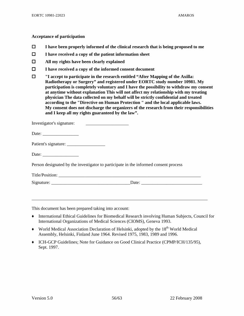
EORTC 10981-22023 AMAROS
Version 5.0 56/63 22 February 2008
Acceptance of participation ���� I have been properly informed of the clinical research that is being proposed to me
���� I have received a copy of the patient information sheet
���� All my rights have been clearly explained
���� I have received a copy of the informed consent document
���� "I accept to participate in the research entitled “After Mapping of the Axilla: Radiotherapy or Surgery” and registered under EORTC study number 10981. My participation is completely voluntary and I have the possibility to withdraw my consent at anytime without explanation This will not affect my relationship with my treating physician The data collected on my behalf will be strictly confidential and treated according to the "Directive on Human Protection " and the local applicable laws. My consent does not discharge the organizers of the research from their responsibilities and I keep all my rights guaranteed by the law”.
Investigator's signature: ___________________ Date: ________________ Patient's signature: _________________ Date: ________________ Person designated by the investigator to participate in the informed consent process Title/Position: _______________________________________________________________
Signature: ___________________________________Date: ___________________________ This document has been prepared taking into account:
♦ International Ethical Guidelines for Biomedical Research involving Human Subjects, Council for International Organizations of Medical Sciences (CIOMS), Geneva 1993.
♦ World Medical Association Declaration of Helsinki, adopted by the 18th World Medical Assembly, Helsinki, Finland June 1964. Revised 1975, 1983, 1989 and 1996.
♦ ICH-GCP Guidelines; Note for Guidance on Good Clinical Practice (CPMP/ICH/135/95), Sept. 1997.

EORTC 10981-22023 AMAROS
Version 5.0 57/63 22 February 2008
APPENDIX VI: EORTC QUALITY OF LIFE QUESTIONNAIRE-C3 0
EORTC QLQ-C30 (version 3)
We are interested in some things about you and your health. Please answer all of the questions yourself by circling the number that best applies to you. There are no "right" or "wrong" answers. The information that you provide will remain strictly confidential. Please fill in your initials: Your birthdate (Day, Month, Year): Today's date (Day, Month, Year): ____________________________________________________________________________________ Not at A Quite Very All Little a Bit Much 1. Do you have any trouble doing strenuous activities, like carrying a heavy shopping bag or a suitcase? 1 2 3 4 2. Do you have any trouble taking a long walk? 1 2 3 4 3. Do you have any trouble taking a short walk outside of the house? 1 2 3 4 4. Do you need to stay in bed or a chair during the day? 1 2 3 4 5. Do you need help with eating, dressing, washing yourself or using the toilet? 1 2 3 4 During the past week: Not at A Quite Very All Little a Bit Much 6. Were you limited in doing either your work or other daily activities? 1 2 3 4 7. Were you limited in pursuing your hobbies or other leisure time activities? 1 2 3 4 8. Were you short of breath? 1 2 3 4 9. Have you had pain? 1 2 3 4 10. Did you need to rest? 1 2 3 4 11. Have you had trouble sleeping? 1 2 3 4 12. Have you felt weak? 1 2 3 4 13. Have you lacked appetite? 1 2 3 4 14. Have you felt nauseated? 1 2 3 4 15. Have you vomited? 1 2 3 4

EORTC 10981-22023 AMAROS
Version 5.0 58/63 22 February 2008
During the past week: Not at A Quite Very
All Little a Bit Much 16. Have you been constipated? 1 2 3 4 17. Have you had diarrhea? 1 2 3 4 18. Were you tired? 1 2 3 4 19. Did pain interfere with your daily activities? 1 2 3 4 20. Have you had difficulty in concentrating on things, like reading a newspaper or watching television? 1 2 3 4 21. Did you feel tense? 1 2 3 4 22. Did you worry? 1 2 3 4 23. Did you feel irritable? 1 2 3 4 24. Did you feel depressed? 1 2 3 4 25. Have you had difficulty remembering things? 1 2 3 4 26. Has your physical condition or medical treatment interfered with your family life? 1 2 3 4 27. Has your physical condition or medical treatment interfered with your social activities? 1 2 3 4 28. Has your physical condition or medical treatment caused you financial difficulties? 1 2 3 4 For the following questions please circle the number between 1 and 7 that best applies to you
29. How would you rate your overall health during the past week? 1 2 3 4 5 6 7 Very poor Excellent 30. How would you rate your overall quality of life during the past week? 1 2 3 4 5 6 7 Very poor Excellent
© Copyright 1995 EORTC Study Group on Quality of Life. All rights reserved. Version 3.0
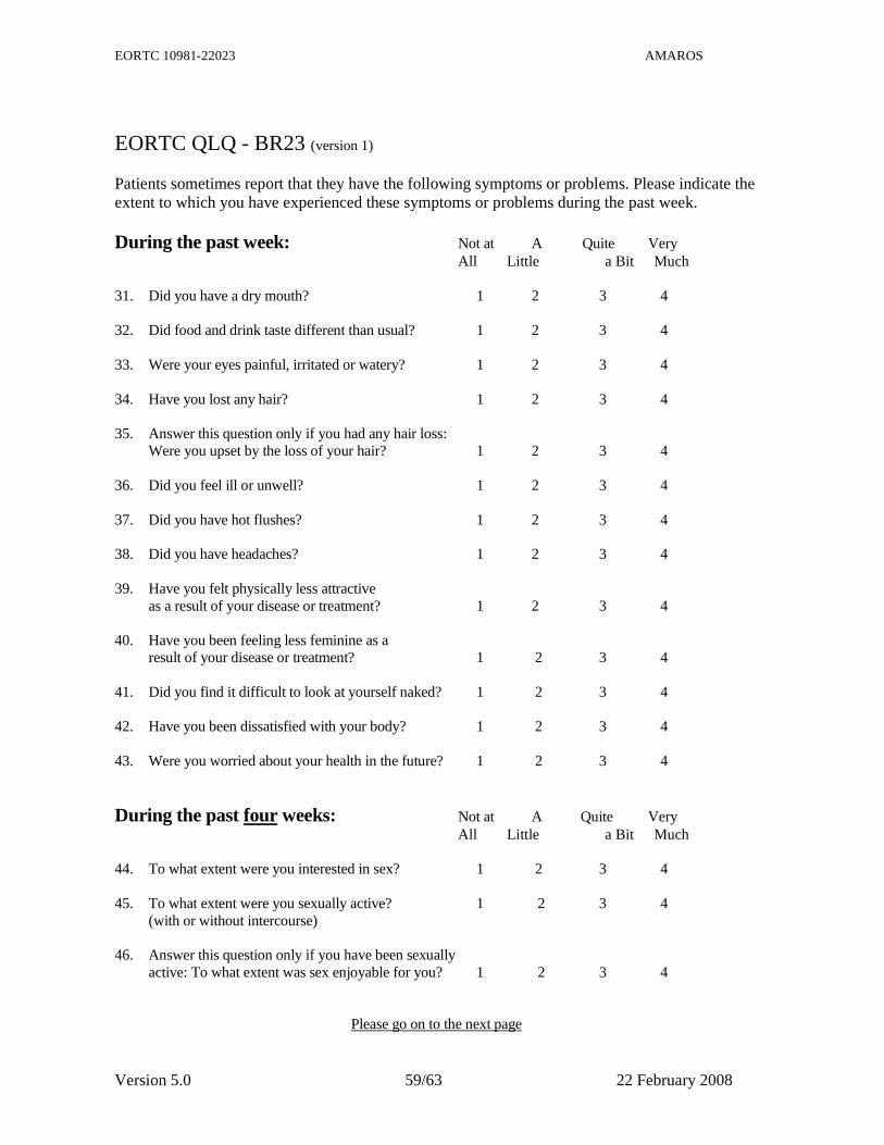
EORTC 10981-22023 AMAROS
Version 5.0 59/63 22 February 2008
EORTC QLQ - BR23 (version 1) Patients sometimes report that they have the following symptoms or problems. Please indicate the extent to which you have experienced these symptoms or problems during the past week. During the past week: Not at A Quite Very
All Little a Bit Much 31. Did you have a dry mouth? 1 2 3 4 32. Did food and drink taste different than usual? 1 2 3 4 33. Were your eyes painful, irritated or watery? 1 2 3 4 34. Have you lost any hair? 1 2 3 4 35. Answer this question only if you had any hair loss: Were you upset by the loss of your hair? 1 2 3 4 36. Did you feel ill or unwell? 1 2 3 4 37. Did you have hot flushes? 1 2 3 4 38. Did you have headaches? 1 2 3 4 39. Have you felt physically less attractive as a result of your disease or treatment? 1 2 3 4 40. Have you been feeling less feminine as a result of your disease or treatment? 1 2 3 4 41. Did you find it difficult to look at yourself naked? 1 2 3 4 42. Have you been dissatisfied with your body? 1 2 3 4 43. Were you worried about your health in the future? 1 2 3 4
During the past four weeks: Not at A Quite Very All Little a Bit Much 44. To what extent were you interested in sex? 1 2 3 4 45. To what extent were you sexually active? 1 2 3 4 (with or without intercourse) 46. Answer this question only if you have been sexually active: To what extent was sex enjoyable for you? 1 2 3 4 Please go on to the next page

EORTC 10981-22023 AMAROS
Version 5.0 60/63 22 February 2008
During the past week: Not at A Quite Very All Little a Bit Much 47. Did you have any pain in your arm or shoulder? 1 2 3 4 48. Did you have a swollen arm or hand? 1 2 3 4 49. Was it difficult to raise your arm or to move it sideways? 1 2 3 4 50. Have you had any pain in the area of your affected breast? 1 2 3 4 51. Was the area of your affected breast swollen? 1 2 3 4 52. Was the area of your affected breast oversensitive? 1 2 3 4 53. Have you had skin problems on or in the area of your affected breast (e.g., itchy, dry, flaky)? 1 2 3 4
© Copyright 1994 EORTC Study Group on Quality of Life. All rights reserved. Version 1.0

EORTC 10981-22023 AMAROS
Version 5.0 61/63 22 February 2008
APPENDIX VII: COMMITMENT STATEMENT / STUDY ACKNOWLE DGMENT
EORTC Headquarters Av. E. Mounier 83/11
1200 Brussels, Belgium Phone: + 32 2 774 16 11
Fax: + 32 2 772 35 45 E-mail: [email protected]
EORTC PROTOCOL 10981-22023: AMAROS After Mapping of the Axilla: Radiotherapy Or Surger y?
I, the undersigned declare that I will participate in the above-mentioned study. I expect to recruit ______________ patients per year.
I have read the protocol and agree that it contains all necessary details for carrying out the study as described. I will conduct this protocol and any subsequent amendments as outlined therein and will make a reasonable effort to complete the study within the time designated. I will provide copies of the protocol and access to all relevant information I receive to study personnel under my supervision. I will discuss this material with them to ensure that they are fully informed about the study and treatment. I understand that the EORTC may terminate the study or suspend enrolment at any time if it becomes necessary to protect the best interests of the study subjects.
I ACCEPT THE FOLLOWING TERMS AND CONDITIONS:
1. As the meetings, correspondence or discussions referred to above may involve matters for which I will be taken into confidence, I will regard as secret and confidential any such information which I may thereby acquire in respect of the manufacturing or commercial interests of the industrial partner, if any and its research. Accordingly I will not disclose such information to a third party.
2. All the trial related minutes of meetings, correspondence or records of discussion together with all other trial documents obtained from the EORTC, other Collaborative groups and/or the industrial partners (if any) are confidential and remain the property of the respective partner. This information will be returned to them if requested.
b I am responsible for Ethics Committee submission.
b The following person is in charge of submission: ____________________________________
b I have no potential conflict of interest, such as a professional interest, a proprietary interest or any other conflict of interest.
b YES, I have a potential conflict of interest (If you have a potential conflict of interest, please indicate this and we will send you the standard of conduct for conflict of interest/confidentiality policy and a conflict of interest/confidentiality disclosure form requesting further clarification).
NAME Principal Investigator: _______________________________________________________ EORTC Institution number: _______________________Date: ___________________________ Signature: _______________________________________________________________________ Please complete and return this form, as soon as possible, to Nicole Duez, responsible data manager at the EORTC Headquarters: Avenue E. Mounier, 83, BP 11, 1200 Brussels, Belgium (Fax: +32 2 771 38 10)

EORTC 10981-22023 AMAROS
Version 5.0 62/63 22 February 2008
APPENDIX VIII: ALMANAC TRIALISTS GROUP SPECIFIC APP ENDIX TO THE EORTC PROTOCOL 10981- 22023
ALMANAC Trialists Group ALMANAC Trialists Group Study Coordinator: Dr. Mansel All chapters of the protocol are common to all participating groups except sections/chapters 5.7, 5.10, 9, 15.2 and 18 that are specific to EORTC and BOOG participants. Those sections/chapters specific to ALMANAC Trialists Group are included in this Group Specific Appendix. Therefore, the content of this document supersedes or complete sections/chapters 5.7, 5.10, 9, 15.2, 18.
Section 5.7: Learning phase The centres from the ALMANAC trialists group are exempted from surgical site visits since quality control is garanteed by the former participation in the ALMANAC trial. It is therefore that the ALMANAC trialists group is also exempted from the conditions as stated in section 5.7 of the main protocol (learning phase). Section 5.10: Radiation of the Axilla.
Since the risk of relapse in axillary lymph nodes is small and there is no existing evidence that delay in radiotherapy to clinically negative lymph nodes will result in a substantial increase in the risk of axillary recurrences, it is unlikely that delay in radiotherapy after chemotherapy will result in a significant difference in axillary recurrence rate. It is therefore that the ALMANAC trialists group is exempted from the conditions as stated in the first paragraph of section 5.10 of the main protocol (radiation of the axilla). Radiotherapy will be given as soon as possible after finishing chemotherapy.
Chapter 9: Forms and procedures for collecting data Chapter 9 of the main protocol and all EORTC documents and guidelines relative to forms and their completion applies to ALMANAC except the address for sending paper forms and answer to queries. Investigators participating on behalf of ALMANAC Trialists Group should send all forms and answer to queries to:
R E Mansel Department of Surgery, University Hospital of Wales, Heath Park, Cardiff, CF14 4XN
Please do not send the data directly to the EORTC Headquarters. The data should be reported on the forms specific to ALMANAC Trialists Group. Section 15.2: On-site quality control

EORTC 10981-22023 AMAROS
Version 5.0 63/63 22 February 2008
The annual on-site visits described in section 15.2 of the main protocol do not apply to ALMANAC Trialists Group. The Quality Control of the radiotherapy is done using the dummy run. This document has to be filled in by each Radiotherapy Department and sent back to:
Marieke Straver, MD Monitor AMAROS study The Netherlands Cancer Institute /Antoni van Leeuwenhoek Hospital Department of Surgery Plesmanlaan 121 1066 CX, Amsterdam The Netherlands
The quality of radiotherapy will be controlled by an annual evaluation of the filed radiation data of 10 randomly chosen patients by the Radiotherapy Coordinator or an independent representative (not
from the site visited centre). Chapter 18: Administrative responsibilities
The study coordinator
The ALMANAC Study Coordinator will be responsible for presenting the protocol to the group and discussing it with the investigators of the Group who will participate in the protocol. The Study Coordinator’s responsibility is to ensure that data are collected and used in accordance with the Group’s policy and quality.
Study coordinator:
Pr. R E Mansel Department of Surgery, University Hospital of Wales, Heath Park, Cardiff, CF14 4XN
The Data Center
The ALMANAC Data Center will act as a "mailbox" in this trial (see forms and procedures for data collection). All methodological questions should be addressed to the Study Coordinator.
The Group
ALMANAC Trialists Group is responsible as a group to guarantee the general compliance of their members with procedures described in this appendix. All questions concerning membership in the ALMANAC should be addressed to the Study Coordinator.