age estimation from computed tomography of cranial suture
TRANSCRIPT

Med & Health Dec 2020; 15(2): 276-289
ORIGINAL ARTICLE
276
https://doi.org/10.17576/MH.2020.1502.24
Address for correspondence and reprint requests: Professor Pasuk Mahakkanukrauh. Excellence Center in Osteology Research and Training Center (ORTC), Chiang Mai University, Chiang Mai, 50200, Thailand. Tel: +66-53-949-474 Email: [email protected]
Age Estimation from Computed Tomography of Cranial Suture Closure in a Thai Population
SITTIPORN R1, NUTTAYA P2, SUKON P3, PASUK M4,5,*
1Department of Forensic Medicine, 2Department of Radiology, Faculty of Medicine, Chiang Mai University, Chiang Mai, 50200, Thailand.
3Department of Statistics, Faculty of Science, Chiang Mai University, Chiang Mai, 50200, Thailand.
4Department of Anatomy, Faculty of Medicine, Chiang Mai University, Chiang Mai, 50200, Thailand.
5Excellence in Osteology Research and Training Center (ORTC), Chiang Mai University, Chiang Mai, 50200, Thailand.
ABSTRAK
Anggaran umur dari sisa tertinggal kerangka manusia adalah langkah penting untuk membina semula profil biologi. Jahitan sutur kranial telah lama dikaji untuk tujuan penutupan yang berkaitan dengan usia. Namun, hingga kini, antropologi forensik masih digunakan untuk menyiasat cara terbaik untuk menganggarkan usia kematian akibat penutupan sutur kranial kerana tengkorak biasanya dijumpai di tempat kejadian kerana mudah dikenali dan kedapatan kerosakan tengkorak pada post-mortem. Atas sebab-sebab ini, dilakukan kajian anggaran usia dari penutupan sutur kranial pada populasi kajian Thailand yang memfokuskan diri untuk mengkaji penampilan dan penglihatan penutupan sutur wajah menggunakan kajian imbasan tomografi (CT). Siri gambar CT sebanyak 140 kes diperolehi untuk menyiasat penutupan ektokranial dari sutur wajah yang dipilih. Hasil analisis gambar CT menunjukkan bahawa imbasan sudut nasomaksilari memberikan pemeriksaan penutupan jahitan yang paling konsisten (52%) sementara sudut frontonasal pula memberikan konsistensi terendah dalam pemeriksaan penutupan sutur (29%). Ketidaklarasan kebanyakan dapatan terjadi dalam menetapkan skor penutupan 1 dan 2. Oleh itu, dapat disarankan agar sistem penutupan sutur berskala 3 dilakukan, iaitu: tutup, meneruskan penutupan, dan menutup keseluruhannya, akan dapat menjadi kaedah yang tepat untuk menilai tahap penutupan ectocranial sutur wajah yang diperoleh daripada pengimejan CT. Maklumat asas penutupan jahitan wajah dari CT dapat menjadi titik awal pengembangan teknik perkiraan usia dari penutupan jahitan dengan menggunakan gambar CT.

277
Computed Tomography of Cranial Suture Closure Med & Health Dec 2020;15(2): 276-289
Kata kunci: anggaran umur, imbasan tomografi, sutur kranial, Thailand
ABSTRACT
Age estimation from human skeletal remains is an important step to reconstruct a biological profile. Cranial suture has long been studied for its age-related closure. However, until now, forensic anthropologists still attempt to investigate the best way of estimating age at death from cranial suture closure because skull is usually found at the crime scene due to its easy recognised-appearance and persistence to post-mortem insults. For these reasons, a study of age estimation from cranial suture closure in a Thai population was conducted, which focussed to study the appearance and visibility of facial suture closure using computed tomography (CT). CT image series of 140 cases were obtained in order to investigate ectocranial closure of the selected facial sutures. The results from CT image analysis revealed that nasomaxillary provided the most consistent examination of suture closure (52%) while frontonasal delivered the lowest consistency in suture closure examination (29%). The inconsistency mostly occurred in assigning the closure score of 1 and 2. Thus, it could be suggested that a 3-scale scoring system of closure: open, closing, and closed, could be an appropriate method of evaluating degree of ectocranial closure of facial sutures obtained from CT imaging. This fundamental information of facial suture closure from CT images could serve as a starting point on development of age estimation technique from suture closure by utilising CT images.
Keywords: age estimation, cranial suture, computed tomography, Thais
appearance and its resistance to post-mortem taphonomic insults (Boldsen et al. 2002; Key et al. 1994). Besides visual assessment of suture closure in dry crania, application of medical imaging through the use of computed tomography (CT) is on the rise in forensic anthropology. Utilisation of CT in autopsy procedures has become more common in American and European forensic contexts for the assessment of trauma of unknown cause (Boyd et al. 2015; Harth et al. 2010; Leth 2007). CT allows acquisition of high-resolution images of bone structures without
INTRODUCTION
Age estimation from human skeletal remains is an essential procedure in reconstructing an individual’s biological profile. With a relatively limited number of age estimation techniques available to the forensic osteologist, many researchers are still attempting to determine the best way to estimate age at death from cranial suture closure. One reason for this continued interest is the fact that the skull is often recovered at crime scenes due to its easy-to-recognise

278
Med & Health Dec 2020;15(2): 276-289 Sittiporn R. et al.
removing soft tissue or damaging the bones, including details of fine bone morphology, fractures, and trauma which are more difficult to investigate using conventional autopsy techniques (Thali et al. 2003; Verhoff et al. 2008). Additionally, defleshing the bone can consume tremendous amounts of time and this procedure is often prohibited by religious practices and beliefs, or even by national laws and regulations, since the removal of soft tissue from bone is considered invasive (Brough et al. 2014). Although some studies have been conducted previously on suture closure using CT (Chiba et al. 2013; Obert et al. 2010), they all focused on vault sutures: coronal sagittal, and lambdoidal. However, knowledge about the appearance and visibility of facial suture closure is still lacking. No research has been conducted on closure of facial sutures located in an area that is difficult to deflesh at autopsy for age estimation using CT. For all of the reasons mentioned above, a thorough study on age estimation from cranial suture closure in a Thai sample is necessary to assist in providing practical and appropriate age estimation approaches for forensic osteologists working in Thailand. Although application of CT is not a routine approach in autopsy procedure in Thailand, this study is expected to provide a foundation of knowledge that can be used as a reference method for aging of Thai skeletons.
MATERIALS AND METHODS
Data Acquisition
All CT images (140 series of images) of facial sutures obtained from 50 crania were evaluated for ectocranial closure of the five facial sutures in order to compare with the closure scores obtained from the visual, morphological study. Fifty crania which composed of 22 males and 28 females with age at the time of death ranging from 28-90 years (mean age of 63.18 + 17.91 years). Crania with anomalies, trauma, disease, or postmortem breakage which affected the facial sutures or facial bones were excluded. Closure of the five facial sutures i.e. frontonasal (FN), nasomaxillary (NML) (left side), internasal (INN), incisive (IN), and median palatine (MP) sutures was scored for all 50 crania in the morphological study using a 4-scale scoring method (0-3) based on the Meindl and Lovejoy method (Meindl & Lovejoy 1985). The entire length of each facial suture was evaluated for its closure. It has to be noted that, for CT, the closure score of the five facial sutures obtained from the morphological study was transformed to the original scale of Meindl & Lovejoy (1985) for comparison. After visual assessment of suture closure, CT scanning was performed on all 50 crania utilising a multidetector CT machine. Various CT studies have been conducted on parts of the cranial skeleton. A range of CT doses have been utilised, e.g. 0.50-2.00 mm slice thickness, 20-200 mA tube current, and 100-120 tube voltage (Boyd et al. 2015; Chiba et al. 2013; Akhlaghi et al. 2017; Appleby et al. 2015; Cesarani et al. 2003; Gufler et al. 2014; Hawass &

279
Computed Tomography of Cranial Suture Closure Med & Health Dec 2020;15(2): 276-289
Saleem 2011; Sakuma et al. 2010). The skulls were scanned on a third generation dual source CT system (SOMATOM Force, Siemens Healthineers, Forchheim, Germany) by placing the bone at the center of CT gantry. The scanning and reconstruction parameters were 200 mAs, 140 kVp, slice thickness 0.4 mm, reconstruction increment 0.2 mm, field of view 20 cm, matrix 512 x 512 and sharp kernel (Ur73) images. The multiplanar two-dimensional reconstruction was performed on coronal and sagittal planes. With these parameters, the image resolution was 0.39 x 0.39 x 0.4 mm3.
Image Interpretation
Before closure of the suture was examined, a total number of CT slices indicating an entire length of the suture was identified. The degree and extent of suture closure on the ectocranial side of the five facial sutures were evaluated on CT images perpendicular to the suture alignment. This is considered to be an appropriate approach for examining the sutures from sectioned images (Furuya et al. 1984). For each
CT slice, the degree of suture closure was considered as open if a lucent line at the outer surface of the suture was found. In contrast, presence of a covering of bony plate at the outer surface of the suture would be scored as closed. Figures 1-5 demonstrate examples of open and closed stages on each suture. Next, the extent of suture closure was evaluated along the CT image series (Figure 6). A four-scale scoring system (0-3) based on the Meindl and Lovejoy method was used (Meindl & Lovejoy 1985). The scale used was as follows: (0) represented a lucent line at the outer surface of suture in all or most CT images of the suture (less than five images show closure of the suture at the outer surface); (1) represented a closed suture at the outer surface on a few CT image slices (<50%); (2) represented most slices (>50%) showing a closed suture at the outer surface; and (3) represented a totally or almost closed suture in all CT images (less than five images show an open suture at the outer surface). Closure scores were calculated for each suture or case. During the image interpretation process, each slice of sectioned suture was assigned a
Figure 1: Frontonasal suture was examined in the sagittal plane. (A) open suture on the ectocranial side (red arrow), (B) closed suture on the ectocranial side (red arrow).

280
Med & Health Dec 2020;15(2): 276-289 Sittiporn R. et al.
Figure 2: Internasal suture was examined in the axial plane. (A) open suture on the ectocranial side (red circle), (B) closed suture on the ectocranial side (red circle).
Figure 3: Nasomaxillary suture was examined in the axial plane. (A) open suture on the ectocranial side (red arrow), (B) closed suture on the ectocranial side (red arrow). The white arrow indicates the
location of the nasomaxillary suture on the right side.
Figure 4: Incisive suture was examined in the sagittal plane. (A) open suture on the ectocranial side (red circle), (B) closed suture on the ectocranial side (red circle).
Figure 5: Median palatine suture was examined in the coronal plane. (A) open suture on the ectocranial side (red circle), (B) closed suture on the ectocranial side (red circle).

281
Computed Tomography of Cranial Suture Closure Med & Health Dec 2020;15(2): 276-289
closure stage (open or closed). Then, the number of slices showing a closed suture was divided by the total number of slices representing the entire length of the suture. After that, the percentage of closure was computed and the closure score assigned according to the Meindl and Lovejoy (1985) method. All CT images were separately examined by two observers (PhD students). Two sessions of image interpretation were conducted by the same observer with one-month interval in between, in order to perform an intra-observer agreement test. If disagreement occurred between two observers, those images would be reviewed by observer 1, 2, and 3 (who is a musculoskeletal radiologist) until a consensus was reached.
Suture Closure Examination Utilising CT
The frequency of the five facial suture closure scores obtained from both the morphological study and the CT images was tabulated. Also, intra-
and inter-observer agreements were analysed using Cohen’s kappa analysis. Evaluation of agreement values was conducted based on the description in the morphological analysis. Accuracy or consistency in facial suture closure examination from CT images was evaluated by comparison to the facial suture closure scores obtained from morphological study of dry crania as the gold standard. All statistical analysis were computed in Microsoft Excel 2013 (Microsoft Corporation, USA), SPSS 20.0 (IBM Corp., Armonk, NY, USA), and R (R Development Core Team, 2017).
RESULTS
Five facial sutures of the selected 50 crania were chosen from the visual assessment study in order to evaluate their closure using CT. All five facial sutures of all selected crania were previously given closure scores by visual assessment. These scores were
Figure 6: Example of some CT images showing sections of internasal suture along its entire length in axial view

282
Med & Health Dec 2020;15(2): 276-289 Sittiporn R. et al.
then used as the gold standard for comparing to closure scores obtained from CT. At first, the samples were selected in an attempt to obtain equally distributed scores, but the score of 0 for incisive was not found among the samples in this study. Additionally, the score of 3 for frontonasal and nasomaxillary suture was rarely found (Table 1). Tables 1-3 list the closure score frequencies obtained from CT images. Consensus scores, the scores given by observer 1 (both original and repeat examinations), and observer 2 were tabulated. Among the 140 suture locations, most of the consensus scores were either a 1 or 2 for closure, except for incisive, for which the highest score frequency was a score of 3 (Table 1). For the scores obtained by observer 1 (original examination), the highest frequency of the given scores was found on the score of 2, except for frontonasal and incisive. Frontonasal had the same frequencies for scores 1 and 2. Incisive, like that of the consensus score, had a score of 3 as the highest frequency given by observer 1 (Table 2). Similar results to those of the consensus and original examination of observer 1 were found for the repeat examination scores of observer 1. Internasal and nasomaxillary exhibited the highest frequency of assigned scores at a score of 2. However, the highest frequency score observed for closure of incisive was found to be a score of 2 (Table 2). For the scores reported by observer 2, score 2 also had the highest frequency found on internasal, nasomaxillary, and median palatine (Table 3). Cross-tabulation between
closure scores obtained from CT images by different observers was also investigated. For each suture, comparisons of consensus scores and scores obtained from the original examination by observer 1, consensus scores and the scores obtained from the repeat examination by observer 1, consensus scores and scores obtained from observer 2, scores obtained from the original and repeat examinations of observer 1, and scores obtained from observer 1 and observer 2 were examined. The results showed that the frontonasal demonstrated a relatively high variation in scoring consistency among observers. Some comparisons exhibited high consistency in some scores compared to the others. Comparison between consensus scores and the scores obtained from observer 2 showed 90.0% consistency in score 1. This score also showed high consistency (63.6 and 70.0%) in the comparison between the original and repeat examinations of observer 1. Furthermore, the score of 3 presented 75.0 and 100% consistency between the consensus scores and the scores obtained from the repeat examination by observer 1. Similarly, high scoring consistency was found in score 1 for most comparisons of internasal closure. The highest consistency (85.7%) was found on the comparisons between consensus scores and scores provided by observer 2 and between the original and repeat examinations of observer 1. The next highest scoring consistency was also found in score 2 on the comparisons between consensus scores and the original examination by

283
Computed Tomography of Cranial Suture Closure Med & Health Dec 2020;15(2): 276-289
Sutu
reV
isua
l as
sess
men
tC
ompu
ted
tom
ogra
phy
(con
sens
us)
Scor
e 0
%Sc
ore
1%
Scor
e 2
%Sc
ore
3%
Tota
lSc
ore
0%
Scor
e 1
%Sc
ore
2%
Scor
e 3
%To
tal
FN8
33.3
729
.28
33.3
14.
224
312
.510
41.7
833
.33
12.5
24
INN
825
.08
25.0
825
.08
25.0
322
6.3
721
.918
56.3
515
.632
NM
L8
32.0
832
.08
32.0
14.
025
14.
06
24.0
1664
.02
8.0
25
IN0*
0*1
6.3
743
.88
50.0
160
00
07
43.8
956
.316
MP
614
.017
39.5
1330
.27
16.3
433
7.0
1330
.217
39.5
1023
.343
Not
e: *
The
re w
ere
no s
ampl
es in
the
pres
ent s
tudy
sho
win
g a
scor
e of
0 (t
otal
ly o
pen)
for I
N
Tabl
e 1:
Fre
quen
cy o
f sut
ure
clos
ure
scor
e ob
tain
ed fr
om v
isua
l ass
essm
ent a
nd c
ompu
ted
tom
ogra
phy
(con
sens
us).
Sutu
reO
bser
ver
1 (O
rigi
nal ex
amin
atio
n)O
bser
ver
1 (R
epea
t ex
amin
atio
n)
Scor
e 0
%Sc
ore
1%
Scor
e 2
%Sc
ore
3%
Tota
lSc
ore
0%
Scor
e 1
%Sc
ore
2%
Scor
e 3
%To
tal
FN3
12.5
1041
.710
41.7
14.
224
520
.811
45.8
416
.74
16.7
24
INN
412
.57
21.9
1959
.42
6.3
323
9.4
721
.913
40.6
928
.132
NM
L1
4.0
520
.019
76.0
00
250
0.0
312
.018
72.0
416
.025
IN0
00
07
43.8
956
.316
00.
00
0.0
1168
.85
31.2
16
MP
37.
013
30.2
2046
.57
16.3
433
7.0
1227
.921
48.8
716
.343
Tabl
e 2:
Fre
quen
cy o
f sut
ure
clos
ure
scor
e ob
tain
ed fr
om c
ompu
ted
tom
ogra
phy
betw
een
obse
rver
1 (O
rigin
al e
xam
inat
ion)
and
ob
serv
er 1
(Rep
eat e
xam
inat
ion)
.

284
Med & Health Dec 2020;15(2): 276-289 Sittiporn R. et al.
observer 1 and between scores given by observers 1 and 2. Regarding nasomaxillary, the highest scoring consistency (92.9 and 81.3%) was found on score 2 in the comparison between the consensus scores and the scores obtained from observer 2. The score of 2 also gave high consistency in other comparisons among observers. On the other hand, it was interesting to note that there was no consistency in the score of 0 for all comparisons. Regarding scores obtained from incisive, scoring consistency varied among scores of 2 and 3 for all comparisons. Percentage of scoring consistency ranged from 37.5 to 100. In contrast, no comparisons revealed consistency in scores of 0 and 1. Lastly, comparisons among scores for midpalatine obtained by observers showed that high scoring consistency for most comparisons was found
for the score of 2. The consistency percentage ranged from 70.0 to 94.1 in three comparisons: consensus score and original examination by observer 1, consensus score and repeat examination by observer 1, and original and repeated examinations by observer 1. However, consistency in scoring was relatively low in the comparison between scores obtained from observers 1 and 2 compared to the others. The consistency percentages ranged from 0 to 100 with most falling around 35.0-40.0%. Results of the intra- and inter-observer agreement analysis were provided in Table 4. Internasal demonstrated the highest agreement (0.63) within observer while frontonasal and nasomaxillary had the lowest agreement value (0.35). In contrast, nasomaxillary exhibited the highest (0.31) agreement in the inter-observer
Suture Score 0 % Score 1 % Score 2 % Score 3 % Total
FN 4 16.7 10 41.7 7 29.2 3 12.5 24
INN 0 0 7 21.9 15 46.9 10 31.3 32
NML 1 4.0 6 24.0 14 56.0 4 16.0 25
IN 0 0 0 0 8 50.0 8 50.0 16
MP 0 0 5 11.6 19 44.2 19 44.2 43
Table 3: Frequency of suture closure score obtained from computed tomography (observer 2).
Table 4: Intra- and inter-observer agreement analysis.
Suture Intra-observer analysis Inter-observer analysis
Kappa statistic p-value Kappa statistic p-value
FN 0.35 0.002 0.02 0.890
INN 0.63 <0.001 0.28 0.005
NML 0.35 0.016 0.31 0.016
IN 0.48 0.029 -0.13 0.614
MP 0.61 <0.001 0.09 0.338
285
Computed Tomography of Cranial Suture Closure Med & Health Dec 2020;15(2): 276-289
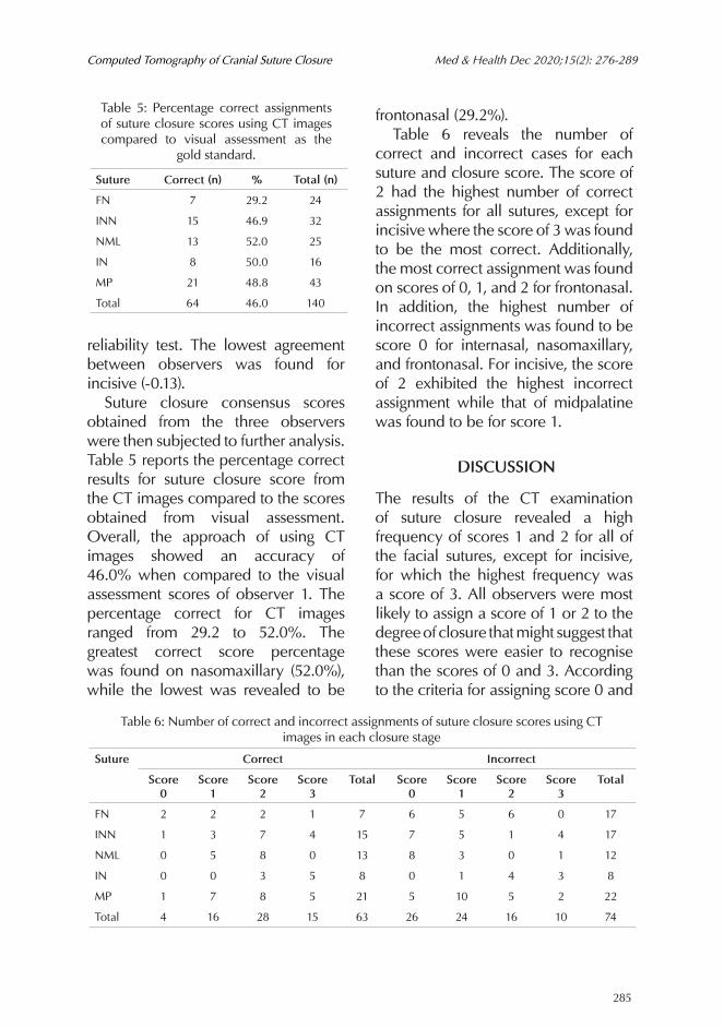
reliability test. The lowest agreement between observers was found for incisive (-0.13). Suture closure consensus scores obtained from the three observers were then subjected to further analysis. Table 5 reports the percentage correct results for suture closure score from the CT images compared to the scores obtained from visual assessment. Overall, the approach of using CT images showed an accuracy of 46.0% when compared to the visual assessment scores of observer 1. The percentage correct for CT images ranged from 29.2 to 52.0%. The greatest correct score percentage was found on nasomaxillary (52.0%), while the lowest was revealed to be
frontonasal (29.2%). Table 6 reveals the number of correct and incorrect cases for each suture and closure score. The score of 2 had the highest number of correct assignments for all sutures, except for incisive where the score of 3 was found to be the most correct. Additionally, the most correct assignment was found on scores of 0, 1, and 2 for frontonasal. In addition, the highest number of incorrect assignments was found to be score 0 for internasal, nasomaxillary, and frontonasal. For incisive, the score of 2 exhibited the highest incorrect assignment while that of midpalatine was found to be for score 1.
DISCUSSION
The results of the CT examination of suture closure revealed a high frequency of scores 1 and 2 for all of the facial sutures, except for incisive, for which the highest frequency was a score of 3. All observers were most likely to assign a score of 1 or 2 to the degree of closure that might suggest that these scores were easier to recognise than the scores of 0 and 3. According to the criteria for assigning score 0 and
Suture Correct (n) % Total (n)
FN 7 29.2 24
INN 15 46.9 32
NML 13 52.0 25
IN 8 50.0 16
MP 21 48.8 43
Total 64 46.0 140
Table 5: Percentage correct assignments of suture closure scores using CT images compared to visual assessment as the
gold standard.
Suture Correct Incorrect
Score 0
Score 1
Score 2
Score 3
Total Score 0
Score 1
Score 2
Score 3
Total
FN 2 2 2 1 7 6 5 6 0 17
INN 1 3 7 4 15 7 5 1 4 17
NML 0 5 8 0 13 8 3 0 1 12
IN 0 0 3 5 8 0 1 4 3 8
MP 1 7 8 5 21 5 10 5 2 22
Total 4 16 28 15 63 26 24 16 10 74
Table 6: Number of correct and incorrect assignments of suture closure scores using CT images in each closure stage

286
Med & Health Dec 2020;15(2): 276-289 Sittiporn R. et al.
3, if less than five images in the total image series showed a closed suture, the closure score would be labeled 0 (totally open). On the other hand, if less than five images in the total image series exhibited an open suture, the closure score would be 3 indicating totally closed. Although five images (CT cuts) were allowed to differ to account for the possible presence of artifacts, the observers occasionally assigned the closure score of 0 and 3 unless it was there were relatively obvious that all CT images in the series depicted opened or closed suture. Besides the effect of artifacts described previously, the present study was limited by low sample size in some closure stages of some sutures. According to the morphological (visual) study, incisive revealed a relatively high frequency of score 3 but a low frequency of scores 0 and 1 since it typically reaches its complete closure very early before adulthood (Mann et al. 1991), and most of the skeletons in the present study were relatively old at the time of death. It was, therefore, difficult to find closure scores of 0 or 1 on incisive. In contrast, the number of cases that presented a closure score of 3 for frontonasal, internasal, and nasomaxillary were relatively low in the present study because these sutures show delayed complete closure; therefore, it was rare to find this stage of closure in the sample as also reported by several previous authors (Beauthier et al. 2010; Wang et al. 2006). According to the results on consistency between closure scores from CT images versus direct examination, frontonasal, as might be
expected, provided the lowest correct assignment accuracy. It produced the same closure score as that from the visual assessment from dry crania only 29.2% of the time. Its morphology was relatively difficult to examine from the sagittal plane since it showed considerable complexity and interdigit-like characteristics. Also, folding-like characteristics could sometimes occur at the edges of the frontal and nasal bones, making it even more difficult to differentiate between open or closed sutures from the CT images. The difficulty in examining frontonasal using CT images is supported by the relatively high variation in closure scores given by different observers. Furthermore, a tremendous amount of variation in closure progression of frontonasal was reported. Its closure seems to be delayed compared to the cranial vault sutures and demonstrates a sporadic pattern of closure. Variations in the closure characteristics could occur on the ectocranial surface of this suture; for instance, an open suture with large gaps and pitting or nearly closed hairline sutures (Alesbury et al. 2013). In contrast, nasomaxillary provided the most accurate assignment (52%) compared to the scores from the visual assessment of the dry crania. One of the reasons that nasomaxillary was easy to examine was that it had a relatively large area of contact between the nasal and maxillary bones. At the interface, the nasal bone usually overlaps the maxillary bone. Gufler and colleagues suggested that the interface between these two adjacent bones affected to the visibility of the suture (Gufler et al.

287
Computed Tomography of Cranial Suture Closure Med & Health Dec 2020;15(2): 276-289
2014). Various studies have been conducted to examine the morphology of midpalatal-median palatine in terms of clinical investigation for rapid maxillary expansion (RME) management (Angelieri et al. 2016; Leonardi et al. 2011; Liu et al. 2015; Woller et al. 2014). In the present study, this suture revealed relatively high intra- and inter-observer variability and demonstrated accurate assignment of 48.8%, compared to that obtained from visual assessment; perhaps because of the effect of torus palatinus, a quite common skeletal variation found in the Thai population (Apinhasmit et al. 2002). In the present study, 24 out of 43 selected cases for CT (56%) exhibited torus palatinus. This morphological variation might also affect the efficiency of anterior median palatine (AMP) as an age indicator that worked fairly well on the Thai sample. Internasal was expected to produce high accuracy when examining its closure using CT images because its alignment is relatively straight in the superior-inferior direction. Nevertheless, this suture delivered only 46.9% consistency with the closure scores from visual assessment. Error resulting from inconsistent examination probably caused by the nature of the interface between two nasal bones, which is relatively narrow anteroposteriorly (Harth et al. 2010; Gufler et al. 2014). Although, the area of contact was long for internasal, the bone edges along the suture area were thin. This problem with scoring of thin bones is supported by findings reported by Gufler et al. (2014). In their study,
sphenofrontal and sphenozygomatica, as well as the orbital sutures located on the medial wall of the orbit, showed poor visibility because these sutures are composed of very thin bones where they articulate (Gufler et al. 2014). In addition, rapid change in suture morphology, e.g. immediate change from open (an empty space) to closed (dense bone), or vice versa, within a very small area along the extent of the suture, could cause some artifacts on CT images called a partial volume effect. This can result from dense objects, compact bone in our circumstance, that intrudes upon the CT section width. This could result in shading artifacts appearing in the CT image (Barrett & Keat 2004). Consequently, this shading artifact could lead to difficulty in scoring suture closure. This might be a reason why there was a low number of correct assignments of the score 0 because observers sometimes assigned score 1 or 2 (partially closed) even when there were totally open sutures. The effect of partial volume might also explain why the assigned closure scores from CT tends to overestimate from those obtained from visual assessment. The results from the present study in terms of the poor match between the CT and visual scoring methods means that CT imaging, based on the scanning method used in the present study, at least, cannot be used as a means to improve visual scoring methodologies, even though it would allow easy examination of skulls without the need for soft tissue removal. If CT is to be used for aging

288
Med & Health Dec 2020;15(2): 276-289 Sittiporn R. et al.
the cranium, the methods would likely need to be CT specific, developed on the living or in autopsy rooms, and applied to skeletons or cadavers. And given the problems with scoring from CT images, the results are likely to not be any better than those from visual scoring. Facial sutures are very fine sutures that need more specific CT methods. Therefore, additional sutures on different areas of the cranium other than the vault should be investigated.
CONCLUSION
Suture closure scores obtained from CT were examined in order to provide fundamental information on cranial suture closure from CT images that can serve as a starting point for the development of age estimation techniques based on suture closure from CT imaging. The results from the CT images analysis revealed that nasomaxiilary provided the most consistent scoring of suture closure (52%) compared to morphological analysis, while frontonasal delivered the lowest consistency in suture closure scoring (29.2%). In conclusion, it is important to note that it would be inappropriate to rely on cranial suture closure as an age indicator unless used in conjunction with other more reliable skeletal part(s). When data from these other skeletal areas are available, cranial suture aging techniques would probably best be applied as a supportive age indicator.
ACKNOWLEDGEMENT
This study was funded by the research
administration section, Faculty of Medicine at Chiang Mai University (Research ID: ANA-2560-04492).The authors also wish to thank the Excellence in Osteology Research and Training Center (ORTC) with partial support by Chiang Mai University.
REFERENCESAkhlaghi, M., Bakhtavar, K., Kamali, A., Maarefdoost,
J., Sheikhazad, A., Mousavi, F., Anary, S.H.S, Sheikhazadi, E. 2017. The diagnostic value of anthropometric indices of maxillary sinuses for sex determination using CT-scan images in Iranian adults; A cross-sectional study. J Forensic Leg Med 49: 94-100.
Alesbury, H.S., Ubelaker, D.H., Bernstein, R. 2013. Utility of the frontonasal suture for estimating age at death in human skeletal remains. J Forensic Sci 58(1): 104-8.
Angelieri, F., Franchi, L., Cevidanes, L.H.S., Bueno-Silva, B., McNamara JA, Jr. 2016. Prediction of rapid maxillary expansion by assessing the maturation of the midpalatal suture on cone beam CT. Dental Press J Orthod 21(6): 115-25.
Apinhasmit, W., Jainkittivong, A., Swasdison, S. 2002. Torus palatinus and torus mandibularis in a Thai population. Sci Asia 28: 105-11.
Appleby, J., Rutty, G.N., Hainsworth, S.V., Woosnam-Savage, R.C., Morgan, B., Brough, A., Earp, R.W., Robinson, C., King, T.E., Morris, M., Buckley, R. 2015. Perimortem trauma in King Richard III: a skeletal analysis. The Lancet 385(9964): 253-9.
Barrett, J.F., Keat, N. 2004. Artifacts in CT: recognition and avoidance. Radiographics 24(6): 1679-91.
Beauthier, J.P., Lefevre, P., Meunier, M., Orban, R., Polet, C., Werquin, J.P., Quatrehomme, G. 2010. Palatine sutures as age indicator: A controlled study in the elderly. J Forensic Sci 55(1): 153-8.
Boldsen, J.L., Milner, G.R., Konigsberg, L.W., Wood, J.W. 2002. Transition analysis: a new method for estimating age from skeletons. In Paleodemography: age distributions from skeletal samples. Edited by Hoppa RD., Vaupel JW. Cambridge University Press; 73-106.
Boyd, K.L., Villa, C., Lynnerup, N. 2015. The use of CT scans in estimating age at death by examining the extent of ectocranial suture closure. J Forensic Sci 60(2): 363-9.
Brough, A.L., Morgan, B., Robinson, C., Black, S., Cunningham, C., Adams, C., Rutty, G.N. 2014. A minimum data set approach to post-mortem computed tomography reporting for anthropological biological profiling. Forensic

289
Computed Tomography of Cranial Suture Closure Med & Health Dec 2020;15(2): 276-289
Sci Med Pathol 10(4): 504-12. Cesarani, F., Martina, M.C., Ferraris, A., Grilletto,
R., Boano, R., Marochetti, E.F., Donadoni, A.M., Gandini, G. 2003. Whole-body three-dimensional multidetector CT of 13 Egyptian human mummies. Am J Roentgenol 180(3): 597-606.
Chiba, F., Makino, Y., Motomura, A., Inokuchi, G., Torimitsu, S., Ishii, N., Sakuma, A., Nagasawa, S., Saitoh, H., Yajima, D., Hayakawa, M., Odo, Y., Suzuki, Y., Iwase, H. 2013. Age estimation by multidetector CT images of the sagittal suture. Int J Legal Med 127(5): 1005-11.
Furuya, Y., Edwards, M.S., Alpers, C.E., Tress, B.M., Ousterhout, D.K., Norman, D. 1984. Computerized tomography of cranial sutures: Part 1: Comparison of suture anatomy in children and adults. J Neurosurg 61(1): 53-8.
Gufler, H., Preiß, M., Koesling, S. 2014. Visibility of sutures of the orbit and periorbital region using multidetector computed tomography. Korean J Radiol 15(6): 802-9.
Harth, S., Obert, M., Ramsthaler, F., Reuss, C., Traupe, H., Verhoff, M.A. 2010. Ossification Degrees of cranial sutures determined with flat-panel computed tomography: Narrowing the age estimate with extrema. J Forensic Sci 55(3): 690-4.
Hawass, Z., Saleem, S.N. 2011. Mummified daughters of King Tutankhamun: archeologic and CT studies. Am J Roentgenol 197(5): W829-36.
Key, C.A., Aiello, L.C., Molleson, T. 1994. Cranial suture closure and its implications for age estimation. Int J Osteoarchaeol 4(3): 193-207.
Leth, P.M. 2007. The use of CT scanning in forensic autopsy. Forensic Sci Med Pathol 3(1): 65-9.
Leonardi, R., Sicurezza, E., Cutrera, A., Barbato, E. 2011. Early post-treatment changes of circumaxillary sutures in young patients treated with rapid maxillary expansion. Angle Orthod 81(1): 36-41.
Liu, S., Xu, T., Zou, W. 2015. Effects of rapid maxillary expansion on the midpalatal suture: a systematic review. Eur J Orthod 37(6): 651-5.
Mann, R.W., Jantz, R.L., Bass, W.M., Willey, P.S. 1991. Maxillary suture obliteration: a visual method for estimating skeletal age. J Forensic Sci 36(3): 781-91.
Meindl, R.S., Lovejoy, C.O. 1985. Ectocranial suture closure: A revised method for the determination of skeletal age at death based on the lateral-anterior sutures. Am J Phys Anthropol 68(1): 57-66.
Obert, M., Schulte-Geers, C., Schilling, R.L., Harth, S., Kläver, M., Traupe, H., Ramsthaler, F., Verhoff, M.A. 2010. High-resolution flat-panel volumetric CT images show no correlation between human age and sagittal suture obliteration-independent of sex. Forensic Sci Int
200(1-3): 180.e1-12. Sakuma, A., Ishii, M., Yamamoto, S., Shimofusa,
R., Kobayashi, K., Motani, H., Hayakawa, M., Yajima, D., Takeichi, H., Iwase, H. 2010. Application of postmortem 3D-CT facial reconstruction for personal identification. J Forensic Sci 55(6): 1624-9.
Thali, M.J., Yen, K., Schweitzer, W., Vock, P., Boesch, C., Ozdoba, C., Schroth, G., Ith, M., Sonnenschein, M., Doernhoefer, T., Scheurer, E., Plattner, T., Dirnhofer, R. 2003. Virtopsy, a new imaging horizon in forensic pathology: virtual autopsy by postmortem multislice computed tomography (MSCT) and magnetic resonance imaging (MRI)-a feasibility study. J Forensic Sci 48(2): 386-403.
Verhoff, M.A., Karger, B., Ramsthaler, F., Obert, M. 2008. Investigations on an isolated skull with gunshot wounds using flat-panel CT. Int J Legal Med 122(5): 441-5.
Wang, Q., Strait, D.S., Dechow, P.C. 2006. Fusion patterns of craniofacial sutures in rhesus monkey skulls of known age and sex from Cayo Santiago. Am J Phys Anthropol 131(4): 469-85.
Woller, J.L., Kim, K.B., Behrents, R.G., Buschang, P.H. 2014. An assessment of the maxilla after rapid maxillary expansion using cone beam computed tomography in growing children. Dental Press J Orthod 19(1): 26-35.
Received: 21 Oct 2020Accepted: 03 Nov 2020